Abstract
Brucella abortus strain RB51 is a stable, rough, attenuated mutant widely used as a live vaccine for bovine brucellosis. Our ultimate goal is to develop strain RB51 as a preferential vector for the delivery of protective antigens of other intracellular pathogens to which the induction of a strong Th1 type of immune response is needed for effective protection. As a first step in that direction, we studied the expression of a foreign reporter protein, β-galactosidase of Escherichia coli, and the 65-kDa heat shock protein (HSP65) of Mycobacterium bovis in strain RB51. We cloned the promoter sequences of Brucella sodC and groE genes in pBBR1MCS to generate plasmids pBBSODpro and pBBgroE, respectively. The genes for β-galactosidase (lacZ) and HSP65 were cloned in these plasmids and used to transform strain RB51. An enzyme assay in the recombinant RB51 strains indicated that the level of β-galactosidase expression is higher under the groE promoter than under the sodC promoter. In strain RB51 containing pBBgroE/lacZ, but not pBBSODpro/lacZ, increased levels of β-galactosidase expression were observed after subjecting the bacteria to heat shock or following internalization into macrophage-like J774A.1 cells. Mice vaccinated with either of the β-galactosidase-expressing recombinant RB51 strains developed specific antibodies of predominantly the immunoglobulin G2a (IgG2a) isotype, and in vitro stimulation of their splenocytes with β-galactosidase induced the secretion of gamma interferon (IFN-γ), but not interleukin-4 (IL-4). A Th1 type of immune response to HSP65, as indicated by the presence of specific serum IgG2a, but not IgG1, antibodies, and IFN-γ, but not IL-4, secretion by the specific-antigen-stimulated splenocytes, was also detected in mice vaccinated with strain RB51 containing pBBgroE/hsp65. Studies with mice indicated that expression of β-galactosidase or HSP65 did not alter either the attenuation characteristics of strain RB51 or its vaccine efficacy against B. abortus 2308 challenge.
Brucella abortus is a faculatatively intracellular, gram-negative bacterial pathogen that can cause abortion in pregnant cattle and undulant fever in humans (1). In the infected host, B. abortus multiplies within the endosomes of phagocytic cells by inhibiting the phago-lysosome fusion (13). Rough mutants which do not contain the O antigen (O polysaccharide chain of the smooth lipopolysaccharide) are attenuated in their virulence compared to their smooth virulent parent B. abortus strains (3, 24, 30, 39). Similar to most of the intracellular bacterial infections, cell-mediated immunity (CMI) appears to play a major role in acquired resistance to brucellosis, although antibodies to surface antigens, especially to the O antigen, can confer a certain level of protection against a challenge infection in some host species, such as mice (4, 5, 13). Attenuated, live B. abortus vaccines have been highly successful in protecting against bovine brucellosis. Recent studies demonstrated that B. abortus induces a Th1 type of immune responses, and inhibits both the primary and secondary Th2 types of immune responses (2, 31).
B. abortus strain RB51 is a stable rough mutant derived from the standard virulent strain 2308 (30). Being a rough strain, vaccination with RB51 does not result in O antigen-specific antibodies, thereby greatly facilitating the serological differentiation of infected and vaccinated animals. This strain is currently employed as the official vaccine for cattle brucellosis in the United States and several other countries. The vaccine efficacy and stability of strain RB51 have been well demonstrated under laboratory as well as field conditions (9, 10, 16, 21, 27). Protection afforded by strain RB51 vaccination is through induction of specific CMI (5). Studies in our laboratory indicate that RB51 preferentially induces the Th1 type of immune responses (35, 36). We reasoned that all of the advantageous vaccinal qualities of strain RB51 could be exploited by developing this vaccine strain as a vector for the delivery of protective proteins of other intracellular pathogens in which Th1 type immune responses are essential for the protection. As a first step in that direction, we constructed two broad-host-range plasmids containing the promoters of the Brucella sodC and groE genes. Utilizing these plasmids, we expressed a foreign reporter protein, β-galactosidase of Escherichia coli, and a mycobacterial protective antigen, the 65-kDa heat shock protein (HSP65), in RB51 and studied the type of specific antibody and CMI responses developed in the mice vaccinated with the recombinant RB51 strains.
MATERIALS AND METHODS
Bacterial strains, plasmids, antigens, and antibodies.
B. abortus strains 2308 and RB51 were from our culture collection. E. coli DH5α was purchased from GIBCO-BRL. All of the bacteria were grown in tryptic soy broth (TSB) or tryptic soy agar (TSA) at 37°C as previously described (30). The plasmids used in this study are listed in Table 1. Bacteria containing plasmids were grown in presence of the appropriate antibiotics (Table 1) at 30- or 100-μg/ml concentrations of chloramphenicol and ampicillin, respectively. β-Galactosidase of E. coli was purchased from Sigma Aldrich, St. Louis, Mo. Recombinant HSP65 and a mouse monoclonal antibody specific for HSP65 were purchased from StressGen Biotechnologies Corp., Victoria, British Columbia, Canada. Rabbit polyclonal antibodies to E. coli GroEL protein were obtained from Epicentre Technologies, Madison, Wis. Mycobacterium bovis BCG was kindly provided by Joseph O. Falkinham III, Fralin Biotechnology Center, Virginia Tech, Blacksburg, Va.
TABLE 1.
Description of the plasmids used in this study
| Plasmid | Description | Source or reference |
|---|---|---|
| pBSSOD | pBluescript plasmid with 1.1-kb insert containing the B. abortus sodC gene and its promoter; Ampr | 26 |
| pBBR1MCS | Broad-host-range vector; Cmr | 18 |
| pMC1871 | Source plasmid for lacZ gene; Tetr | Pharmacia Biotech |
| pBA2131 | pUC19 vector with a 4.5-kb insert containing the B. abortus groE operon and its promoter; Ampr | 29 |
| pRSETB | Expression vector; Ampr | Invitrogen, Inc. |
| pBBSODpro | pBBR1MCS plasmid containing the B. abortus sodC promoter sequences; Cmr | This study |
| pBBgroE | pBBR1MCS plasmid containing the B. abortus groE promoter sequences; Cmr | This study |
Cloning of the gene encoding HSP65 of M. bovis BCG.
The gene for HSP65 was amplified via PCR from the genomic DNA of M. bovis BCG. A primer pair consisting of one forward primer (5′ AGA TCT CCC CCG GTT TCA CCC CG 3′) and one reverse primer (5′ TCT AGA ACT TCT CGC CGG GGT CAG 3′) were designed based on the nucleotide sequence (GenBank accession no. M17705). The forward primer was selected from the region immediately upstream of the ribosomal binding site (RBS) so that the amplified fragment contained the RBS and open reading frame of hsp65. A restriction site was engineered into each primer (BglII in the forward primer, and XbaI in the reverse primer) to facilitate directional cloning in the expression vector pBBgroE. Ready-To-Go PCR beads (Pharmacia Biotech) were used for the PCR. Amplification was performed in an Omni Gene thermocycler (Hybaid, Franklin, Mass.) at 95°C for 5 min, followed by 35 cycles that each included 1 min of denaturation at 95°C, 2 min of annealing at 62°C, and 2 min of extension at 72°C. The amplified gene fragment was cloned into the pCR2.1 vector of the TA cloning system (Invitrogen, Inc., San Diego, Calif.).
Construction of plasmids for β-galactosidase and HSP65 expression.
The strategy depicted in Fig. 1A and B was followed to clone the promoter sequences of B. abortus sodC and groE genes in plasmid pBBR1MCS to generate plasmids pBBSODpro and pBBgroE, respectively. In both plasmids, a foreign gene along with its RBS can be cloned in the indicated restriction sites to achieve the expression of complete protein. In addition, in pBBSODpro, a foreign gene can be cloned in frame with the signal peptide-coding nucleotide sequences of sodC to obtain a fusion protein that can be translocated to the periplasmic space. As shown in Fig. 1C, the truncated lacZ gene containing a deletion of eight codons at the 5′ end was excised from pMC1871 and subcloned in pRSETB to generate pRSET/βgal. This cloning step was performed to create the RBS and start codon at the 5′ end of the lacZ open reading frame. From pRSET/βgal, the insert encompassing the lacZ gene and RBS was excised by an XbaI-XhoI double digestion, blunt ended with the Klenow fragment, and cloned in BamHI-digested and blunt-ended pBBSODpro or pBBgroE. E. coli DH5α cells transformed with pBBSODpro/lacZ or pBBgroE/lacZ were grown on TSA plates containing chloramphenicol and X-Gal (5-bromo-4-chloro-3-indolyl-β-d-galactopyranoside). Blue colonies expressing β-galactosidase were selected for plasmid extraction. The gene for HSP65 was excised from the pCR2.1 plasmid with BglII and XbaI digestion and subcloned into BamHI and XbaI sites of pBBgroE to generate pBBgroE/hsp65. The recombinant plasmids constructed were first transformed into E. coli DH5α cells. Subsequently, the purified plasmids were used to transform B. abortus RB51.
FIG. 1.
Schematic diagrams depicting the cloning strategy for construction of plasmids pBBSODpro (A), pBBgroE (B), and pRSET/βgal (C). In pBBSODpro and pBBgroE, the nucleotide sequences of the regions containing the available restriction endonuclease sites for cloning foreign genes are shown.
Transformation of B. abortus RB51.
Recombinant plasmids that were extracted from the E. coli cells were electroporated into strain RB51 according to methods previously described (23). The chloramphenicol-resistant B. abortus RB51 colonies were analyzed by the enzyme assay for the expression of β-galactosidase and by Western blot analysis for the expression of HSP65. B. abortus RB51 strains containing plasmids pBBgroE, pBBSODpro, pBBgroE/lacZ, pBBSODpro/lacZ, and pBBgroE/hsp65 were designated RB51G, RB51S, RB51G/LacZ, RB51S/LacZ, and RB51G/HSP, respectively.
β-Galactosidase enzyme assay.
β-Galactosidase was assayed in B. abortus RB51 by the methods, with some modifications, described by Miller for E. coli (25). Briefly, 100 μl of RB51 cells in the late-log phase was added to 900 μl of Z buffer (6 mM Na2HPO4, 40 mM NaH2PO4, 10 mM KCl, 1 mM MgSO4, 50 mM β-mercaptoethanol) and permeabilized with 20 μl of chloroform and 10 μl of 0.1% sodium dodecyl sulfate. The mixtures were mixed by vortexing for 20 s and incubated for 5 min at room temperature. Two hundred microliters of substrate solution (4 mg of o-nitrophenyl galactosidase per ml in Z buffer) was added, and the mixture was incubated for 3 min at room temperature before the reaction was stopped by adding 0.5 ml of 1 M Na2CO3. The solutions were centrifuged for 5 min at 15,000 × g, the A420 was measured, and the β-galactosidase activity in modified Miller units was calculated with the following formula: (OD420 × 1,000)/(t × v × log10 CFU/ml), where OD420 is the optical density at 420 nm, t is the incubation time in minutes, and v is the volume of culture used in milliliters.
Expression of β-galactosidase by the intracellularly located recombinant strains of RB51.
The effect of intracellular localization of recombinant strains of RB51 on the expression of β-galactosidase was examined with J774A.1 cells (American Type Culture Collection, Manassas, Va.). Previously described methods were used to infect J774A.1 cells with the recombinant strains of RB51 (40). Briefly, J774A.1 cells cultured in 75-cm2 flasks for 24 h with antibiotic-free medium were infected with 5 × 10⁹ CFU (1/100 ratio) of RB51, RB51G, RB51S, RB51G/LacZ, or RB51S/LacZ. After 1 h of incubation at 37°C, the bacterial suspension was removed, and the monolayers were washed with medium containing gentamicin to kill the extracellular bacteria. After being cultured for 12 more hours in medium containing gentamicin, the monlayers were washed three times, and the cells were lysed with 0.1% deoxycholate solution. The lysates were assayed for the β-galactosidase activity. The CFU of Brucella in the lysates were determined by plating the 10-fold serial dilutions onto TSA.
Mouse experiments.
Female BALB/c mice 4 to 6 weeks of age were used. Thirteen mice of each group were each vaccinated with ∼4 × 10⁸ CFU of RB51S/LacZ, RB51G/LacZ, RB51G/HSP, or RB51. As a negative control, another group of eight mice was injected with saline alone. Five mice from each group, except the saline-inoculated group, were sacrificed at 4 weeks postinoculation (p.i.) to determine the bacterial CFU in their spleens. Three mice from each group were bled at 4 and 6 weeks p.i. to obtain sera for enzyme-linked immunosorbent assay (ELISA) and Western blot analyses. Also at 6 weeks p.i., three mice from each group were sacrificed, and their splenocytes were used for in vitro culture to determine cytokine production. At 6 weeks p.i., five mice from each group were challenge infected intraperitoneally with 2 × 10⁴ CFU of B. abortus strain 2308. Two weeks after the challenge infection, the mice were killed, bacteria from their spleens were recovered, and CFU were determined.
ELISA.
In the inoculated mice, the presence of serum immunoglobulin G (IgG), IgG1, and IgG2a isotypes with specificity for β-galactosidase or HSP65 was determined by indirect ELISA. The β-galactosidase and HSP65 were diluted to a 5-μg/ml concentration in carbonate buffer (pH 9.6) and used to coat the wells of polystyrene plates (100 μl/well; Nunc-Immuno plate with a MaxiSorp surface). After overnight incubation at 4°C, the plates were washed four times in wash buffer (Tris-buffered saline [TBS] at pH 7.4, 0.05% Tween 20) and blocked with 2% bovine serum albumin (BSA) in TBS. After 1 h at room temperature, the blocking solution was discarded, and the diluted mouse serum samples (1:100 dilution in blocking solution) were added to the wells (50 μl/well). Each serum sample was tested in triplicate wells. The plates were incubated for 4 h at room temperature and washed four times, and isotype-specific goat anti-mouse horseradish peroxidase conjugates (Caltag Laboratories, San Francisco, Calif.) were added (50 μl/well) at an appropriate dilution. After 1 h of incubation at room temperature, the plates were washed four times, and 100 μl of substrate solution (TMB Microwell peroxidase substrate; Kirkegaard & Perry Laboratories, Gaithersburg, Md.) was added to each well. After 20 min of incubation at room temperature, the enzyme reaction was stopped by adding 100 μl of stop solution (0.185 M sulfuric acid), and the A450 was recorded with a microplate reader (Molecular Devices, Sunnyvale, Calif.).
Cytokine quantitation.
Splenocytes from the inoculated mice were obtained according to methods previously described (36) and were cultured in the presence of 0.5 μg of β-galactosidase, 0.25 μg of mycobacterial HSP65, 5 μg of B. abortus RB51 crude extract (25), 0.5 μg of concanavalin A, or no additives (unstimulated control). The cells were cultured for 5 days, and their supernatants were tested for the presence of gamma interferon (IFN-γ) and interleukin-4 (IL-4) by previously described sandwich ELISAs (36) with recombinant mouse IFN-γ and IL-4 as standards (PharMingen, San Diego, Calif.). In these assays, the lower detection limits were 100 and 10 pg for the IFN-γ and IL-4 assays, respectively. The assays were performed in triplicate.
Statistical analyses.
The data for ELISA and IFN-γ production were subjected to analysis of variance, and the means were compared by using Tukey's honest significant difference procedure (20). The data for β-galactosidase activity and bacterial numbers in the spleens of mice were analyzed by Student's t test.
RESULTS
Expression of β-galactosidase and HSP65 in RB51.
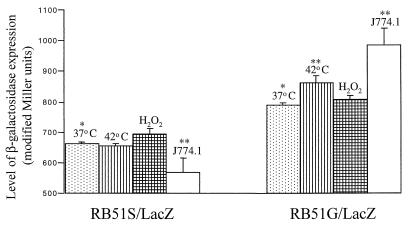
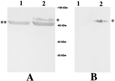
B. abortus strains RB51G/LacZ and RB51S/LacZ expressed β-galactosidase, as determined first by the appearance of blue colonies on TSA plates containing X-Gal and later by assaying the enzyme in the bacteria grown in TSB cultures (Fig. 2). Comparison of the β-galactosidase activity in strains RB51G/LacZ and RB51S/LacZ revealed that the expression under the groE promoter was significantly higher than that under the sodC promoter. No enzymatic activity was detected either in strains RB51, RB51G, and RB51S or in culture supernatants of strains RB51G/LacZ and RB51S/LacZ (data not shown). Expression of HSP65 in strain RB51G/HSP was detected by Western blotting with polyclonal antisera to E. coli GroEL and a monoclonal antibody to the mycobacterial HSP65. As expected, the polyclonal sera reacted with both Brucella GroEL and mycobacterial HSP65, whereas the monoclonal antibody reacted with the latter protein only (Fig. 3).
FIG. 2.
β-Galactosidase activity in strains RB51S/LacZ and RB51G/LacZ at 37°C and in response to the indicated stress conditions. Groups marked with one asterisk are significantly different from each other. In both strains, groups marked with two asterisks are significantly different from the 37°C group of their respective strain. P < 0.05 is considered significant.
FIG. 3.
Demonstration of the expression of HSP65 of M. bovis in strain RB51G/HSP by Western blot analysis. Lanes 1 and 2 contain the whole antigens of strains RB51 and RB51G/HSP, respectively. The antigens were separated by 12.5% sodium dodecyl sulfate–polyacrylamide gel electrophoresis and analyzed by Western blotting as previously described (36). Panel A was reacted with rabbit sera against the GroEL protein of E. coli. Panel B was reacted with mouse monoclonal antibody specific for HSP65. ∗, HSP65 of M. bovis; ∗∗, GroEL homolog of B. abortus.
Influence of stress stimuli on the activity of Brucella promoters.
In order to determine the activity of the cloned promoter sequences, we examined the effect of specific stress stimuli on the level of β-galactosidase expression and compared it with the level of expression determined at 37°C. Heat shocking by incubating the bacterial cultures at 42°C for 20 min resulted in a significant increase in the β-galactosidase activity in strain RB51G/LacZ; no change in activity was observed in strain RB51S/LacZ (Fig. 2). Addition of H2O2 to the bacterial cultures at a concentration of 0.005% slightly, but not significantly, increased the β-galactosidase activity only in strain RB51S/LacZ (Fig. 2). The heat shock and H2O2 treatments did not affect the viability of either strain. Upon intracellular localization in J774A.1 cells, β-galactosidase activity in strain RB51G/LacZ increased significantly, whereas in strain RB51S/LacZ, it decreased significantly (Fig. 2).
Induction of Th1 type immune responses in mice.
Specific antibody and CMI responses of the vaccinated mice were determined by ELISA and cytokine quantitation, respectively. Mice vaccinated with strain RB51G/LacZ and RB51S/LacZ, but not those immunized with strain RB51 or inoculated with saline, developed β-galactosidase-specific IgG (Fig. 4A). Subisotype analysis indicated that the developed antibodies were predominantly IgG2a. Very low levels of IgG1 antibodies specific to β-galactosidase were detected (Fig. 4A). Comparison between the two groups demonstrate that mice immunized with strain RB51G/LacZ developed a higher concentration of β-galactosidase-specific antibodies. Mice in all groups, except the saline-inoculated one, developed similar concentrations of antibodies specific to the antigens of strain RB51 (Fig. 4B; data for the group immunized with strain RB51G/HSP are not shown). Again, these antibodies were predominantly of IgG2a subisotype, and no IgG1 was detected. Mice immunized with strain RB51G/HSP developed HSP65-specific antibodies of the IgG2a subisotype (Fig. 5), but not IgG1 (data not shown). Sera from strain RB51-vaccinated mice, but not saline-inoculated ones, also reacted with HSP65, indicating a cross-reactivity between HSP65 and an antigen, most probably GroEL, of strain RB51 (Fig. 5).
FIG. 4.
ELISA detection of β-galactosidase-specific (A) and strain RB51-specific (B) IgG, IgG1, and IgG2a antibodies in serum of mice vaccinated with strain RB51S/LacZ, RB51G/LacZ, and RB51 or inoculated with saline alone. Sera collected from three mice of each group at 4 and 6 weeks postvaccination were diluted 1/100 and assayed for the presence of specific antibodies. Results are shown as the mean ± standard deviation of OD450 of the color developed.
FIG. 5.
ELISA detection of HSP65 protein-specific IgG and IgG2a antibodies in serum of mice vaccinated with strains RB51G/HSP and RB51 or inoculated with saline alone. Sera collected from three mice of each group at 4 and 6 weeks postvaccination were diluted 1/100 and assayed for the presence of antibodies to HSP65 of M. bovis. Results are shown as the mean ± standard deviation of OD450 of the color developed.
After stimulation with specific antigens, IFN-γ, but not IL-4 (data not shown), was detected in the culture supernatants of splenocytes obtained from the vaccinated mice. Splenocytes stimulated with antigen extracts of strain RB51 secreted similar concentrations of IFN-γ (Table 2). However, when stimulated with β-galactosidase, splenocytes from mice vaccinated with strain RB51G/LacZ produced significantly more IFN-γ than the splenocytes from mice vaccinated with strain RB51S/LacZ. Stimulation with β-galactosidase did not induce the secretion of IFN-γ from splenocytes of strain RB51- or saline-inoculated mice. Splenocytes from strain RB51G/HSP, but not strain RB51- or saline-inoculated mice, secreted IFN-γ upon stimulation with HSP65. Splenocytes from all groups produced similar concentrations of IFN-γ and IL-4 when stimulated with concanavalin A (data not shown).
TABLE 2.
Production of IFN-γ by splenocytes of naive and vaccinated mice after in vitro stimulation with specific antigens
| Stimulant | Concn of IFN-γ (ng/ml) in micea
|
||||
|---|---|---|---|---|---|
| Naive | RB51 vaccinated | RB51G/LacZ vaccinated | RB51S/LacZ vaccinated | RB51G/HSP vaccinated | |
| Media | — | — | — | — | — |
| RB51 extract | — | 11.44 ± 0.60∗ | 11.85 ± 0.78∗ | 11.71 ± 0.75∗ | 12.04 ± 0.94∗ |
| β-Galactosidase | — | — | 1.58 ± 0.41∗∗ | 0.43 ± 0.13∗∗ | NT |
| HSP | — | — | NT | NT | 5.04 ± 0.35 |
Values are means ± standard deviations (n = 3). Means with one asterisk were not significantly different from each other (P > 0.5); means with two asterisks were significantly different from each other (P < 0.01). —, below the detection limit; NT, not tested.
Attenuation and vaccine efficacy of the recombinant RB51 strains.
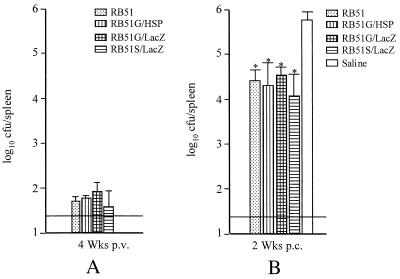
No significant difference in the number of Brucella present in the spleens of vaccinated mice at 4 weeks was observed (Fig. 6). Also, the levels of protection against a challenge infection with B. abortus 2308 were similar in all of the vaccinated mice (Fig. 6).
FIG. 6.
(A) Persistence of strain RB51 and its recombinants in spleens of vaccinated mice. Four weeks postvaccination (p.v.), five mice from each group were euthanized and the number of CFU in their spleens was determined as described in Materials and Methods. (B) Resistance to B. abortus 2308 challenge infection in mice vaccinated with strain RB51 and its recombinants. Mice were vaccinated 6 weeks prior to the challenge infection. Two weeks postchallenge (p.c.) infection, the number of strain 2308 CFU in their spleens was determined. The horizontal line above the x axis in panels A and B indicates the lower detection limit. In panel B, groups with one asterisk were significantly different from the saline group (P < 0.001), but not from each other.
DISCUSSION
In this study, we constructed two expression plasmids containing the promoters of Brucella sodC and groE genes. Using these plasmids and the lacZ and hsp65 genes of E. coli and M. bovis, respectively, we demonstrated that (i) B. abortus vaccine strain RB51 can be used as a vector for the expression of heterologous bacterial proteins, (ii) vaccination of mice with the recombinant RB51 strains results in the preferential induction of a Th1 type of immune response specific to the foreign protein, and (iii) expression of β-galactosidase or mycobacterial HSP65 does not alter either the attenuation characteristic of strain RB51 or its protective efficacy against virulent B. abortus infection. First, in order to achieve the expression of a foreign protein in quantities sufficient to induce an immune response, we examined the activity of Brucella sodC and groE promoters by determining the levels of β-galactosidase expression in strains RB51G/LacZ and RB51S/LacZ. In the case of the sodC promoter, no significant increase in its activity was observed under the stress conditions tested. However, we observed a significant decrease in the sodC promoter's activity when the bacteria were localized within the macrophage-like J774A.1 cells. This observation supports a previous finding indicating that the expression of Cu/Zn superoxide dismutase in B. abortus is decreased during intracellular growth in bovine macrophages (28). In contrast, a significant increase in the activity of groE promoter under heat shock conditions and upon intracellular localization of RB51G/LacZ was observed. This was expected based on the previously published characterization of groE promoters of other intracellular bacteria, which also demonstrated similar activities (6, 14, 15, 19). In addition, increased expression of GroEL protein in B. abortus was documented when the bacteria were subjected to different stress stimuli (28); this can be attributed to the enhanced activity of the groE promoter. Although vaccination of mice with either RB51G/LacZ or RB51S/LacZ induced β-galactosidase-specific immune responses, these responses were stronger in the case of RB51G/LacZ vaccination. This can be directly correlated with the higher levels of β-galactosidase expression under the groE promoter, especially after intracellular localization of the bacteria within the macrophages. In the case of HSP65, it should be mentioned that we were unable to detect significant levels of HSP65-specific immune responses in mice vaccinated with a recombinant RB51 strain expressing HSP65 under the sodC promoter, suggesting that expression of heterologous proteins in sufficient quantities is essential for the induction of detectable immune responses. The presence of IgG2a, but not IgG1, antibodies specific for β-galactosidase or HSP65 as well as strain RB51 antigens in the serum of vaccinated mice indicates the preferential development of a Th1 type of immune response (34). This is further corroborated by the secretion of IFN-γ, but not IL-4, by the splenocytes of vaccinated mice upon in vitro stimulation with the specific antigens. In our ELISA, sera from mice vaccinated with RB51 showed reactivity with HSP65 (Fig. 5). This could be due to serological cross-reactivity between HSP65 and Brucella GroEL, since there is ∼60% amino acid sequence homology between these two proteins (29). Similar cross-reactivity has been reported in the literature for other bacterial GroEL proteins and HSP65 (19, 32). However, it is interesting to find in our study that the splenocytes from RB51-vaccinated mice did not produce detectable levels of IFN-γ when stimulated with HSP65. Although the actual reason for this is not known, the dose of HSP65 (0.25 μg/well) we used for the in vitro stimulation may not be sufficient to activate the cross-reactive T cells, and/or such T cells could be present at very low frequency. Using selected synthetic peptides for immunization of mice, Brett et al. (7) have shown that it is possible to elicit T cells that can cross-react with HSP65 and E. coli GroEL protein.
The protective capabilities of several mycobacterial proteins, including that of HSP65, have been well studied and documented (22, 33). Also, it is well known that the development of a Th1 type of immune response is essential for immunoprotection against infections by Mycobacterium spp. As we demonstrated in this study, along with other reports on B. abortus, it is clear that strain RB51 is a strong inducer of a Th1-biased CMI. The feasibility of expressing foreign proteins in strain RB51 makes it a strong candidate vector for the development of a live, attenuated multivalent vaccine that can provide simultaneous protection against brucellosis and infection with heterologous pathogens, such as Mycobacterium spp. Recently, several attenuated bacterial strains, including B. abortus strain 19, have been used for the expression of heterologous proteins (8, 11, 12, 17, 38, 41). However, strain RB51 has many advantages over all other live bacterial vectors for use in several domestic and wild animals where vaccination against brucellosis is required. We have also been successful in expressing several other bacterial, parasitic, and fungal proteins of interest in strain RB51 (unpublished data), suggesting a broad scope for the utilization of RB51 as a live vaccine vector. Recently, we demonstrated that the overexpression of a protective protein of B. abortus in strain RB51 significantly increased its vaccine efficacy against brucellosis (37). A recombinant strain of RB51 that can overproduce homologous protective antigen(s) and simultaneously express protective proteins of other intracellular pathogens may become a very effective multivalent vaccine.
ACKNOWLEDGMENT
This work was supported by U.S. Department of Agriculture grant 97-35204-4483.
REFERENCES
- 1.Acha P, Szyfres B. Zoonoses and communicable diseases common to man and animals. Washington, D.C.: Pan American Health Organization; 1980. pp. 28–45. [Google Scholar]
- 2.Agranovich I, Scott D E, Terle D, Lee K, Golding B. Down-regulation of Th2 responses by Brucella abortus, a strong Th1 stimulus, correlates with alterations in the B7.2-CD28 pathway. Infect Immun. 1999;67:4418–4426. doi: 10.1128/iai.67.9.4418-4426.1999. [DOI] [PMC free article] [PubMed] [Google Scholar]
- 3.Allen C A, Garry Adams L, Ficht T A. Transposon-derived Brucella abortus rough mutants are attenuated and exhibit reduced intracellular survival. Infect Immun. 1998;66:1008–1016. doi: 10.1128/iai.66.3.1008-1016.1998. [DOI] [PMC free article] [PubMed] [Google Scholar]
- 4.Araya L N, Elzer P H, Rowe G E, Enright F M, Winter A J. Temporal development of protective cell-mediated and humoral immunity in BALB/c mice infected with Brucella abortus. J Immunol. 1989;143:3330–3337. [PubMed] [Google Scholar]
- 5.Araya L N, Winter A J. Comparative protection of mice against virulent and attenuated strains of Brucella abortus by passive transfer of immune T cells or serum. Infect Immun. 1990;58:254–256. doi: 10.1128/iai.58.1.254-256.1990. [DOI] [PMC free article] [PubMed] [Google Scholar]
- 6.Batoni G, Maisetta G, Florio W, Freer G, Campa M, Senesi S. Analysis of the Mycobacterium bovis hsp60 promoter activity in recombinant Mycobacterium avium. FEMS Microbiol Lett. 1998;169:117–124. doi: 10.1111/j.1574-6968.1998.tb13307.x. [DOI] [PubMed] [Google Scholar]
- 7.Brett S J, Lamb J R, Cox J H, Rothbard J B, Mehlert A, Ivanyi J. Differential pattern of T cell recognition of the 65-kDa mycobacterial antigen following immunization with the whole protein or peptides. Eur J Immunol. 1989;19:1303–1310. doi: 10.1002/eji.1830190723. [DOI] [PubMed] [Google Scholar]
- 8.Brossier F, Mock M, Sirard J. Antigen delivery by attenuated Bacillus anthracis: new prospects in veterinary vaccines. J Appl Microbiol. 1999;87:298–302. doi: 10.1046/j.1365-2672.1999.00895.x. [DOI] [PubMed] [Google Scholar]
- 9.Cheville N F, Jensen A E, Halling S M, Tatum F M, Morfitt D C, Hennager S G, Frerichs W M, Schurig G G. Bacterial survival, lymph node changes, and immunologic responses of cattle vaccinated with standard and mutant strains of Brucella abortus. Am J Vet Res. 1992;53:1881–1888. [PubMed] [Google Scholar]
- 10.Cheville N F, Stevens M G, Jensen A E, Tatum F M, Halling S M. Immune responses and protection against infection and abortion in cattle experimentally vaccinated with mutant strains of Brucella abortus. Am J Vet Res. 1993;54:1591–1597. [PubMed] [Google Scholar]
- 11.Cirillo J D, Stover C K, Bloom B R, Jacobs W R, Jr, Barletta R G. Bacterial vaccine vectors and bacillus Calmette-Guerin. Clin Infect Dis. 1995;20:1001–1009. doi: 10.1093/clinids/20.4.1001. [DOI] [PubMed] [Google Scholar]
- 12.Comerci D J, Pollevick G D, Vigliocco A M, Frasch A C C, Ugalde R A. Vector development for the expression of foreign proteins in the vaccine strain Brucella abortus S19. Infect Immun. 1998;66:3862–3866. doi: 10.1128/iai.66.8.3862-3866.1998. [DOI] [PMC free article] [PubMed] [Google Scholar]
- 13.Corbel M J. Brucellosis: an overview. Emerg Infect Dis. 1997;3:213–221. doi: 10.3201/eid0302.970219. [DOI] [PMC free article] [PubMed] [Google Scholar]
- 14.Dellagostin O A, Esposito G, Eales L J, Dale J W, McFadden J. Activity of mycobacterial promoters during intracellular and extracellular growth. Microbiology. 1995;141:1785–1792. doi: 10.1099/13500872-141-8-1785. [DOI] [PubMed] [Google Scholar]
- 15.Hoffman P S, Houston L, Butler C A. Legionella pneumophila htpAB heat shock operon: nucleotide sequence and expression of the 60-kilodalton antigen in L. pneumophila-infected HeLa cells. Infect Immun. 1990;58:3380–3387. doi: 10.1128/iai.58.10.3380-3387.1990. [DOI] [PMC free article] [PubMed] [Google Scholar]
- 16.Jensen A E, Ewalt D R, Cheville N F, Thoen C O, Payeur J B. Determination of stability of Brucella abortus RB51 by use of genomic fingerprint, oxidative metabolism, and colonial morphology and differentiation of strain RB51 from B. abortus isolates from bison and elk. J Clin Microbiol. 1996;34:628–633. doi: 10.1128/jcm.34.3.628-633.1996. [DOI] [PMC free article] [PubMed] [Google Scholar]
- 17.Killeen K, Spriggs D, Mekalanos J. Bacterial mucosal vaccines: Vibrio cholerae as a live attenuated vaccine/vector paradigm. Curr Top Microbiol Immunol. 1999;236:237–254. doi: 10.1007/978-3-642-59951-4_12. [DOI] [PubMed] [Google Scholar]
- 18.Kovach M E, Phillips R W, Elzer P H, Roop II R M, Peterson K M. pBBR1MCS: a broad-host-range cloning vector. BioTechniques. 1994;16:800–802. [PubMed] [Google Scholar]
- 19.Lindquist S, Craig E A. The heat-shock proteins. Annu Rev Genet. 1988;22:631–677. doi: 10.1146/annurev.ge.22.120188.003215. [DOI] [PubMed] [Google Scholar]
- 20.Littell R C, Milliken G A, Stroup W W, Wolfinger R D. SAS system for mixed models. Cary, N.C: SAS Institute, Inc.; 1996. [Google Scholar]
- 21.Lord V R, Schurig G G, Cherwonogrodzky J W, Marcano M J, Melendez G E. Field study of vaccination of cattle with Brucella abortus strains RB51 and 19 under high and low disease prevalence. Am J Vet Res. 1998;59:1016–1020. [PubMed] [Google Scholar]
- 22.Lowrie D B, Silva C L, Colston M J, Ragno S, Tascon R E. Protection against tuberculosis by a plasmid DNA vaccine. Vaccine. 1997;15:834–838. doi: 10.1016/s0264-410x(97)00073-x. [DOI] [PubMed] [Google Scholar]
- 23.McQuiston J R, Schurig G G, Sriranganathan N, Boyle S M. Transformation of Brucella species with suicide and broad host-range plasmids. Methods Mol Biol. 1995;47:143–148. doi: 10.1385/0-89603-310-4:143. [DOI] [PubMed] [Google Scholar]
- 24.McQuiston J R, Vemulapalli R, Inzana T J, Schurig G G, Sriranganathan N, Fritzinger D, Hadfield T L, Warren R A, Snellings N, Hoover D, Halling S M, Boyle S M. Genetic characterization of a Tn5-disrupted glycosyltransferase gene homolog in Brucella abortus and its effect on lipopolysaccharide composition and virulence. Infect Immun. 1999;67:3830–3835. doi: 10.1128/iai.67.8.3830-3835.1999. [DOI] [PMC free article] [PubMed] [Google Scholar]
- 25.Miller J H. A short course in bacterial genetics: a laboratory manual and handbook for Escherichia coli and related bacteria. Cold Spring Harbor, N.Y: Cold Spring Harbor Laboratory; 1992. [Google Scholar]
- 26.Oñate A A, Vemulapalli R, Andrews E, Schurig G G, Boyle S, Folch H. Vaccination with live Escherichia coli expressing Brucella abortus Cu/Zn superoxide dismutase protects mice against virulent B. abortus. Infect Immun. 1999;67:986–988. doi: 10.1128/iai.67.2.986-988.1999. [DOI] [PMC free article] [PubMed] [Google Scholar]
- 27.Palmer M V, Olsen S C, Cheville N F. Safety and immunogenicity of Brucella abortus strain RB51 vaccine in pregnant cattle. Am J Vet Res. 1997;58:472–477. [PubMed] [Google Scholar]
- 28.Rafie-Kolpin M, Essenberg R C, Wyckoff J H., III Identification and comparison of macrophage-induced proteins and proteins induced under various stress conditions in Brucella abortus. Infect Immun. 1996;64:5274–5283. doi: 10.1128/iai.64.12.5274-5283.1996. [DOI] [PMC free article] [PubMed] [Google Scholar]
- 29.Roop R M, II, Price M L, Dunn B E, Boyle S M, Sriranganathan N, Schurig G G. Molecular cloning and nucleotide sequence analysis of the gene encoding the immunoreactive Brucella abortus Hsp60 protein, BA60K. Microb Pathog. 1992;12:47–62. doi: 10.1016/0882-4010(92)90065-v. [DOI] [PubMed] [Google Scholar]
- 30.Schurig G G, Roop R M, Bagchi T, Boyle S, Buhrman D, Sriranganathan N. Biological properties of RB51: a stable rough strain of Brucella abortus. Vet Microbiol. 1991;28:171–188. doi: 10.1016/0378-1135(91)90091-s. [DOI] [PubMed] [Google Scholar]
- 31.Scott D E, Agranovich I, Inman J, Gober M, Golding B. Inhibition of primary and recall allergen-specific T helper cell type 2-mediated responses by a T helper cell type 1 stimulus. J Immunol. 1997;159:107–116. [PubMed] [Google Scholar]
- 32.Shinnick T M. Heat shock proteins as antigens of bacterial and parasitic pathogens. Curr Top Microbiol Immunol. 1991;167:145–150. doi: 10.1007/978-3-642-75875-1_9. [DOI] [PubMed] [Google Scholar]
- 33.Silva C L, Silva M F, Pietro R C L R, Lowrie D B. Characterization of T cells that confer a high degree of protective immunity against tuberculosis in mice after vaccination with tumor cells expressing mycobacterial hsp65. Infect Immun. 1996;64:2400–2407. doi: 10.1128/iai.64.7.2400-2407.1996. [DOI] [PMC free article] [PubMed] [Google Scholar]
- 34.Stevens T L, Bossie A, Sanders V M, Fernandez-Botran R, Coffman R L, Mosmann T R, Vitetta E S. Regulation of antibody isotype secretion by subsets of antigen-specific helper T cells. Nature. 1988;334:255–258. doi: 10.1038/334255a0. [DOI] [PubMed] [Google Scholar]
- 35.Vemulapalli R, Cravero S, Calvert C L, Toth T E, Boyle S M, Sriranganathan N, Schurig G G. Characterization of specific immune responses of mice inoculated with recombinant vaccinia virus expressing an 18-kilodalton outer membrane protein of Brucella abortus. Clin Diagn Lab Immunol. 2000;7:114–118. doi: 10.1128/cdli.7.1.114-118.2000. [DOI] [PMC free article] [PubMed] [Google Scholar]
- 36.Vemulapalli R, Duncan A J, Boyle S M, Sriranganathan N, Toth T E, Schurig G G. Cloning and sequencing of yajC and secD homologs of Brucella abortus and demonstration of immune responses to YajC in mice vaccinated with B. abortus RB51. Infect Immun. 1998;66:5684–5691. doi: 10.1128/iai.66.12.5684-5691.1998. [DOI] [PMC free article] [PubMed] [Google Scholar]
- 37.Vemulapalli R, He Y, Cravero S, Sriranganathan N, Boyle S M, Schurig G G. Overexpression of protective antigen as a novel approach to enhance vaccine efficacy of Brucella abortus strain RB51. Infect Immun. 2000;68:3286–3289. doi: 10.1128/iai.68.6.3286-3289.2000. [DOI] [PMC free article] [PubMed] [Google Scholar]
- 38.Weiskirch L M, Paterson Y. Listeria monocytogenes: a potent vaccine vector for neoplastic and infectious disease. Immunol Rev. 1997;158:159–169. doi: 10.1111/j.1600-065x.1997.tb01002.x. [DOI] [PubMed] [Google Scholar]
- 39.Winter A J, Schurig G G, Boyle S M, Sriranganathan N, Bevins J S, Enright F M, Elzer P H, Kope J D. Protection of BALB/c mice against homologous and heterologous species of Brucella by rough strain vaccines derived from Brucella melitensis and Brucella suis biovar 4. Am J Vet Res. 1996;57:677–683. [PubMed] [Google Scholar]
- 40.Wise D J, Sriranganathan N, Boyle S M, Schurig G G. Evaluation of the intracellular growth of various Brucella species in J774.A1 and PU5-1.8 macrophage-like cell lines as an in vitro model of assessing attenuation in vivo. In: Frank J F, editor. Networking in brucellosis research II. Proceedings of the UNU/BIOLAC Brucellosis Workshop. Tokyo, Japan: United Nations University Press; 1998. pp. 93–110. [Google Scholar]
- 41.Zegers N D, Kluter E, Van Der Stap H, Van Dura E, Van Dalen P, Shaw M, Baillie L. Expression of the protective antigen of Bacillus anthracis by Lactobacillus casei: towards the development of an oral vaccine against anthrax. J Appl Microbiol. 1999;87:309–314. doi: 10.1046/j.1365-2672.1999.00900.x. [DOI] [PubMed] [Google Scholar]


