Abstract
Background
Currently, the optimum surgical approach for treating adherent otitis media is debatable. The traditional treatment is usually performed by microscopic tympanoplasty combined with temporal myofascial tympanic tube placement. In recent years, the application of whole ear endoscopic surgery in the treatment of middle ear diseases has gradually increased, otoendoscopy has been used in the operation of adhesive otitis media, but its safety and effectiveness are still controversial.
Methods
This study retrospectively analyzed 17 patients with adhesive otitis media treated by endoscopic ear surgery (EES) in our hospital from January 2018 to July 2021 over a 6-month period post-surgery. Of the 17 patients, 8 were males and 9 were females (mean age, 53 years; age range, 24–70 years). There were 12 and 5 cases of adhesive otitis media involving the left and right ear, respectively. The patients had follow-up evaluations 1 week, 2 weeks, 1 month, 3 months, and 6 months after surgery.
Results
A total of 17 patients with adhesive otitis were enrolled, including 1 patient with Dornhoffer stage II; 6 patients with stage III; and 10 patients with stage IV. Adhesive otitis media was combined with middle ear cholesteatoma in 4 patients (24%). Fourteen patients (82%) had disruption or interruption of the ossicular chain (8 malleus, 14 incus, and 4 stapes lesions), 11 of whom had artificial ossicular chain reconstruction [8 with partial ossicular reconstruction prosthesis (PORP) and 3 with total ossicular reconstruction prosthesis (TORP) implantation]. All patients had good tympanic membrane and graft morphology, no invaginations, and no perforations. The mean postoperative air-conduction hearing threshold [49.06±22.15 dB hearing level (dB HL)] and mean air-bone gap (19.94±10.00 dB HL) were significantly improved compared with the preoperative values (65.29±21.53 and 32.53±8.21 dB HL, respectively; P<0.05). No recurrences, secondary cholesteatomas, or secondary surgeries were reported.
Conclusions
EES seems to be a safe and effective surgical method for the management of adhesive otitis media. The study has limitations due to its small sample size and lack of controlled studies. It still needs to be proven in clinical randomized controlled trials.
Keywords: Endoscopic ear surgery (EES), adhesive otitis media, tympanoplasty
Introduction
Adhesive otitis media is a chronic suppurative otitis media in which the middle ear tissues become adhesive due to persistent inflammation. It is one of the most frequent otolaryngologic conditions. Patients with adhesive otitis media frequently present with complete or partial adhesions between the thin retracted and atrophic pars tensa and the middle ear medial wall. Moreover, the middle ear ossicles may be encased in soft tissue debris (1-3). Adhesive otitis media may be unilateral or bilateral, and is characterized as a chunky, sticky secretion similar to glue that collects in the middle ear and is thus generally termed “glue ear” (4,5). Fibrosis and adhesions form in the middle ear due to various causes, resulting in impaired movement of the middle ear sound transmission system, leading to conductive deafness (6,7). Fibrosis and adhesions are common sequelae of otitis media, which can lead to malperfusion of the middle ear, negative pressure in the tympanic chamber, fluid accumulation in the middle ear, invagination of the tympanic membrane and adhesion to the tympanic capsule and anvil, proliferation and chemosis of the middle ear mucosa, granulation formation, and eventually adhesions. It is often complicated by middle ear diseases, such as middle ear cholesteatoma, middle ear cholesterol granuloma, and tympanosclerosis, and patients often have nasal disorders, such as dysphagia, chronic sinusitis with/without nasal polyps, allergic rhinitis, and nasopharyngeal lesions (1,8,9).
Currently, the optimum surgical approach for treating adherent otitis media remains controversial (10). Endoscopic ear surgery (EES) provides a better view of the surgical field. The distal half of the device is lit up and has angled lenses to provide a better view of the surgical area. The external auditory canal is converted into a surgical channel that provides a wider field of view, greater imaging capabilities, higher magnification, and the technology to study otherwise inaccessible areas of the middle ear. EES also allows surgeons to use a minimally invasive approach in otologic procedures. Endoscopic tympanoplasty has been shown to require less operative time than microscope-assisted surgery in some cases (11). In the traditional treatment of adhesive otitis media, microscopic tympanoplasty is generally performed. In recent years, otoendoscopic surgery and eustachian tube balloon dilatation have been gradually applied. There is still a lack of large sample studies on the surgical efficacy of otoendoscopic tympanoplasty for adhesive otitis media. This study intends to collect the cases of endoscopic endoscopic surgery of adhesive otitis media, and evaluate the efficacy of endoscopic treatment of adhesive otitis media by analyzing the operation and prognosis of postoperative endoscopic examination and audiological examination data. Herein, we discussed the clinical indications, surgical characteristics, and postoperative effects of EES in the treatment of adhesive otitis media. We present the following article in accordance with the STROBE reporting checklist (available at https://atm.amegroups.com/article/view/10.21037/atm-22-4831/rc).
Methods
General data
We retrospectively analyzed the surgical efficacy of EES in 17 patients with adhesive otitis media (17 ears) who were admitted to the Department of Otolaryngology at our hospital from January 2018 to April 2021. The inclusion criteria were as follows: patients aged 18 years and older who were diagnosed with adhesive otitis media; preoperative endoscopy, pure tone audiometry, and a sound conductivity examination were performed; a postoperative review was completed with endoscopy, pure tone audiometry, and a sound conductivity examination; and follow-up evaluations were performed up to 6 months postoperatively. The exclusion criteria were as follows: co-presentation of multiple underlying diseases; and a combination of external auditory canal cholesteatoma and external auditory canal stenosis.
The study was conducted in accordance with the Declaration of Helsinki (as revised in 2013). Individual consent for this retrospective analysis was waived. The study was approved by the ethics committee of Ren Ji Hospital, Shanghai Jiao Tong University School of Medicine (No. LY-2022-075-B).
Preoperative evaluation
All patients underwent routine preoperative endoscopy, a computed tomography (CT) plain scan of the middle ear mastoid, a pure tone hearing threshold test (125 Hz–8 kHz), an air-conduction bone conduction, and an acoustic conduction resistance test.
Surgical equipment
The 0°/30° endoscope was 1.9 mm in diameter and 10 cm in length (Storz, Tuttlingen, Germany). The display and video were purchased from Storz, and all surgical steps were saved on video. The surgical instruments included conventional otologic microscopic instruments, some of which were self-modified to adapt to the operative angle of the ear microscopic instruments, including a peeler, suction device, and other apical attachments.
Preoperative preparation
After the patient was placed under local or general anesthesia, the external ear canal was cleaned of cerumen and secretions via endoscopy, and the overgrown ear hairs on the side of the tragus at the ear canal opening were trimmed. No temporal skin preparation was required.
Surgical steps
Patients were placed in the supine position with the head turned 30°–45° to the healthy side and the operator was seated on the affected side. Local block anesthesia was used in 5 patients, and general anesthesia was used in 12 patients. After anesthesia was established, the operative field was disinfected and surgical towels were laid.
Creation of the external auditory canal skin-tympanic flap
The skin of the posterior wall of the external auditory canal was cut in an arc 1–1.5 cm from the tympanic ring in a counter-clockwise direction from 1 to 6 o’clock (right ear) or a clockwise direction from 11 to 6 o’clock (left ear) to reach the bone surface. The flap was peeled off with epinephrine cotton pellets to reach the tympanic ring, and the tympanic ring was peeled off with a right-angle hook or peeler. The flap was lifted off to enter the tympanum.
Exploration of lesions and release of adhesions
Upper tympanic chamber-lateral wall dissection was performed after separating and exposing the tympanum. The extent of the adhesion foci was probed, such as stage III–IV, to perform upper tympanum-lateral wall dissection. Part of the upper tympanic chamber lateral wall was removed with an osteotome and scraping spoon to reveal the upper tympanum, ossicular chain, and fully expose the adhesion foci.
To release the adhesions and remove the lesions, a peeler was used to advance from posterior-to-anterior and parallel to release the adhesions. In cases with cholesteatomas, the epithelium of the cholesteatoma was completely removed to fully expose the epitympanic antrum direction, and explore with an elbow suction, an elbow peeler, and a long crochet. To probe the ossicular chain, the integrity of the auditory chain and the presence of the round window reflection must be assured by probing. To remove the residual incus, the caput mallei was cut, the manubrium of the malleus and stapes (if present) was preserved, and a partial ossicular reconstruction prosthesis (PORP) or total ossicular reconstruction prosthesis (TORP) was placed to reconstruct the ossicular chain. The eustachian tube was explored intraoperatively using an epidural catheter.
Preparation of the graft
The graft was prepared as follows: (I) excision of the tragus cartilage-chondral membrane complex; and (II) the removed tragus cartilage-chondral membrane complex must be trimmed, and part of the cartilage was trimmed for reconstruction of the lateral wall of the superior tympanic chamber, leaving a V-shaped incision for placement of the manubrium of the malleus.
Implantation of grafts and artificial auditory bone
The epidural catheter was removed, the tragus cartilage-chondral membrane complex was placed internally, a PORP was placed between the stirrup head and the manubrium of the malleus/tragus cartilage, or a TORP was placed between the round window and the manubrium of the malleus/tragus cartilage. The tympanic chamber was filled with a dexamethasone-infiltrated gelatin sponge and anti-adhesive membrane, the graft was adjusted, the external auditory canal skin-tympanic flap was repositioned, and the ear canal was filled with a gelatin sponge.
Closure of the incision and filling
Hemostasis of the tragus incision was assured and glued together, and the ear canal opening was filled with chlortetracycline gauze to compress the tragus incision.
Postoperative treatment
Postoperatively, patients were observed for facial palsy, vertigo, hearing loss, and other manifestations, and routinely discharged on the first postoperative day. Oral antibiotics were prescribed for 2 weeks after surgery.
Follow-up
Patients had follow-up evaluations at 1 week, 2 weeks, 1 month, 3 months, and 6 months postoperatively. At the first postoperative follow-up, the external auditory canal gauze was withdrawn, the external auditory canal gelatin sponge was cleaned, and the graft growth was observed and evaluated. Audiologic examinations, such as pure tone audiometry (Otometrics Madsen Astera 2; GN Otometrics A/S, Copenhagen, Denmark) and acoustic conductance (Madsen Zodiac; GN Otometrics A/S) were performed in the first, third, and sixth months after surgery.
Statistical analysis
The SPSS 21.0 software was used for data analysis, and all measurement data are expressed as the mean ± standard deviation (SD). A paired t-test was used for comparison of pre- and postoperative data. A P value <0.05 was considered statistically significant.
Results
A total of 17 ears were analyzed in this study, including 8 males and 9 females with a mean age of 53 years (range, 24–70 years). There were 12 cases involving the left ear and 5 cases involving the right ear. According to Dornhoffer’s staging of adhesive otitis media (12), 1 patient was stage II, 6 patients were stage III, and 10 patients were stage IV. The mean duration of adhesive otitis media was 18 years. The mean preoperative air-conduction hearing threshold was 65.29±21.53 dB hearing level (dB HL), and the mean preoperative air-bone gap was 32.53±8.21 dB HL.
Analysis of clinical data
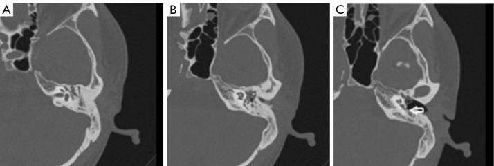
All patients had a history of adhesive otitis media with a duration of 1 month to 70 years (average duration of 18 years). The symptoms included hearing loss (n=12), aural fullness (n=4), otopyorrhea (n=10), otalgia (n=3), and tinnitus (n=2). There were 2 patients with perforations in the pars flaccida, 6 patients with perforations in the pars tensa, and 9 patients with intact tympanic membranes. There were 4, 2, and 3 patients presenting with adhesive otitis media combined with a middle ear cholesteatoma, middle ear cholesterol granuloma, or tympanosclerosis, respectively. Among the 17 patients with adhesive otitis media, 3 had sinusitis and 3 had nasopharyngitis. One patient was stage II, 6 were stage III, and 10 were stage IV (Figure 1). Hearing reconstruction with artificial ossiculoplasty was performed in 11 patients (PORP in 8 patients and TORP in 3 patients), and 6 patients did not undergo hearing reconstruction (Table 1).
Figure 1.
CT of a patient with stage III adhesive otitis media. (A) A patient with stage III adhesive otitis media. Computed tomography suggests a soft tissue shadow of the right epitympanum and tympanic antrum, and the ossicular chain is surrounded by a soft tissue shadow. (B) The mesotympanum-hypotympanum was invaginated with a soft tissue shadow. (C) A mesotympanum-hypotympanum invagination and a tight fit with the promontory (white arrow).
Table 1. Basic clinical characteristics of the patients.
| Characteristics | Cases (n=17), n [%] |
|---|---|
| Sex | |
| Male | 8 [47] |
| Female | 9 [53] |
| Sides | |
| Left | 12 [71] |
| Right | 5 [29] |
| Dornhoffer’s staging | |
| I | 0 [0] |
| II | 1 [6] |
| III | 6 [35] |
| IV | 10 [59] |
| Symptoms | |
| Hearing loss | 12 [71] |
| Aural fullness | 4 [24] |
| Otopyorrhea | 10 [59] |
| Ear pain | 3 [18] |
| Tinnitus | 2 [12] |
| Complications of perforation | |
| Perforation in the pars tensa | 6 [35] |
| perforation in the pars flaccida | 2 [12] |
| No tympanic membrane perforation | 9 [53] |
| Ossicular chain disruption | |
| Total | 14 [82] |
| Malleus | 8 [47] |
| Incus | 14 [82] |
| Stapes | 4 [24] |
| Other middle ear diseases | |
| Chronic suppurative otitis media | 6 [35] |
| Middle ear cholesteatoma | 4 [24] |
| Middle ear cholesterol granuloma | 2 [12] |
| Tympanosclerosis | 3 [18] |
| Combined sinus or nasopharyngeal disorders | |
| Sinusitis | 3 [18] |
| Nasopharyngitis | 3 [18] |
| Anesthesia method | |
| Local anesthesia | 5 [29] |
| General anesthesia | 12 [71] |
| Reconstruction of ossicular chain | |
| No | 6 [35] |
| PORP | 8 [47] |
| TORP | 3 [18] |
PORP, partial ossicular reconstruction prosthesis; TORP, total ossicular reconstruction prosthesis.
Postoperative follow-up
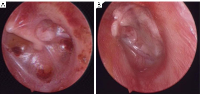
The median follow-up time was 2.9 years (range, 1–4 years). All patients had dry ears within 1 month after surgery, and no recurrences were noted at the 1-year follow-up evaluations (Figure 2A,2B).
Figure 2.
Preoperative and postoperative comparison of the patient with stage III adhesive otitis media. (A) A preoperative endoscopy of a patient with stage III adhesive otitis media showing tympanic membrane invagination with incudo-stapedial joints and promontory adhesions. (B) An endoscopy 6 months after tympanoplasty type II surgery in a patient with stage III adhesive otitis media, showing good tragus cartilage-chondrogenic healing.
Postoperative tympanic membrane condition
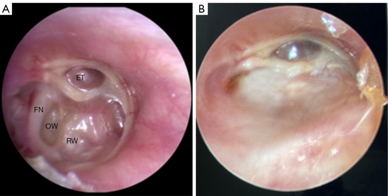
The grafts healed well and the tympanic membranes were in good shape without invaginations or re-adhesions (Figure 3A,3B).
Figure 3.
Preoperative and postoperative comparison of the patient with stage IV adhesive otitis media. (A) A preoperative endoscopy of a patient with stage IV adhesive otitis media, showing that the tympanic membrane was thin and invaginated. The endoscopy was unable to visualize the full extent of the adhesions. (B) An endoscopy of a stage IV adhesive otitis media patient at 3 months after tympanoplasty type III surgery, showing good tragus cartilage-chondrogenic healing. ET, eustachian tube-tympanum opening; FN, tympanic segment of the facial nerve; OW, oval window; RW, round window.
Audiologic status
The postoperative mean air-conduction hearing threshold at 0.5, 1.0, and 2.0 kHz and mean air-bone gap at 0.5, 1.0, and 2.0 kHz were significantly improved (49.06±22.15 and 19.94±10.00 dB HL, respectively) compared with the preoperative mean air-bone conduction threshold and mean air-bone gap (65.29±21.53 and 32.53±8.21 dB HL, respectively) in 17 patients (P<0.05; Table 2).
Table 2. Preoperative and postoperative hearing results in 17 patients.
| Variables | Preoperative | Postoperative | P value |
|---|---|---|---|
| Air-conduction hearing threshold (dB HL) | 65.29±21.53 | 49.06±22.15 | 0.038 |
| Air-bone gap (dB HL) | 32.53±8.21 | 19.94±10.00 | 0.00034 |
Data are expressed as mean ± SD. SD, standard deviation; dB HL, dB hearing level.
Discussion
Etiology of adhesive otitis media and the traditional surgical approach
Adhesive otitis media is caused by long-term ventilation dysfunction of the middle ear. The main causes of chronic deficient ventilation of the middle ear are gas diffusion dysfunction of the middle ear and mastoid process mucosa, auditory tube ventilation dysfunction, tympanic membrane and mastoid process air chamber pressure cushioning dysfunction, and tympanic membrane structural and functional dysfunction (13). Traditional treatment modalities usually include tympanoplasty in conjunction with simultaneous temporal muscle fascia-tympanic membrane tube placement (4,14). The EES allows for better cholesteatoma screening in cases of chronic otitis media, better visualization of anterior poor ventilation of the mesotympanum, improved access to selective epitympanic poor ventilation and secondary selective chronic otitis media, superior vision and reconstruction of anterior tympanic membrane perforations, Sheehy’s lateral graft tympanoplasty via a trans-canal style, and increases the likelihood of preoperative recognition of ossicular chain disruption accompanied by perforations (15).
Indications for endoscopic management of adhesive otitis media
EES offers similar surgical results compared to a traditional microscopic technique, with a shorter operative time and hospital stay after surgery being the main advantages of this technique (16,17).
Surgical repair of adhesive otitis media in patients with stage I–III lesions can be performed endoscopically. Specifically, dry lesions limited to the tympanic cavity and not involving the tympanic antrum have apparent surgical advantages, such as linear or angled endoscopic surgery without an incision.
Endoscopic removal of the full tympanic membrane invagination cannot be observed in patients with stage IV adhesive otitis media. The adhesion foci can be completely cleared by epitympanum chiseling, and the lateral wall of the epitympanum can be reconstructed as needed. In patients with adhesive otitis media and a middle ear cholesteatoma or a middle ear cholesterol granuloma involving the tympanic antrum and mastoid process, there may be an inability to completely clear the lesion and microscopic open surgery may be required. In this group, 4 and 2 patients with adhesive otitis media and a middle ear cholesteatoma or a middle ear cholesterol granuloma, respectively, had lesion clearance under EES.
Treatment of the ossicular chain
There were 14 patients with ossicular chain disruption, which was mostly located in the long crus of the incus (n=12). Because the course of adhesive otitis media depends on the stage (invagination-contact and incudo-stapedial joints-contact promontory), it is not possible to peer into the adhesions.
Extent of the adhesions
The long crus of the incus is easily disrupted because it is a loose bone and the first site of invasion. The artificial ossicular chain can be used to restore the outer wall of the epitympanum, conserving as much malleus and tragus cartilage as feasible to protect the tympanic space.
Experience summary of surgical techniques and management to avoid adhesion reformation
Intraoperative procedure
During the process of loosening adhesions, simultaneous injury causing postoperative adhesion reformation should be avoided.
Production of the external auditory canal tympanomeatal flap
During preparation of the tympanomeatal flap, attention should be paid to the preparation of the tympanomeatal flap to preserve as much of the epitympanum lateral wall corresponding to the tympanomeatal flap as possible so that the lateral wall bone can still be used after removal of the tympanomeatal flap coverage.
Separation range
In the 6 o’clock direction before separation, and exposing the short process and manubrium of the malleus, the chorda tympani nerve and ossicular chain should be protected during separation to avoid a short-term postoperative taste disorder caused by damage to the chorda tympani nerve, or postoperative vertigo, nausea, and tinnitus caused by the ossicular chain operation.
Epitympanum lateral wall dissection
After separating and exposing the epitympanum, the extent of the adhesion foci was explored, and there was no need to perform epitympanum chiseling for stage I–III lesions. Stage IV lesions required epitympanum lateral wall resection to preserve as much of the original bone of the epitympanum lateral wall as possible and to avoid later collapse.
The lateral wall of the epitympanum was partially removed with an osteotome and scraper to reveal the epitympanum and ossicular chain and fully exposed the adhesion foci. Attention should be paid to the delicate operation, mastering the percussion strength, angle, and attachment position of the osteotome to avoid damage to the ossicular chain, facial nerve canal, and other important structures due to slippage of the osteotome or excessive force. If conditions permit, an otolaryngologic drill can be used to grind away the bone, and the operation is recommended to grind the bone under water to reduce thermal and kinetic stimulation.
Releasing the adhesions and removing the lesions
Care should be taken to keep the residual tympanic membrane intact and to avoid damage to the promontory mucosa during the separation process because of the thin and invasive tympanic membrane and adhesions to the promontory in patients with adhesive otitis media. In patients with adhesive otitis media and a cholesteatoma, the epithelium of the cholesteatoma should be thoroughly removed, the epitympanum/tympanic antrum direction should be fully exposed, and exploration should be performed with an elbow suction, elbow peeler, and long crochet.
Exploration of the auditory tube
The auditory tube was routinely explored using an epidural anesthesia catheter with gentle movements to avoid damaging the mucosa of the tympanic opening and causing adhesions. Cartilage repair tympanoplasty combined with auditory tube balloon dilation is effective in the treatment of adhesive otitis media. However, there was no statistical difference with respect to the Eustachian Tube Scores (ETS), Tinnitus Handicap Inventory (THI), Visual Analog Scale (VAS), nor Chronic Otitis Media Outcome Test-15 (COMOT-15) scores when compared with the cartilage repair tympanoplasty group alone (8,14).
Preparation and implantation of grafts
The tragus cartilage-chondral membrane complex was trimmed and repositioned, and the cartilage edges were precisely embedded in the lateral aspect of the manubrium of the malleus, the tympanic ring, and the lateral wall of the epitympanum to prevent postoperative re-collapse.
Application of anti-adhesive materials
The process of releasing adhesions in patients with stage IV adhesive otitis media can cause damage to the tympanic mucosa and the residual tympanic epithelium, and the probability of postoperative re-adhesions is high. In our study, anti-adhesive films were used intraoperatively, placed on the surface of the promontory and around the ossicular chain, and on the surface of the facial nerve canal. Silicone sheets were primarily used in the traditional way as anti-adhesion materials. Gelatin sponges have been widely used as middle ear filling materials in recent years, but the anti-adhesion effect is not satisfactory (18,19). Anti-adhesive films are prepared from regenerated oxidized cellulose and are effective in preventing adhesions and reducing inflammatory reactions. Hyaluronic acid (HA) and carboxymethyl cellulose (CMC) used for middle ear cavity filling have a better effect as an anti-adhesion material (20).
Selection of grafts
Use of the tragus cartilage-chondral membrane complex, which has some tension, is less likely to collapse (21) compared to the traditional approach that mostly uses tympanoplasty and temporalis fascia as a graft combined with simultaneous tympanic placement. Lou reported that the stiffness and rigidity of the full tragus cartilage-chondral membrane complex as a graft could play a role in resisting retraction and negative middle ear pressure caused by eustachian tube dysfunction (ETD) (22).
Postoperative management
Pinching and puffing maneuvers were performed from 2 weeks postoperatively to prevent adhesion reformation.
Patients operated under local anesthesia
In patients with stages I, II, and III adhesive otitis media with a clear preoperative diagnosis, EES can be performed entirely under local anesthesia. Local and general anesthesia are two different modalities of anesthesia used in ear surgery. Although each method of anesthesia has advantages and disadvantages, the choice of anesthesia for ear surgery usually depends largely on the surgeon’s preference. General anesthesia provides comfort for the patient and convenience for the surgeon. However, local anesthesia, in addition to the short anesthetic time and cost savings of the visit, also reduces operative time, improves hemostasis, and allows intraoperative assessment of hearing. Local anesthesia can be used for a wide range of otologic procedures, including mastoidectomy, myringoplasty, tympanoplasty, ossiculoplasty, and stapes surgery (23).
Patients under local anesthesia who are nervous and have a combination of cardiovascular diseases, such as hypertension, may have an elevated intraoperative blood pressure and increased blood loss during intraoperative flap turning. Although a local block anesthesia is ideal and most patients have no discomfort, some patients complain of pain when the skin of the anterior wall of the external auditory canal is touched intraoperatively. During the separation of the adhesions on the surface of the promontory, the patient may feel pain because the Jacobson nerve on the surface of the promontory is touched. When chiseling the lateral wall of the epitympanum, the patient does not feel any pain, but the “percussion vibration” may cause fear, so the patient should be reassured. If there is an exposed facial nerve and surface adhesion foci, pain and tingling may occur when the facial nerve is touched during the operation to release the adhesions. At the first follow-up evaluation, the external auditory canal tympanomeatal flap is slightly swollen, the graft is red and healing well, and a second follow-up is scheduled after receiving Ciprodex ear drops for 1 week. After the second follow-up evaluation, the patient starts pinching and puffing exercises, after which the ear is kept dry and the follow-up evaluation is performed as usual.
Advantages and disadvantages of EES
EES significantly shortens the operative and hospitalization times and saves medical costs. Adherent otitis media is mostly a dry lesion and the lesion is confined to the epitympanum, which rarely involves the tympanic antrum and is suitable for EES. The most common perceived benefits are reduced recurrence and residual disease rates (59%), followed by ease of teaching trainees (36%), faster patient recovery (31%), ease of use (25%), and less post-operative pain (25%) (24). Only a tragus incision is required, and the location is hidden, resulting in minimal damage, a wide field of view, and the ability to observe the epitympanum and aditus using an angled mirror (25).
There are several disadvantages to EES, including the fact that EES is a one-handed procedure, the light source may cause thermal injury, and vision with the endoscope is severely limited if bleeding is profuse (11,25). The scope of surgery is microscopically-limited. In patients with adhesive otitis media and a middle ear cholesterol granuloma or middle ear cholesteatoma, and lesions involving the mastoid process or intraoperative lesion cleaning difficulties, intermediate open microscopic surgery may be required. In addition, the lens generates heat and may cause thermal damage if it is too close to the tissues. Currently, special instruments for endoscopy are not commonly used. Indeed, each surgical unit mostly uses ear microscope instruments and homemade angular instruments. For some angular lesions, there is a dilemma of “visible-undesirable”.
Conclusions
In conclusion, patients with adhesive otitis media can be treated by EES under local anesthesia, which shortens the operative time and reduces trauma while providing clearer intraoperative patient perception and communication of hearing changes.
Supplementary
The article’s supplementary files as
Acknowledgments
Funding: None.
Ethical Statement: The authors are accountable for all aspects of the work in ensuring that questions related to the accuracy or integrity of any part of the work are appropriately investigated and resolved. The study was conducted in accordance with the Declaration of Helsinki (as revised in 2013). Individual consent for this retrospective analysis was waived. The study was approved by the ethics committee of Ren Ji Hospital, Shanghai Jiao Tong University School of Medicine (No. LY-2022-075-B).
Reporting Checklist: The authors have completed the STROBE reporting checklist. Available at https://atm.amegroups.com/article/view/10.21037/atm-22-4831/rc
Data Sharing Statement: Available at https://atm.amegroups.com/article/view/10.21037/atm-22-4831/dss
Conflicts of Interest: All authors have completed the ICMJE uniform disclosure form (available at https://atm.amegroups.com/article/view/10.21037/atm-22-4831/coif). The authors have no conflicts of interest to declare.
(English Language Editor: J. Teoh)
References
- 1.Yoshida S, Seki S, Sugiyama T, et al. Comparative study on adhesive otitis media and pars tensa cholesteatoma in children. Auris Nasus Larynx 2022;49:790-6. 10.1016/j.anl.2022.02.007 [DOI] [PubMed] [Google Scholar]
- 2.Yoshida S, Seki S, Sugiyama T, et al. Clinical characteristics of atelectatic eardrums and adhesive otitis media in children. Int J Pediatr Otorhinolaryngol 2022;159:111188. 10.1016/j.ijporl.2022.111188 [DOI] [PubMed] [Google Scholar]
- 3.Jung SY, Kim D, Park DC, et al. Immunoglobulins and Transcription Factors in Otitis Media. Int J Mol Sci 2021;22:3201. 10.3390/ijms22063201 [DOI] [PMC free article] [PubMed] [Google Scholar]
- 4.Li W, Du Q, Wang W. Treatment of adhesive otitis media by tympanoplasty combined with fascia grafting catheterization. Eur Arch Otorhinolaryngol 2019;276:2721-7. 10.1007/s00405-019-05514-5 [DOI] [PubMed] [Google Scholar]
- 5.Ciprandi G, Tosca MA. Turbinate Hypertrophy, Allergic Rhinitis, and Otitis Media. Curr Allergy Asthma Rep 2021;21:44. 10.1007/s11882-021-01021-y [DOI] [PubMed] [Google Scholar]
- 6.Zou YH, Huang DL, Yang WY. Pathogenesis of adhesive otitis media. Medical Journal of Chinese People's Liberation Army 2000;25:75-8. [Google Scholar]
- 7.Wald ER. Management of Recurrent Acute Otitis Media. N Engl J Med 2021;384:1859-60. 10.1056/NEJMe2104952 [DOI] [PubMed] [Google Scholar]
- 8.Alanazy S, Kim H, Nam GS, et al. Radiologic dimensions of the Eustachian tube in patients with adhesive otitis media. J Laryngol Otol 2022. [Epub ahead of print]. doi: . 10.1017/S0022215122001645 [DOI] [PubMed] [Google Scholar]
- 9.Hampton T, Whitehall E, Beasley C, et al. Recurrent acute otitis media: a survey of current management in England. J Laryngol Otol 2021;135:855-7. 10.1017/S0022215121001924 [DOI] [PubMed] [Google Scholar]
- 10.Valtonen HJ, Qvarnberg YH, Nuutinen J. Otological and audiological outcomes five years after tympanostomy in early childhood. Laryngoscope 2002;112:669-75. 10.1097/00005537-200204000-00014 [DOI] [PubMed] [Google Scholar]
- 11.Emre IE, Cingi C, Bayar Muluk N, et al. Endoscopic ear surgery. J Otol 2020;15:27-32. 10.1016/j.joto.2019.11.004 [DOI] [PMC free article] [PubMed] [Google Scholar]
- 12.Dornhoffer JL. Surgical management of the atelectatic ear. Am J Otol 2000;21:315-21. 10.1016/S0196-0709(00)80038-1 [DOI] [PubMed] [Google Scholar]
- 13.Chen M, Li SF. Advances in the diagnosis and treatment of adhesive otitis media. Chinese Journal of Ophthalmology and Otorhinolaryngology 2020;20:493-7. [Google Scholar]
- 14.Zhang ZG. Study on the effectiveness and safety of different treatment modalities for adhesive otitis media. In: Proceedings of the 2016 National Young and Middle-aged Conference on Otolaryngology and Head and Neck Surgery of the Chinese Medical Association. Guangzhou; China. 2016:159-60. [Google Scholar]
- 15.Tarabichi M, Ayache S, Nogueira JF, et al. Endoscopic management of chronic otitis media and tympanoplasty. Otolaryngol Clin North Am 2013;46:155-63. 10.1016/j.otc.2012.12.002 [DOI] [PubMed] [Google Scholar]
- 16.Lee S, Cho HH. Transcanal Endoscopic Tympanoplasty for Pediatric Patients Under 15 Years of Age With Chronic Otitis Media. Clin Exp Otorhinolaryngol 2020;13:41-6. 10.21053/ceo.2019.00318 [DOI] [PMC free article] [PubMed] [Google Scholar]
- 17.Karhuketo TS, Ilomäki JH, Puhakka HJ. Tympanoscope-assisted myringoplasty. ORL J Otorhinolaryngol Relat Spec 2001;63:353-7; discussion 358. 10.1159/000055773 [DOI] [PubMed] [Google Scholar]
- 18.Dogru S, Haholu A, Gungor A, et al. Histologic analysis of the effects of three different support materials within rat middle ear. Otolaryngol Head Neck Surg 2009;140:177-82. 10.1016/j.otohns.2008.10.023 [DOI] [PubMed] [Google Scholar]
- 19.Yamamoto K, Morino T, Kasai Y, et al. Cell sheet transplantation prevents inflammatory adhesions: A new treatment for adhesive otitis media. Regen Ther 2021;18:457-63. 10.1016/j.reth.2021.10.001 [DOI] [PMC free article] [PubMed] [Google Scholar]
- 20.Jang CH, Cho YB, Choi CH, et al. Effect of anti-adhesion barrier solution containing ciprofloxacin-hydrocortisone on abraded mucosa with otitis media. Int J Pediatr Otorhinolaryngol 2013;77:19-24. 10.1016/j.ijporl.2012.09.018 [DOI] [PubMed] [Google Scholar]
- 21.Zhao YX, Yu LS. The application of cartilage in middle ear surgery. Lin Chung Er Bi Yan Hou Tou Jing Wai Ke Za Zhi 2018;32:1912-6. [DOI] [PubMed] [Google Scholar]
- 22.Lou Z. Endoscopic full-thickness cartilage-perichondrium double graft myringoplasty in adhesive perforation: retrospective case series. Acta Otolaryngol 2021;141:14-8. 10.1080/00016489.2020.1814406 [DOI] [PubMed] [Google Scholar]
- 23.Caner G, Olgun L, Gültekin G, et al. Local anesthesia for middle ear surgery. Otolaryngol Head Neck Surg 2005;133:295-7. 10.1016/j.otohns.2004.09.112 [DOI] [PubMed] [Google Scholar]
- 24.Yong M, Mijovic T, Lea J. Endoscopic ear surgery in Canada: a cross-sectional study. J Otolaryngol Head Neck Surg 2016;45:4. 10.1186/s40463-016-0117-7 [DOI] [PMC free article] [PubMed] [Google Scholar]
- 25.Kozin ED, Gulati S, Kaplan AB, et al. Systematic review of outcomes following observational and operative endoscopic middle ear surgery. Laryngoscope 2015;125:1205-14. 10.1002/lary.25048 [DOI] [PMC free article] [PubMed] [Google Scholar]
Associated Data
This section collects any data citations, data availability statements, or supplementary materials included in this article.
Supplementary Materials
The article’s supplementary files as


