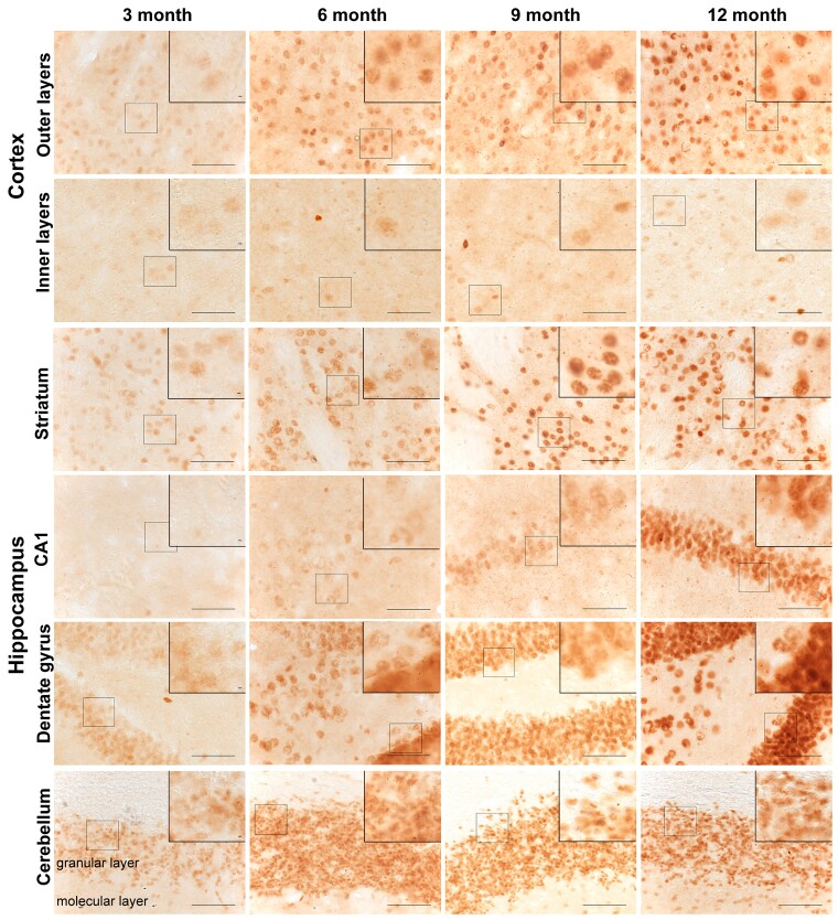
Figure 6.
Spatiotemporal appearance of HTT aggregation in YAC128 mice. Coronal sections from 3-, 6-, 9- and 12-month-old YAC128 mice were immunostained with the S830 antibody to visualize HTT aggregates. A diffuse nuclear S830 immunostain, indicative of HTT aggregation, was readily identified in the striatum, outer cortical layers, dentate gyrus of the hippocampus and granular layer of the cerebellum at 3 months of age, but not in the CA1 region of the hippocampus until 9 months. This nuclear immunostain increased in intensity with disease progression and small nuclear inclusions were observed only rarely (see zoomed sections). By 6 months of age, extranuclear inclusions were readily apparent in the outer and inner layers of the cortex and the striatum and in the CA1 region of the hippocampus by 9 months. The location of the images from within the brain sections is illustrated in Supplementary Figs 6 and 7. The wild-type control sections from mice at 12 months of age are shown in Supplementary Fig. 8. YAC128 (n = 3), wild-type (n = 1). Scale bar = 20 μm.

