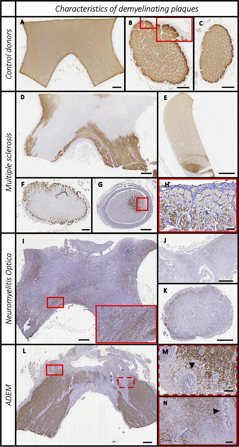
Figure 1.
Features of demyelinating plaques (PLP staining) in the pre-geniculate optic pathway. Non-neurological donors (A–C), multiple sclerosis (D–G), neuromyelitis optica spectrum disorder (H–J) and acute disseminated encephalomyelitis (K–M) cases. Controls display densely packed PLP-positive fibres, and a rim of absent PLP staining in the glia limitans of the subpial zone and in the connective tissue sheaths surrounding axonal bundles (A–C). Multiple sclerosis samples often presented multiple and confluent plaques (D) and a very heterogenous patter of involvement with either central (F) or sub-pial demyelination (G, with rotated magnification image in H). In 13% of samples, remyelinated shadow plaques were also observed (E, faint PLP stained plaque at the top). The neuromyelitis optica spectrum disorder cases showed extensive demyelination, with some sparing of PLP-positive fibres invariably located in the sub-pial area (I–K). Acute disseminated encephalomyelitis (ADEM) cases displayed a typical perivenular demyelination around hyper-cellular perivascular spaces (L, indicated by arrowheads in the two magnifications M and N). Scale bar = 1 mm, except H, M and N where scale bar = 100 µm.

