Abstract
The great potential of zinc oxide nanoparticles (ZnO NPs) for biomedical applications is attributed to their physicochemical properties. In this work, pure and Ag and Ce dual-doped ZnO NPs were synthesized through a facile and green route to examine their cytotoxicity in breast cancer and normal cells. The initial preparation of dual-doped nanoparticles was completed by the usage of taranjabin. The synthesis of Ag and Ce dual-doped ZnO NPs was started with preparing the Ce:Ag ratios of 1:1, 1:2, and 1:4. The cytotoxicity effects of synthesized nanoparticles against breast normal cells (MCF-10A) and breast cancer cells (MDA-MB-231) were examined. The hexagonal structure of synthesized nanoparticles was observed through the results of X-ray diffraction (XRD). Scanning electron microscopy (SEM) images exhibited the spherical shape and smooth surfaces of prepared particles along with the homogeneous distribution of Ag and Ce in ZnO with high-quality lattice fringes without any distortions. According to the cytotoxic results, the effects of Ag/Ce dual-doped ZnO NPs on breast cancer (MDA-MB-231) cells were significantly more than of pure ZnO NPs, while dual-doped and pure nanoparticles remained indifferent towards breast normal (MCF-10A) cells. In addition, we investigated the antimicrobial activity against harmful bacteria.
Graphical Abstract
Keywords: Ag and Ce dual-doped ZnO NPs, Green synthesis, MDA-MB-231 cells, MTT assay
Introduction
As a novel type of widely used mineral particles [1], metal–organic framework [2] and metallic nanoparticles [3–5] such as zinc oxide were noticed and explored by researchers due to their suitable mechanical [6, 7], physical [8, 9] and chemical properties [10–13] that are combined with a higher adsorption power than other zinc-containing compounds [14, 15]. Zinc oxide is one of the compounds of zinc that was recognized as a safe substance by the US Department of Food and Drug Administration [16]. The properties of nanostructures led to their various applications such as anticancer [17, 18], tissue engineering [19–21], antimicrobial [22, 23], degradation [24], photocatalyst [25–29], antioxidant [30], sensor [31–36], sensing [37–39], agriculture [40–42], absorption [43], purification [44, 45], energy [46–49], anti-inflammatory therapy [50], food analysis [51], and drug carriers [52–54]. Among the notable properties of zinc oxide nanoparticles, one can point out their high chemical stability, low dielectric constant, high catalytic activity, absorption of infrared and ultraviolet light, and most importantly their antibacterial properties [55]. Confirming the therapeutic and toxic effects of these compounds can stand as a significant step throughout the advancements of cancer [56–63] and fungal/bacterial infection disease [64–66] treatments [67–69] such as COVID 19 [70, 71]. The primary prevention of infection [72–74] and cancers’ diseases [75–77], new development in research [78–80] and innovation [81–84], such as nanotechnology [85, 86], materials [87–89] and digital technologies [90], have the need to improve our understanding of diseases [91–93] such as cancer [94–97]. In fact, recent developments [98] in all field of science [99–101] and technology [102–104] have impact on human health [105–108] and life [109–112].
Although the main action mechanism of nanoparticles remains unknown[113–115], yet the results of various studies on in in vivo and in vitro environments [116, 117] were indicative of their ability to produce reactive oxygen species (ROS) [118–120], which consequently points out their potent functionality in intracellular calcium concentration, activation of transcription factors, and alterations in cytokines [121–123]. The various approaches of ROS in damaging cells include DNA damage [124–126], interference with cellular signaling pathways, changes in gene transcription, etc. [127–129].
There are different physical [130], chemical [61, 131], and biological methods [132–137] for synthesizing nanostructures, while the exertion of each technique is dependent on the available conditions and purpose of the synthesis [138]. The most common synthesizing methods for the prepare of zinc oxide nanoparticles are observed in the form of sol–gel [139], microemulsion, mechanical–chemical process, direct solvent evaporation, hydrothermal, and spark deposition, which is selected depending on certain factors such as surface chemistry, size distribution, particle morphology, and particle reaction in solution [140]. Next to the advantages of these procedures, there are disadvantages as well since the involved substances are toxic and their usage in medical research is limited [141, 142]. In addition, some of the applied materials remain insoluble and can cause environmental pollution [143–146]. Therefore, in recent years, the application of biological or green methods was noticeably highlighted in order to overcome the disadvantages [147–151]. Green synthesis is defined as the exertion of biological organisms, such as microorganisms, for completing the synthesizing processes that are composed of different bacteria species, actinomycetes [152], algae, fungi, bacteria [153], and biomass [154] or plant extracts [155–159]. Green synthesizing techniques lack the hazardous aspects of physical and chemical methods, and on the other hand, they were confirmed to be environmentally friendly [160] and cost-effective [161] without requiring the usage of high pressure, high energy, high temperature, and toxic chemicals [162–165]. The application of plant extracts for the synthesis of nanoparticles may be a better option than other biological methods since it is suitable for conducting large-scale synthesis while being more cost-effective, as well as capable of accurately preserving the cellular environment [166–168].
Alhagi persarum is a shrub with thin, branched, and prickly stems with an average height of 50 cm. The leaves of this plant are small, oval, pointed, and simple that grows at intervals from the stems. This plant mainly grows in hot and deserted areas (deserts) of Iran, especially in southern regions. A sugary substance is secreted from the stems of this plant that is known as taranjabin in Iran, which turns into white, yellow, or brownish-yellow droplets upon being exposed to air. The chemical composition of taranjabin includes 47.7% of melezitose, 26.44% of sucrose, 11.64% of fructose reducing sugar, 12.4% of gum, and mucilage and 5.1% of ash. Taranjabin is recognized as a laxative that can relieve rheumatic, chest, cough, fever, and biliary pains, which is used in traditional medicine for the treatment of jaundice in infants, as well as children with rubella and infectious fevers.
In order to discover fast and effective treatment pathways or to produce materials with high therapeutic effects for the treatment of cancer, this study attempted to synthesize Ag and Ce dual-doped ZnO NPs by the usage of taranjabin for the very first time and evaluated the cytotoxic activity of synthesized nanoparticles on human breast cancer (MDA-MB-231) and breast normal (MCF-10A) cells lines. In addition, we investigated the antimicrobial activity against harmful bacteria.
Materials and methods
Synthesis of pure and dual-doped ZnO NPs
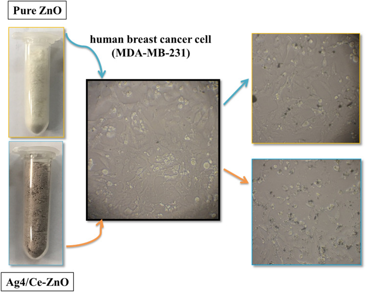
The synthesis of Ag and Ce dual-doped ZnO NPs was started with preparing the Ce:Ag ratios of 1:1, 1:2, and 1:4. Then, 0.3 gr of taranjabin was dissolved in 50 mL of distilled water within four Erlenmeyer flasks to arrange one sample of un-doped and three samples of dual-doped nanoparticles, respectively. In the following, subsequent to the addition of zinc nitrate hexahydrate (0.02 M, Zn(NO3)2.6H2O, Merck) to all the four taranjabin solutions, silver nitrate (AgNO3, Merck) and cerium nitrate hexahydrate (Ce(NO3)3.6H2O, Merck) were appended in accordance with the specified ratios, respectively. Once the solutions were mixed by a heater stirrer at 70 °C for 3 h, they were dried in an oven at 80 °C for 24 h. The resulting raw material was calcined at 600 °C for 2 h. The un-doped and cerium and silver dual-doped ZnO NPs were labeled as ZnO, Ag1/Ce–ZnO, Ag2/Ce–ZnO, and Ag4/Ce–ZnO, respectively (Fig. 1).
Fig. 1.
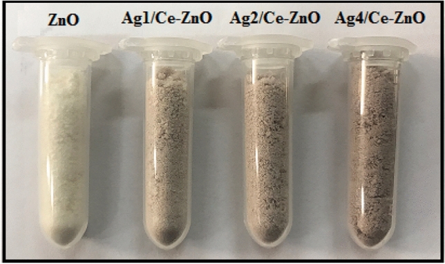
Images of biosynthesized pure ZnO, Ag1/Ce–ZnO, Ag2/Ce–ZnO, and Ag4/Ce–ZnO nanoparticles using taranjabin
Characterization
The size, morphology, and other physical–chemical properties of synthesized nanoparticles were examined through the performance of PXRD (Netherlands, PANalyticalX’Pert PRO MPD system, Cu Kα), UV–Vis (Rayleigh: UV-2100, China), Raman spectra that were captured by a Raman Takram P50C0R10 device at the laser wavelength of 532 nm, FESEM (MIRA3 TESCAN, Czech), and UV–visible spectroscopy (UV–Vis, UV-1800, SHIMADZU) analyses.
Cytotoxic
Cells’ culture
In this study, human breast cancer (MDA-MB-231) and breast normal (MCF-10A) cells were used to evaluate the cytotoxicity of synthesized nanoparticles. MCF-10A and MDA-MB-231 cells were obtained from the Pasteur Institute of Iran and thawed in prior to being cultured. The cells were transferred to Falcon tubes and centrifuged at 833 rpm for 9 min. Once the supernatant was removed, a complete culture medium was added to the cells to have the prepared suspensions poured into flasks. High-glucose DMEM culture medium was exerted for the process of cells culturing and the next step required the addition of 10% fetal bovine serum (FBS), 100 μg/mL of streptomycin, and 100 international units/mL of penicillin to each culture medium to prevent the inducement of microbial growth. In order to proliferate and grow the cells, the culture medium was incubated under 5% CO2 at 37 °C.
MTT assay
Human breast cancer (MDA-MB-231) and breast normal (MCF-10A) cells were cultured in an incubator with a high glucose DMEM that was supplemented with 10% fetal bovine serum and 1% penicillin/streptomycin solution (37 ◦C, 5% CO2) until the cells count of each well of 96-well plate reached 10,000. The culture medium was replaced with 100 μL of the DMEM that contained the formulations at different concentrations (1, 10, 50, 100, and 500 μg/mL) to be seeded for another 24 h. Three duplications were considered for each concentration. In the following, the culture medium was changed after 24 h along with the replacement of fresh high glucose DMEM. Then, 20 μL of 5 mg/mL 3-(4, 5-dimethylthiazol-2-yl)-2, 5-diphenyl tetrazolium bromide (MTT) solution was added to each well and another course of incubation was performed for 4 h. Once 100 µL of DMSO was added to each well of 96-well plate, the resulting mixture was shaken for about 15 min at room temperature to dissolve the formazan. A microplate reader was exerted to measure the optical density (OD) at 570 nm. In addition, the cells viability rate (VR) was calculated according to the following equation:
in which A represents the absorbance of the cells that were treated with formulations and A0 refers to the absorbance of control group.
Antibacterial assay
The antibacterial test was studied on Pseudomonas aeruginosa using macrodilution method. The P. aeruginosa were cultured on these culture media in contact with nanoparticles. The concentrations of 1–250 mg/mL of nanoparticles were prepared in the Mueller Hinton culture medium. Then, the samples were placed in an incubator at 37 °C for 24 h. Finally, bacterial turbidity in the culture media was observed. The turbidity was a sign of the growth of the microbial strain in that concentration of nanoparticles.
IC50
The conduction of probit test was completed through the exertion of SPSS software for two purposes including the calculation of drug and nanoparticles concentrations that could limit the growth of 50% of cells (IC50) and to measure the restriction percentage of cells growth against concentration.
Results and discussion
XRD analysis
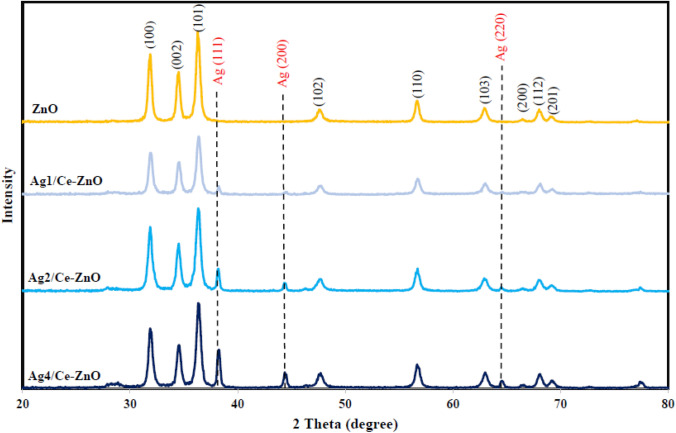
Figure 2 presents the XRD pattern of biosynthesized pure ZnO, Ag1/Ce–ZnO, Ag2/Ce–ZnO, and Ag4/Ce–ZnO nanoparticles. The observed peaks in pure ZnO nanoparticles and Ag and Ce dual-doped ZnO nanoparticles were indexed to (100), (002), (101), (102), (110), (103), (200), (112), and (201), which is comparable with the hexagonal structure of ZnO (JCPDS-36–1451). In conformity to Fig. 2, increasing the ratio of Ag resulted in the appearance of peaks related to the silver-doped nanoparticles throughout the PXRD pattern. The purity and high crystalline form of synthesized nanoparticles was confirmed by the lack of observing any other additional peaks. The crystalline size of synthesized nanoparticles was calculated through the Debye–Scherer formula as given in the following equation:
| 2 |
Fig. 2.
PXRD pattern of biosynthesized pure ZnO, Ag1/Ce–ZnO, Ag2/Ce–ZnO, and Ag4/Ce–ZnO nanoparticles using taranjabin
where D refers to the crystallite size of nanoparticles, K represents the shape factor, λ is the wavelength of applied radiation, β would be full width at half maxima (FWHM) in radians, and θ stands for the diffraction angle. The average crystallite size of synthesized nanoparticles was estimated by considering the full width at half maxima (FWHM) of XRD peak (101) through the usage of Debye–Scherer formula, which was obtained to be 19.14, 19.73, 22.05, and 22.20 nm for ZnO, Ag1/Ce–ZnO, Ag2/Ce–ZnO, and Ag4/Ce–ZnO nanoparticles, respectively. The data in Fig. 2 indicate that the doping of Ce and Ag metals to the crystalline network of ZnO nanoparticles caused an increasing in the crystalline size of synthesized doped nanoparticles due to the difference in ionic radius of zinc atom (1.38 Å) when compared to silver (1.26 Å) and cerium (1.037 Å).
FESEM and EDX analyses
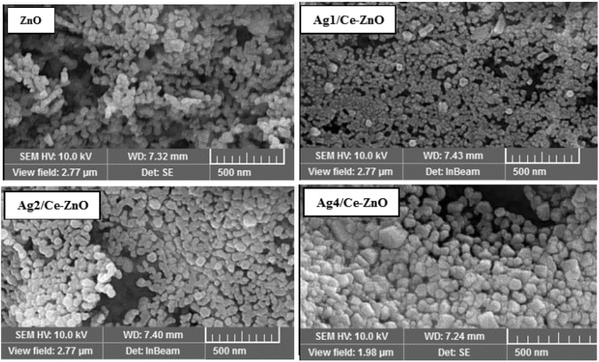
Figure 3 presents the FESEM images of biosynthesized pure ZnO, Ag1/Ce–ZnO, Ag2/Ce–ZnO, and Ag4/Ce–ZnO nanoparticles obtained by the usage of taranjabin, which displays the approximately spherical shape of ZnO particles. The recorded doped nanoparticles throughout the FESEM images were also spherical, while observations indicated the inducement of an increasing in the size of synthesized particles due to the doping of Ag and Ce metals into the structure of ZnO. The mean particle size distribution of synthesized ZnO, Ag1/Ce–ZnO, Ag2/Ce–ZnO, and Ag4/Ce–ZnO nanoparticles, which were estimated to be 31.59, 31.93, 36.89, and 38.44 nm, exhibits the satisfying growth of particles as a result of increasing the percentage of doped metals. In conformity to the provided EDX profiles of biosynthesized ZnO and Ag4/Ce–ZnO nanoparticles in Fig. 4, the synthesized nanoparticles contained a high-purity content with the composition of Zn and O elements for ZnO and Zn, as well as O, Ag, and Ce elements for Ag4/Ce–ZnO nanoparticles. The table form of elemental composition is inserted in Fig. 4.
Fig. 3.
FESEM images and particle size distribution of biosynthesized pure ZnO, Ag1/Ce–ZnO, Ag2/Ce–ZnO, and Ag4/Ce–ZnO nanoparticles using taranjabin
Fig. 4.
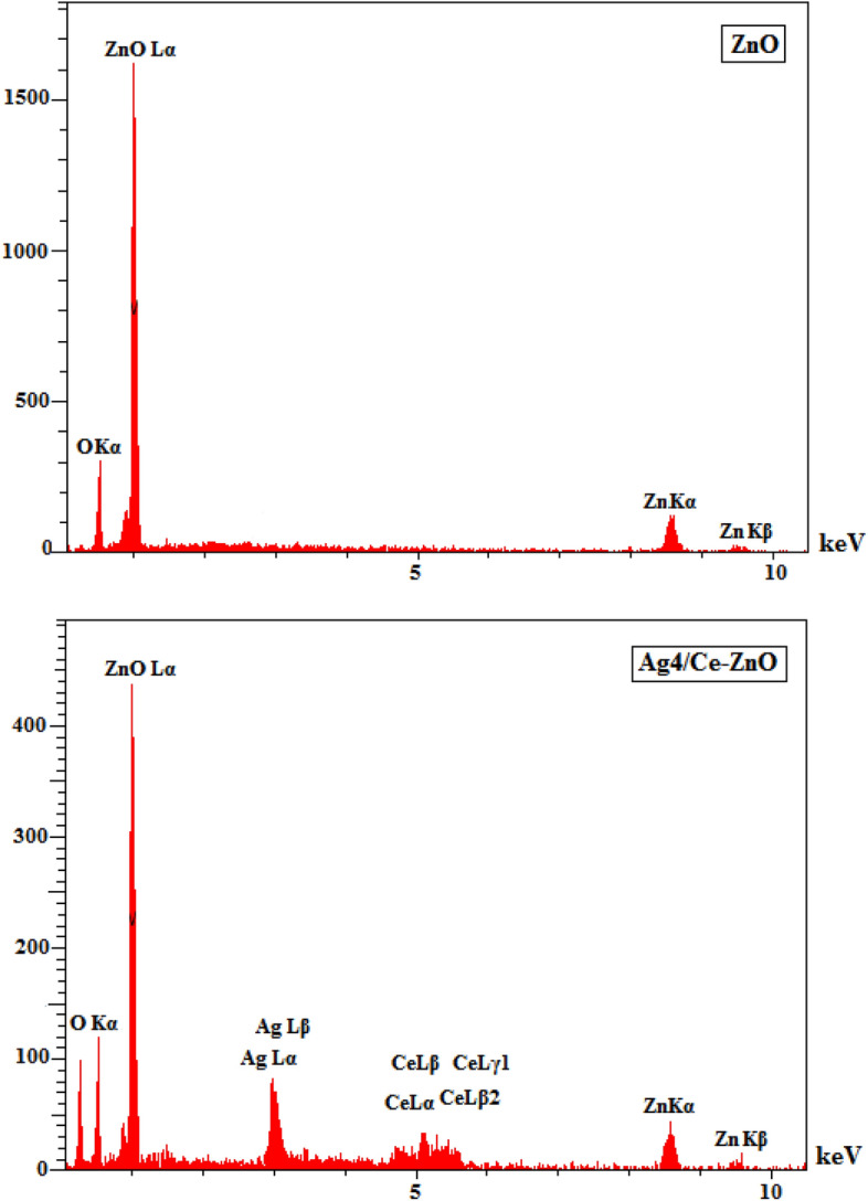
EDX profiles of biosynthesized pure ZnO and Ag4/Ce–ZnO nanoparticles using taranjabin
Raman analysis
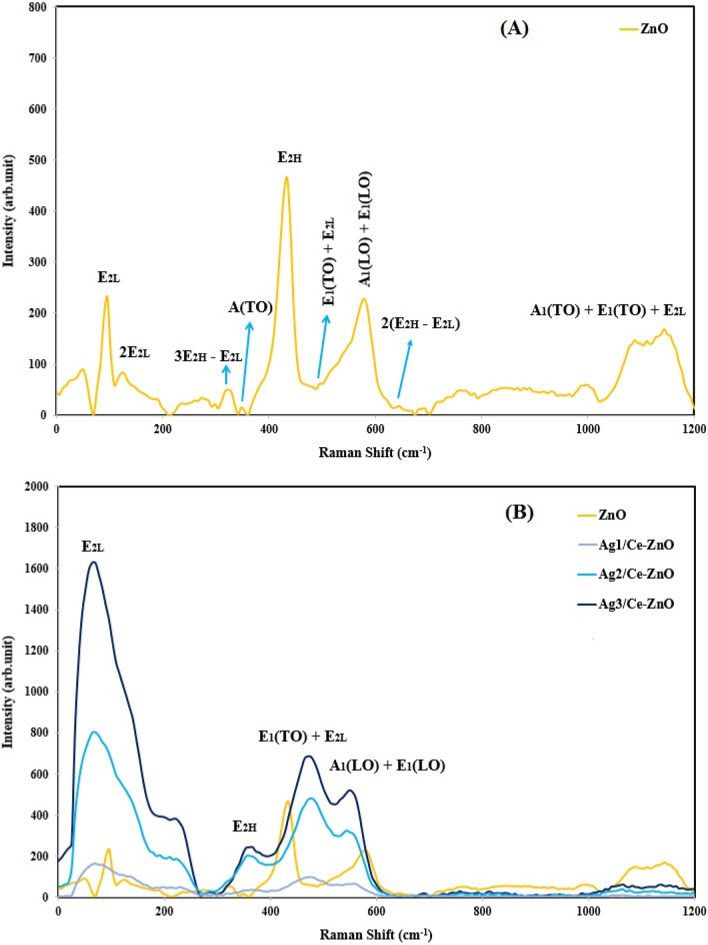
Raman spectroscopy is a non-destructive chemical analysis technique for providing detailed information about chemical structure, phase and polymorphy, crystallinity, and molecular interactions, which is based upon the interaction of light with chemical bonds within a material. According to group theory, ZnO nanoparticles contain a hexagonal wurtzite structure with a space group of P63mc. The optical modes of A1 + 2B2 + E1 + 2E2 imply the wurtzite structure of ZnO, which includes A1 + E1 + 2E2 as the active Raman mode, A1 + E1 as the active infrared mode, and 2B1 as the silent Raman mode. The A1 and E1 modes are two polar branches that are divided into longitudinal optical (LO) and transverse optical (TO). The A1, E1, and E2 modes are recognized as the first-order Raman active and based on Raman law, B1 modes are usually inactive throughout the Raman spectrum and are known as the silent modes. The Raman spectra of biosynthesized pure ZnO, Ag1/Ce–ZnO, Ag2/Ce–ZnO, and Ag4/Ce–ZnO nanoparticles are represented in Fig. 5.
Fig. 5.
Raman spectra of biosynthesized (A) pure ZnO nanoparticles, and (B) ZnO, Ag1/Ce–ZnO, Ag2/Ce–ZnO, and Ag4/Ce–ZnO nanoparticles using taranjabin
The main phonon states of ZnO nanoparticles with a hexagonal structure appeared in the regions of 583, 441, 345, 91 cm−1, which were in correspondence to the A1(LO)–E1(LO), E2H, A1(TO), and E2H modes, respectively. The 2E2L mode was in correlation to the second-order phonon mode that appeared in the region of 132 cm−1. Moreover, the modes of 3E2H-E2L, E1(TO) + E2L, 2(E2H-E2L), and A1(TO) + E1(TO) + E2L were related to the poly-phonon scattering that was detected in the points of 324, 475, 658, and 1105 cm−1, respectively.
As it is displayed in Fig. 5, the doping factor (both Ag and Ce) of ZnO matrix caused significant changes in the polar and non-polar states. The E2H state involves the oxygen motion, while being sensitive to internal stress, and containing the characteristics of hexagonal structure of zinc oxide nanoparticles. Due to the decomposition of impurities and defects, the E2H mode faced a sharp decrease in the peak intensities of doped samples. In addition, this mode was observed to be steadily decreased and expanded as the doping concentrations of silver and cerium were increased. The detected polarity of A1(LO)–E1(LO) at around 583 cm−1 was related to the doping of silver and cerium that can expand a peak and also force its shifting towards lower energies. All the variations and extensions of phonon modes were obtained by scattering contributions outside the center of Brillouin area. The phonon state of A1 (LO)–E1 (LO) is usually attributed to the interfacial defect of zinc and oxygen vacancy throughout the network of ZnO. Due to the combination of Ag and Ce ions with ZnO nanoparticles, the intensity of ZnO Raman peaks can be greatly increased through the doping of silver and cerium. In addition, further results confirmed the crystallization of ZnO nanoparticles with few defects due to the presence of Ag and Ce ions.
UV–Vis analysis
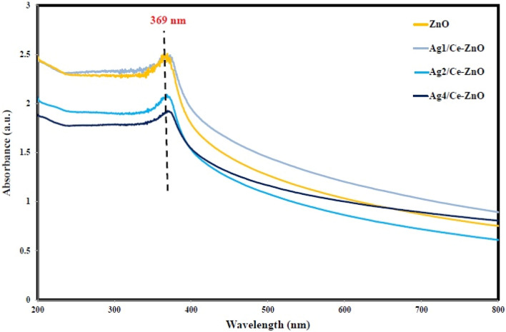
Electron spectroscopy is a technique for investigating the energy distribution of ejected electrons from a material as a result of being irradiated by a source of ionizing irradiation. Figure 6 presents the electronic spectra of biosynthesized pure ZnO, Ag1/Ce–ZnO, Ag2/Ce–ZnO, and Ag4/Ce–ZnO nanoparticles obtained by the usage of taranjabin. The maximum wavelength of pure ZnO, Ag1/Ce–ZnO, Ag2/Ce–ZnO, and Ag4/Ce–ZnO nanoparticles were observed at the regions of 396, 372, 383, and 385 nm, respectively.
Fig. 6.
Electronic graph of biosynthesized pure ZnO, Ag1/Ce–ZnO, Ag2/Ce–ZnO, and Ag4/Ce–ZnO nanoparticles using taranjabin
An increase in the concentration of Ce and Ag throughout the structure of ZnO causes a shifting in the absorption spectra towards higher wavelengths (red shift) due to the induced alteration in the amount of optical bandgap. This red shift represents the increasing crystallization and the effects of quantum confinement. An enlargement in the electron population during the doping of Ce and Ag into ZnO can lead to quantum constraints and finally cause a red shift in optical absorption behavior.
Cytotoxic performance
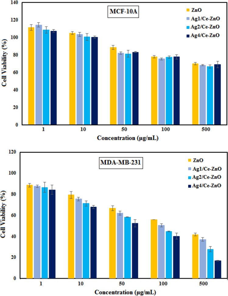
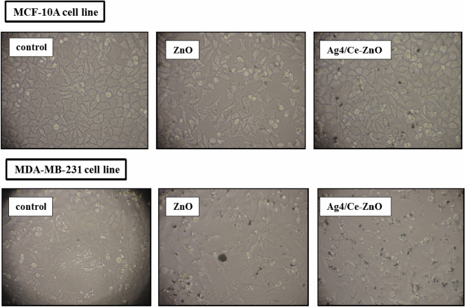
In this study, we examined the cytotoxicity effects of biosynthesized ZnO, Ag1/Ce–ZnO, Ag2/Ce–ZnO, and Ag4/Ce–ZnO nanoparticles obtained by the usage of taranjabin against breast normal cells (MCF-10A) and breast cancer cells (MDA-MB-231). For this purpose, the cells were exposed for 24 h at different concentrations (1–500 μg/mL) of un-doped and dual-doped ZnO nanoparticles through the means of MTT assay (Fig. 7). In conformity to Fig. 6, the pure and Ag and Ce dual-doped ZnO nanoparticles did not cause any significant toxicity effects on the normal cell line (MCF-10A), while the doped nanoparticles resulted in almost similar toxicity impacts to that of un-doped nanoparticles. Furthermore, increasing the concentrations of doped and un-doped nanoparticles did not cause any significant toxicity effects. The assessment results of cytotoxic activity of synthesized nanoparticles on breast cancer cell line (MDA-MB-231) are presented in Fig. 7. According to observations, increasing the applied concentration intensified the effects of cytotoxicity, which reached a significant point at the concentration of 500 µg/mL. The cytotoxic effect of doped nanoparticles was more that of un-doped nanoparticles. As, 80% of the cells were killed from being treated with Ag4/Ce–ZnO nanoparticles at the concentration of 500 µg/mL. In addition, IC50 data strongly confirmed the obtained results (Table 1), which less IC50 was attributed to Ag4/Ce–ZnO nanoparticles. Hence, Ag4/Ce–ZnO nanoparticles show the greatest effect of toxicity. Figure 8 depicts the effect of synthesized nanoparticles being treated with breast normal cell and breast cancer cell lines. This figure clearly displays the difference in the cytotoxic activity of synthesized pure and dual-doped ZnO nanoparticles against these two cell lines. These results suggested that the synthesized nanoparticles induced cytotoxicity in cancer cells without affecting the normal cells.
Fig. 7.
The cytotoxic activity of biosynthesized pure ZnO, Ag1/Ce–ZnO, Ag2/Ce–ZnO, and Ag4/Ce–ZnO nanoparticles using taranjabin
Table 1.
IC50 values of biosynthesized pure ZnO, Ag1/Ce–ZnO, Ag2/Ce–ZnO, and Ag4/Ce–ZnO nanoparticles using taranjabin
| Cell lines | IC50 values (µg/mL) | |||
|---|---|---|---|---|
| ZnO | Ag1/Ce–ZnO | Ag2/Ce–ZnO | Ag4/Ce–ZnO | |
| MCF-10A | 604.2647 | 799.8132 | 778.4796 | 878.4803 |
| MDA-MB-231 | 447.3 | 418.113 | 325.833 | 220.461 |
Fig. 8.
Images of breast normal cells (MCF-10A) and breast cancer cells (MDA-MB-231) lines treated with pure ZnO and Ag4/Ce–ZnO nanoparticles after 24 h treatment
The cytotoxic effects of green synthesized doped ZnO NPs on cancer cells were mentioned in the work of many authors. For example, MJ Akhtar et al. studied the oxidative stress-mediated cytotoxicity of Al-doped ZnO nanoparticles against MCF-7 cells. According to their report, Al-doping was able to enhance the cytotoxicity and oxidative stress responses of ZnO nanoparticles against MCF-7 cells. In addition, they obtained an IC50 of 44 μg/mL for un-doped ZnO nanoparticles and 31 μg/ml for the Al-doped ZnO counterparts. It was suggested by their results that Al-doped ZnO nanoparticles can induce apoptosis in MCF-7 cells through the mitochondrial pathway [169]. In another work, G. Vijayakumar et al. investigated the cells viability, ROS generation, and nanoparticle cells penetration rate of PEG encapsulated bare and Mn-doped ZnO nanoparticles against human liver carcinoma Huh7 cell lines. Based on their findings, un-doped the Mn-doped ZnO nanoparticles exhibited a higher cells annihilation effect, which may be due to the combined effects of Zn2+ ion release and intracellular ROS generation; therefore, the inducement of apoptosis can be expected due to oxidative stress and ROS generation [170]. Considering these facts, doped metal oxide nanoparticles can stand as an attractive research topic for biomedical applications. Nano-sized materials have enabled many developments in biomedicine and other biological applications such as drug delivery, anticancer activity, gene delivery, fluorescent biological labels, protein detection, MRI contrast enhancement, probing of DNA, tissue engineering, phagokinetic studies, hyperthermia, and filtration of biological based molecular cell.
The antibacterial test of doped and non-doped nanoparticles was on P. aeruginosa and E.coli. The IC50 was at 50 μg/mL.
Conclusion
Un-doped and Ag and Ce dual-doped ZnO NPs were synthesized through a facile green method by exerting the extract of taranjabin. The obtained PXRD spectra displayed the hexagonal phase of un-doped and dual-doped ZnO NPs. SEM mapping demonstrated the homogeneous distribution of Ag and Ce in ZnO with high-quality lattice fringes while lacking any distortions. According to cytotoxicity results, the un-doped ZnO NPs displayed a similar toxicity effect on breast cancer cells (MDA-MB-231) to that of dual-doped ZnO NPs. Considering the comparable toxicity effect of doped nanoparticles with the un-doped nanoparticles, it can be stated that the simultaneous doping of cerium and silver did cause significant alterations in the cytotoxic properties of zinc oxide nanoparticles. However, this discovery requires further investigation since it may affect certain physical and biological properties such as luminescence, UV absorption, or antibacterial features of zinc oxide nanoparticles. Therefore, this attempt can stand as a useful approach due to the cosmetic and even industrial applications of zinc oxide nanoparticles.
Acknowledgements
The authors extend their appreciation to the Deputyship for Research & Innovation, Ministry of Education in Saudi Arabia for funding this research work through the project number (IF2/PSAU/2022/01/22623).
Declarations
Conflict of interest
The authors confirm that the content of this article involves no competing interests.
Footnotes
Publisher's Note
Springer Nature remains neutral with regard to jurisdictional claims in published maps and institutional affiliations.
References
- 1.He X, et al. MgFe-LDH nanoparticles: a promising leukemia inhibitory factor replacement for self-renewal and pluripotency maintenance in cultured mouse embryonic stem cells. Advanced Science. 2021;8(9):2003535. doi: 10.1002/advs.202003535. [DOI] [PMC free article] [PubMed] [Google Scholar]
- 2.Zeng Q, et al. Hyperpolarized Xe NMR signal advancement by metal-organic framework entrapment in aqueous solution. Proc Natl Acad Sci. 2020;117(30):17558–17563. doi: 10.1073/pnas.2004121117. [DOI] [PMC free article] [PubMed] [Google Scholar]
- 3.Jasim SA, et al. Investigation of crotonaldehyde adsorption on pure and Pd-decorated GaN nanotubes: A density functional theory study. Solid State Commun. 2022;348:114741. doi: 10.1016/j.ssc.2022.114741. [DOI] [Google Scholar]
- 4.Xia J, Majdi A, Toghraie D. Molecular dynamics simulation of friction process in atomic structures with spherical nanoparticles. Solid State Commun. 2022;346:114717. doi: 10.1016/j.ssc.2022.114717. [DOI] [Google Scholar]
- 5.Jia D, et al. Experimental verification of nanoparticle jet minimum quantity lubrication effectiveness in grinding. J Nanopart Res. 2014;16(12):2758. doi: 10.1007/s11051-014-2758-7. [DOI] [Google Scholar]
- 6.Yang Y, et al. Machinability of ultrasonic vibration assisted micro-grinding in biological bone using nanolubricant. Front Mech Eng. 2022 doi: 10.1007/s11465-022-0717-z. [DOI] [Google Scholar]
- 7.Duan Z et al. (2022) Mechanical behavior and Semiempirical force model of aerospace aluminum alloy milling using nano biological lubricant. Front Mech Eng
- 8.Gao T, et al. Grindability of carbon fiber reinforced polymer using CNT biological lubricant. Sci Rep. 2021;11(1):22535. doi: 10.1038/s41598-021-02071-y. [DOI] [PMC free article] [PubMed] [Google Scholar]
- 9.Duan Z, et al. Milling surface roughness for 7050 aluminum alloy cavity influenced by nozzle position of nanofluid minimum quantity lubrication. Chin J Aeronaut. 2021;34(6):33–53. doi: 10.1016/j.cja.2020.04.029. [DOI] [Google Scholar]
- 10.Jeevanandam J, et al. Review on nanoparticles and nanostructured materials: history, sources, toxicity and regulations. Beilstein J Nanotechnol. 2018;9(1):1050–1074. doi: 10.3762/bjnano.9.98. [DOI] [PMC free article] [PubMed] [Google Scholar]
- 11.Raya I, et al. Role of compositional changes on thermal, magnetic, and mechanical properties of Fe-PC-based amorphous alloys. Chin Phys B. 2022;31(1):016401. doi: 10.1088/1674-1056/ac3655. [DOI] [Google Scholar]
- 12.Chupradit S et al Use of organic and copper-based nanoparticles on the turbulator installment in a shell tube heat exchanger: a CFD-based simulation approach by using nanofluids. J Nanomater 2021. 2021.
- 13.Hu X, et al. The microchannel type effects on water-Fe3O4 nanofluid atomic behavior: Molecular dynamics approach. J Taiwan Inst Chem Eng. 2022;135:104396. doi: 10.1016/j.jtice.2022.104396. [DOI] [Google Scholar]
- 14.Khatami M, et al. Rectangular shaped zinc oxide nanoparticles: Green synthesis by Stevia and its biomedical efficiency. Ceram Int. 2018;44(13):15596–15602. doi: 10.1016/j.ceramint.2018.05.224. [DOI] [Google Scholar]
- 15.Miri A, et al. Zinc oxide nanoparticles: Biosynthesis, characterization, antifungal and cytotoxic activity. Mater Sci Eng C. 2019;104:109981. doi: 10.1016/j.msec.2019.109981. [DOI] [PubMed] [Google Scholar]
- 16.Indhira D, et al. Biomimetic facile synthesis of zinc oxide and copper oxide nanoparticles from Elaeagnus indica for enhanced photocatalytic activity. Environ Res. 2022;212:113323. doi: 10.1016/j.envres.2022.113323. [DOI] [PubMed] [Google Scholar]
- 17.Cao Y, et al. Green synthesis of bimetallic ZnO–CuO nanoparticles and their cytotoxicity properties. Sci Rep. 2021;11(1):1–8. doi: 10.1038/s41598-021-02937-1. [DOI] [PMC free article] [PubMed] [Google Scholar]
- 18.Vidhya MS, et al. Anti-cancer applications of Zr Co, Ni-doped ZnO thin nanoplates. Mater Lett. 2021;283:128760. doi: 10.1016/j.matlet.2020.128760. [DOI] [Google Scholar]
- 19.Xue X et al. Rational Design of Multifunctional CuS Nanoparticle-PEG Composite Soft Hydrogel-Coated 3D Hard Polycaprolactone Scaffolds for Efficient Bone Regeneration. Advanced Functional Materials. n/a(n/a): p. 2202470.
- 20.Liu J, et al. Electrospun strong, bioactive, and bioabsorbable silk fibroin/poly (L-lactic-acid) nanoyarns for constructing advanced nanotextile tissue scaffolds. Materials Today Bio. 2022;14:100243. doi: 10.1016/j.mtbio.2022.100243. [DOI] [PMC free article] [PubMed] [Google Scholar]
- 21.Arkaban H, et al. Polyacrylic Acid Nanoplatforms: Antimicrobial, Tissue Engineering, and Cancer Theranostic Applications. Polymers. 2022;14(6):1259. doi: 10.3390/polym14061259. [DOI] [PMC free article] [PubMed] [Google Scholar]
- 22.Hachem K, et al. Adsorption of Pb (II) and Cd (II) by magnetic chitosan-salicylaldehyde Schiff base: Synthesis, characterization, thermal study and antibacterial activity. J Chin Chem Soc. 2022;69(3):512–521. doi: 10.1002/jccs.202100507. [DOI] [Google Scholar]
- 23.Obaid RF, et al. Antibacterial activity, anti-adherence and anti-biofilm activities of plants extracts against Aggregatibacter actinomycetemcomitans: An in vitro study in Hilla City Iraq. Caspian J Environment Sci. 2022;20(2):367–372. [Google Scholar]
- 24.Turki Jalil A, et al. CuO/ZrO2 nanocomposites: facile synthesis, characterization and photocatalytic degradation of tetracycline antibiotic. J Nanostruct. 2021;11(2):333–346. [Google Scholar]
- 25.Ameen F, Dawoud T, AlNadhari S. Ecofriendly and low-cost synthesis of ZnO nanoparticles from Acremonium potronii for the photocatalytic degradation of azo dyes. Environ Res. 2021;202:111700. doi: 10.1016/j.envres.2021.111700. [DOI] [PubMed] [Google Scholar]
- 26.Selvam K et al. (2022) Laccase production from Bacillus aestuarii KSK using Borassus flabellifer empty fruit bunch waste as a substrate and assessing their malachite green dye degradation. J Appl Microbiol. [DOI] [PubMed]
- 27.Aljumaily MM, et al. Modification of Poly (vinylidene fluoride-co-hexafluoropropylene) Membranes with DES-Functionalized Carbon Nanospheres for Removal of Methyl Orange by Membrane Distillation. Water. 2022;14(9):1396. doi: 10.3390/w14091396. [DOI] [Google Scholar]
- 28.Jasim SA, et al. Nanomagnetic Salamo-based-Pd (0) Complex: an efficient heterogeneous catalyst for Suzuki-Miyaura and Heck cross-coupling reactions in aqueous medium. J Mol Struct. 2022;1261:132930. doi: 10.1016/j.molstruc.2022.132930. [DOI] [Google Scholar]
- 29.Al-Nayili A, et al. Formic acid dehydrogenation using noble-metal nanoheterogeneous catalysts: towards sustainable hydrogen-based energy. Catalysts. 2022;12(3):324. doi: 10.3390/catal12030324. [DOI] [Google Scholar]
- 30.Gangalla R, et al. Optimization and characterization of exopolysaccharide produced by Bacillus aerophilus rk1 and its in vitro antioxidant activities. J King Saud Univ Sci. 2021;33(5):101470. doi: 10.1016/j.jksus.2021.101470. [DOI] [Google Scholar]
- 31.Yumashev AV, et al. (2022) Optical-based biosensor for detection of oncomarker CA 125, recent progress and current status. Anal Biochem 114750. [DOI] [PubMed]
- 32.Chupradit S, et al. (2022) Various types of electrochemical biosensors for leukemia detection and therapeutic approaches. Anal Biochem, 114736. [DOI] [PubMed]
- 33.Jalil AT, et al. High-sensitivity biosensor based on glass resonance PhC cavities for detection of blood component and glucose concentration in human urine. Coatings. 2021;11(12):1555. doi: 10.3390/coatings11121555. [DOI] [Google Scholar]
- 34.Chupradit S, et al. Ultra-sensitive biosensor with simultaneous detection (of cancer and diabetes) and analysis of deformation effects on dielectric rods in optical microstructure. Coatings. 2021;11(12):1564. doi: 10.3390/coatings11121564. [DOI] [Google Scholar]
- 35.Kartika R, et al. Ca12O12 nanocluster as highly sensitive material for the detection of hazardous mustard gas: Density-functional theory. Inorg Chem Commun. 2022;137:109174. doi: 10.1016/j.inoche.2021.109174. [DOI] [Google Scholar]
- 36.Olegovich Bokov D, et al. Ir-decorated gallium nitride nanotubes as a chemical sensor for recognition of mesalamine drug: a DFT study. Mol Simul. 2022;48(5):438–447. doi: 10.1080/08927022.2021.2025234. [DOI] [Google Scholar]
- 37.Khaki N, et al. Sensing of acetaminophen drug using Zn-doped boron nitride nanocones: a DFT inspection. Appl Biochem Biotechnol. 2022;194(6):2481–2491. doi: 10.1007/s12010-022-03830-x. [DOI] [PubMed] [Google Scholar]
- 38.Sun S, et al. Selection and identification of a novel ssDNA aptamer targeting human skeletal muscle. Bioactive Mater. 2023;20:166–178. doi: 10.1016/j.bioactmat.2022.05.016. [DOI] [PMC free article] [PubMed] [Google Scholar]
- 39.Hou Q. et al. (2022) Role of Nutrient-sensing Receptor GPRC6A in Regulating Colonic Group 3 Innate Lymphoid Cells and Inflamed Mucosal Healing. J Crohn's and Colitis [DOI] [PubMed]
- 40.Gao T, et al. Dispersing mechanism and tribological performance of vegetable oil-based CNT nanofluids with different surfactants. Tribol Int. 2019;131:51–63. doi: 10.1016/j.triboint.2018.10.025. [DOI] [Google Scholar]
- 41.Lei J, et al. Intestinal microbiota regulate certain meat quality parameters in chicken. Front Nutr. 2022;9:747705. doi: 10.3389/fnut.2022.747705. [DOI] [PMC free article] [PubMed] [Google Scholar]
- 42.Nazaripour E, et al. Ferromagnetic nickel (II) oxide (NiO) nanoparticles: biosynthesis, characterization and their antibacterial activities. Rendiconti Lincei Scienze Fisiche e Naturali. 2022;33(1):127–134. doi: 10.1007/s12210-021-01042-9. [DOI] [Google Scholar]
- 43.Sadeghi M, et al. Dichlorosilane adsorption on the Al, Ga, and Zn-doped fullerenes. Monatshefte für Chemie-Chemical Monthly. 2022;153:427–434. doi: 10.1007/s00706-022-02926-8. [DOI] [Google Scholar]
- 44.ANZUM R. et al. (2022) A review on separation and detection of copper, cadmium, and chromium in food based on cloud point extraction technology. Food Sci Technol 2022. 42.
- 45.Wu X, et al. Circulating purification of cutting fluid: an overview. Int J Adv Manufact Technol. 2021;117(9):2565–2600. doi: 10.1007/s00170-021-07854-1. [DOI] [PMC free article] [PubMed] [Google Scholar]
- 46.Sivaraman R. et al (2022) Evaluating the potential of graphene-like boron nitride as a promising cathode for Mg-ion batteries. J Electroanal Chem 116413.
- 47.Li B, et al. Heat transfer performance of MQL grinding with different nanofluids for Ni-based alloys using vegetable oil. J Clean Prod. 2017;154:1–11. doi: 10.1016/j.jclepro.2017.03.213. [DOI] [Google Scholar]
- 48.Li B, et al. Grinding temperature and energy ratio coefficient in MQL grinding of high-temperature nickel-base alloy by using different vegetable oils as base oil. Chin J Aeronaut. 2016;29(4):1084–1095. doi: 10.1016/j.cja.2015.10.012. [DOI] [Google Scholar]
- 49.Anqi AE, et al. Effect of combined air cooling and nano enhanced phase change materials on thermal management of lithium-ion batteries. J Energy Storage. 2022;52:104906. doi: 10.1016/j.est.2022.104906. [DOI] [Google Scholar]
- 50.Xue X, et al. Neutrophil-erythrocyte hybrid membrane-coated hollow copper sulfide nanoparticles for targeted and photothermal/ anti-inflammatory therapy of osteoarthritis. Compos B Eng. 2022;237:109855. doi: 10.1016/j.compositesb.2022.109855. [DOI] [Google Scholar]
- 51.Ngafwan N, et al. Study on novel fluorescent carbon nanomaterials in food analysis. Food Sci Technol. 2021;42(37821):1–6. [Google Scholar]
- 52.Jasim SA, et al. MXene/metal and polymer nanocomposites: preparation, properties, and applications. J Alloy Compound. 2022;61:80–115. [Google Scholar]
- 53.Saleh RO, et al. Application of aluminum nitride nanotubes as a promising nanocarriers for anticancer drug 5-aminosalicylic acid in drug delivery system. J Mol Liq. 2022;352:118676. doi: 10.1016/j.molliq.2022.118676. [DOI] [Google Scholar]
- 54.Zhuo Z, et al. A Loop-based and AGO-incorporated virtual screening model targeting AGO-Mediated miRNA–mRNA interactions for drug discovery to rescue bone phenotype in genetically modified mice. Adv Sci. 2020;7(13):1903451. doi: 10.1002/advs.201903451. [DOI] [PMC free article] [PubMed] [Google Scholar]
- 55.Saravanan M, et al. Green synthesis of anisotropic zinc oxide nanoparticles with antibacterial and cytofriendly properties. Microb Pathog. 2018;115:57–63. doi: 10.1016/j.micpath.2017.12.039. [DOI] [PubMed] [Google Scholar]
- 56.Swathi S, et al. Cancer targeting potential of bioinspired chain like magnetite (Fe3O4) nanostructures. Curr Appl Phys. 2020;20(8):982–987. doi: 10.1016/j.cap.2020.06.013. [DOI] [Google Scholar]
- 57.Isacfranklin M, et al. Single-phase Cr2O3 nanoparticles for biomedical applications. Ceram Int. 2020;46(12):19890–19895. doi: 10.1016/j.ceramint.2020.05.050. [DOI] [Google Scholar]
- 58.Isacfranklin M, et al. Y2O3 nanorods for cytotoxicity evaluation. Ceram Int. 2020;46(12):20553–20557. doi: 10.1016/j.ceramint.2020.05.172. [DOI] [Google Scholar]
- 59.Sonbol H, et al. Padina boryana mediated green synthesis of crystalline palladium nanoparticles as potential nanodrug against multidrug resistant bacteria and cancer cells. Sci Rep. 2021;11(1):1–19. doi: 10.1038/s41598-021-84794-6. [DOI] [PMC free article] [PubMed] [Google Scholar]
- 60.Ameen F, et al. Anti-oxidant, anti-fungal and cytotoxic effects of silver nanoparticles synthesized using marine fungus Cladosporium halotolerans. Appl Nanosci. 2021;12:1–9. [Google Scholar]
- 61.Iram S, et al. Gold nanoconjugates reinforce the potency of conjugated cisplatin and doxorubicin. Colloids Surf, B. 2017;160:254–264. doi: 10.1016/j.colsurfb.2017.09.017. [DOI] [PubMed] [Google Scholar]
- 62.Kumar V, et al. Evaluation of cytotoxicity and genotoxicity effects of refractory pollutants of untreated and biomethanated distillery effluent using Allium cepa. Environ Pollut. 2022;300:118975. doi: 10.1016/j.envpol.2022.118975. [DOI] [PubMed] [Google Scholar]
- 63.Jalil AT, et al. Cancer stages and demographical study of HPV16 in gene L2 isolated from cervical cancer in Dhi-Qar province, Iraq. Appl Nanosci. 2021;12:1–7. [Google Scholar]
- 64.Mostafa AA-F, et al. In vitro evaluation of antifungal activity of some agricultural fungicides against two saprolegnoid fungi infecting cultured fish. J King Saud Univer Sci. 2020;32(7):3091–3096. doi: 10.1016/j.jksus.2020.08.019. [DOI] [Google Scholar]
- 65.Widjaja G, et al. Humoral immune mechanisms involved in protective and pathological immunity during COVID-19. Hum Immunol. 2021;82(10):733–745. doi: 10.1016/j.humimm.2021.06.011. [DOI] [PMC free article] [PubMed] [Google Scholar]
- 66.Jalil AT, et al (2021) Polymerase chain reaction technique for molecular detection of HPV16 infections among women with cervical cancer in Dhi-Qar Province. Materials Today: Proceedings.
- 67.Sarika K, et al. Antimicrobial and antifungal activity of soil actinomycetes isolated from coal mine sites. Saudi J Biol Sci. 2021;28(6):3553–3558. doi: 10.1016/j.sjbs.2021.03.029. [DOI] [PMC free article] [PubMed] [Google Scholar]
- 68.Saleh MM, et al. Evaluation of immunoglobulins, CD4/CD8 T lymphocyte ratio and interleukin-6 in COVID-19 patients. Turkish J Immunol. 2020;8(3):129–134. doi: 10.25002/tji.2020.1347. [DOI] [Google Scholar]
- 69.Akter F, et al. Cocos nucifera endocarp extract exhibits anti-diabetic and antilipidemic activities in diabetic rat model. Int J Sci Res Dental Med Sci. 2022;4(1):8–15. [Google Scholar]
- 70.Kausikan SP, et al. The impact of COVID-19 on auditory and visual choice reaction time of non-hospitalized patients: an observational study. Int J Sci Res Dental Med Sci. 2022;4(1):21–25. [Google Scholar]
- 71.Li H, et al. Damaged lung gas exchange function of discharged COVID-19 patients detected by hyperpolarized 129Xe MRI. Sci Adv. 2021;7(1):eabc8180. doi: 10.1126/sciadv.abc8180. [DOI] [PMC free article] [PubMed] [Google Scholar]
- 72.Jalil AT, et al. Viral hepatitis in Dhi-Qar Province: demographics and hematological characteristics of patients. Int J Pharmaceutical Res. 2020;12(1):2081–2087. [Google Scholar]
- 73.Hanan ZK, et al. Detection of human genetic variation in VAC14 gene by ARMA-PCR technique and relation with typhoid fever infection in patients with gallbladder diseases in Thi-Qar province/Iraq. Mater Today Proc. 2021 doi: 10.1016/j.matpr.2021.05.236. [DOI] [Google Scholar]
- 74.Sotelo Núñez N, et al. Evaluation the effect of micro-osteoperforation on the tooth movement rate and the level of pain on miniscrew-supported maxillary molar distalization: a systematic review and meta-analysis. Int J Sci Res Dental Med Sci. 2020;2(3):81–86. [Google Scholar]
- 75.Marofi F, et al. CAR-NK cell in cancer immunotherapy A promising frontier. Cancer Sci. 2021;112(9):3427–3436. doi: 10.1111/cas.14993. [DOI] [PMC free article] [PubMed] [Google Scholar]
- 76.Gowhari Shabgah A, et al. Does CCL19 act as a double-edged sword in cancer development? Clin Exp Immunol. 2021;207(2):164–175. doi: 10.1093/cei/uxab039. [DOI] [PMC free article] [PubMed] [Google Scholar]
- 77.Yan J, et al. Chiral protein supraparticles for tumor suppression and synergistic immunotherapy: an enabling strategy for bioactive supramolecular chirality construction. Nano Lett. 2020;20(8):5844–5852. doi: 10.1021/acs.nanolett.0c01757. [DOI] [PubMed] [Google Scholar]
- 78.Moghadasi S, et al. A paradigm shift in cell-free approach: the emerging role of MSCs-derived exosomes in regenerative medicine. J Transl Med. 2021;19(1):1–21. doi: 10.1186/s12967-021-02980-6. [DOI] [PMC free article] [PubMed] [Google Scholar]
- 79.Jalil AT, et al. HEMATOLOGICAL AND SEROLOGICAL PARAMETERS FOR DETECTION OF COVID-19. J Microbiol Biotechnol Food Sci. 2022;11(4):e4229. doi: 10.55251/jmbfs.4229. [DOI] [Google Scholar]
- 80.Widjaja G, et al. Effect of tomato consumption on inflammatory markers in health and disease status: A systematic review and meta-analysis of clinical trials. Clin Nutr ESPEN. 2022;50:93–100. doi: 10.1016/j.clnesp.2022.04.019. [DOI] [PubMed] [Google Scholar]
- 81.Marofi F, et al. Novel CAR T therapy is a ray of hope in the treatment of seriously ill AML patients. Stem Cell Res Ther. 2021;12(1):465. doi: 10.1186/s13287-021-02420-8. [DOI] [PMC free article] [PubMed] [Google Scholar]
- 82.Vakili-Samiani S, et al. Targeting Wee1 kinase as a therapeutic approach in Hematological Malignancies. DNA Repair. 2021;107:103203. doi: 10.1016/j.dnarep.2021.103203. [DOI] [PubMed] [Google Scholar]
- 83.Xu Y, et al. Prediction of COVID-19 manipulation by selective ACE inhibitory compounds of Potentilla reptant root: In silico study and ADMET profile. Arab J Chem. 2022;15(7):103942. doi: 10.1016/j.arabjc.2022.103942. [DOI] [PMC free article] [PubMed] [Google Scholar]
- 84.Zou M, et al. Gut microbiota on admission as predictive biomarker for acute necrotizing pancreatitis. Front Immunol. 2022;13:988326. doi: 10.3389/fimmu.2022.988326. [DOI] [PMC free article] [PubMed] [Google Scholar]
- 85.Zhang Y, et al. Experimental study on the effect of nanoparticle concentration on the lubricating property of nanofluids for MQL grinding of Ni-based alloy. J Mater Process Technol. 2016;232:100–115. doi: 10.1016/j.jmatprotec.2016.01.031. [DOI] [Google Scholar]
- 86.Gao T, et al. Surface morphology assessment of CFRP transverse grinding using CNT nanofluid minimum quantity lubrication. J Clean Prod. 2020;277:123328. doi: 10.1016/j.jclepro.2020.123328. [DOI] [Google Scholar]
- 87.Wang Y, et al. Processing Characteristics of Vegetable Oil-based Nanofluid MQL for Grinding Different Workpiece Materials. Int J Precis Eng Manuf - Green Technol. 2018;5(2):327–339. doi: 10.1007/s40684-018-0035-4. [DOI] [Google Scholar]
- 88.Cui X, et al. Grindability of titanium alloy using cryogenic nanolubricant minimum quantity lubrication. J Manuf Process. 2022;80:273–286. doi: 10.1016/j.jmapro.2022.06.003. [DOI] [Google Scholar]
- 89.Duan C, et al. Accelerate gas diffusion-weighted MRI for lung morphometry with deep learning. Eur Radiol. 2022;32(1):702–713. doi: 10.1007/s00330-021-08126-y. [DOI] [PMC free article] [PubMed] [Google Scholar]
- 90.Liu Q, Peng H, Wang Z-A. Convergence to nonlinear diffusion waves for a hyperbolic-parabolic chemotaxis system modelling vasculogenesis. J Differ Equ. 2022;314:251–286. doi: 10.1016/j.jde.2022.01.021. [DOI] [Google Scholar]
- 91.Alimadadi H, Ashraf H, Nasrabadi N. Dens invaginatus with palatal expansion and buccal sinus tract: a case report. Int J Sci Res Dent Med Sci. 2019;1(3):52–56. [Google Scholar]
- 92.Volodymyr A, Sergii K, Kozyk O. Evaluation of the effectiveness of mini-screw-facilitated micro-osteoperforation interventions on the treatment process in patients with orthodontic treatment: a systematic review and meta-analysis. Int J Sci Res Dent Med Sci. 2021;3(3):147–152. [Google Scholar]
- 93.Endriani R, et al. Aerobic bacteria and antibiotic sensitivity on odontectomy wound in RSUD Arifin Achmad Riau. Int J Sci Res Dent Med Sci. 2022;4(1):26–32. [Google Scholar]
- 94.Qu Y-Y, et al. Inactivation of the AMPK–GATA3–ECHS1 pathway induces fatty acid synthesis that promotes clear cell renal cell carcinoma growth. Can Res. 2020;80(2):319–333. doi: 10.1158/0008-5472.CAN-19-1023. [DOI] [PubMed] [Google Scholar]
- 95.Li Y, et al. APC/CCDH1 synchronizes ribose-5-phosphate levels and DNA synthesis to cell cycle progression. Nat Commun. 2019;10(1):1–16. doi: 10.1038/s41467-019-10375-x. [DOI] [PMC free article] [PubMed] [Google Scholar]
- 96.Wang D, et al. (2018) Colonic lysine homocysteinylation induced by high-fat diet suppresses DNA damage repair. Cell Rep 25(2): 398–412.e6. [DOI] [PubMed]
- 97.Wang D, et al. Lower circulating folate induced by a fidgetin intronic variant is associated with reduced congenital heart disease susceptibility. Circulation. 2017;135(18):1733–1748. doi: 10.1161/CIRCULATIONAHA.116.025164. [DOI] [PubMed] [Google Scholar]
- 98.Ding W, et al. Metabolic engineering of threonine catabolism enables Saccharomyces cerevisiae to produce propionate under aerobic conditions. Biotechnol J. 2022;17(3):2100579. doi: 10.1002/biot.202100579. [DOI] [PubMed] [Google Scholar]
- 99.Assi LN, et al. Early properties of concrete with alkali-activated fly ash as partial cement replacement. Proc Ins Civ Eng Constr Mater. 2021;174(1):13–20. doi: 10.1680/jcoma.19.00092. [DOI] [Google Scholar]
- 100.Rahbaran M, et al. Cloning and embryo splitting in mammalians: brief history, methods, and achievements. Stem Cells Int. 2021;2021:2347506. doi: 10.1155/2021/2347506. [DOI] [PMC free article] [PubMed] [Google Scholar]
- 101.Zhang J, Lv J, Wang J. The crystal structure of (E)-1-(4-aminophenyl)-3-(p-tolyl) prop-2-en-1-one, C16H15NO. Z für Krist-New Cryst Struct. 2022;237(3):385–387. [Google Scholar]
- 102.Yang W, et al. Turning chiral peptides into a racemic supraparticle to induce the self-degradation of MDM2. J Adv Res. 2022;3:S2090-1232(22)00121-7. doi: 10.1016/j.jare.2022.05.009. [DOI] [PMC free article] [PubMed] [Google Scholar]
- 103.Liu P, Shi J, Wang Z-A. Pattern formation of the attraction-repulsion Keller-Segel system. Discrete & Continuous Dynamical Systems-B. 2013;18(10):2597. doi: 10.3934/dcdsb.2013.18.2597. [DOI] [Google Scholar]
- 104.Jin H-Y, Wang Z-A. Global stabilization of the full attraction-repulsion Keller-Segel system. Discret Contin Dyn Syst. 2020;40(6):3509–3527. doi: 10.3934/dcds.2020027. [DOI] [Google Scholar]
- 105.Abosaooda M, et al. Role of vitamin C in the protection of the gum and implants in the human body: theoretical and experimental studies. Int J Corros Scale Inhib. 2021;10(3):1213–1229. [Google Scholar]
- 106.Widjaja G, et al. Mesenchymal stromal/stem cells and their exosomes application in the treatment of intervertebral disc disease: A promising frontier. Int Immunopharmacol. 2022;105:108537. doi: 10.1016/j.intimp.2022.108537. [DOI] [PubMed] [Google Scholar]
- 107.Mahawar R, et al. Nasal cavity malignant solitary fibrous tumor: a case report. Int J Sci Res Dent Med Sci. 2022;4(1):42–44. [Google Scholar]
- 108.Zheng J, et al. Visualization of Zika virus infection via a light-initiated bio-orthogonal cycloaddition labeling strategy. Front Bioeng Biotechnol. 2022;10:940511. doi: 10.3389/fbioe.2022.940511. [DOI] [PMC free article] [PubMed] [Google Scholar]
- 109.Zhang X et al. (2022) Gestational Leucylation Suppresses Embryonic T‐Box Transcription Factor 5 Signal and Causes Congenital Heart Disease. Adv Sci p. 2201034. [DOI] [PMC free article] [PubMed]
- 110.Cai K, et al. Nicotinamide mononucleotide alleviates cardiomyopathy phenotypes caused by short-chain enoyl-CoA hydratase 1 deficiency. Basic Translat Sci. 2022;7(4):348–362. doi: 10.1016/j.jacbts.2021.12.007. [DOI] [PMC free article] [PubMed] [Google Scholar]
- 111.Zhang X, et al. Homocysteine inhibits pro-insulin receptor cleavage and causes insulin resistance via protein cysteine-homocysteinylation. Cell Rep. 2021;37(2):109821. doi: 10.1016/j.celrep.2021.109821. [DOI] [PubMed] [Google Scholar]
- 112.Xu S, et al. Ketogenic diets inhibit mitochondrial biogenesis and induce cardiac fibrosis. Signal Transduct Target Ther. 2021;6(1):1–13. doi: 10.1038/s41392-020-00411-4. [DOI] [PMC free article] [PubMed] [Google Scholar]
- 113.Kim D-Y, et al. Green synthesis of silver nanoparticles using Laminaria japonica extract: Characterization and seedling growth assessment. J Clean Prod. 2018;172:2910–2918. doi: 10.1016/j.jclepro.2017.11.123. [DOI] [Google Scholar]
- 114.Rao MP, et al. Synthesis of N-doped potassium tantalate perovskite material for environmental applications. J Solid State Chem. 2018;258:647–655. doi: 10.1016/j.jssc.2017.11.031. [DOI] [Google Scholar]
- 115.Mohanta YK, et al. Bio-inspired synthesis of silver nanoparticles from leaf extracts of Cleistanthus collinus (Roxb.): its potential antibacterial and anticancer activities. IET Nanobiotechnol. 2018;12:343–348. doi: 10.1049/iet-nbt.2017.0203. [DOI] [Google Scholar]
- 116.Megarajan S, et al. Synthesis of N-myristoyltaurine stabilized gold and silver nanoparticles: assessment of their catalytic activity, antimicrobial effectiveness and toxicity in zebrafish. Environ Res. 2022;212:113159. doi: 10.1016/j.envres.2022.113159. [DOI] [PubMed] [Google Scholar]
- 117.Subramaniyan SB, et al. Phytolectin-cationic lipid complex revive ciprofloxacin efficacy against multi-drug resistant uropathogenic Escherichia coli. Colloids Surf, A. 2022;647:128970. doi: 10.1016/j.colsurfa.2022.128970. [DOI] [Google Scholar]
- 118.Mythili R, et al. Utilization of market vegetable waste for silver nanoparticle synthesis and its antibacterial activity. Mater Lett. 2018;225:101–104. doi: 10.1016/j.matlet.2018.04.111. [DOI] [Google Scholar]
- 119.Ameen F, et al. Soil bacteria Cupriavidus sp. mediates the extracellular synthesis of antibacterial silver nanoparticles. J Mol Struct. 2020;1202:127233. doi: 10.1016/j.molstruc.2019.127233. [DOI] [Google Scholar]
- 120.Salahdin OD, et al. Oxygen reduction reaction on metal-doped nanotubes and nanocages for fuel cells. Ionics. 2022;28:3409–3419. doi: 10.1007/s11581-022-04564-w. [DOI] [Google Scholar]
- 121.Khan A, et al. Fabrication and antibacterial activity of nanoenhanced conjugate of silver (I) oxide with graphene oxide. Mater Today Commun. 2020;25:101667. doi: 10.1016/j.mtcomm.2020.101667. [DOI] [Google Scholar]
- 122.Begum I, et al. Facile fabrication of malonic acid capped silver nanoparticles and their antibacterial activity. J King Saud Univ Sci. 2021;33(1):101231. doi: 10.1016/j.jksus.2020.101231. [DOI] [Google Scholar]
- 123.Sonbol H, et al. Bioinspired synthesize of CuO nanoparticles using Cylindrospermum stagnale for antibacterial, anticancer and larvicidal applications. Appl Nanosci. 2021;12:1–11. [Google Scholar]
- 124.Rajadurai UM, et al. Assessment of behavioral changes and antitumor effects of silver nanoparticles synthesized using diosgenin in mice model. J Drug Delivery Sci Technol. 2021;66:102766. doi: 10.1016/j.jddst.2021.102766. [DOI] [Google Scholar]
- 125.Ameen F, et al. Antioxidant, antibacterial and anticancer efficacy of Alternaria chlamydospora-mediated gold nanoparticles. Appl Nanosci. 2022;12:1–8. [Google Scholar]
- 126.Tang W, et al. Tumor origin detection with tissue-specific miRNA and DNA methylation markers. Bioinformatics. 2017;34(3):398–406. doi: 10.1093/bioinformatics/btx622. [DOI] [PubMed] [Google Scholar]
- 127.Ameen F, et al. Flavonoid dihydromyricetin-mediated silver nanoparticles as potential nanomedicine for biomedical treatment of infections caused by opportunistic fungal pathogens. Res Chem Intermed. 2018;44(9):5063–5073. doi: 10.1007/s11164-018-3409-x. [DOI] [Google Scholar]
- 128.AlYahya S, et al. Size dependent magnetic and antibacterial properties of solvothermally synthesized cuprous oxide (Cu2O) nanocubes. J Mater Sci: Mater Electron. 2018;29(20):17622–17629. [Google Scholar]
- 129.Khosravikia M, et al. A simulation study of an applied approach to enhance drug recovery through electromembrane extraction. J Mol Liq. 2022;15:119210. doi: 10.1016/j.molliq.2022.119210. [DOI] [Google Scholar]
- 130.Naveenraj S, et al. A general microwave synthesis of metal (Ni, Cu, Zn) selenide nanoparticles and their competitive interaction with human serum albumin. New J Chem. 2018;42(8):5759–5766. doi: 10.1039/C7NJ04316C. [DOI] [Google Scholar]
- 131.Jasni MJF, et al. Fabrication, characterization and application of laccase–nylon 6,6/Fe3+ composite nanofibrous membrane for 3,3′-dimethoxybenzidine detoxification. Bioprocess Biosyst Eng. 2017;40(2):191–200. doi: 10.1007/s00449-016-1686-6. [DOI] [PubMed] [Google Scholar]
- 132.AlNadhari S, et al. A review on biogenic synthesis of metal nanoparticles using marine algae and its applications. Environ Res. 2021;194:110672. doi: 10.1016/j.envres.2020.110672. [DOI] [PubMed] [Google Scholar]
- 133.Wang K, et al. Upgrading wood biorefinery: an integration strategy for sugar production and reactive lignin preparation. Indust Crop Prod. 2022;187(1):115366–115368. doi: 10.1016/j.indcrop.2022.115366. [DOI] [Google Scholar]
- 134.Valarmathi N, et al. Utilization of marine seaweed Spyridia filamentosa for silver nanoparticles synthesis and its clinical applications. Mater Lett. 2020;263:127244. doi: 10.1016/j.matlet.2019.127244. [DOI] [Google Scholar]
- 135.Ahmed B, et al. Destruction of cell topography, morphology, membrane, inhibition of respiration, biofilm formation, and bioactive molecule production by nanoparticles of Ag, ZnO, CuO, TiO2, and Al2O3 toward beneficial soil bacteria. ACS Omega. 2020;5(14):7861–7876. doi: 10.1021/acsomega.9b04084. [DOI] [PMC free article] [PubMed] [Google Scholar]
- 136.Alsamhary K, et al. Gold nanoparticles synthesised by flavonoid tricetin as a potential antibacterial nanomedicine to treat respiratory infections causing opportunistic bacterial pathogens. Microb Pathog. 2020;139:103928. doi: 10.1016/j.micpath.2019.103928. [DOI] [PubMed] [Google Scholar]
- 137.Mythili R, et al. Biogenic synthesis, characterization and antibacterial activity of gold nanoparticles synthesised from vegetable waste. J Mol Liq. 2018;262:318–321. doi: 10.1016/j.molliq.2018.04.087. [DOI] [Google Scholar]
- 138.Hajimiri M, et al. Rational design, synthesis, in vitro, and in silico studies of chlorophenylquinazolin-4(3H)-one containing different aryl acetohydrazides as tyrosinase inhibitors. Chem Biodivers. 2022;19(7):e202100964. doi: 10.1002/cbdv.202100964. [DOI] [PubMed] [Google Scholar]
- 139.Bokov D, et al. Nanomaterial by sol-gel method: synthesis and application. Adv Mater Sci Eng. 2021;2021:5102014. doi: 10.1155/2021/5102014. [DOI] [Google Scholar]
- 140.Cao Y, et al. Ceramic magnetic ferrite nanoribbons: Eco-friendly synthesis and their antifungal and parasiticidal activity. Ceram Int. 2022;48(3):3448–3454. doi: 10.1016/j.ceramint.2021.10.121. [DOI] [Google Scholar]
- 141.Guo S, et al. Experimental evaluation of the lubrication performance of mixtures of castor oil with other vegetable oils in MQL grinding of nickel-based alloy. J Clean Prod. 2017;140:1060–1076. doi: 10.1016/j.jclepro.2016.10.073. [DOI] [Google Scholar]
- 142.Sadeghi H, et al. Iron oxyhydroxide nanoparticles: green synthesis and their cytotoxicity activity against A549 human lung adenocarcinoma cells. Rendiconti Lincei Scienze Fisiche e Naturali. 2022;33(2):461–469. doi: 10.1007/s12210-022-01065-w. [DOI] [Google Scholar]
- 143.Awad ES, et al. Groundwater Hydrogeochemical and Quality Appraisal for Agriculture Irrigation in Greenbelt Area, Iraq. Environments. 2022;9(4):43. doi: 10.3390/environments9040043. [DOI] [Google Scholar]
- 144.Fitriyah A, et al. Exposure to ambient air pollution and osteoarthritis; an animal study. Chemosphere. 2022;301:134698. doi: 10.1016/j.chemosphere.2022.134698. [DOI] [PubMed] [Google Scholar]
- 145.Sarjito ME, et al. CFD-based simulation to reduce greenhouse gas emissions from industrial plants. Int J Chem Reactor Eng. 2021;19(11):1179–1186. doi: 10.1515/ijcre-2021-0063. [DOI] [Google Scholar]
- 146.Zhang J, et al. Experimental assessment of an environmentally friendly grinding process using nanofluid minimum quantity lubrication with cryogenic air. J Clean Prod. 2018;193:236–248. doi: 10.1016/j.jclepro.2018.05.009. [DOI] [Google Scholar]
- 147.Moghadam NCZ, et al. Nickel oxide nanoparticles synthesis using plant extract and evaluation of their antibacterial effects on Streptococcus mutans. Bioprocess Biosyst Eng. 2022;45(7):1201–1210. doi: 10.1007/s00449-022-02736-6. [DOI] [PubMed] [Google Scholar]
- 148.Xie Y, et al. A multiscale biomimetic strategy to design strong, tough hydrogels by tuning the self-assembly behavior of cellulose. J Mat Chem A. 2022;10(45):1–6. [Google Scholar]
- 149.Al-Enazi NM, et al. Tin oxide nanoparticles (SnO2-NPs) synthesis using Galaxaura elongata and its anti-microbial and cytotoxicity study: a greenery approach. Appl Nanosci. 2021;12:1–9. [Google Scholar]
- 150.Rahim M, et al. Nutratherapeutics approach against cancer: tomato-mediated synthesised gold nanoparticles. IET Nanobiotechnol. 2018;12(1):1–5. doi: 10.1049/iet-nbt.2017.0068. [DOI] [Google Scholar]
- 151.Ghodake SG, et al. Colorimetric detection of Cu2+ based on the formation of peptide–copper complexes on silver nanoparticle surfaces. Beilstein J Nanotechnol. 2018;9:1414–1422. doi: 10.3762/bjnano.9.134. [DOI] [PMC free article] [PubMed] [Google Scholar]
- 152.Soltani Nejad M, Khatami M, Shahidi Bonjar GH (2016) Extracellular synthesis gold nanotriangles using biomass of Streptomyces microflavus. IET nanobiotechnol 10(1):33–38 [DOI] [PMC free article] [PubMed]
- 153.Mortazavi SM, et al. (2017) Bacterial biosynthesis of gold nanoparticles using Salmonella enterica subsp. enterica serovar Typhi isolated from blood and stool specimens of patients. J Cluster Sci. 28(5): 2997–3007.
- 154.Alshehrei F, Al-Enazi NM, Ameen F. Vermicomposting amended with microalgal biomass and biochar produce phytopathogen-resistant seedbeds for vegetables. Biomass Convers Biorefinery. 2021;13:1–8. [Google Scholar]
- 155.Ghaffar S, et al. What is the influence of grape products on liver enzymes? A systematic review and meta-analysis of randomized controlled trials. Complement Ther Med. 2022;69:102845. doi: 10.1016/j.ctim.2022.102845. [DOI] [PubMed] [Google Scholar]
- 156.Rudiansyah M, et al. Beneficial alterations in growth performance, blood biochemicals, immune responses, and antioxidant capacity of common carp (Cyprinus carpio) fed a blend of Thymus vulgaris, Origanum majorana, and Satureja hortensis extracts. Aquaculture. 2022;555:738254. doi: 10.1016/j.aquaculture.2022.738254. [DOI] [Google Scholar]
- 157.Hou S, et al. Understanding of promoting enzymatic hydrolysis of combined hydrothermal and deep eutectic solvent pretreated poplars by Tween 80. Biores Technol. 2022;362(1):127825. doi: 10.1016/j.biortech.2022.127825. [DOI] [PubMed] [Google Scholar]
- 158.Khatami M, Iravani S. Green and eco-friendly synthesis of nanophotocatalysts: an overview. Comments Inorg Chem. 2021;41(3):133–187. doi: 10.1080/02603594.2021.1895127. [DOI] [Google Scholar]
- 159.Haghighat M, et al. Cytotoxicity properties of plant-mediated synthesized K-doped ZnO nanostructures. Bioprocess Biosyst Eng. 2022;45(1):97–105. doi: 10.1007/s00449-021-02643-2. [DOI] [PubMed] [Google Scholar]
- 160.Jasim SA, et al. Green synthesis of spinel copper ferrite (CuFe2O4) nanoparticles and their toxicity. Nanotechnol Rev. 2022;11(1):2483–2492. doi: 10.1515/ntrev-2022-0143. [DOI] [Google Scholar]
- 161.Akbarizadeh MR, et al. Cytotoxic activity and Magnetic Behavior of green synthesized iron oxide nanoparticles on brain glioblastoma cells. Nanomed Res J. 2022;7(1):99–106. [Google Scholar]
- 162.Almansob A, et al. Effective treatment of resistant opportunistic fungi associated with immuno-compromised individuals using silver biosynthesized nanoparticles. Appl Nanosci. 2022;12:1–12. doi: 10.1007/s13204-022-02539-x. [DOI] [PMC free article] [PubMed] [Google Scholar]
- 163.Mohammed AE, et al. In-silico predicting as a tool to develop plant-based biomedicines and nanoparticles: Lycium shawii metabolites. Biomed Pharmacother. 2022;150:113008. doi: 10.1016/j.biopha.2022.113008. [DOI] [PubMed] [Google Scholar]
- 164.Begum I, et al. A combinatorial approach towards antibacterial and antioxidant activity using tartaric acid capped silver nanoparticles. Processes. 2022;10(4):716. doi: 10.3390/pr10040716. [DOI] [Google Scholar]
- 165.Satarzadeh N, et al. Facile microwave-assisted biosynthesis of arsenic nanoparticles and evaluation their antioxidant properties and cytotoxic effects: a preliminary in vitro study. J Clust Sci. 2022;33(1):1. [Google Scholar]
- 166.Mortezagholi B, et al. Plant-mediated synthesis of silver-doped zinc oxide nanoparticles and evaluation of their antimicrobial activity against bacteria cause tooth decay. Microsc Res Tech. 2022;85(11):3553–3564. doi: 10.1002/jemt.24207. [DOI] [PubMed] [Google Scholar]
- 167.Sabouri Z, et al. Plant-based synthesis of cerium oxide nanoparticles using Rheum turkestanicum extract and evaluation of their cytotoxicity and photocatalytic properties. Mater Technol. 2022;37(8):555–568. doi: 10.1080/10667857.2020.1863573. [DOI] [Google Scholar]
- 168.Akbarizadeh MR, Sarani M, Darijani S. Study of antibacterial performance of biosynthesized pure and Ag-doped ZnO nanoparticles. Rendiconti Lincei. Scienze Fisiche e Naturali. 2022;33:613–621. doi: 10.1007/s12210-022-01079-4. [DOI] [Google Scholar]
- 169.Akhtar MJ, et al. Aluminum doping tunes band gap energy level as well as oxidative stress-mediated cytotoxicity of ZnO nanoparticles in MCF-7 cells. Sci Rep. 2015;5(1):1–16. doi: 10.1038/srep13876. [DOI] [PMC free article] [PubMed] [Google Scholar]
- 170.Vijayakumar G, Boopathi G, Elango M. In vitro cytotoxic efficacy of PEG encapsulated manganese-doped zinc oxide nanoparticles on hepatocellular carcinoma cells. Mater Technol. 2019;34(13):807–817. doi: 10.1080/10667857.2019.1633787. [DOI] [Google Scholar]


