Abstract
We and others have previously shown that nematodes or nematode products can stimulate or inhibit the generation of lymphocyte responses, suggesting that nematodes exert diverse effects on the developing immune responses of their host. In this study we examined the immunomodulatory effect of a soluble extract of Nippostrongylus brasiliensis (adult worm homogenate [AWH]) on B-cell responsiveness. We found that the extract inhibited the proliferation of B cells to lipopolysaccharide (LPS) stimulation in a dose-dependent manner. This effect was specific to B cells, since the extract did not inhibit T-cell proliferation to concanavalin A or anti-CD3 stimulation. The data presented here confirm that the extract is not toxic to B cells. We present evidence that the active factor is proteinaceous in nature and that the inhibitory activity is restricted to the adult stage of Nb. The extract does not appear to interfere with early activation events since it can be added up to 48 h after LPS stimulation, and it inhibited responses to phorbol myristate acetate and ionomycin. Furthermore, the proliferation of B cells to other activators was also inhibited by AWH. This observation shows that the inhibitory activity of AWH is not restricted to LPS-mediated B-cell proliferation. We present evidence that, in the absence of accessory cells, the inhibitory effect of the extract was ablated. This observation shows that the activity of AWH is not mediated directly on B cells but is mediated via the production of negative signals from accessory cells (macrophages), which affect a downstream pathway required by all B-cell activators tested. These effects on B-cell and accessory cell function are likely to have a significant effect on the outcome of infections experienced concurrently.
To ensure survival and continuation of their life cycle, nematodes have evolved a unique relationship with their hosts. Part of this accommodation is the ability to modulate host immune responsiveness (25). Modulation of lymphocyte function has been widely reported among nematodes (2, 3, 19, 21, 22, 28) and other parasites (6, 15, 27, 36). For example, we have previously shown a dramatic potentiation of antibody responses to third party antigens by treatment with a body fluid extract of Ascaris suum (19). This observation is significant in that it suggests that the modulatory effect of nematodes on immune responses is far-reaching and may have profound effects on developing immune responses to unrelated antigenic challenge. We have also reported the presence of a B cell mitogen in the body fluid of A. suum, which stimulated G0 B cells to enter the cell cycle (20). This activity of the Ascaris factor was dependent on viable accessory cells.
The effect of helminth products on the developing immune response is likely to be complex. For example, in contrast to the immunostimulatory activity of the body fluid of Ascaris reported above, Ferreira and coworkers (13) and Soares and coworkers (39, 40) have reported immunomodulatory factors in secretions of A. suum, which ablate B-cell responses. Similarly, the nematode Heligmosomoides polygyrus has been reported to contain immunomodulatory factors capable of either stimulating (37, 38) or inhibiting (33, 34) T- and B-cell responses. These studies demonstrate the diverse modulatory effects that nematodes or nematode factors have on developing immune responses. In Nippostrongylus brasiliensis infection, studies have focussed on the stimulatory effect of N. brasiliensis on B and T cells (18, 22, 46). For example, Liwski and Lee (22) demonstrated the induction of hyper-responsiveness of T cells to activators (concanavalin A [ConA] and anti-CD3 monoclonal antibody [MAb]) in mice infected with N. brasiliensis. This increase in T-cell response was associated with the modulation of accessory cell function by N. brasiliensis. Work by Ledingham and coworkers (18) reported the modulation of T-cell responses to kidney allografts by N. brasiliensis. These studies clearly demonstrate the ability of N. brasiliensis to mediate a change in the host response by modulating T-cell function.
The prevailing evidence supports a significant role for accessory cells in the activation of T and B cells both in vivo and in vitro (7, 41). This has prompted numerous investigations into the role of macrophages in helminth-mediated immunomodulation of lymphocyte function (2, 15, 17, 22, 27, 28, 36). These studies have demonstrated a significant role for macrophages in the effects of helminths on developing immune responses. For example, Liwski and Lee (22) demonstrated increased production of interleukin-6 (IL-6) by splenic accessory cells from N. brasiliensis infected mice, which mediated the hyper-responsiveness of T cells to activators of proliferation. Moreover, N. brasiliensis larvae activate alveolar macrophages to produce cytokines postulated to contribute to the robust type 2 immune response associated with nematode infection (12). Other studies have reported parasite factors that either alter accessory cell costimulation or cause accessory cells to produce factors (such as prostaglandins, nitric oxide, hydrogen peroxide, and superoxide anion), which mediate a change in lymphocyte function (2, 15, 17, 27, 28, 36). These effects of parasites on accessory cell function are likely to have a significant effect on the outcome of infections experienced concurrently. This is particularly important in areas where helminth infection is endemic.
In this study, we examined the effects of a soluble protein extract of N. brasiliensis on in vitro lymphocyte proliferative responses to mitogens. We found that B-cell proliferation in response to lipopolysaccharide (LPS) activation was inhibited by N. brasiliensis extract (adult worm homogenate; AWH). We also demonstrated that the inhibitory effect was specific to B-cell activation, since T-cell activation was not inhibited by the extract. We confirmed that the inhibitory activities of the soluble N. brasiliensis extract were mediated by negative signals produced by accessory cells (macrophages). Further, we present evidence that the proliferative response of B cells to other activators, including phorbol myristate acetate (PMA) and ionomycin, was also inhibited. This demonstrates that the macrophage-mediated AWH effects are exhibited downstream of initial signaling events and of protein kinase C (PKC) activation and calcium mobilization.
MATERIALS AND METHODS
Animals.
Female nude (nu−/nu−) BALB/cBYJ mice and control littermates (nu+/nu+) were purchased from Jackson Laboratories (Bar Harbor, Maine). All mice were used at 8 to 12 weeks of age. Male Sprague-Dawley rats (220 to 250 g) used in the maintenance of N. brasiliensis and in the preparation of AWH were purchased from Harlan Sprague-Dawley (Indianapolis, Ind.). All animals were maintained in compliance with the Canadian Council on Animal Care guidelines, with food and water provided ad libitum.
Nippostrongylus adult and larval worm extract preparation.
A whole adult worm extract (AWH) was prepared essentially as previously described by Nawa et al. (29). Briefly, Sprague-Dawley rats were infected by subcutaneous injection of 5,000 third-stage larvae of N. brasiliensis in 0.5 ml of phosphate-buffered saline (PBS) containing 100 U of penicillin and 100 μg of streptomycin per ml (PBS-PS). Rats were sacrificed 8 days later, the abdominal cavity was exposed, and the adult worms recovered from the small intestine using a modified Baermann apparatus. To prepare the larval worm extract (LWH), infective larvae were harvested from the fecal cultures as described previously (18, 33). The larvae and adult worms were washed at least 10 times with sterile PBS-PS. The last two washes were done in PBS alone. After the washing, the worms were counted, transferred into a glass tube and homogenized in 1 to 2 ml of PBS with a glass tissue homogenizer. The homogenate, in addition to 3 to 5 ml of PBS used to rinse the homogenizer, was transferred into 15-ml polypropylene tubes. To eliminate large particles, the homogenate was centrifuged at 1,000 × g for 15 min at 4°C. The supernatant was collected and then further clarified (to eliminate fine particles) by centrifugation at 15,000 × g for 30 min at 4°C. The homogenate was sterilized through syringe filters (0.22 μm [pore size]; Millipore Corp.), aliquoted into 1.5-ml microfuge tubes (Fisher Scientific, Nepean, Ontario, Canada), and stored at −20°C. No protease inhibitors were added at any stage of the procedure. Protein content of the extract was determined with the bicinchoninic acid protein assay kit (Pierce Laboratory, Rockford, Ill.) according to the manufacturer's instructions.
Heat and proteolytic treatment of AWH.
Heat treatment of AWH was carried out by placing 500 μl of AWH in a 1.5-ml microcentrifuge tube, which was then suspended in a beaker of boiling water for 5 to 10 min. The tube was cooled on ice and stored at −20°C. Before use, boiled AWH was always vortex mixed to ensure that it was completely in solution.
Proteolytic treatment of AWH involved the incubation of AWH with the insoluble agarose matrix-bound enzymes, trypsin (10 U/ml) or chymotrypsin (10 U/ml) (Sigma-Aldrich Co., Oakville, Ontario, Canada), in 1.5-ml microcentrifuge tubes for 2 to 3 h at 37°C. Prior to use, the enzymes were prewashed with PBS. After treatment, the preparation was centrifuged to separate the insoluble bound enzymes from AWH. The supernatant (AWH) was then filter sterilized and stored at −20°C until used.
Cell preparation.
Single spleen cell suspensions from either naive or treated mice were prepared in RPMI 1640 (ICN, Aurora, Ohio) supplemented with 10% fetal bovine serum, 20 mM HEPES, 100 U of penicillin per ml, 100 μg of streptomycin per ml, 2 mM l-glutamine (cRPMI; Life Technologies, Burlington, Ontario, Canada), and 50 μM 2-mercaptoethanol (Sigma-Aldrich Co.). The cell suspension was purged of red blood cells by hypotonic lysis with ammonium chloride lysing buffer. Spleen cells from nude mice consist mainly of B cells with a minimal T-cell population. To isolate the B cells, the cell suspension after red blood cell lysis was washed three times in cRPMI, and adherent cells were depleted by two rounds of incubation in 75-cm2 tissue culture flasks (Falcon) for 1 h at 37°C. From control littermates, B cells were isolated as previously described (20). Briefly, T cells were depleted by treatment of the cell suspension with anti-Thy1.2 antibody followed by rabbit low-tox complement (both from Cedarlane Laboratories, Hornby, Ontario, Canada). Adherent cells were depleted as described above. This treatment consistently yielded a B-cell purity of >95% as determined by flow cytometry. The viability of the cells was determined by trypan blue exclusion.
Proliferation assays.
B cells (2 × 10⁵/well) were cultured in 96-well flat-bottom plates (Nalge Nunc, Inc.) in a total volume of 200 μl per well. Each well was stimulated with LPS (5 μg/ml, from E. coli serotype O55.55; Sigma-Aldrich Co.) in the presence or absence of AWH at different concentrations. In other experiments, B cells were also stimulated in proliferation assays with hamster anti-mouse CD40 MAb (0.5 μg/ml; Pharmingen), pokeweed mitogen (PWM; 5 μg/ml; Sigma-Aldrich Co.), Staphylococcus aureus cowan strain I (SAC; 1:2,000, final dilution in the well; Calbiochem), PMA (1 ng/ml; Sigma-Aldrich Co.), and ionomycin (0.5 μg/ml; Sigma-Aldrich Co.). For T-cell proliferation assays, spleen single cell suspensions (106/ml) were cultured in 96-well flat-bottom plates in a total volume of 200 μl per well. Each well was stimulated with either ConA (5 μg/ml; Sigma-Aldrich Co.) or anti-CD3 MAb (hybridoma supernatant; 1:100 dilution in the well) in the presence or absence of AWH at different concentrations. To assess the involvement of adherent accessory cells (macrophages) in the inhibitory activity of AWH, enriched B-cell populations (with residual macrophages) were preincubated in the presence of LPS (10 μg/ml) for 24 h in a flask. Preincubating the cells with LPS prior to purging residual macrophages was done to ensure the survival of B cells during the overnight incubation (at 37°C). Incubation of unstimulated B cells for this time period results in a dramatic loss of viability. Also, preincubating the cells allowed for strong adherence of macrophages and enhanced depletion of macrophages from the B-cell population. After the overnight incubation, nonadherent cells were then harvested from this flask and passed through a B-cell enrichment column to remove any remaining macrophages and to remove dead cells. This procedure eliminated detectable macrophages in the B-cell preparation by fluorescence-activated cell-sorting analysis. These purified, LPS-prestimulated B cells (purged of macrophages) were then washed in fresh media and restimulated with LPS in the presence or absence of AWH. To assess whether the inhibition of B-cell proliferation by AWH was mediated via the production of soluble factors such as IL-10, transforming growth factor β (TGF-β), prostaglandins, hydrogen peroxide, and nitric oxide, varied concentrations of inhibitors of these factors were added at the beginning of LPS- and AWH-stimulated B-cell cultures. The effectiveness of these inhibitors was tested before use. The inhibitors used were as follows: anti-IL-10 MAb (rat immunoglobulin G1 [IgG1] anti-mouse antibody; Genzyme Corp., Cambridge, Mass.) to block IL-10 activity, anti-TGF-β MAb (rat IgG1 anti-mouse antibody; Genzyme) to block TGF-β activity, indomethacin (Sigma) to suppress prostaglandins; catalase (Sigma) to suppress hydrogen peroxide, and l-NG-monomethyl-arginine (L-NMMA; Sigma) to inhibit nitric oxide production. The cultures were incubated for 72 h at 37°C, followed by an additional 18 h after the cells were pulsed with 1 μCi of [3H]thymidine ([3H]TdR; ICN Radiochemicals, Aurora, Ohio) per ml. In time course experiments, the cells were pulsed with [3H]TdR at various times (0, 24, 48, and 72 h). The cells were harvested onto glass fiber mats with a cell harvester (Skatron Instrument, Inc., Sterling, Va.). The proliferative response of the cells was assessed by measuring [3H]TdR incorporation with a scintillation counter (Beckman, Mississauga, Ontario, Canada). In all experiments, wells were set up in triplicate. The data are expressed as disintegrations per minute (dpm) ± the standard deviation (SD) in each triplicate.
Statistics.
Statistical analyses were performed by using the one-way analysis of variance (ANOVA) for comparisons between multiple treatments and Student's t test for comparisons between two treatments. Comparisons with a probability value of <0.05 were considered to be significant.
RESULTS
AWH inhibits LPS-induced murine B-cell proliferation.
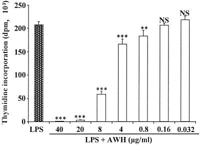
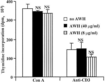
To investigate the modulatory effect of N. brasiliensis on B cell function, we examined the activity of a soluble extract of N. brasiliensis (AWH) on mitogen-induced proliferative responses of splenic B cells in vitro. Stimulation of highly enriched (>95%) B cells with LPS induces significant proliferation. However, the addition of AWH at the initiation of culture with LPS significantly (P < 0.001) inhibited the proliferative response of these B cells in a dose-dependent fashion. Maximum inhibition (90%) was seen at 20 μg/ml, but significant inhibition was seen with as little as 0.8 μg/ml (Fig. 1). In contrast to the effect on LPS-stimulated B cells, AWH had no inhibitory effect on the proliferative response of T cells stimulated with either ConA or anti-CD3 MAb (Fig. 2). These data confirm that the inhibitory activity of AWH is not nonspecific and suggests that B cells are uniquely sensitive to the effects of AWH.
FIG. 1.
AWH inhibits B-cell proliferation in response to LPS stimulation. Purified splenic B cells (from BALB/c nude mice) were activated with LPS (5 μg/ml) alone (filled bar) or in the presence of various concentrations of AWH (open bars). After 72 h of incubation at 37°C, the cultures were pulsed for 18 h with [3H]TdR. Data shown are expressed as the mean dpm of triplicate wells ± the SD. The results are from a single experiment and are representative of 10 separate experiments (∗∗, P < 0.01; ∗∗∗, P < 0.001, NS, not significant; P > 0.05, one-way ANOVA).
FIG. 2.
AWH has no inhibitory effect on the proliferative response of spleen cells to various T-cell activators. Spleen cells were activated with ConA (5 μg/ml) or anti-CD3 MAb (final dilution, 1:10) alone or in the presence of AWH. The cultures were pulsed for 18 h with [3H]TdR after initial 72 h of incubation. The data shown are expressed as the mean dpm of triplicate wells ± the SD. The results are from one experiment and are representative of five independent experiments (NS, not significant; P > 0.05, one-way ANOVA).
AWH does not block early activation events.
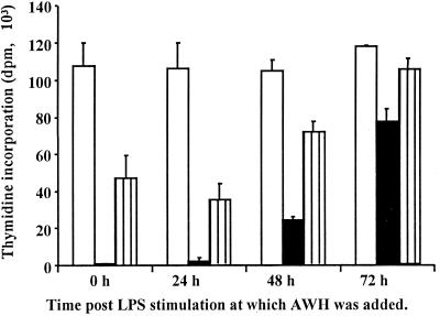
To address the mechanism of inhibition, we first assessed whether AWH prevented LPS activation of its cell-bound receptor. To accomplish this, LPS was added to cultures of highly enriched B cells 24, 48, and 72 h before the addition of AWH. The cultures were incubated for a total of 72 h and then pulsed with [3H]TdR for an additional 18 h. As shown in Fig. 3, the level of inhibition when AWH was added 24 h after LPS was comparable to that observed when both factors were added simultaneously. The inhibitory effect decreased somewhat when AWH was added at 48 h, and minimal inhibitory activity was detected when AWH was added 72 h after LPS stimulation. These data demonstrate that AWH still exhibits inhibitory activity after LPS has bound to its receptor and initiated early activation events. In a further experiment, cells were preincubated with AWH for 4 h, washed, and assessed for proliferation. These pretreated cells proliferated, as well as untreated controls, in response to stimulation with LPS (data not shown), confirming that AWH is not acting as a stable antagonist to LPS. Further, increasing the concentration of LPS (to 100 μg/ml) in the B cell culture, which increased [3H]TdR incorporation, did not change the inhibitory activity of AWH on B-cell proliferation (data not shown), confirming that AWH does not competitively inhibit LPS binding and that AWH inhibits both LPS receptor-dependent and receptor-independent LPS activation. Taken together, these data indicate that AWH does not act by interfering with LPS binding to cells or by inhibiting early activation events.
FIG. 3.
Effect of AWH added after LPS stimulation of B cells. B cells were stimulated with LPS (5 mg/ml) 24, 48, or 72 h prior to the addition of AWH (either 40 or 8 μg/ml). Cells were incubated for 72 h from the initial time of LPS stimulation and then were pulsed with [3H]TdR for an additional 18 h. The data are expressed as the mean dpm of triplicate wells ± the SD. The results are from a single experiment and are representative of five separate experiments. White bars, no AWH; black bars, AWH (40 μg/ml); striped bars (8 μg/ml).
AWH inhibitory activity appears to be mediated by modulation of macrophage (accessory cell) activation.
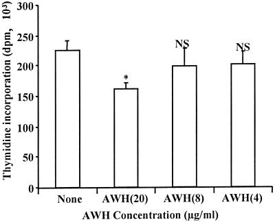
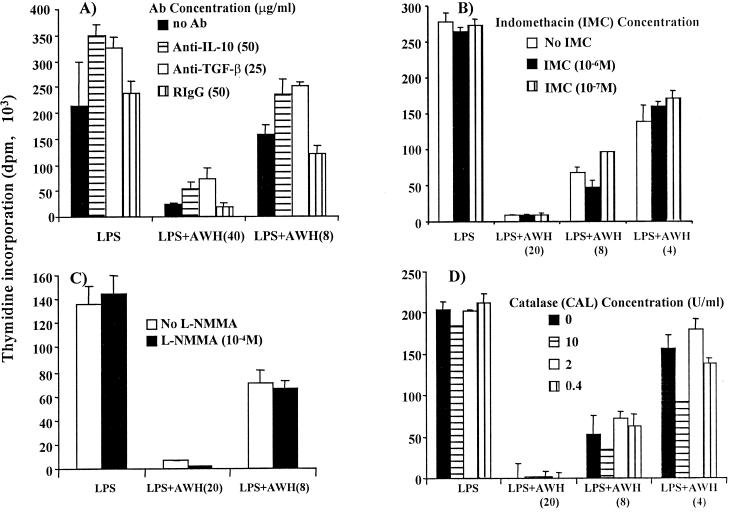
For maximal stimulation of B-cell proliferation in response to LPS activation, additional signals from accessory macrophages are essential (7). Since AWH can have an effect 24 h after LPS activation of B cells, it is possible that AWH could be mediating its inhibitory effect by modulating the function of the residual macrophages in our B-cell cultures (<5% macrophages), potentially blocking late activation signals or inducing late suppressive signals from these residual macrophages. To address this issue, enriched B-cell populations (with residual macrophages) were purged of macrophages as described in Materials and Methods and restimulated with LPS in the presence or absence of AWH. In these macrophage-purged cultures, the effect of AWH was almost completely ablated (Fig. 4). The proliferation was only slightly lower (but significant P < 0.05) compared to the level observed in B cells stimulated with LPS alone even at the highest dose of AWH, which normally gives >90% inhibition of proliferation. The small effect of AWH at the highest concentration may be attributed to a very few contaminating macrophages, which may still be capable of mediating some small inhibitory effect. These data confirm that AWH mediates its inhibitory activity on LPS-stimulated B-cell proliferation indirectly, through the modulation of macrophages to produce negative signals. This experiment confirms that AWH does not act to interfere with late-activating signals produced by macrophages since clearly no late-activating signals are required for continued B-cell proliferation in the absence of macrophages. To ascertain whether the inhibition of B-cell proliferation mediated by the modulatory effect of AWH on macrophages was associated with the release of mediators such as IL-10, TGF-β, prostaglandins, hydrogen peroxide, and nitric oxide, which could suppress the B-cell response we analyzed the contribution of these molecules by using specific inhibitors. The results presented in Fig. 5 show that the addition of inhibitors of IL-10 (anti-IL-10 MAb; Fig. 5A), TGF-β (anti-TGF-β MAb; Fig. 5A), prostaglandins (indomethacin; Fig. 5B), nitric oxide (L-NMMA; Fig. 5C), and hydrogen peroxide (catalase; Fig. 5D) to cultures of B cells stimulated with LPS and AWH did not reverse the proliferative suppression caused by AWH modulatory activity on the macrophages present in the B-cell cultures. This observation suggests that the inhibitory effect of AWH is not mediated by soluble factors that have been demonstrated in other studies to mediate the inhibition of B-cell proliferation.
FIG. 4.
Removal of macrophages from B cells reverses the inhibitory effect of AWH. B cells were activated with LPS (10 μg/ml) in a flask for 24 h. Adherent accessory cells were removed, and the remaining cells were then restimulated with LPS (5 μg/ml) in the presence or absence of AWH. After 72 h of incubation at 37°C, the cells were pulsed with [3H]TdR for an additional 18 h. The data are expressed as the mean dpm of triplicate wells ± the SD (∗, P < 0.05; NS, not significant; P > 0.05, one-way ANOVA). The results are from one experiment and are representative of five separate experiments. The control for the suppressive activity of AWH in this data set can only be the consistent results of AWH in all the other experiments reported here. The same batch of AWH used in these experiments was used in the experiment shown in this figure.
FIG. 5.
Inhibitory activity of AWH is not reversed by blocking antibodies to IL-10 and TGF-β and inhibitors of prostaglandins, nitric oxide, and hydrogen peroxide. B cells were stimulated with LPS (5 μg/ml) and cultured in the presence or absence of different concentrations of AWH. The cultures in panel A received either 50 μg of anti-IL-10 MAb or 25 μg of anti-TGF-β MAb per ml. The cultures in panel B received either 10−6 or 10−7 M indomethacin (prostaglandins inhibitor). The cultures in panel C received 10−4 M L-NMMA (nitric oxide inhibitor). The cultures in panel D received 0, 10, 2, or 0.4 U of catalase (hydrogen peroxide inhibitor) per ml at the beginning of the culture. The effectiveness of these inhibitors was tested before use. The culture was incubated for 72 h at 37°C and then pulsed with [3H]TdR for an additional 18 h. The data are expressed as the mean dpm of triplicate wells ± the SD.
AWH inhibits proliferation mediated by other activators.
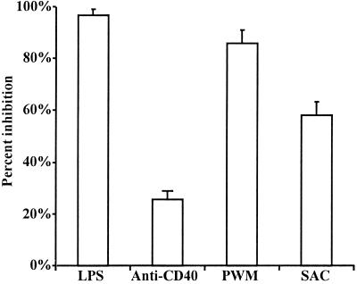
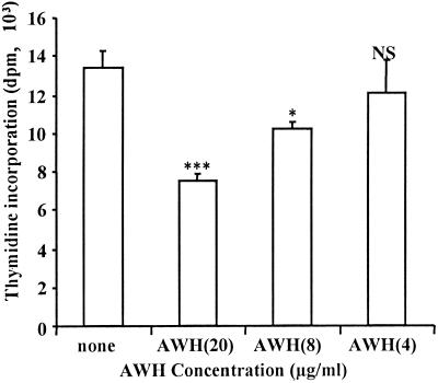
The proliferative response of B cells stimulated with mitogens using different signaling pathways was also inhibited by AWH. B cells stimulated with anti-CD40 MAb, PWM, or SAC were sensitive to the effects of AWH, but at variable levels (Fig. 6). B-cell proliferation mediated by anti-CD40 was the least susceptible, followed by that mediated by SAC and then PWM. PWM-stimulated B-cell proliferation was inhibited to approximately similar levels as LPS stimulation. This observation shows that the inhibitory activity of AWH is not restricted to LPS-mediated B-cell proliferation. Thus, the negative signals induced by AWH appear to have an effect on a downstream pathway required by all the B-cell activators tested. This notion is supported by the observation that AWH inhibited the proliferative response of B cells stimulated with the combination of the pharmacological B-cell activating agents PMA (PKC activator) and ionomycin (stimulator of calcium influx; Fig. 7). This demonstrates that the pathway which is effected, indirectly, by the macrophage-mediated inhibitory effect of AWH is downstream to initial signaling events and to PKC activation and calcium mobilization.
FIG. 6.
Percent inhibition by AWH of B-cell stimulation by other agents. Purified B cells were stimulated with LPS (5 mg/ml), anti-CD40 MAb (0.5 mg/ml), PWM (5 μg/ml) or SAC (1:2,000 dilution) with or without AWH (40 μg/ml). After 72 h of incubation, the cultures were pulsed for 18 h with [3H]TdR. The data shown are expressed as the mean percent inhibition of proliferation ± the SD. The data shown are representative of four similar experiments.
FIG. 7.
AWH inhibitory effect targets signaling pathway downstream of PKC. Purified B cells were activated with PMA (10 ng/ml) in combination with ionomycin (1 μg/ml) in the presence of different concentrations of AWH. After 72 h of incubation at 37°C, the cultures were pulsed for 18 h with [3H]TdR. The data are expressed as the mean dpm of triplicate wells ± the SD. The results are from one experiment and are representative of three separate experiments (∗, P < 0.05; ∗∗∗, P < 0.001; NS, not significant; P > 0.05, one-way ANOVA).
Active factor in AWH is proteinaceous in nature.
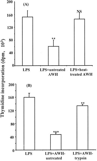
To ascertain the nature of the active factor in AWH, which mediates the inhibition of B-cell proliferation, AWH was subjected to denaturation either by heat treatment or by digestion with proteolytic enzymes (trypsin or chymotrypsin; bound to insoluble agarose matrix). Figure 8A confirms that heat treatment ablated the inhibitory effect of AWH on LPS-induced B-cell proliferation. In addition, proteolytic cleavage of AWH by solid matrix bound trypsin resulted in a reduction of the inhibitory effect by 80% (Fig. 8B). A similar result was obtained when AWH was cleaved with chymotrypsin (data not shown). These data provide strong evidence that the factor mediating this effect is proteinaceous in nature.
FIG. 8.
Heat and proteolytic enzyme treatment reverses the inhibitory effect of AWH on LPS-induced B-cell proliferation. B cells were stimulated with LPS (5 μg/ml) and cultured in the presence of AWH (10 μg/ml) that had either been heat treated by boiling (100°C) for 5 to 10 min (A) or digested with the proteolytic enzyme trypsin (10 U/ml) bound to an insoluble agarose matrix (B). The culture was incubated for 72 h at 37°C and then pulsed with [3H]TdR for additional 18 h. The data are expressed as the mean dpm of triplicate wells ± the SD (∗∗, P < 0.01; ∗∗∗, P < 0.001; NS, not significant; P > 0.05, one-way ANOVA). The results are from a single experiment and are representative of five separate experiments.
Inhibitory factor in N. brasiliensis extract is specific to the adult stage.
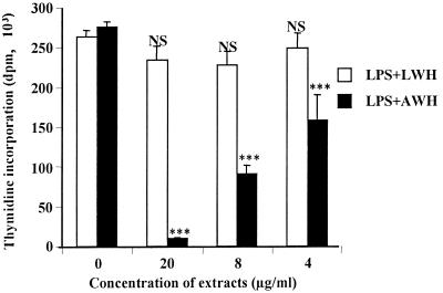
Because of the changes in protein composition associated with the developmental life cycle of nematodes (11, 24), the effect of an extract from the infective larvae (third-stage larvae) of N. brasiliensis was compared to the adult extract. B cells stimulated with LPS were cultured in the presence of extracts from either the infective larvae of N. brasiliensis (LWH) or the adult stage (AWH), and proliferation was assessed as described previously. Data from this experiment reveal that, unlike AWH, LWH had no inhibitory effect on the proliferative response of B cells to LPS, even at the highest concentration of 20 μg/ml (Fig. 9), a level at which AWH inhibited B-cell proliferation by >90%. This observation confirms that the factor responsible for the inhibitory activity of AWH is specific to the adult stage in the developmental life cycle of N. brasiliensis. This will aid in the future characterization of this factor since shared constituents between the larvae and adult can be eliminated from consideration as candidate molecules.
FIG. 9.
Inhibitory effect of N. brasiliensis extract is specific to the adult stage. B cells were activated with LPS (5 μg/ml) alone or in the presence of various concentrations of extract of the adult stage (AWH) or extract of the larvae stage (LWH). After 72 h of incubation at 37°C, the cultures were pulsed for 18 h with [3H]TdR. The data are expressed as the mean dpm of triplicate wells ± the SD (∗∗∗, P < 0.001; NS, not significant; P > 0.05, one-way ANOVA). The results are from a single experiment and are representative of three separate experiments.
DISCUSSION
The ability of helminths, particularly nematodes, to modify host immune responses has been an area of considerable research interest. The study of this modulation could elucidate basic immune regulatory pathways, result in the discovery of novel therapeutics, and influence the delivery of vaccines in areas where helminth infection is endemic. Previously, N. brasiliensis has been investigated with respect to stimulatory activities (18, 22, 46). However, suppression of mitogen responses in both B and T cells has been reported in a number of helminth parasites (2, 3, 6, 9, 14, 16, 17, 21). In the present study a reductive approach was taken by examining the effect of a soluble extract of N. brasiliensis (AWH) on mitogen-induced lymphocyte proliferation. The results presented here showed that AWH, added at the onset of culture, mediated a dose-dependent inhibition of LPS-stimulated B-cell proliferation. At the highest concentration of AWH, the proliferative response of B cells was inhibited by >90%, demonstrating potent inhibitory activity. The inhibitory activity of AWH appears to be consistent over time in culture. Evidence of AWH inhibition of LPS-mediated B-cell proliferation could be seen as early as 24 h after initiation of culture (data not shown). However, the inhibition was greater in cultures incubated for 72 h prior to the addition of TdR.
Although AWH strongly inhibited B-cell proliferation in response to LPS stimulation, it did not have any effect on the proliferation of T cells to activators of proliferation. This confirms that the lymphocyte inhibitory effect is restricted to B cells. This observation is unusual in that, in most cases, both B and T cells are subject to the effects of inhibitory factors from helminths. For example, a phosphorylcholine-containing preparation of Brugia malayi (16) and an extract of A. suum (13, 39, 40) have been demonstrated to inhibit the functional activities of both B and T cells. Interestingly, an earlier report by Price and Turner (33) indicated that extracts from H. polygyrus and N. brasiliensis inhibited proliferative responses of spleen cells to ConA (T-cell mitogen) and LPS (B-cell mitogen). The inhibition was observed optimally when the extract was added to the cells 24 h prior to the addition of mitogens. In contrast, the experiments in the present study revealed that the inhibitory effect of N. brasiliensis AWH is specific to B cells, with no effect on T cells, and that AWH is able to inhibit LPS-induced B-cell proliferation long after the inductive phase, as indicated by the inhibition observed when the extract was added as late as 48 h after exposure to LPS. The reasons for this discrepancy are unclear but may relate to the manner in which the N. brasiliensis extract was prepared by Price and Turner (33). Price and Turner prepared their extract from adult worms stored frozen in distilled water, and the final product was stored lyophilized. The extract used in our study was prepared from freshly isolated worms, and the final product was stored frozen. These differences are sufficient to affect the profile of the modulatory factors, which would ultimately affect the nature of the response.
The data presented in this study show that AWH is not toxic to cells. This was immediately evident in the T-cell culture experiments in which AWH had no inhibitory effect on T-cell proliferation. The data also show that AWH is not selectively toxic to B cells. This was clearly demonstrated by the observation that AWH exhibited no inhibitory effect on B cells purged on macrophages. Additional evidence that AWH was not toxic to B cells is provided by the observation that short-time exposure of B cells to AWH for 4 to 6 h did not have an effect on their proliferative response upon restimulation with LPS (data not shown). Similar short exposure of B cells to agents with cytotoxic potential induces cell death (31, 45). These observations are in congruence with other reports which have shown that immunomodulation by helminths is not mediated by cytotoxicity. For example, Cervi and Masih (6) showed that the suppressive effect of the trematode Fasciola hepatica glycoprotein on spleen cell proliferation was not attributable to cytotoxic action. They found that the viabilities of cells cultured in the presence of the glycoprotein were higher than those of control cells that were not exposed to the parasite extract. Studies with nematode extracts (9, 14, 16, 21, 33) have shown similar findings.
In addressing the mode of action of AWH on B cells, it was important to investigate whether the effect was mediated during the early or late stages of B-cell activation. Our data demonstrate that the effect of AWH does not appear to be mediated by either interfering with the binding of LPS to its receptor, or by downregulating early stages of B-cell activation, but rather appears to act on events downstream (during the late stages of B-cell activation), probably in an indirect fashion. This observation is consistent with those of other investigators who showed that nematode products did not suppress lymphocyte proliferation by directly downregulating the early stages of lymphocyte activation but rather appear to act indirectly on downstream activation pathways (2, 3, 6, 16, 21).
Although the exact mechanisms through which AWH induces the inhibition of LPS-mediated B-cell proliferation remains unclear, the data presented in this study confirm that AWH does not appear to have a direct suppressive effect on B cells but rather suggest that it acts through the modulation of macrophages to produce negative signals that impair the proliferative response of B cells. Macrophages, as a source of costimulatory signals, are essential for the generation of robust proliferation of B and T cells (7). It is therefore not surprising that they appear to be primary targets for factors modulating lymphocyte function. A number of reports have demonstrated the effects of parasite factors on macrophage activities (2, 6, 15, 27, 28, 35, 36). The data presented in this study show that macrophages are involved in the inhibitory activity of AWH on LPS-stimulated B cells. This is demonstrated by the observation that a B-cell population that had been purged of macrophages was resistant to the inhibitory effect of AWH. To rule out dendritic cells as the accessory cell, we allowed the cells to adhere 24 h, at which time splenic dendritic cells become nonadherent and macrophages remain adherent (41). The data set obtained from this experiment was identical to that obtained from the 4-h adherence experiment (in which both macrophages and dendritic cells would be removed), thus implicating macrophages as the cell of importance.
The involvement of macrophages in AWH inhibitory activity appears to be mediated via the production of a negative signal(s) by macrophages, under the influence of AWH. The signal(s) then targets a downstream pathway in B cells. This observation is not restricted to extracts from N. brasiliensis. Products of other helminths have been reported to induce the development of suppressive peritoneal exudate cells by modifying macrophage accessory functions and the production of nonspecific inhibitory factors, which inhibit both B- and T-cell proliferation in vitro (2, 3, 15, 23, 35, 36). These studies demonstrate that interference with the normal accessory role of macrophages, by parasites, will have profound effects on lymphocyte responses. However, these studies do not account for the B-cell-specific effects seen with AWH. Such an effect on B cells, sparing T cells, argues for the effect being mediated by a specific factor rather than a more nonspecific effect mediated by increased production of suppressive factors such as prostaglandins, nitric oxide, hydrogen peroxide, and arginase (1, 2, 23, 47). This is true in our study, since blocking the activity of these nonspecific suppressive factors did not reverse the inhibitory effect of AWH on B-cell proliferation. In addition, cytokines such as IL-10 (10, 26, 30) and TGF-β (5) that have been reported in other studies to inhibit lymphocyte proliferation were not involved in mediating the inhibitory activity of AWH. This observation suggests that the inhibitory effect of AWH is not mediated by any of the obvious soluble factors, demonstrated in other studies to mediate the inhibition of B-cell proliferation. Although the specific AWH-induced macrophage factor(s) mediating the inhibition of B cells could not be identified, the observation that the inhibitory effect of AWH is reversed by the removal of macrophages clearly shows the effect of AWH is mediated by negative signals from the macrophages.
AWH also inhibited B-cell proliferation to a variety of stimuli, including PMA and ionomycin. B cells stimulated with mitogens (LPS and PWM) were more susceptible to the inhibitory effect of AWH, but cells activated by cross-linking either CD40 (by anti-CD40 MAb) or surface immunoglobulin (by SAC) or by the combination of PMA (PKC activator) and ionomycin (stimulator of calcium influx) also showed decreased proliferation in the presence of AWH. Such a pleotrophic effect confirms that the effect of AWH is not restricted to LPS-mediated proliferation and suggests a downstream activity of the negative signals induced from accessory cells by AWH and that the pathway targeted is common to the activators tested in this study. However, the difference in the effect of AWH on the activators tested suggests a variation in their sensitivity to the negative signals from accessory cells, possibly because of the existence of alternate activation pathways. The fact that AWH inhibited B-cell proliferation stimulated by the combination of PMA (PKC activator) and ionomycin (stimulator of calcium influx) demonstrates that the pathway targeted by AWH-mediated suppression is downstream of initial signaling events and of PKC activation and calcium mobilization. Ras/mitogen-activated protein kinase and the phosphatidylinositol 3-kinase pathways are two major downstream (late-activation) signaling pathways during B-cell activation (4, 42) and have been associated with LPS-mediated cell activation (8, 42, 43). Since these pathways are significant to B-cell proliferation, they are likely targets of the negative signals produced by accessory cells in response to AWH stimulation.
AWH is a complex mixture of proteins. A preliminary characterization of the nature of the factor in AWH responsible for inducing the production of the accessory-cell-mediated negative signal revealed that it is sensitive to denaturation by boiling and proteolytic enzyme treatments. This suggests that the active factor(s) is proteinaceous in nature. This result is in congruence with evidence with respect to other helminth immunomodulatory factors (6, 14, 16, 33, 40). The finding that the inhibitory factor was present in the adult, but not the larval, stage of N. brasiliensis is particularly important because it provides direction for further characterization of the factor. It is possible that the stage-specific expression of the immunomodulatory factor could be an adaptation by the adult nematode to immunosuppress the host during the critical egg-producing stage. This shift in protein composition as nematodes develop from larvae to adults has been well documented, including in the nematode N. brasiliensis (11, 24, 32). Because the extract used in this study is a whole-worm homogenate, we do not know if the modulatory factor is secreted (thus raising the question of clinical relevance). However, it is important to note that we also do not know that the modulatory factor is not secreted and it will be complex to determine whether it is. In addition, there is a paucity of evidence relating the exposure of hosts to somatic molecules from nematodes, and it is very reasonable to assume that somatic products from dead or dying worms can influence the host. Indeed, antibodies to somatic antigens are found in a variety of nematode infections, thus confirming exposure to somatic products in these infections (11, 24, 32, 44).
This study provides strong evidence of the diversity in the modulatory activity of N. brasiliensis in its hosts. It demonstrates the ability of an N. brasiliensis extract to specifically inhibit B-cell proliferation by affecting macrophage function, which is essential for maximal proliferation of B cells. This observation is in apparent contrast to the stimulation of reaginic immunoglobulin responses widely reported in nematode-infected animals. It is important to note that both the immune response to nematodes and the modulation of that immune response by nematode factors are likely to be very complex. For example, Price and Turner (33) have shown that a somatic extract of Heligmosomoides polygyrus inhibits B-cell proliferation but, in contrast, Robinson and coworkers (38) reported that a similar extract stimulated IgG1 production in vitro. Moreover, extracts from A. suum have been reported to stimulate (20) and to inhibit (13, 39) B-cell function. Some of the discrepancies and apparent inconsistencies in reports regarding the modulation of immune function in response to nematode extracts are undoubtedly due to variation in the profile of the modulatory factors between nematodes, life cycle stages, and/or methods of isolation. This view is underscored by the significant differences we observed in the modulatory activities of the adult versus larval N. brasiliensis extracts in our experimentation. Although the data reported here are from in vitro experimentation, it is likely that this B-cell inhibitory activity has significant in vivo relevance. The role of antibody in the clearance of nematode infection is still controversial, but clearly any dysfunction of B-cell expansion will have profound effects on the progress of the immune response elicited by the infection. In addition to antiparasite immunity, this modulation could also have profound effects on the development of immune responses to vaccines and other infectious agents in nematode-infected individuals.
ACKNOWLEDGMENTS
This work was supported by grants from the Natural Sciences and Engineering Research Council of Canada and Novartis, Inc.
REFERENCES
- 1.Albina J E, Abate J A, Henry W., Jr Nitric oxide production is required for murine resident peritoneal macrophages to suppress mitogen-stimulated T cell proliferation: role of IFN-γ in the induction of the nitric oxide synthesizing pathway. J Immunol. 1991;147:144–148. [PubMed] [Google Scholar]
- 2.Allen J E, Lawrence R A, Maizels R M. APC from mice harbouring the filarial nematode, Brugia malayi, prevents cellular proliferation but not cytokine production. Int Immunol. 1996;8:143–151. doi: 10.1093/intimm/8.1.143. [DOI] [PubMed] [Google Scholar]
- 3.Allen J E, Macdonald A S. Profound suppression of cellular proliferation mediated by the secretions of nematodes. Parasite Immunol. 1998;20:241–247. doi: 10.1046/j.1365-3024.1998.00151.x. [DOI] [PubMed] [Google Scholar]
- 4.Armitage R J, Alderson M R. B cell stimulation. Curr Opin Immunol. 1995;7:243–247. doi: 10.1016/0952-7915(95)80009-3. [DOI] [PubMed] [Google Scholar]
- 5.Bouchard C, Fridman W H, Sautè C. Mechanism of inhibition of lipopolysaccharide-stimulated mouse B cell responses by transforming growth factor-β1. Immunol Lett. 1994;40:105–110. doi: 10.1016/0165-2478(94)90180-5. [DOI] [PubMed] [Google Scholar]
- 6.Cervi L, Masih D T. Inhibition of spleen cell proliferative response to mitogens by excretory-secretory antigens of Fasciola hepatica. Int J Parasitol. 1997;27:573–579. doi: 10.1016/s0020-7519(96)00188-9. [DOI] [PubMed] [Google Scholar]
- 7.Corbel C, Melchers F. Requirement for macrophages or for macrophage- or T cell-derived factors in the mitogenic stimulation of murine B lymphocytes by lipopolysaccharides. Eur J Immunol. 1983;13:528–533. doi: 10.1002/eji.1830130703. [DOI] [PubMed] [Google Scholar]
- 8.Dearden-Badet M-T, Revillard J-P. Requirement for tyrosine phosphorylation in lipopolysaccharide-induced murine B-cell proliferation. Immunology. 1993;80:658–660. [PMC free article] [PubMed] [Google Scholar]
- 9.Deehan M R, Frame M J, Parkhouse R M E, Seatter S D, Reid S D, Harnett M M, Harnett W. A phosphorylcholine-containing filarial nematode-secreted product disrupts B lymphocyte activation by targetting key proliferative signaling pathways. J Immunol. 1998;160:2692–2699. [PubMed] [Google Scholar]
- 10.Ding L, Shevach E M. IL-10 inhibits mitogen-induced T cell proliferation by selectively inhibiting macrophage costimulatory function. J Immunol. 1992;148:3133–3139. [PubMed] [Google Scholar]
- 11.Dorzok U, Bohn A, König W. Proteins of Nippostrongylus brasiliensis analyzed by immunoblotting. Parasitol Res. 1989;75:482–487. doi: 10.1007/BF00930977. [DOI] [PubMed] [Google Scholar]
- 12.Egwang T G, Befus A D, Gauldie J. Activation of alveolar macrophages following infection with the parasitic nematode Nippostrongylus brasiliensis. Immunology. 1985;54:581–588. [PMC free article] [PubMed] [Google Scholar]
- 13.Ferreira A P, Faquim E S, Abrahamson I A, Macedo M S. Immunization with Ascaris suum extract impairs T cell functions in mice. Cell Immunol. 1995;162:202–210. doi: 10.1006/cimm.1995.1070. [DOI] [PubMed] [Google Scholar]
- 14.Harnett W, Harnett M M. Inhibition of murine B cell proliferation and downregulation of protein kinase-C levels by a phosphorylcholine-containing filarial excretory-secretory product. J Immunol. 1993;151:4829–4837. [PubMed] [Google Scholar]
- 15.Kadian S K, Dixon J B, Green J R, Carter S D, Jenkins P. Modification of macrophage-T cell interaction during infection of mice with Mesocestoides corti (cestoda) Parasitol. 1994;109:591–597. doi: 10.1017/s0031182000076472. [DOI] [PubMed] [Google Scholar]
- 16.Lal R B, Kumaraswami V, Steel C, Nutman T B. Phosphocholine-containing antigens of Brugia malayi nonspecifically suppress lymphocyte function. Am J Trop Med Hyg. 1990;42:56–64. doi: 10.4269/ajtmh.1990.42.56. [DOI] [PubMed] [Google Scholar]
- 17.Lammie P J, Katz S P. Immunoregulation in experimental filariasis. I. In vitro suppression of mitogen-induced blastogenesis by adherent cells from jirds chronically infected with Brugia pahangi. J Immunol. 1983;130:1386–1389. [PubMed] [Google Scholar]
- 18.Ledingham D L, McAlister V C, Ehigiator H N, Giacomantonio C, Theal M, Lee T D G. Prolongation of rat kidney allograft survival by nematodes. Transplantation. 1996;61:184–188. doi: 10.1097/00007890-199601270-00003. [DOI] [PubMed] [Google Scholar]
- 19.Lee T D G, McGibbon A M. Potentiation of IgE responses to third party antigens mediated by A. suum soluble products. Int Arch Allergy Immunol. 1993;102:185–190. doi: 10.1159/000236570. [DOI] [PubMed] [Google Scholar]
- 20.Lee T D G, Xie C Y. Polyclonal stimulation of murine B lymphocytes by Ascaris soluble product. J Allergy Clin Immunol. 1995;95:1246–1254. doi: 10.1016/s0091-6749(95)70082-x. [DOI] [PubMed] [Google Scholar]
- 21.Leiva L E, Lammie P J. Modulation of lymphocyte activation by soluble Brugia pahangi extracts. Trop Med Parasitol. 1989;40:327–331. [PubMed] [Google Scholar]
- 22.Liwski R S, Lee T D G. Nematode infection enhances survival of activated T cells by modulating accessory cell function. J Immunol. 1999;163:5005–5012. [PubMed] [Google Scholar]
- 23.MacDonald A S, Maizels R M, Lawrence R A, Dransfield I, Allen J E. Requirement for in vivo production of IL-4, but not IL-10, in the induction of proliferative suppression by filarial parasites. J Immunol. 1998;160:4124–4132. [PubMed] [Google Scholar]
- 24.Maizels R M, Meghji M, Ogilvie B M. Restricted sets of parasite antigens from the surface of different stages and sexes of the nematode Nippostrongylus brasiliensis. Immunology. 1983;48:107–121. [PMC free article] [PubMed] [Google Scholar]
- 25.Maizels R M, Bundy D A P, Selkirk M E, Smith D F, Anderson R M. Immunological modulation and evasion by helminth parasites in human populations. Nature. 1993;365:7497–7508. doi: 10.1038/365797a0. [DOI] [PubMed] [Google Scholar]
- 26.Marcelletti J F. IL-10 inhibits lipopolysaccharide-induced murine B cell proliferation and cross-linking of surface antigen receptors or ligation of CD40 restores the response. J Immunol. 1996;157:3323–3333. [PubMed] [Google Scholar]
- 27.Motran C, Gruppi A, Vullo C M, Pistoresi-Palencia M C, Serra H M. Involvement of accessory cells in the Trypanosoma cruzi-induced inhibition of the polyclonal response of T lymphocytes. Parasite Immunol. 1996;18:43–48. doi: 10.1046/j.1365-3024.1996.d01-5.x. [DOI] [PubMed] [Google Scholar]
- 28.Nasarre C, Krahenbuhl J L, Klei T R. Downregulation of macrophage activation in Brugia pahangi-infected jirds (Meriones unguiculatus) Infect Immun. 1998;66:1063–1069. doi: 10.1128/iai.66.3.1063-1069.1998. [DOI] [PMC free article] [PubMed] [Google Scholar]
- 29.Nawa Y, Miller H R P, Hall E, Jarrett E E E. Adoptive transfer of total and parasite-specific IgE responses in rats infected with Nippostrongylus brasiliensis. Immunology. 1981;44:119–123. [PMC free article] [PubMed] [Google Scholar]
- 30.Peçanha L M T, Snapper C M, Lees A, Yamaguchi H, Mond J J. IL-10 inhibits T cell-independent but not T cell-dependent responses in vitro. J Immunol. 1993;150:3215–3223. [PubMed] [Google Scholar]
- 31.Perreault J, Lemieux R. Rapid apoptotic cell death of B cell hybridomas in absence of gene expression. J Cell Physiol. 1993;156:286–293. doi: 10.1002/jcp.1041560210. [DOI] [PubMed] [Google Scholar]
- 32.Phillip M, Parkhouse R M E, Ogilvie B M. Changing proteins on the surface of a parasitic nematode. Nature. 1980;287:538–540. doi: 10.1038/287538a0. [DOI] [PubMed] [Google Scholar]
- 33.Price P, Turner K J. Immunosuppressive effects of extracts of helminthic parasites in C57BL mice. Int J Parasitol. 1986;16:607–615. doi: 10.1016/0020-7519(86)90028-7. [DOI] [PubMed] [Google Scholar]
- 34.Pritchard D I, Lawrence C E, Appleby P, Gibb A, Glover K. Immunosuppressive proteins secreted by the gastrointestinal nematode parasite Heligmosomoides polygyrus. Int J Parasitol. 1994;24:495–500. doi: 10.1016/0020-7519(94)90140-6. [DOI] [PubMed] [Google Scholar]
- 35.Rakha N K, Dixon J B, Skerritt G C, Carter S D, Jenkins P, Marshall-Clarke S. Lymphoreticular responses to metacestodes: Taenia multiceps (cestoda) can modify interaction between accessory cells and responder cells during lymphocyte activation. Parasitology. 1991;102:133–140. doi: 10.1017/s0031182000060443. [DOI] [PubMed] [Google Scholar]
- 36.Rakha N K, Dixon J B, Carter S D. Taenia multiceps (cestoda): Ia antigen expression and prostaglandin secretion by parasite-modified, murine peritoneal macrophages. Parasite. 1996;3:135–141. doi: 10.1051/parasite/1996032135. [DOI] [PubMed] [Google Scholar]
- 37.Robinson M, Gustad T R, Wei F-Y, David C S, Storey N. Adult worm homogenate of the nematode parasite Heligmosomoides polygyrus induces proliferation of naive T lymphocytes without MHC restriction. Cell Immunol. 1994;158:157–166. doi: 10.1006/cimm.1994.1264. [DOI] [PubMed] [Google Scholar]
- 38.Robinson M, Gustad T R. In vitro stimulation of naive mouse lymphocytes by Heligmosomoides polygyrus adult worm antigens induces the production of IgG1. Parasite Immunol. 1996;18:87–93. doi: 10.1046/j.1365-3024.1996.d01-52.x. [DOI] [PubMed] [Google Scholar]
- 39.Soares M F M, Macedo M S, Mota I. Suppressive effect of an Ascaris suum extract on IgE and IgG antibody responses in mice. Brazilian J Med Biol Res. 1987;20:203–211. [PubMed] [Google Scholar]
- 40.Soares M F M, Mota I, Macedo M S. Isolation of Ascaris suum components, which suppress IgE antibody responses. Int Arch Allergy Immunol. 1992;97:37–43. doi: 10.1159/000236093. [DOI] [PubMed] [Google Scholar]
- 41.Steinman R M. The dendritic cell system and its role in immunogenicity. Annu Rev Immunol. 1991;9:271–296. doi: 10.1146/annurev.iy.09.040191.001415. [DOI] [PubMed] [Google Scholar]
- 42.Venkataraman C, Shankar G, Sen G, Bondada S. Bacterial lipopolysaccharide induced B cell activation is mediated via a phosphatidylinositol 3-kinase dependent signaling pathway. Immunol Lett. 1999;69:233–238. doi: 10.1016/s0165-2478(99)00068-1. [DOI] [PubMed] [Google Scholar]
- 43.Weinstein S L, Sanghera J S, Lemke K, Defranco A L, Pelech S L. Bacterial lipopolysaccharide induces tyrosine phosphorylation and activation of mitogen-activated protein kinases in macrophages. J Biol Chem. 1992;267:14955–14962. [PubMed] [Google Scholar]
- 44.Yamada M, Nakazawa M, Matsumoto Y, Arizono N. IgE antibody production in rats against multiple components of excretory-secretory products of the nematode Nippostrongylus brasiliensis. Immunology. 1991;72:104. [PMC free article] [PubMed] [Google Scholar]
- 45.Yankelevich B, Soldatenkov V A, Hodgson J, Polotsky A J, Creswell K, Mazumder A. Differential induction of programmed cell death in CD8+ and CD4+ T cells by the subunit of cholera toxin. Cell Immunol. 1996;168:229–234. doi: 10.1006/cimm.1996.0070. [DOI] [PubMed] [Google Scholar]
- 46.Zakroff S G H, Beck L, Platzer E G, Spiegelberg H L. The IgE and IgG subclass responses of mice to four helminth parasites. Cell Immunol. 1989;119:193–201. doi: 10.1016/0008-8749(89)90235-9. [DOI] [PubMed] [Google Scholar]
- 47.Zwadlo-Klarwasser G, Platen S, Schmutzler W. Inhibition of spontaneous and mitogen-induced lymphocyte proliferation by murine bone marrow-derived macrophages: role of prostaglandins, nitric oxides and cell-to-cell contact. Scand J Immunol. 1994;40:10–15. doi: 10.1111/j.1365-3083.1994.tb03426.x. [DOI] [PubMed] [Google Scholar]


