Abstract
Purpose
To analyze the characteristics of ocular surface microbial composition in children and adolescents with diabetes mellitus and dry eye (DE) by tear analysis.
Methods
We selected 65 children and adolescents aged 8 to 16 years with DE and non–DE diabetes mellitus and 33 healthy children in the same age group from the Shanghai Children and Adolescent Diabetes Eye Study. Tears were collected for high-throughput sequencing of the V3 and V4 region of 16S rRNA. The ocular surface microbiota in diabetic DE (DM-DE; n = 31), diabetic with non–DE (DM-NDE; n = 34), and healthy (NDM; n = 33) groups were studied. QIIME2 software was used to analyze the microbiota of each group.
Results
The DM-DE group had the highest amplicon sequence variants, and the differences in α-diversity and β-diversity of micro-organisms in the ocular surfaces of DM-DE, diabetic with non-DE, and healthy eyes were statistically significant (P < 0.05). Bacteroidetes (15.6%), Tenericutes (9.3%), Firmicutes (21.8%), and Lactococcus (7.9%), Bacteroides (7.8%), Acinetobacter (3.9%), Clostridium (0.8%), Lactobacillus (0.8%) and Streptococcus (0.2%) were the specific phyla and genera, respectively, in the DM-DE group.
Conclusions
Compared with the patients with non–DE and healthy children, the microbial diversity of the ocular surface in children and adolescents with diabetes mellitus and DE was higher with unique bacterial phyla and genera composition.
Keywords: children and adolescents, diabetes mellitus, dry eye, tears, 16S rRNA, microbiome
In recent years, due to changes in lifestyle and everyday habits that affect the eyes, the incidence of dry eye (DE) has been increasing every year, and more causes of DE have been identified. DE resulting from systemic diseases, especially diabetes mellitus (DM), has been gaining attention, and the prevalence of diabetic DE (DM-DE) is higher.1,2 In 2018, our research group established the Shanghai Children and Adolescent Diabetes Eye (SCADE) study. The cross-sectional study1 found that the prevalence of DE in children with diabetes (28.95%) was much higher than that in healthy children (5.00%). Subsequently, our research group followed up the children with diabetes regularly. In January 2021, the 3-year incidence of DE in children with diabetes was 22.5%, and the annual average incidence was 7.5%,3 which was the first report on the incidence of DE in diabetic children. DE in children with diabetes causes prolonged ocular damage and has a long-term impact on the quality of life. However, the pathogenesis of DE in this group remains unknown, and the corresponding targeted treatment is also unclear.
In 2008, the Human Microbiome Project by the National Institutes of Health of the United States revealed that the human body is inhabited by extremely rich and diverse microbial species communities.4 In recent years, the characteristics of ocular surface microbiota (OSM) have increasingly gained attention.5,6 Growing numbers of studies have shown that OSM may be closely related to DE, and Staphylococcus aureus, coagulase negative Staphylococcus and Corynebacterium are related to the prevalence of DE.7,8 OSM dysregulation is also involved in the pathogenesis and development of DE.9–16 The possible mechanism of OSM dysregulation in the DM population and high incidence of DE in them is as follows: the abundant glucose in the tears of patients with DM provides sufficient energy for the growth and reproduction of OSM, resulting in a high diversity and number of OSM in patients with DM.17,18 A high glucose environment can activate immune cells and induce ocular surface inflammation by increasing tear osmotic pressure and promoting the formation of advanced glycation end products,19–21 followed by an enhanced immune response at the ocular surface to OSM, leading to OSM dysregulation. In a high-glucose environment, inflammatory factors damage nerve fibers22; meanwhile, nerve growth factor, sphingomyelin, and other neuronutrients are down-regulated,23,24 advanced glycation end products damage microvessels, decreasing nerve blood supply,25 and eventually ocular surface neuropathy occurs, which decreases corneal perception and blink movement, and delays the removal of ocular surface substances.26 This facilitates the colonization and propagation of micro-organisms on the ocular surface. However, along with decreases in lacrimal gland secretion and the antimicrobial substances in tears, the antibacterial property of tear film dereases.26
Better than traditional culture methods, detection methods based on molecular biology techniques, such as 16S rRNA sequencing, can more comprehensively and accurately identify the species composition of OSM.27 Kittipibul et al.,16 using 16S rRNA gene detection, revealed that the increased abundance of Acinetobacter, Bacillus, Bacteroides, Lactobacillus, Pseudomonas, Staphylococcus, and Streptococcus may participate in the development of DE in adults. Ham et al.28 found that Acinetobacter was more prevalent in patients with diabetes, whereas Bradyrhizobium and Streptophylococcus were prevalent in healthy subjects. Li et al.17 found that Acinetobacter and Pseudomonas were the main OSM in DM. Our previous study9 using the 16S rRNA gene to detect the microbiota on the ocular surface found that unclassified Clostridium and Lactobacillus were related to DM-DE in adults. However, there is no report on the microbiome associated with DE in children with diabetes. The aim of this study was to ascertain the differences in the population and number of micro-organisms on the ocular surface among children with diabetes with and without DE and healthy children to provide a basis for exploring the mechanism underlying high incidence of DE in children with diabetes from an OSM perspective.
Materials and Methods
In the SCADE study, 65 children and adolescents with diabetes, comprising 31 and 34 children with DE (DM-DE group) and non-DE (DM-NDE group), respectively, and 33 healthy controls of the same age without diabetes in this scenario (NDM group) who visited the outpatient department in January 2022 were included in the study.
The inclusion criteria of study participants were as follows: (1) all participants and their guardians were fully informed about the study and they provided signed written informed consent, (2) participants were aged 8 to 16 years, (3) participants with diabetes diagnosed with type 1 diabetes according to the diagnostic criteria of the World Health Organization,29 (4) the healthy control group had no other systemic diseases or history of DE, and (5) all participants cooperated to complete the eye examination. The exclusion criteria were as follows: other ocular and systemic diseases affecting the secretion and quality of tears, namely (1) eyelid diseases trichiasis, entropion, ectropion, ptosis, incomplete eyelid closure, and so on; (2) conjunctival diseases: acute and chronic infectious conjunctivitis, allergic conjunctivitis, conjunctival stones, and so on; (3) severe ocular chemical injury complications and ocular trauma history within 6 months; (4) ocular surgery history within 6 months; (5) severe ocular complications caused by diabetes: cataract, retinopathy, optic atrophy, and so on; (6) treatment with eye drops; and (7) the use of contact lenses for more than 1 month.
This study conformed to the ethical principles of the Declaration of Helsinki and was approved by the ethics committees of the First People's Hospital Affiliated to Shanghai Jiaotong University School of Medicine and Children's Hospital of Fudan University, Shanghai (approval numbers were 2016KY005 and No. 01 [2018]).
According to the International Dry Eye Workshop II criteria for DE examination and diagnosis,30 DE was diagnosed when one of the two eyes had ocular dryness, foreign body sensation, burning sensation, fatigue, discomfort, eye redness, vision fluctuation, and other subjective symptom complaints and an ocular surface disease index of 13 or more points; tear break-up time of 5 seconds or less or a Schirmer I test of 5 mm/5 min or less; and a positive fluorescein sodium corneal staining in case of DE-related symptoms, an ocular surface disease index of 13 or greater, a tear break-up time of more than 5 and 10 or fewer seconds, or a Schirmer I test of more than 5 mm/5 min and 10 mm/5 min or less.
On the day before specimen collection, the guardians were instructed to ensure that the teenagers and children slept well and obtained samples between 8 am and 10 am each time. Before sampling, a saline swab was used to wipe the participants’ eyelid skin twice; subsequently, a tear test paper was used to collect tears from the eyes. The entire procedure was performed aseptically. The collected samples were quickly placed in a tube containing DNA protecting fluid (GeWei Bio-Tech, Shanghai, China) and stored in a refrigerator at –20 °C (Haier, Qingdao, China) for cryopreservation, then used for testing and DNA extraction.
All children were arranged for specimen collection at Shanghai Eye Disease Prevention and Treatment Center/Shanghai Eye Hospital. Eye specimens were collected by a trained ophthalmologist (Z.C.) for consistency of results.
Extraction of Total DNA From Tear Samples, PCR Amplification, Product Purification and Quantification, and 16S rRNA High-Throughput Sequencing
After sample collection, total microbial genomic DNA was extracted from all specimens using the DNeasy PowerSoil Kit (Qiagen Inc, Venlo, the Netherlands) according to the Kit extraction instructions. The total microbial genomic DNA was stored in a refrigerator at −20 °C for further analysis. The quality and quantity of DNA were determined by 0.8% agarose gel (Biowest, Spain) electrophoresis (61 Instruments, Beijing, China) and NanoDrop ND-1000 spectrophotometer (Thermo Scientific, Waltham, MA), respectively.
NEB Q5 DNA high-fidelity polymerase (New England Biolabs, Ipswich, MA) was used for PCR amplification, and the number of amplification cycles was strictly controlled. PCR amplification of V3-V4 region of bacterial 16S rRNA gene was performed with forward primer 338F(5′-barcode+ACTCCTACGGGAGGCAGCA-3′) and reverse primer 806R (5′-GGACTACHVGGGTWTCTAAT-3′). The specific tag sequences (7-bp) that distinguish the samples were integrated into the primers for multiplex sequencing. After the purification and quantification steps as described elsewhere in this article, equal amounts of the amplified products were pooled and double-terminal 2 × 300 bp sequencing was performed using the Illumina MiSeq platform and MiSeq Reagent Kit v3 program.
Sequencing Data Processing
(1) QIIME2 2019.4 version was used, the original sequence data were decoded and processed by demux plug-in, cutadapt plug-in was used to excise primers, and DADA2 plug-in was used to process the sequence quality filtering, denoising, splicing, and chimera removal. The sequences obtained above were merged with 100% sequence similarity to generate amplicon sequence variants (ASVs) and abundance data tables.
(2) For ASV classification and taxonomic status identification, the corresponding taxonomic information of each ASV was obtained by comparing ASV characteristic sequences with the reference sequences in the database using Greengenes database. ASVs with abundance values of less than 0.001% of the total sequencing volume were removed, and the resulting abundance matrix, with rare ASVs removed, was used for subsequent series of analyses. At the same time, R software was used to draw the identification results of each sample at each classification level into bar charts, and the differences of ASV number and classification status identification results of different samples were visually compared.
QIIME2 software was used to calculate seven diversity indexes for each sample, namely the Chao1 index, observed species index, Shannon index, Simpson index, Faith's PD index, Pielou's evenness index, and Good's coverage index and then the boxplots were drawn. The richness, diversity, and evenness of ASVs among different samples were compared.
β-Diversity analysis was performed by R software and QIIME2 software using UniFrac distance metric to investigate the changes in microbial community structure among samples, and principal coordinate analysis was used to show the differences in species composition among groups.
According to the results of the ASV division and taxonomic status identification, the specific-species composition of each sample at each taxonomic level was obtained. Identification of different taxonomic levels namely phylum, class, order, family, genus, and species are equivalent to viewing community composition at different resolutions. The 10 most abundant micro-organisms were included.
QIIME2 software was used to obtain the composition and abundance table of each sample at six taxonomic levels: phylum, class, order, family, genus, and species, and the analysis results were presented as bar graphs. Permutational multivariate ANOVA was used to evaluate the significance of differences in microbial community structure among the groups. Linear discriminant analysis effect size method was used to detect the taxon with rich differences between groups.
Based on the power and pairwise sample size estimation of the permutational multivariate ANOVA application Micropower,31 low-abundance 16S rRNA datasets similar to previous ocular microbiome studies32 require a minimum sample size of 30 to produce a discriminant power of 0.8 with a significance level of 0.05. Therefore, we aimed for at least 30 subjects in each study group, and all three groups in this study met the required sample size for the study.
Statistical Analysis
SPSS 22.0 (SPSS, Inc., Armonk, NY) software was used for statistical analysis. One-way ANOVA was used to compare the ages of the three groups; the χ2 test was used for gender comparison. The difference of diabetes course between DM-DE group and DM-NDE group was compared by a nonparametric test. Fasting blood glucose and hemoglobin A1c were compared between the two groups using an independent samples t test; and a P value of less than 0.05 was considered statistically significant.
Results
Basic Information of the Study Subjects
The average age of children and adolescents in the NDM, DM-DE, and DM-NDE groups was 13.28 ± 2.86, 13.19 ± 2.67, and 13.90 ± 2.76 years, respectively, and there was no significant difference in age among the groups (F = 0.61; P = 0.96). There were 10, 11, and 12 boys and 23, 20, and 22 girls in the NDM, DM-DE, and DM-NDE groups, respectively; no significant differences were observed among the groups with respect to the sexes (χ2 = 0.25; P = 0.88). All 65 diabetic children were treated with insulin, and their fasting blood glucose was stable in the past year. We compared the diabetes duration (U = 437.5; P = 0.24), fasting blood glucose levels (t = 0.21; P = 0.84), and hemoglobin A1c (t = 0.26; P = 0.79) between DM-DE and DM-NDE groups and found no statistical significance.
Statistics of Valid Sequences and ASVs/Operational Taxonomic Units (OTU)s
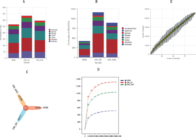
A total of 7,873,887 (Reads) valid sequences were obtained by DADA2 method for priming, quality filtering, and denoising with an average of 80,346 (Reads) valid sequences per child specimen. Among all the children's specimens, the highest and lowest number of sequences was 117,850 (Reads) and 62,198 (Reads) with an average of 80,346 (Reads). The average length of each sequence was 426 bp, and 99.5% of valid sequences were distributed between 405 bp and 431 bp in length. Species taxonomy annotation results showed that the number of ASVs/OTUs in the NDM, DM-DE, and DM-NDE groups were, respectively, annotated as 13, 15, and 13 phyla; 26, 32, and 27 classes; 39, 48, and 41 orders; 68, 79, and 72 families; 82, 105, and 95 genera; 39, 54, and 48 species, respectively, and the DM-DE group was annotated with the highest number of species at the phylum, class, order, family, genus, and species level. A total of 41291 ASVs/OTUs were obtained by combining high-quality sequences according to 97% sequence similarity, including 19,974 ASVs/OTUs in the DM-DE group, 15,971 ASVs/OTUs in the DM-NDE group, and 7072 ASVs/OTUs in the NDM group. There were 863 ASVs/OTUs shared by the three groups, and the results showed that the most ASVs/OTUs were found in the DM-DE group, indicating that the species richness and diversity of ocular surface micro-organisms in this group were the highest. The common ASVs/OTUs of the three groups represented the core microbiota of children's eyes (Figs. 1A, B, and C).
Figure 1.
(A) The number of taxa in the three groups. (B) The number of ASVs/OTUs in the three study groups. (C) ASV/OTU Venn diagram. (D) Rarefaction curve. (E) Species accumulation curves.
Assessment of Sequencing Depth and Sample Size
The flattening degree of the rarefaction curve reflects the effect of sequencing depth on microbial community diversity. In this study, with the increase of the amount of sequencing data, the rarefaction curve gradually flattened out, indicating that the current sequencing depth was sufficient to reflect the richness and uniformity of micro-organisms contained in this sample. The species accumulation curves tended to be flat to reflect the number of samples, which met the requirements of this study (Figs. 1C and D).
α-Diversity Analysis
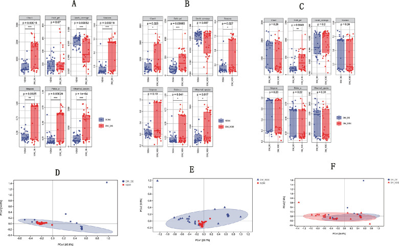
α-Diversity indexes, that is, the Chao1 index, Faith's PD index, Good's coverage index, Shannon index, Simpson index, Pielou's evenness index, and observed species index, were compared among the NDM, DM-DE, and DM-NDE groups. Significant differences were observed in the indexes between the NDM and DM-DE groups except in the case Faith's PD index (P < 0.05). The differences between NDM and DM-NDE groups were statistically significant except for Good's coverage index and the Simpson index (P < 0.05). There was no significant difference between DM-DE group and DM-NDE group, except for Faith's PD index (P > 0.05). The results showed that the microbial richness, diversity, evenness, and coverage of the ocular surface in the DM-DE group were higher than those in the healthy control group (Figs. 2A, B, and C).
Figure 2.
α-Diversity index analysis of the three study groups: (A) NDM versus DM-DE. (B) NDM versus DM-NDE. (C) DM-DE versus DM-NDE. Principal coordinate analysis of the three study groups: (A) NDM versus DM-DE (R2 = 0.352383; P = 0.001). (B) NDM versus DM-NDE (R2 = 0.180094; P = 0.001). (C) DM-DE versus DM-NDE (R2 = 0.041521; P = 0.067).
β-Diversity Analysis
The partial least squares-discriminant analysis model based on the relative abundances of species was constructed, and the dimension value was 2 and the elliptic confidence was 0.95 by the unweighted UniFrac algorithm. The principal coordinate analysis is shown in Figs. 2D, E, and F. In this study, there were significant differences in the microbial compositions on the eye surface among the children in the DM-DE, DM-NDE, and healthy control groups.
Taxonomic Analysis of Micro-Organisms
Microbial Profile
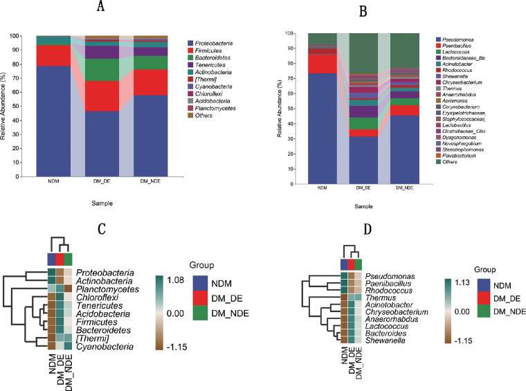
The bacterial composition on the ocular surfaces of the participants of the three groups was compared, and the bacterial 16S rRNA sequences of individual subjects were divided into the phylum and genus levels. The 16S rRNA gene sequencing of the three groups of samples could be divided into 10 dominant phyla (Fig. 3A), and most of the sequences mainly belonged to five phyla. In the DM-DE, DM-NDE, and NDM samples they comprised Proteobacteria (78.4%, 46.3%, and 57.8%), Firmicutes (14.6%, 21.8%, and 18.4%), Bacteroidetes (1.2%, 15.6%, and 9.5%), Tenericutes (0%, 9.3%, and 6.0%), and Actinobacteria (5.1%, 2.8%, and 3.8%), with respective figures provided for each phylum.
Figure 3.
(A) The relative abundance of three groups at phyla level (top 10). (B) The relative abundance of three groups at genus level (top 20). (C) Pheatmap of three groups at phyla level (top 10). (D). Pheatmap of three groups at genus level (top 10).
At the genus level, the 16S rRNA gene sequencing results of the three groups of samples were mostly classified into 10 bacterial genera (Fig. 3B): Pseudomonas (73.4%, 31.4%, and 45.4%), Paenibacillus (12.6%, 4.8%, and 6.6%), Lactococcus (0.0%, 7.9%, and 4.7%), Bacteroides (0.0%, 7.8%, and 4.7%), Acinetobacter (0.2%, 3.9%, and 2.2%), Rhodococcus (3.5%, 1.2%, and 1.6%), Shewanella (0.0%, 3.8%, and 2.3%), Chryseobacterium (0.1%, 3.7%, and 2.1%), Thermus (0.0%, 1.2%, and 1.2%), and Anaerorhabdus (0.0%, 1.4%, and 0.9%). Further analysis showed that the common Clostridium, Staphylococcus, and Lactobacillus were highly abundant in the DM-DE group.
In all samples, Proteobacteria, Firmicutes, Bacteroidetes, Tenericutes, and Actinobacteria were the dominant phyla, whereas Pseudomonas, Paenibacillus, Lactococcus, Bacteroidetes, Acinetobacter, and Rhodococcus were the dominant genera, which together constituted the core microbiota of the ocular surface.
The results of microbial composition heat map showed that the abundance of Proteobacteria and Actinobacteria was higher in the NDM group at the phylum level. In the DM-DE group, Firmicutes, Bacteroidetes, and Tenericutes were more abundant than in the DM group. The abundance of Cyanobacteria was higher in DM-NDE group. At the genus level, the abundance of Pseudomonas, Paenibacillus, and Rhodococcus was the highest in the NDM group. The abundance of Acinetobacter, Lactococcus, and Bacteroides was the highest in the DM-DE group (Figs. 3C and D). All of the above observed differences were statistically significant (P < 0.05).
Difference Analysis Between Groups
Linear Discriminant Analysis Effect Size Analysis
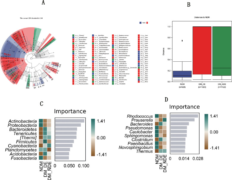
Bacteroidetes, Tenericutes, and Firmicutes were identified by linear discriminant analysis effect size analysis. Lactococcus, Bacteroides, Acinetobacter, Clostridium, Lactobacillus, and Streptococcus are the unique flora of the DM-DE group. Actinobacteria namely Pseudomonas, Paenibacillus, and Rhodococcus were found in the ocular surface of the healthy control group, and there were significant differences between the DM-DE group and healthy control groups (P < 0.05) (Fig. 4A).
Figure 4.
(A) Linear discriminant analysis effect size analysis in three groups. (B) Difference analysis among three groups (F = 12.862; P = 0.001), NDM versus DM-DE (F = 31.020; P = 0.001), NDM versus DM-NDE (F = 14.277; P = 0.001), DM-DE versus DM-NDE (F = 2.301; P = 0.115). (C) Different marker species in three groups at phylum level. (D) Different marker species in three groups at genus level.
Comparison of Differences Between Groups
Using the Bray–Curits distance matrix file, the permutational multivariate ANOVA difference analysis was conducted by Python's Scikit-bio package to obtain the differences in the presence and abundance of different species in the tear samples of the three groups. Difference between the DM-DE and NDM groups was statistically significant (P < 0.05). The difference between the DM-NDE and NDM groups was statistically significant (P < 0.05).There was no significant difference between the DM-DE and DM-NDE group (P > 0.05) (Fig. 4B). This finding indicated that there were differences in the composition of ocular micro-organisms between diabetic children and healthy children.
Species Differences and Marker Species
Random forest analysis revealed significant differences among the three groups (P < 0.05) (Figs. 4C, D). Bacteroidetes, Tenericutes, Firmicutes, and Acidobacteria were more abundant in DM-DE group. Bacteroides and Clostridium were highly abundant in the DM-DE group.
Discussion
According to PubMed search results, DE ocular surface microbe detections and analyses are currently performed only in healthy children and adults, but there is no study on DE surface microbe in children with diabetes. In our project, 16S rRNA was investigated in the tears of children with diabetes and DE, and the specific composition of core micro-organisms on the ocular surface of these patients was determined.
This study found that there were significant differences in the α- and β-diversities of OSM in the DM-DE group compared with the healthy control group, and both α- and β-diversities were higher than those of the healthy control group. This finding is similar to the previously reported conclusion that the α-diversity of OSM in non-DM adults is higher than that in the healthy population,10,12,16,33 and a previous study of our project found that the α-diversity and β-diversity of OSM in adults with DM-DE9 are higher than those in the healthy control group. Shimizu et al. believe that this may be related to the ocular surface epithelial damage and the reduced secretion of mucins and antimicrobial substances in patients with DE, resulting in the reduced elimination of ocular surface bacteria.32
In this study, we found that the main bacterial phyla on the ocular surface of healthy children were composed of Proteobacteria, Firmicutes, and Actinobacteria, supporting previous studies on the ocular surface of healthy adults,34–36 but there are differences in the abundances of the main bacterial phyla, which may be due to the age difference among the participants and also related to different health behaviors, immune status, and living habits of people of different ages.37 This study showed that in children with diabetic ocular surface deformation the bacteria flora were composed of the main phyla: Proteobacteria, Firmicutes, Bacteroidetes, Tenericutes, and Actinobacteria, and the main genera: Pseudomonas, Paenibacillus, Rhodococcus, Exiguobacterium, Corynebacterium, and Staphylococcus, which are greatly different from those in adults with diabetic ocular surface in terms of composition and main species.9,34–36 The reason for this difference, in addition to age, may also be related to the different sampling methods. Considering that the participants of this study were children, the tear test paper with small trauma was selected, which has been reported in previous DE OSM studies9 and is reasonable, but the OSM biological load in tears is low.38 In most other studies, OSM samples are obtained using swabs, which are also affected by sampling site and wiping force,34,39 all leading to differences at the genus level.
More Tenericutes were identified in the OSM of the children with diabetes included in this study, which has not been reported in healthy children and adults in the past. Tenericutes are harmless to humans. The increased abundance of Tenericutes in the OSM of the children with DM may play a certain role in preventing ocular surface damage. Children’s OSM is dissimilar to the species composition in adult diabetic OSM found in past research.9 These differences suggest that the possible mechanism underlying the high incidence of DE in children with diabetes could be flora diversity and composition change, causing abnormality of immunity and substance metabolism on the ocular surface, ocular surface barrier, and immune tolerance and, thus, promoting immune cells to secrete inflammatory factor. In addition, pathogenic bacteria secrete toxic substances and aggravate ocular surface damage.
The phyla and genera (Bacteroidetes, Tenericutes, and Firmicutes and Lactococcus, Bacteroides, Acinetobacter, Clostridium, Lactobacillus, and Streptococcus) were observed to be significantly different between the DM-DE and other two groups. These phyla and genera have been reported to be involved in the pathogenesis of DE in the past,9,10,15,16 but the specific mechanism remains unclear. Interestingly, we found that the abundance of Lactobacillus in OSM of the DM-DE group was higher and significantly different from those of the DM-NDE and NDM groups. Lactobacillus is generally used as a probiotic, and animal studies have shown that local supplementation of Lactobacillus in the eye can treat DE.40 Previously, we believed that increased Lactobacillus in the DM-DE group may be involved in the pathogenesis of DE in children with diabetes, and the possible mechanism is that these bacteria participate in the regulation of nuclear factor-κB and STAT-3 signaling pathways,41,42 or participate in the reduction of lysozyme C and Zn-α-2-glycoprotein in tears.9 In this study, we also found that the ocular surface abundance of Staphylococcus was higher in children with DM-DE. Staphylococcus epidermidis can secrete cholesterol esterase and wax esterase,14 and Staphylococcus aureus can secrete fatty acid–modifying enzyme, lipase, and phospholipase,43 which affects the secretion of lipids from the meibomian glands. Further effects on tear film composition lead to DE. Pseudomonas spp., a normal bacterial group on the ocular surface, have a low dominance in children with DM-DE, and the dominant bacteria are important in protecting the microenvironment of the ocular surface. The decrease and change of normal dominant bacteria on the ocular surface in patients with DM-DE may play a role in the occurrence and pathology of DE. There were significant differences in the dominant bacteria of the ocular surface among the three groups (P < 0.05), and the abundance of OSM in children with DM-DE was higher than that in children with NDM. Compared with that in the children with NDM, the microbial diversity was increased in children with DM-DE, and more diverse communities were likely to cause diseases, which may be the reason for the high incidence of DE in children with diabetes.
This study has some limitations. First, given the very low prevalence of DE in children without diabetes, only 5.00%,1 it was difficult to collect a sufficient sample size of DE in healthy children, and we did not set up a DE group in healthy children. Second, owing to the small number of children with diabetes, we could not specifically compare the gender differences and prepubescent group/postpubescent group within each group. Third, using 16S rRNA sequencing, the difference between viable and nonviable bacteria could be flawed.44,45 At last, the tear test paper used for the collection of tears may have a degree of invasiveness that can easily cause reflective tear secretion and thus affecting the accuracy of the results. The stable presence of commensal bacteria at the ocular surface level has long been a topic of debate owing to the constant flushing of tear films and the antimicrobial properties of eye secretions. The erosion of tears and the damage of lysozymes in tears to micro-organisms on the eye surface diminish the content of effective micro-organisms that can be extracted, affecting the actual composition of micro-organisms on the eye surface. However, tear dipstick collection has the advantages of minor trauma and better suitability for children. At last, the assessment of the ocular surface microbiome in this study was based on 97% homologous OTU clustering, but clustering of bacterial 16S rRNA genes using Illumina MiSeq was only able to determine the relative abundance of specific OTUs. In future studies, metagenomic sequencing or insertion of standardized microbial communities can be used to better understand the absolute abundance of micro-organisms. However, metagenomic detection is expensive and not suitable for the study of a large number of samples, and the microbial content in tears is very low, which does not meet the conditions of metagenomic detection.
In conclusion, this study found that DE in children with DM was accompanied by characteristic OSM changes. In the future, multicenter clinical studies can be performed to expand the sample size, increase the sampling at multiple time points, and screen the bacteria closely related to the development of DM-DE. Metagenomic sequencing can also be used to detect the composition and function of OSM in patients with DM-DE and explore the pathogenesis of DE.
Acknowledgments
Supported by Chinese National key research and development programme (Project number 2021YFC2702100), Shanghai engineering research center of precise diagnosis and treatment of eye diseases, Shanghai, China (Project No. 19DZ2250100),Shanghai General Hospital, Clinical Research CTCCR-2018Z01.
Disclosure: Z. Chen, None; Y. Jia, None; Y. Xiao, None; Q. Lin, None; Y. Qian, None; Z. Xiang, None; L. Cui, None; X. Qin, None; S. Chen, None; C. Yang, None; H. Zou, None
References
- 1. Wang S, Jia Y, Li T, et al.. Dry eye disease is more prevalent in children with diabetes than in those without diabetes. Curr Eye Res. 2019; 44(12): 1299–1305. [DOI] [PubMed] [Google Scholar]
- 2. Manaviat MR, Rashidi M, Afkhami-Ardekani M, et al.. Prevalence of dry eye syndrome and diabetic retinopathy in type 2 diabetic patients. BMC Ophthalmol. 2008; 8(10): 284–291. [DOI] [PMC free article] [PubMed] [Google Scholar]
- 3. Chen Z, Xiao Y, Qian Y, et al.. Incidence and risk factors of dry eye in children and adolescents with diabetes mellitus: a 3-year follow-up study. Front Med (Lausanne). 2021; 11(8): 760006. [DOI] [PMC free article] [PubMed] [Google Scholar]
- 4. Gevers KR, Petrosino JF, Huang K, et al.. The Human Microbiome Project: a community resource for the healthy human microbiome. PLoS Biology. 2012; 10(8): e1001377. [DOI] [PMC free article] [PubMed] [Google Scholar]
- 5. Willcox M. Characterization of the normal microbiota of the ocular surface. Exp Eye Res. 2013; 117: 99–105. [DOI] [PubMed] [Google Scholar]
- 6. KV Gao X, Antonopoulos DA, et al.. Diversity of bacteria at healthy human conjunctiva. Invest Ophthalmol Vis Sci. 2011; 52(8): 5408–5413. [DOI] [PMC free article] [PubMed] [Google Scholar]
- 7. Gupta P.C, Ram J.. Conjunctival microbial flora in ocular Stevens-Johnson syndrome sequelae patients at a tertiary eye care center. Cornea. 2016; 35(8): 1117–1121. [DOI] [PubMed] [Google Scholar]
- 8. Miller D, Iovieno A.. The role of microbial flora on the ocular surface. Curr Opin Allergy Clin Immunol. 2009; 9(5): 466–470. [DOI] [PubMed] [Google Scholar]
- 9. Zhang Z, Zou X, Xue W, et al.. Ocular surface microbiota in diabetic patients with dry eye disease. Invest Ophthalmol Vis Sci. 2021; 62(12): 13. [DOI] [PMC free article] [PubMed] [Google Scholar]
- 10. Li Z, Gong Y, Chen S, et al.. Comparative portrayal of ocular surface microbe with and without dry eye. J Microbiol. 2019; 57(11): 1025–1032. [DOI] [PubMed] [Google Scholar]
- 11. Andersson J, Vogt JK, Dalgaard MD, et al.. Ocular surface microbiota in patients with aqueous tear-deficient dry eye. Ocul Surf. 2021; 19: 210–217. [DOI] [PubMed] [Google Scholar]
- 12. Willis KA, Postnikoff CK, Freeman A, et al.. The closed eye harbours a unique microbiome in dry eye disease. Sci Rep. 2020; 10(1): 12035. [DOI] [PMC free article] [PubMed] [Google Scholar]
- 13. Graham JE, Moore JE, Jiru X, et al.. Ocular pathogen or commensal: a PCR-based study of surface bacterial flora in normal and dry eyes. Invest Ophthalmol Vis Sci. 2007; 48(12): 5616–5623. [DOI] [PubMed] [Google Scholar]
- 14. Zhao F, Zhang D, Ge C, et al.. Metagenomic profiling of ocular surface microbiome changes in meibomian gland dysfunction. Invest Ophthalmol Vis Sci. 2020; 61(8): 22. [DOI] [PMC free article] [PubMed] [Google Scholar]
- 15. Dong X, Wang Y, Wang W, et al.. Composition and diversity of bacterial community on the ocular surface of patients with meibomian gland dysfunction. Invest Ophthalmol Vis Sci. 2019; 60(14): 4774–4783. [DOI] [PubMed] [Google Scholar]
- 16. Kittipibul T, Puangsricharern V, Chatsuwan T.. Comparison of the ocular microbiome between chronic Stevens-Johnson syndrome patients and healthy subjects. Sci Rep. 2020; 10(1): 4353. [DOI] [PMC free article] [PubMed] [Google Scholar]
- 17. Li S, Yi G, Peng H, et al.. How ocular surface microbiota debuts in type 2 diabetes mellitus. Front Cell Infect Microbiol. 2019; 9: 202. [DOI] [PMC free article] [PubMed] [Google Scholar]
- 18. Zhu X, Wei L, Rong X, et al.. Conjunctival microbiota in patients with type 2 diabetes mellitus and influences of perioperative use of topical levofloxacin in ocular surgery. Front Med (Lausanne). 2021; 8: 605639. [DOI] [PMC free article] [PubMed] [Google Scholar]
- 19. Shih KC, Lam KS, Tong L.. A systematic review on the impact of diabetes mellitus on the ocular surface. Nutr Diabetes. 2017; 7(3): e251. [DOI] [PMC free article] [PubMed] [Google Scholar]
- 20. Clayton JA. Dry eye. N Engl J Med. 2018; 378(23): 2212–2223. [DOI] [PubMed] [Google Scholar]
- 21. Shi L, Yu X, Yang H, et al.. Advanced glycation end products induce human corneal epithelial cells apoptosis through generation of reactive oxygen species and activation of JNK and p38 MAPK pathways. PLoS One. 2013; 8(6): e66781. [DOI] [PMC free article] [PubMed] [Google Scholar]
- 22. Leppin K, Behrendt AK, Reichard M, et al.. Diabetes mellitus leads to accumulation of dendritic cells and nerve fiber damage of the subbasal nerve plexus in the cornea. Invest Ophthalmol Vis Sci. 2014; 55(6): 3603–3615. [DOI] [PubMed] [Google Scholar]
- 23. Kim HC, Cho YJ, Ahn CW, et al.. Nerve growth factor and expression of its receptors in patients with diabetic neuropathy. Diabet Med. 2009; 26(12): 1228–1234. [DOI] [PubMed] [Google Scholar]
- 24. Priyadarsini S, Sarker-Nag A, Allegood J, et al.. Description of the sphingolipid content and subspecies in the diabetic cornea. Curr Eye Res. 2015; 40(12): 1204–1210. [DOI] [PMC free article] [PubMed] [Google Scholar]
- 25. Goldin A, Beckman JA, Schmidt AM, et al.. Advanced glycation end products: sparking the development of diabetic vascular injury. Circulation. 2006; 114(6): 597–605. [DOI] [PubMed] [Google Scholar]
- 26. Zhang X, VJ M, Qu Y, et al.. Dry eye management: targeting the ocular surface microenvironment. Int J Mol Sci. 2017; 18(7): 1398. [DOI] [PMC free article] [PubMed] [Google Scholar]
- 27. Qin J, Li R, Raes J, et al.. A human gut microbial gene catalogue established by metagenomic sequencing. Nature. 2010; 464(7285): 59–65. [DOI] [PMC free article] [PubMed] [Google Scholar]
- 28. Ham B, Hwang HB, Jung SH, et al.. Distribution and diversity of ocular microbial communities in diabetic patients compared with healthy subjects. Curr Eye Res. 2018; 43: 314–324. [DOI] [PubMed] [Google Scholar]
- 29. Diabetes Mellitus. Report of a WHO study group. World Health Organ Tech Rep Ser. 1985; 727: 1–113. [PubMed] [Google Scholar]
- 30. Wolffsohn JS, Arita R, Chalmers R, et al.. TFOS DEWSII diagnostic methodology report. Ocul Surf. 2017; 15(3): 539–574. [DOI] [PubMed] [Google Scholar]
- 31. Kelly B J, Gross R, Bittinger K, et al.. Power and sample-size estimation for microbiome studies using pairwise distances and PERMANOVA. Bioinformatics. 2015; 31(15): 2461–2468. [DOI] [PMC free article] [PubMed] [Google Scholar]
- 32. Baim A D, Movahedan A, Farooq A V, et al.. The microbiome and ophthalmic disease. Exp Biol Med (Maywood). 2019; 244(6): 419–429. [DOI] [PMC free article] [PubMed] [Google Scholar]
- 33. Shimizu E, Ogawa Y, Saijo Y, et al.. Commensal microflora in human conjunctiva: characteristics of microflora in the patients with chronic ocular graft-versus-host disease. Ocul Surf. 2019; 17(2): 265–271. [DOI] [PubMed] [Google Scholar]
- 34. Dong Q, Brulc JM, Iovieno A, et al.. Diversity of bacteria at healthy human conjunctiva. Invest Ophthalmol Vis Sci. 2011; 52(8): 5408–5413. [DOI] [PMC free article] [PubMed] [Google Scholar]
- 35. Huang Y, Yang B, Li W. Defining the normal core microbiome of conjunctival microbial communities. Clin Microbiol Infect. 2016; 22(7): 643.e7–643.e12. [DOI] [PubMed] [Google Scholar]
- 36. Zhou Y, Holland M J, Makalo P, et al.. The conjunctival microbiome in health and trachomatous disease: a case control study. Genome Med. 2014; 6(11): 99. [DOI] [PMC free article] [PubMed] [Google Scholar]
- 37. Wen X, Miao L, Deng Y, et al.. The influence of age and sex on ocular surface microbiota in healthy adults. Invest Ophthalmol Vis Sci. 2017; 58(14): 6030–6037. [DOI] [PubMed] [Google Scholar]
- 38. Ozkan J, Willcox MD.. The ocular microbiome: molecular characterisation of a unique and low microbial environment. Curr Eye Res. 2019; 44(7): 685–694. [DOI] [PubMed] [Google Scholar]
- 39. Ozkan J, Willcox M, Wemheuer B, et al.. Biogeography of the human ocular microbiota. Ocul Surf. 2019; 17(1): 111–118. [DOI] [PubMed] [Google Scholar]
- 40. Kim J, Choi S H, Kim Y J, et al.. Clinical effect of IRT-5 probiotics on immune modulation of autoimmunity or alloimmunity in the eye. Nutrients. 2017; 9(11): 1166. [DOI] [PMC free article] [PubMed] [Google Scholar]
- 41. Kumar A, Wu H, Collier-Hyams LS, et al.. Commensal bacteria modulate cullin-dependent signaling via generation of reactive oxygen species. EMBO J. 2007; 26(21): 4457–4466. [DOI] [PMC free article] [PubMed] [Google Scholar]
- 42. Lim SM, Jang HM, Jang SE, et al.. Lactobacillus fermentum IM12 attenuates inflammation in mice by inhibiting NF-kappaB-STAT3 signalling pathway. Beneficial Microbes. 2017; 8(3): 407–419. [DOI] [PubMed] [Google Scholar]
- 43. Tam K, Torres VJ.. Staphylococcus aureus secreted toxins and extracellular enzymes. Microbiol Spectr. 2019; 7(2): 10. [DOI] [PMC free article] [PubMed] [Google Scholar]
- 44. Ozkan J, Nielsen S, Diez-Vives C, et al.. Temporal stability and composition of the ocular surface microbiome. Sci Rep. 2017; 7(1): 9880. [DOI] [PMC free article] [PubMed] [Google Scholar]
- 45. Doan T, Akileswaran L, Andersen D, et al.. Paucibacterial microbiome and resident DNA virome of the healthy conjunctiva. Invest Ophthalmol Vis Sci. 2016; 57(13): 5116–5126. [DOI] [PMC free article] [PubMed] [Google Scholar]


