Abstract
Calvarium and skull base can be affected by a variety of benign, tumor-like, and malignant processes. Skull metastases (SMs) may be located in any layer of the skull and may be incidental or present with neurological symptoms during the diagnostic workup. In the present study, we discuss the occurrence of SMs from various index malignancies and their myriad clinical presentation. This data-based study includes patients of bone metastases between June 2018 and July 2020. Patients with skull bone metastases were recognized, and location of primary site, their clinical presentation, and management strategy were noted. Ten patients with skull bone metastases were identified during this period. Four patients had skull base location with clinical manifestation as syndromes. Six patients had primary from breast cancer, three from Ewing's sarcoma, and one from lung cancer. Management varied according to the primary site and symptoms of each patient. SM, though not rare, is often diagnosed incidentally but presents diagnostic and management challenges in the patient with cancer.
Keywords: calvarial, metastases, skull base, disseminated disease
Introduction
Bone metastases are relatively common in many malignancies. Breast, prostate, and lung account for approximately 85% of metastatic lesions to the bone. Kidney, thyroid, and melanoma are other sites with the propensity for bone metastasis. Lumbar spine is the most common site of bone metastasis in the axial skeleton, while the proximal femur is most common in the appendicular skeleton. Skull metastasis (SM) though not rare is often diagnosed incidentally. 1 2 SM may be located in any layer of the skull and may be either osteolytic or sclerotic. When they present with symptoms such as increased intracranial pressure due to dural sinus involvement, the patient almost always has disseminated disease. 3 Management varies from conservative to surgical resection to palliation of symptoms. Stark et al and Hong et al reported surgical resection in 12 and 36 patients with SM, respectively. 4 5 Stark et al proposed that surgical option in SM should be considered if there is neurological deficit, massive destruction of bone with dura infiltration, mass is causing pain, and lesion is solitary and for confirmation of diagnosis. Overall prognosis in SM remains poor. Here, we present a series of 10 cases from various primary histologies and varied clinical presentation.
Materials and Methods
We retrospectively reviewed medical records of patients of bone metastases between June 2018 and July 2020. Patients with skull bone metastases were identified, and location of primary site, their clinical presentation, and management strategy were noted. The clinical data included age, gender presenting symptoms and signs, index malignancy, radiological findings, and management. Any associated clinical and laboratory findings were also observed.
Results
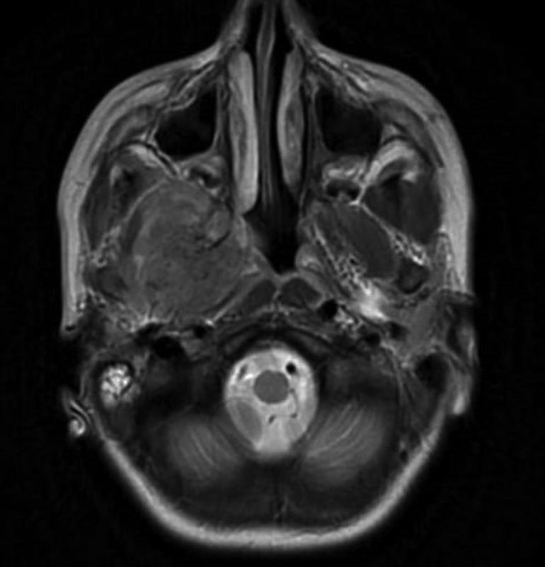
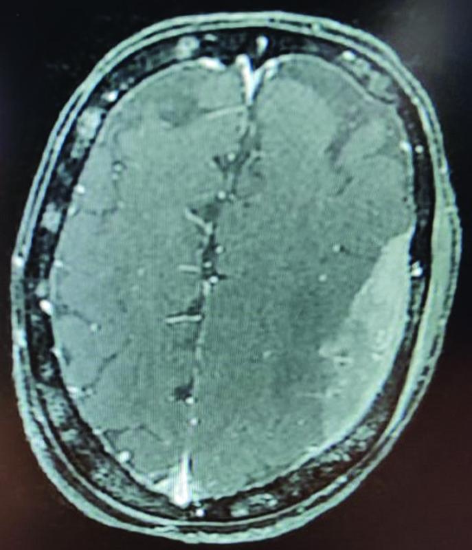
Ten cases of calvarial metastasis from different anatomical sites were identified during this period. Out of 10 cases of calvarial metastasis, 3 were from Ewing's sarcoma, 6 of them from breast cancer, and 1 from lung cancer. Out of 10 cases, 2 were males and 8 were females. The details of the clinical characteristics of patient and treatment are summarized in Table 1 . Three patients had metastases in the base of the skull region, one sphenoid and two temporal region, three had multiple calvarial metastases, and four had single calvarial metastasis ( Figs. 1 , 2 ). Four patients presented with the neurological deficit: ipsilateral facial palsy, lower limb weakness, slurring of speech, and angle of mouth deviation. Also, one patient presented with hypercalcemia (serum Ca 13.9 mg/dL). Patients with neurological deficits were given steroids to reduce tumoral edema, and it leads to symptomatic improvement in their deficit. All the patients had received chemotherapy according to the primary tumor site. One patient with hypercalcemia received bisphosphonates and improved gradually. Three patients received palliative whole brain radiotherapy because of multiple calvarial metastases and brain parenchyma infiltration, three patients received involved-field radiation to localized region, three patients received palliative chemotherapy only as they were not symptomatic for calvarial lesions, and one patient defaulted from further treatment.
Table 1. Clinical presentation and management details of patients with calvarial metastases.
| Sl. no. | Age/gender | Symptoms | Primary malignancy | Brain imaging | Management |
|---|---|---|---|---|---|
| 1 | 8 y/F | Right lower limb swelling and pain from 5 mo | Right thigh Ewing's sarcoma | CECT of the brain shows bone metastasis to skull base greater wing of sphenoid | Received two courses of VAC chemotherapy |
| 2 | 8 y/M | Right thigh swelling and pain in right thigh from 4 mo | Right proximal thigh Ewing's sarcoma | CEMRI showed metastatic lesion in the base of the skull (temporal) | Received VAC and IE chemotherapies |
| 3 | 13 y/F | Pain and swelling in the left gluteal region | Left iliac bone Ewing's sarcoma | CEMRI of the brain showed mass in right temporal bone region involving right infratemporal fossa | Received VAC/IE-based chemotherapy followed by local RT to left ilium and parietal bone metastasis |
| 4 | 67 y/F | Lump breast × 3 mo and later developed slurring of speech and angle of mouth deviation | Left breast invasive ductal carcinoma | CEMRI of the brain ( Fig. 1 ) showed calvarial and extradural metastases with infiltration into the brain parenchyma | Post-MRM received palliative WBRT 20 Gy in five fractions followed by palliative chemotherapy |
| 5 | 60 y/F | Breast lump × 5 mo later presented with complaints of the decreased sensorium, pain, and weakness in right lower limb | Right breast invasive ductal carcinoma | Noncontrast CT suggestive of a lytic lesion with associated soft tissue in occipital bone with multiple lytic lesions in calvaria | Palliative WBRT 20 Gy in five fractions |
| 6 | 42 y/F | Breast lump, headache | Left breast invasive ductal carcinoma | CECT of the brain showed multiple calvarial metastases | Palliative WBRT 20 Gy in five fractions |
| 7 | 45 y/F | Breast lump and left facial weakness | Left breast invasive ductal carcinoma | CECT of the brain showed occipital bone metastasis | Palliative radiation 20 Gy in five fractions to occipital region |
| 8 | 70 y/F | Breast lump with ulceration and bleeding, headache | Right breast invasive carcinoma | CECT of the brain showed occipital bone metastasis | Palliative RT to occipital region |
| 9 | 65 y/M | Hemoptysis, pain over shoulder region, and swelling over scalp | Left lung squamous cell carcinoma | CECT of the brain showed a single solitary deposit in calvarial bone | Defaulted for treatment |
| 10 | 45 y/F | Headache, vomiting, and generalized weakness | Carcinoma left breast with hypercalcemia | CEMRI of the brain suggestive of cavarial metastases without parenchyma involvement | Bisphosphonates and palliative chemotherapy |
Abbreviations: CECT, contrast-enhanced computed tomography; CEMRI, contrast-enhanced magnetic resonance imaging; CT, computed tomography; IE, ifosfamide and etoposide; RT, radiotherapy; VAC, vincristine, Adriamycin, cyclophosphamide; WBRT, whole brain radiotherapy.
Fig. 1.

Contrast-enhanced magnetic resonance imaging showing mass in right temporal region involving infratemporal fossa.
Fig. 2.

Contrast-enhanced magnetic resonance imaging showing calvarial and extradural metastases infiltrating into the brain parenchyma.
Discussion
The true incidence of SMs is not known, but reported incidence is seen in 15 to 25% of patients with advanced systemic cancer. 6 Occasionally, it might be the initial presentation of index malignancy. When we encounter a patient with multiple osteolytic lesions in the skull, the first assumption is multiple myeloma. 7 Osteolytic metastases are most frequently encountered in breast and lung carcinomas. Other rare tumors, such as reticulum cell sarcoma, angiosarcoma, and malignant fibrous histiocytoma, were also reported to show osteolytic bone lesions. 8 The calvaria consists of an inner table, bone marrow space, and an outer table. Metastases to the calvarial bones usually involve all the three skull layers. The spread is usually hematogenous and less likely through extension of cranial nerves. The most likely mechanism is retrograde spread via Batson's valveless venous plexus. 9 Approximately 50% of the patients with calvarial metastases are asymptomatic, and presentation of patients with outer table, periosteal, or dural involvement may include localized pain in the setting of a palpable mass. Inner table of the skull can result in neurological symptoms including headaches, neurological deficits, meningeal irritation, and seizures. When these neurological sequelae are present, aggressive management is warranted for lesions invading the inner table of the bone and with dural involvement. 10 Skull base metastases usually show symptoms due to engrossment of cranial nerves. Dysphagia, diplopia, trigeminal and occipital neuralgia are extremely incapacitating symptoms and are alarming indicators of skull base involvement in cancer patients. Five syndromes mentioned for skull base metastases are the orbital, parasellar, middle fossa, jugular foramen, and occipital condyle syndromes, and these are stated according to site of metastases. 11 A retrospective cohort study of 175 patients with SMs found breast as the most common site of origin (55%) of SMs, followed by lung (14%) from lung carcinoma, prostate cancer (6%), and rest sites constituted 25%. 12 Ewing's sarcoma can also metastasize to the central nervous system (CNS) with a relatively low incidence (6.3% of cases), and in this case series, there are three cases. There are two reported principal modes of metastatic spread of Ewing's sarcoma to the CNS. The first is a direct extension from the skull, which may be the site of both primary and secondary Ewing's sarcomas. 13 Computed tomography scan with bone window is helpful to establish the diagnosis, but in patients with soft tissue extension or coexisting brain metastases, magnetic resonance imaging is more informative. Bone scan has poor diagnostic ability in the case of osteolytic bone metastases. 6 Patients with SM are usually unsuitable for surgical intervention, due to the nature of primary malignancy, extent of disease, and quantity of lesions. The difficulty of surgery depends on the involvement of vital structures such as the dural sinuses. However, the surgical technique is forthright and involves complete excision of the lesion and cranioplasty. Thus, radiotherapy is another option for treatment. Gamma Knife surgery is one of the emerging modalities in cases of superficial calvarial lesions and had tried in the group of patients by Kotecha et al with a significant amount of success. Systemic chemotherapy with cytotoxic agents has historically induced a modest reduction in tumor mass and will likely have a role in the management of CNS metastases alongside surgery and radiation. 14 Overall prognosis is poor for patients with skull base involvement as these metastases appear late during the course of the disease.
Conclusion
According to our knowledge, the literature is not so robust for large osteolytic metastases involving the skull. These metastases may have varied clinical symptoms ranging from bone pain to neurological deficits. Management should be individualized depending on location in skull and symptoms of the patient.
Funding Statement
Funding None.
Footnotes
Conflict of Interest None declared.
References
- 1.Chin H, Kim J. Bone metastasis: concise overview. Fed Pract. 2015;32(02):24–30. [PMC free article] [PubMed] [Google Scholar]
- 2.Shen J, Wang S, Zhao X. Skull metastasis from follicular thyroid carcinoma: report of three cases and review of literature. Int J Clin Exp Pathol. 2015;8(11):15285–15293. [PMC free article] [PubMed] [Google Scholar]
- 3.Takeda H, Ohe R, Fukui T. Rapid progression of intracranial dural metastases in a patient with carcinoma of unknown primary site. Case Rep Oncol. 2019;12(02):666–670. doi: 10.1159/000502416. [DOI] [PMC free article] [PubMed] [Google Scholar]
- 4.Stark A M, Eichmann T, Mehdorn H M.Skull metastases: clinical features, differential diagnosis, and review of the literature Surg Neurol 20036003219–225., discussion 225–226 [DOI] [PubMed] [Google Scholar]
- 5.Hong B, Hermann E J, Klein R, Krauss J K, Nakamura M. Surgical resection of osteolytic calvarial lesions: clinicopathological features. Clin Neurol Neurosurg. 2010;112(10):865–869. doi: 10.1016/j.clineuro.2010.07.010. [DOI] [PubMed] [Google Scholar]
- 6.Mitsuya K, Nakasu Y, Horiguchi S. Metastatic skull tumors: MRI features and a new conventional classification. J Neurooncol. 2011;104(01):239–245. doi: 10.1007/s11060-010-0465-5. [DOI] [PMC free article] [PubMed] [Google Scholar]
- 7.Ugga L, Cuocolo R, Cocozza S. Spectrum of lytic lesions of the skull: a pictorial essay. Insights Imaging. 2018;9(05):845–856. doi: 10.1007/s13244-018-0653-y. [DOI] [PMC free article] [PubMed] [Google Scholar]
- 8.Kang Y M, Lee H J, Kim S J. Metastatic breast cancer with osteolytic skull lesions suspected to be multiple myeloma. Korean J Clin Oncol. 2017;13(02):152–155. [Google Scholar]
- 9.Laigle-Donadey F, Taillibert S, Martin-Duverneuil N, Hildebrand J, Delattre J Y. Skull-base metastases. J Neurooncol. 2005;75(01):63–69. doi: 10.1007/s11060-004-8099-0. [DOI] [PubMed] [Google Scholar]
- 10.Kotecha R, Angelov L, Barnett G H.Calvarial and skull base metastases: expanding the clinical utility of Gamma Knife surgery J Neurosurg 2014121(Suppl):91–101. [DOI] [PubMed] [Google Scholar]
- 11.Mitsuya K, Nakasu Y. Metastatic skull tumours: diagnosis and management. Eur Assoc Neuro Oncol Mag. 2014;4(02):71–74. [Google Scholar]
- 12.Altalhy A, Maghrabi Y, Almansouri Z, Baeesa S S. Solitary skull metastasis as the first presentation of a metachronous primary lung cancer in a survivor from pancreatic cancer. Case Rep Oncol Med. 2017;2017:5.674749E6. doi: 10.1155/2017/5674749. [DOI] [PMC free article] [PubMed] [Google Scholar]
- 13.Ben Nsir A, Boughamoura M, Maatouk M, Kilani M, Hattab N. Dural metastasis of Ewing's sarcoma. Surg Neurol Int. 2013;4:96. doi: 10.4103/2152-7806.115487. [DOI] [PMC free article] [PubMed] [Google Scholar]
- 14.Rick J W, Shahin M, Chandra A. Systemic therapy for brain metastases. Crit Rev Oncol Hematol. 2019;142:44–50. doi: 10.1016/j.critrevonc.2019.07.012. [DOI] [PMC free article] [PubMed] [Google Scholar]


