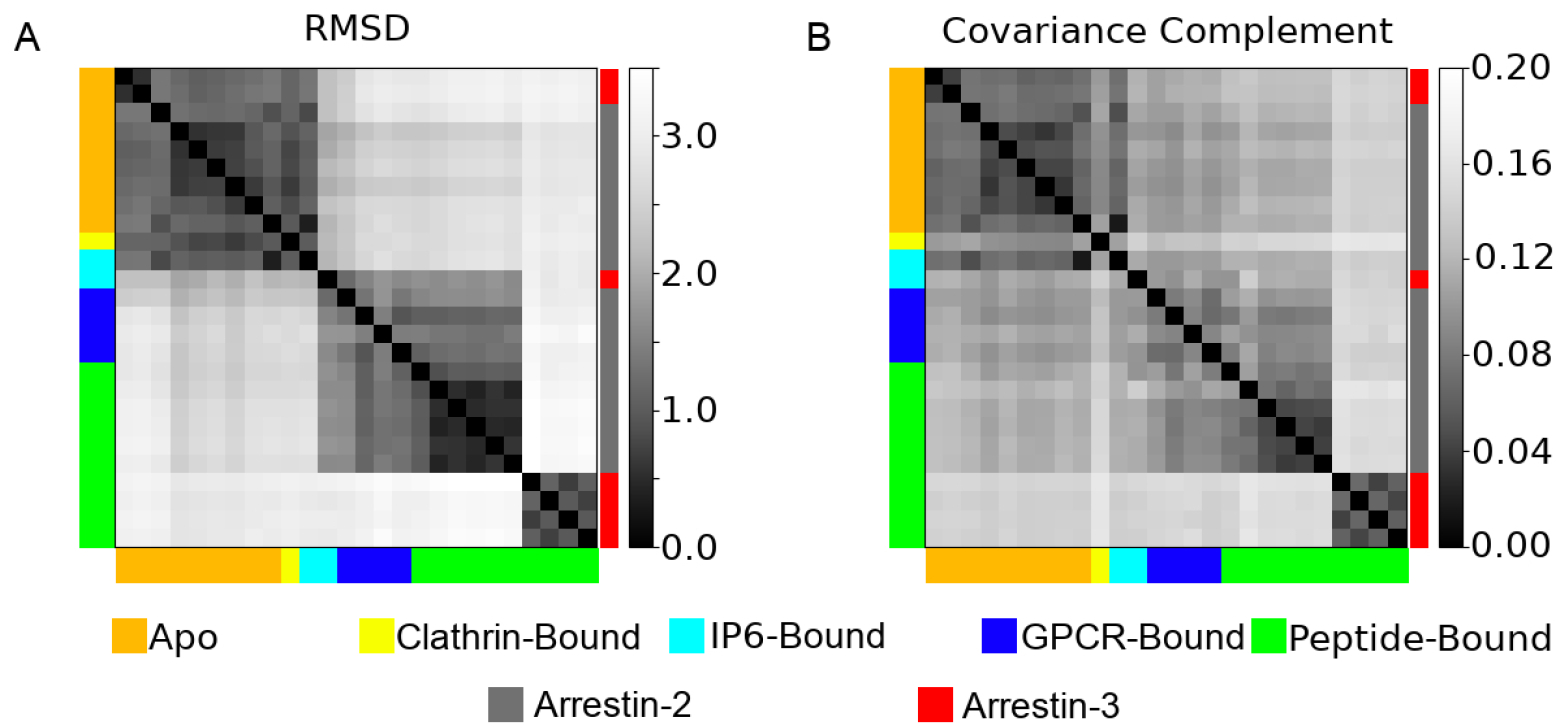
Figure 3:

A pairwise comparison of the A) root mean squared deviation (RMSD) and B) the covariance complement derived from the 26 different structures analyzed in this study. Structures are color coded both by their ligands (left and bottom of graph), or whether they are arrestin-2 (gray) or arrestin-3 (red) (right of graph).
