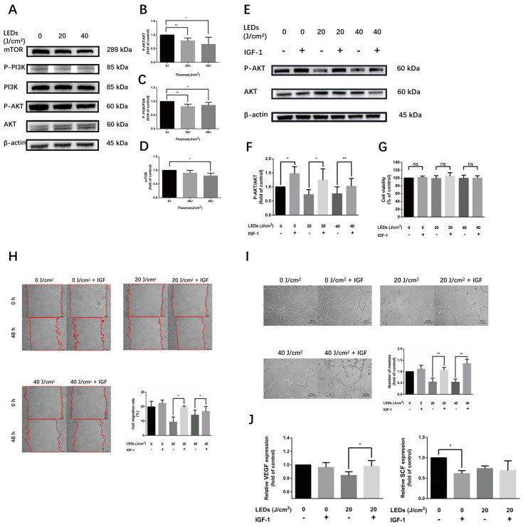
Figure 5.
590 nm LED inhibited angiogenesis of HMEC-1 predominantly via AKT/PI3K/mTOR pathway. (A–D) The phosphorylation level of AKT/PI3K/mTOR pathway after LED illumination (20, 40 J/cm2, 0.5 h). (E,F) The phosphorylation level of AKT after LED irradiation (20, 40 J/cm2) alone or pretreated with IGF-1 (50 ng/mL, 2 h). (G) Cell viability of irradiated cells or with IGF-1 pretreatment. (H) Cell migration (48 h) of wound healing assay after LED illumination alone or pretreated with IGF-1. Bar, 100 μm. (I) Number of meshes (8 h) of HMEC-1 after LED irradiation alone or with IGF-1 pretreatment. Bar, 100 μm. (J) The secretion of VEGF and SCF from cells irradiated by 20 J/cm2 LED alone or with IGF-1 pretreatment detected by ELISA. (B–D,F,G,I,J) Results of irradiated groups are normalized to the control. Data are shown as means ± SD. (A–F) n = 5; (G–J) n = 3. *, p < 0.05; **, p < 0.01. LED, light-emitting diodes; IGF-1, insulin-like growth factor 1; VEGF, vascular endothelial growth factor; SCF, stem cell factor; n.s., no significance.

