Abstract
Simple Summary
Cell-free DNA RAS mutation is being increasingly monitored in metastatic colorectal cancer (mCRC) for disease molecular characterization and selecting eligible patients for anti-EGFR initiation and rechallenge. Here, we monitored a homogeneous mCRC RAS wild-type (as per baseline solid biopsy) population starting first-line treatment using a BEAMing technique at three different mutant allele fraction (MAF) sensitivity cut-offs and we characterized the role of each MAF threshold and its correlation with clinical variables.
Abstract
The serial analysis of cell-free DNA (cfDNA) enables minimally invasive monitoring of tumor evolution, providing continuous genetic information. PERSEIDA was an observational, prospective study assessing the cfDNA RAS (KRAS/NRAS) mutational status evolution in first-line, metastatic CRC, RAS wild-type (according to baseline tumor tissue biopsy) patients. Plasma samples were collected before first-line treatment, after 20 ± 2 weeks, and at disease progression. One hundred and nineteen patients were included (102 received panitumumab and chemotherapy as first-line treatment—panitumumab subpopulation). Fifteen (12.6%) patients presented baseline cfDNA RAS mutations (n = 14 [13.7%], panitumumab subpopulation) (mutant allele fraction ≥0.02 for all results). No patients presented emergent mutations (cfDNA RAS mutations not present at baseline) at 20 weeks. At disease progression, 11 patients (n = 9; panitumumab subpopulation) presented emergent mutations (RAS conversion rate: 19.0% [11/58]; 17.7% [9/51], panitumumab subpopulation). In contrast, three (5.2%) patients presenting baseline cfDNA RAS mutations were RAS wild-type at disease progression. No significant associations were observed between overall response rate or progression-free survival and cfDNA RAS mutational status in the total panitumumab subpopulation. Although, in patients with left-sided tumors, a significantly longer progression-free survival was observed in cfDNA RAS wild-type patients compared to those presenting cfDNA RAS mutations at any time. Continuous evaluation of RAS mutations may provide valuable insights on tumor molecular dynamics that can help clinical practice.
Keywords: colorectal cancer, cell-free DNA, RAS mutations, solid biopsy
1. Introduction
Colorectal cancer (CRC) is the third most diagnosed cancer in men and the second most diagnosed cancer in women, and it is the second most common cause of cancer death in men and the third most common cause in women globally [1]. In Europe, the incidence in 2020 was 150,000 and 191,000 new cases in women and men, respectively [2], whereas in Spain, over 43,370 new cases were predicted for 2022 in the total population [3]. The recent advances in cytotoxic chemotherapy and targeted agents have improved overall survival, doubling it over the last 20 years up to 30 months [4].
In patients with metastatic CRC (mCRC), RAS (KRAS/NRAS) mutational status currently guides the therapeutic use of epidermal growth factor receptor (EGFR) inhibitors [4]. Tumor tissue biopsy testing is the standard of care to assess RAS (KRAS/NRAS) mutation in these patients [5,6,7,8]. Its determination must be performed in the primary tumor or the metastatic tissue upon the diagnosis of metastatic disease according to current treatment guidelines [4].
Detection of genetic alterations in tumor DNA is increasingly used for diagnostic, prognostic, and treatment purposes. Usually, genetic alterations are detected from archived tumor samples collected at a specific time, which do not provide information on disease progression or heterogeneity. Colorectal cancer harbors a considerable heterogenicity, with temporal and spatial differences in genetic mutations [9]. Tumor cells release 150–~200 base pair fragments of circulating tumor DNA (ctDNA) into the bloodstream [10]. This ctDNA normal half-life is less than an hour and contains the same genetic and epigenetic characteristics and mutations of the tumor [10], providing key information related to tumor development, progression, and resistance to treatment. The analysis of plasma ctDNA, also called “liquid biopsy”, has been actively studied and tested as a possible alternative to the invasive techniques for obtaining tumor insight. Liquid biopsy enables minimally invasive monitoring of tumor evolution which could provide continuous genetic information about the tumor while the patients is being treated and may overcome some of the challenges associated with tumor heterogeneity, such as the spatial and temporal heterogeneity [11,12,13].
Despite all of the liquid biopsy advantages, one of the main inconveniences is the sensitivity of the techniques used. ctDNA may vary between 0.01% and 93% of the total circulating free DNA (cfDNA) [14,15]. Some studies to detect KRAS mutations in cfDNA by PCR have not provided adequate concordance with the results obtained from solid biopsies [16,17,18]. Thus, alternative techniques such as BEAMing, which are highly sensitive for detecting the presence of point mutations in cfDNA even when they are uncommon, are required [19,20]. Aiming to provide new evidence on the potential added value of baseline liquid biopsy genotyping in the first-line setting, the primary objective of this observational, prospective study was to assess the concordance of the RAS (KRAS/NRAS) mutational status assessed in tissue samples and plasma samples (using the BEAMing technique) at baseline in RAS wild-type mCRC patients starting their standard first-line treatment. Additionally, the RAS (KRAS/NRAS) mutational status was assessed at 20 weeks after treatment initiation and at disease progression to first-line treatment. Moreover, the associations between RAS mutation status and different outcomes according to different mutant allele fraction (MAF) cut-offs and the predictive factors of progression-free survival (PFS) and tumor burden were explored in the panitumumab-treated subpopulation.
2. Materials and Methods
PERSEIDA (NCT02792478) was a nationwide, observational, multi-center, prospective study designed to evaluate the RAS (KRAS/NRAS) mutational status in liquid biopsies in first-line, mCRC, RAS wild-type (according to baseline tumor tissue biopsy) patients. The participants were managed following standard clinical practice, including the selection of the first-line treatment and the baseline tumor tissue biopsy.
Blood samples were collected before starting first-line treatment (baseline), at 20 ± 2 weeks after starting the treatment (prior to the second radiological assessment of the tumor response) and at disease progression coinciding with routine blood withdrawals. The samples were centrifuged to obtain plasma. The plasma was then frozen and maintained at −80 °C until it was shipped to Sysmex Inostics GmbH (Hamburg, Germany) for BEAMing analysis [21] at the end of the study. Accordingly, all of the investigators were blind to the BEAMing results during the study. The 20 weeks after starting treatment timepoint was selected following the Diaz et al. estimations of mutant KRAS fragments predicted to become evident after anti-EGFR initiation [22].
Tumor response was evaluated approximately every 3 months following RECIST version 1.1 criteria [23] until tumor progression, following clinical practice.
The inclusion criteria were: patients ≥18 years, with mCRC measurable by RECIST, who have started first-line treatment and with a histologically confirmed diagnosis of mCRC and wild-type RAS (according to baseline tumor tissue biopsy). The exclusion criteria were: pregnant or breastfeeding women, patients who have previously received monoclonal antibodies against EGFR (cetuximab or panitumumab), small-molecule EGFR inhibitors (such as erlotinib) or other biological cancer treatments, patients with a history of another solid or hematological tumor in the previous 5 years (except a history of basal cell carcinoma of the skin or pre-invasive cervical cancer), and patients who were participating or had participated in a clinical trial in the 30 days prior to inclusion.
The protocol was approved by an independent ethics committee, and all of the patients gave their written informed consent before enrollment.
The patients were recruited consecutively and were followed-up until disease progression. At baseline, the following variables were collected: demographic data, relevant medical history (including CRC-related data: date of histological diagnosis, primary location, previous surgery and outcome, previous treatments, adjuvant or neoadjuvant intention, and affected organs), physical examination, tumor lesions according to RECIST criteria, ECOG performance status, first-line treatment, and laboratory parameters (hematologic and biochemical data and serum carcinoembryonic antigen concentrations). Tumor assessment and safety data were collected during the treatment, at the end of first-line treatment, and at follow-up visits.
Statistical Analysis
The primary endpoint was the detection rate of RAS (KRAS/NRAS) mutations in liquid biopsies at baseline. Secondary endpoints included description of RAS (KRAS/NRAS) mutations in liquid biopsies at disease progression and at 20 ± 2 weeks. The detection rate was defined as the percentage of patients who had RAS mutations in liquid biopsies in patients with wild-type RAS according to solid biopsy (percentage of discordant patients, together with its 95% confidence interval [CI]). This detection rate was calculated considering three different MAF cut-off points (≥1%, ≥0.1% and ≥0.02%). The percentages of patients with mutations on different KRAS and NRAS exons (KRAS: exon 2, codons 12 [mutations: c.34G>A, c.34G>C, c.34G>T, c.35G>A, c.35G>C, c.35G>T] and 13 [c.38G>A]; exon 3, codon 61 [c.182A>T, c.183A>C, c.183A>T]; exon 4, codon 146 [c.436G>A]; NRAS: exon 2, codons 12 [c.35G>A] and 13 [c.38G>A]; exon 3, codon 61 [c.181C>A, c.182A>G, c.182A>T, c.183A>T, c.183A>C]) were also obtained.
Moreover, the conversion rate at disease progression was calculated, defined as the percentage of patients who had RAS wild-type status at baseline (in solid and liquid biopsy) that converted to RAS mutated in liquid biopsy at disease progression (emergent mutations).
In addition, the association between cfDNA RAS mutational status and overall response rate (ORR), PFS, and overall survival (OS) was explored in the panitumumab-treated subpopulation. PFS was defined as the time from the start date of treatment until objective tumor progression, initiation of second-line treatment, or all-cause death, whichever occurred first. Progression was derived from the response according to RECIST version 1.1 criteria [23]. OS was defined as the time from the start date of treatment until all-cause death. ORR was defined as the proportion of patients who had a partial (PR) or complete response (CR) to therapy according to the RECIST criteria, not including stable disease (SD). The best overall response for CR and PR was considered confirmed if assessed in at least two consecutive evaluations, performed no less than 28 days after the response criteria was met for the first time. SD required an SD response or better at a visit at least 49 days after the start of treatment.
PFS and OS analyses were performed using the Kaplan–Meier method, and a multivariable Cox regression analysis was also used to explore the predictive factors of PFS (providing hazard ratios [HR] and 95% CIs). The following variables were included in the univariable model: RAS status at any time by MAF cut-off (wild-type/mutant), number of affected organs (1/>1), ECOG performance status (0/>0), age, primary tumor location (left/right colon), primary tumor surgery (yes/no), number of metastasis localizations, and Köhne prognostic score (high/intermediate/low risk). Those variables with p-value < 0.15 at univariable model were included into the multivariable model, along with RAS status at any time (MAF ≥ 0.02%). Furthermore, a multivariable linear regression model was used to explore the predictive factors of tumor burden.
Changes in continuous variables over time were analyzed using paired t-tests. Differences between subgroups of patients were tested using Student’s t-tests, Mann–Whitney tests, or Chi-squared tests, as applicable. Descriptive analyses were provided for each variable at all the study visits.
Statistical analyses were performed with the SAS statistical software package (SAS Institute, Inc., Cary, NC, USA).
3. Results
3.1. Baseline Characteristics
One hundred and twenty-nine (n = 129) patients were screened in 25 Spanish hospitals between May 2016 and March 2020, of which 119 were included (evaluable population). In the evaluable population, 113 patients received chemotherapy plus anti-EGFR, 4 patients received chemotherapy plus anti-VEGF, and 2 patients received chemotherapy alone. The most frequently initiated first-line treatment was panitumumab plus chemotherapy (n = 102), constituting the panitumumab subpopulation. Regarding chemotherapy, the most frequently initiated regimen was FOLFOX in 94 patients (in combination with panitumumab [n = 85], cetuximab [n = 8] or alone [n = 1]) followed by FOLFIRI in 14 patients (in combination with panitumumab [n = 11] or cetuximab [n = 3]). Table 1 displays the main demographic and clinical characteristics of both populations. Most patients were male, with a mean age of 62 years and an ECOG of 0 or 1. The mean time since CRC diagnosis was 6 months, with 80% of patients presenting left tumor location and 37% of patients presenting at least one previous CRC surgery. The mean time between RAS determination in solid biopsy and the baseline liquid biopsy was 1.03 months in the evaluable population and 1.17 months in the panitumumab subpopulation.
Table 1.
Baseline demographic and clinical characteristics.
| Panitumumab Subpopulation 1 (n = 102) | Evaluable Population (n = 119) |
|
|---|---|---|
| Male, n (%) | 63 (61.8) | 73 (61.3) |
| Age (years), mean (SD) | 62.2 (10.6) | 62.3 (10.6) |
| BMI (Kg/m2), mean (SD) | 26.0 (4.0) | 25.8 (4.3) |
| ECOG performance status, n (%) | ||
| 0 | 48 (47.1) | 56 (47.1) |
| 1 | 50 (49.0) | 59 (49.6) |
| 2 | 1 (1.0) | 1 (0.84) |
| Not available | 3 (2.9) | 3 (2.5) |
| Köhne prognostic score, n (%) | ||
| Low risk | 44 (43.1) | 50 (42.0) |
| Medium risk | 45 (44.1) | 55 (46.2) |
| High risk | 9 (8.8) | 10 (8.4) |
| Not available | 4 (3.9) | 4 (3.4) |
| Time (months) since histological diagnosis, mean (SD) | 6.0 (10.4) | 6.2 (11.1) |
| Primary tumor location, n (%) | ||
| Left colon | 82 (80.4) | 95 (79.8) |
| Right colon | 20 (19.6) | 24 (20.2) |
| Previous surgeries for colorectal cancer, n (%) | 37 (36.3) | 45 (37.8) |
| Prior treatment for colorectal cancer, n (%) | 17 (16.7) | 20 (16.8) |
| Radiotherapy | 1 (1.0) | 1 (0.8) |
| Chemotherapy | 11 (10.8) | 12 (10.1) |
| Radiotherapy and chemotherapy | 5 (4.9) | 7 (5.9) |
| No prior treatment | 84 (82.4) | 98 (82.4) |
| Affected organs, n (%) | ||
| Liver | 68 (66.7) | 60 (67.2) |
| Lung | 39 (38.2) | 42 (35.3) |
| Basal ganglia | 28 (27.5) | 35 (29.4) |
| Peritoneum | 18 (17.7) | 22 (18.5) |
| Adrenal | 8 (7.8) | 8 (6.7) |
| Bone | 4 (3.9) | 4 (3.4) |
| Other | 20 (19.6) | 26 (21.9) |
| Sum of diameters of target lesions (mm), mean (SD) | 89.9 (75.1) | 88.1 (71.8) |
| Serum carcinoembryonic antigen (ng/mL), median (Q1, Q3) | 38.6 (7.6, 176.5) | 33.8 (6.8, 170.8) |
| Lactate dehydrogenase, ULN, median (Q1, Q3) | 326.5 (211.0, 498.0) | 312.0 (207.0, 478.0) |
| Time (months) since RAS wild-type determination by solid biopsy, mean (SD) | 1.03 (3.29) | 1.17 (3.47) |
| Solid biopsy extraction localization, n (%) | ||
| Primary | 88 (86.3) | 104 (87.4) |
| Metastasis | 14 (13.7) | 15 (12.6) |
1. Evaluable population treated with chemotherapy + panitumumab. BMI: body mass index; Q1: 25th percentile; Q3: 75th percentile; SD: standard deviation; ULN: upper limit of normality.
3.2. Primary Endpoint
A total of 15 (12.6%) patients presented RAS (KRAS/NRAS) mutations (MAF ≥ 0.02) in liquid biopsies at baseline in the evaluable population (n = 14 [13.7%] in the panitumumab subpopulation), with decreased rates at higher MAF cut-offs (Table 2). Accordingly, the percentage of RAS mutational status concordance between solid and liquid biopsies was 87.4% in the evaluable population and 86.3% in the panitumumab subpopulation at baseline (MAF ≥ 0.02) (Table 2). A logistic regression analysis did not find any variables associated with discordant cases at baseline (data not shown).
Table 2.
Percentage of RAS mutations in liquid biopsies at baseline and conversion rate at disease progression according to MAF cut-offs.
| Panitumumab Subpopulation 1 (n = 102) | Evaluable Population 1 (n = 119) | |||||
|---|---|---|---|---|---|---|
| MAF ≥ 1% | MAF ≥ 0.1% | MAF ≥ 0.02% | MAF ≥ 1% | MAF ≥ 0.1% | MAF ≥ 0.02% | |
| At baseline | ||||||
| RAS mutant detection rate, % (95% CI) 2 | 2.9 (0.6–8.4) | 5.9 (2.2–12.4) | 13.7 (7.7–22.0) | 2.5 (0.5–7.2) | 5.0 (1.9–10.7) | 12.6 (7.2–19.9) |
| Negative percent agreement (RAS), % (95% CI) 3 | 97.1 (91.6–99.4) | 94.1 (87.6–97.8) | 86.3 (78.0–92.3) | 97.5 (92.8–99.5) | 95.0 (89.4–98.1) | 87.4 (80.1–92.8) |
| At disease progression | ||||||
| Patients that converted to RAS mutant at progression, n (%) 4 | 1 (1.0) | 9 (8.8) | 9 (8.8) | 1 (0.8) | 10 (8.4) | 11 (9.2) |
| Conversion rate, % (95% CI) 5 | 1.7 (0.04–9.2) | 15.8 (7.5–27.9) | 17.7 (8.4–30.9) | 1.5 (0.04–8.3) | 15.6 (7.8–26.9) | 19.0 (9.9–31.4) |
1 At baseline, one patient had both KRAS and NRAS mutations (MAF ≥ 0.1% and MAF ≥ 0.02%). At disease progression, one patient had both KRAS and NRAS mutations (MAF ≥ 0.1%) and three patients had both KRAS and NRAS mutations (MAF ≥ 0.02%). 2 Percentage of discordant patients. 3 Percentage of concordant patients in RAS wild-type patients according to solid biopsy. 4 Patients who initially had RAS wild-type status (by solid and liquid biopsy) that converted to RAS mutant at disease progression (liquid biopsy, any mutation). 5 Percentages calculated on patients with baseline RAS wild-type status (by solid and liquid biopsy) and blood sample available at disease progression (n = 58/57/51 in the panitumumab subpopulation and n = 65/64/58 in the evaluable population for MAF ≥ 1%/≥0.1%/≥0.02%, respectively). CI: confidence interval using the Clopper–Pearson exact method; MAF: mutant allele fraction.
3.3. Secondary Endpoints
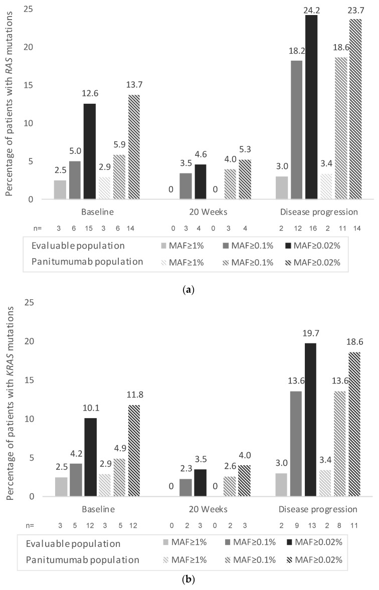
A total of 4 (4.6%) patients presented cfDNA RAS (KRAS/NRAS) mutations at 20 weeks and 16 (24.2%) patients presented cfDNA RAS (KRAS/NRAS) mutations at disease progression (both percentages calculated at MAF ≥ 0.02) in the evaluable population (Figure 1A). The percentages of patients with RAS (KRAS and NRAS) mutations in liquid biopsies at 20 weeks and at disease progression according to the different MAF cut-offs in the evaluable population and panitumumab subpopulation are shown in Figure 1.
Figure 1.
Percentage of patients with (a) RAS, (b) KRAS, and (c) NRAS mutations in liquid biopsies at baseline, at 20 weeks (±2 weeks), and at disease progression according to mutant allele fraction (MAF) cut-offs. One (n = 1) patient had both KRAS and NRAS mutations (MAF ≥ 0.1% and MAF ≥ 0.02%). n: number of patients with mutations. Percentages calculated based on patients with available samples.
At disease progression, a total of 11 patients (n = 9 in the panitumumab subpopulation) presented RAS mutations in liquid biopsies (MAF ≥ 0.02%) that were not present at baseline (by solid and liquid biopsy). Accordingly, the RAS conversion rate (emergent mutations) at disease progression was 19.0% (11/58 patients with baseline RAS wild-type status [by solid and liquid biopsy] and blood samples available at disease progression) in the evaluable population and 17.7% (9/51 patients) in the panitumumab subpopulation (MAF ≥ 0.02%). The conversion rates according to the different MAF cut-offs are displayed in Table 2 for both populations. As expected, the conversion rates were higher at lower MAF cut-offs. No patients presented RAS mutations in liquid biopsies at 20 weeks that were not present at baseline (0% conversion rate at 20 weeks).
The characteristics of patients with RAS mutations at any time (per liquid biopsy), including codon–exon–amino acid position/change, primary tumor location, site of metastasis, first-line treatment, best overall response, and PFS are shown in Supplementary Table S1. Ten (n = 10) out of the eleven patients presenting RAS mutations in liquid biopsies (MAF ≥ 0.02%) at disease progression that were not present at baseline achieved PR as the best overall response, while one achieved SD. In these patients, the most frequent metastatic site was the liver (n = 8). Moreover, three (5.2%) patients presenting RAS mutations in liquid biopsies (MAF ≥ 0.02%) at baseline were RAS wild-type at disease progression (Supplementary Table S1, patients #1, #2 and #5). These three patients reached PR or CR, received FOLFOX plus panitumumab, and had liver or liver plus lung metastases.
3.4. Exploratory Endpoints (Only Assessed in the Panitumumab Subpopulation)
A total of 93 patients had data available for the response (not confirmed) in the panitumumab subpopulation. The ORR was 75.3% (n = 70; CR: 18.3% [n = 17], PR: 57.0% [n = 53]) in the total panitumumab subpopulation, with the rate being 78.1% (57/73) and 65.0% (13/20) in patients with left- and right-sided primary tumor location, respectively.
The ORR according to RAS (KRAS/NRAS) mutational status at baseline and at any time and by tumor location is shown in Table 3. Considering the RAS status at baseline, the ORR was numerically higher in the left-sided, RAS wild-type tumors compared to the right-sided (all right-sided tumors were RAS wild-type) and the RAS mutant tumors (in all of the different MAF cut-offs, not significative). Similar results were observed when considering the RAS mutational status at any time. Among patients with right-sided tumors, there were only one (MAF ≥ 0.1%) and two patients (MAF ≥ 0.02%) with RAS mutant status (both achieving ORR), thus preventing direct comparisons (Table 3). A tendency to increased ORR rates was observed at diminishing MAF cut-offs in RAS mutant patients at baseline and at any time.
Table 3.
Overall response rate according to RAS mutational status in liquid biopsy at baseline and at any time (panitumumab subpopulation, classified by primary tumor location).
| RAS Wild-Type | RAS Mutant | Odds Ratio (95% CI) | ||
|---|---|---|---|---|
| At baseline | ||||
| Total population (n = 93) | ||||
| MAF ≥ 1% | ORR 1, % (95% CI) | 76.7% (66.6–84.9%) | 33.3% (0.8–90.6%) | 6.6 (0.6–76.1) |
| n/N 2 | 69/90 | 1/3 | ||
| MAF ≥ 0.1% | ORR 1, % (95% CI) | 76.1% (65.9–84.6%) | 60.0% (14.7–94.7%) | 2.1 (0.3–13.6) |
| n/N 2 | 67/88 | 3/5 | ||
| MAF ≥ 0.02% | ORR 1, % (95% CI) | 77.5% (66.8–86.1%) | 61.5% (31.6–86.1%) | 2.2 (0.6–7.4) |
| n/N 2 | 62/80 | 8/13 | ||
| Left-sided tumors (n = 73) | ||||
| MAF ≥ 1% | ORR 1, % (95% CI) | 80.0% (68.7–88.6%) | 33.3% (0.8–90.6%) | 8.0 (0.7–94.7) |
| n/N 2 | 56/70 | 1/3 | ||
| MAF ≥ 0.1% | ORR 1, % (95% CI) | 79.4% (67.9–88.3%) | 60.0% (14.7–94.7%) | 2.6 (0.4–16.9) |
| n/N 2 | 54/68 | 3/5 | ||
| MAF ≥ 0.02% | ORR 1, % (95% CI) | 81.7% (69.6–90.5%) | 61.5% (31.6–86.1%) | 2.8 (0.8–10.2) |
| n/N 2 | 49/60 | 8/13 | ||
| Right-sided tumors (n = 20) | ||||
| MAF ≥ 1% | ORR 1, % (95% CI) | 65.0% (40.8–84.6%) | 0% | - |
| n/N 2 | 13/20 | 0/0 | ||
| MAF ≥ 0.1% | ORR 1, % (95% CI) | 65.0% (40.8–84.6%) | 0% | - |
| n/N 2 | 13/20 | 0/0 | ||
| MAF ≥ 0.02% | ORR 1, % (95% CI) | 65.0% (40.8–84.6%) | 0% | - |
| n/N 2 | 13/20 | 0/0 | ||
| At any time | ||||
| Total population (n = 93) | ||||
| MAF ≥ 1% | ORR 1, % (95% CI) | 76.4% (66.2–84.8%) | 50.0% (6.8–93.2%) | 3.2 (0.4–24.4) |
| n/N 2 | 68/89 | 2/4 | ||
| MAF ≥ 0.1% | ORR 1, % (95% CI) | 74.7% (63.6–83.8%) | 78.6% (49.2–95.3%) | 0.8 (0.2–3.2) |
| n/N 2 | 59/79 | 11/14 | ||
| MAF ≥ 0.02% | ORR 1, % (95% CI) | 74.7% (62.9–84.2%) | 77.3% (54.6–92.2%) | 0.9 (0.3–2.7) |
| n/N 2 | 53/71 | 17/22 | ||
| Left-sided tumors (n = 73) | ||||
| MAF ≥ 1% | ORR 1, % (95% CI) | 79.7% (68.3–88.4%) | 50.0% (6.8–93.2%) | 3.9 (0.5–30.4) |
| n/N 2 | 55/69 | 2/4 | ||
| MAF ≥ 0.1% | ORR 1, % (95% CI) | 78.3% (65.8–87.9%) | 76.9% (46.2–95.0%) | 1.1 (0.3–4.5) |
| n/N 2 | 47/60 | 10/13 | ||
| MAF ≥ 0.02% | ORR 1, % (95% CI) | 79.6% (65.9–89.2%) | 75.0% (50.9–91.3%) | 1.3 (0.4–4.3) |
| n/N 2 | 42/53 | 15/20 | ||
| Right-sided tumors (n = 20) | ||||
| MAF ≥ 1% | ORR 1, % (95% CI) | 65.0% (40.8–84.6%) | 0% | - |
| n/N 2 | 13/20 | 0/0 | ||
| MAF ≥ 0.1% | ORR 1, % (95% CI) | 63.2% (38.4–83.7%) | 100% (2.5–100%) | - |
| n/N 2 | 12/19 | 1/1 | ||
| MAF ≥ 0.02% | ORR 1, % (95% CI) | 61.1% (35.8–82.7%) | 100% (15.8–100%) | - |
| n/N 2 | 11/18 | 2/2 | ||
1 Not confirmed. A total of 93 patients had available response data. 2 n: number of patients with partial response and complete response; N: number of patients with available response data. CI: confidence interval using the Clopper–Pearson exact method; MAF: mutant allele fraction; ORR: overall response rate.
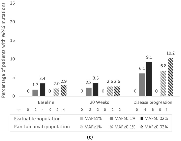
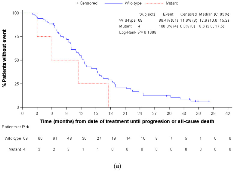
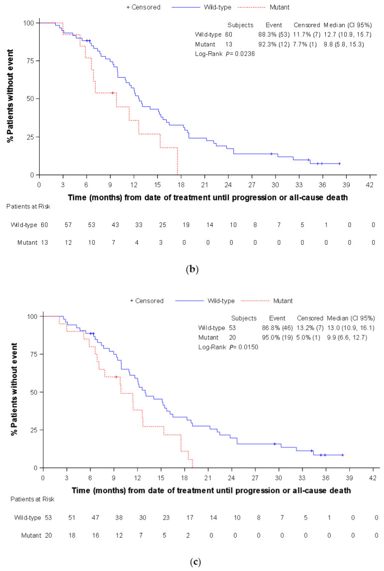
Regarding PFS, the median (CI 95%) time in the total panitumumab subpopulation was 12.1 (10.0–13.8) months. There were no statistically significant differences in PFS between patients with RAS wild-type and RAS mutated in the different MAF cut-offs, both at baseline and at any time (data not shown). Similar results were observed by RAS status at baseline in the subgroup of patients with left-sided tumors. By contrast, in this subgroup there were statistically significant differences in the median PFS between patients with RAS wild-type and RAS mutated at any time in MAF ≥ 0.02% cut-off (13.0 [IC 95%: 10.9–16.1] vs. 9.9 [6.6–12.7] months, respectively; p = 0.015), and MAF ≥ 0.1% cut-off (12.7 [IC 95%: 10.9–15.7] vs. 9.8 [5.9–15.3] months, respectively; p = 0.024) (Figure 2).
Figure 2.
Progression free survival according to RAS mutational status in liquid biopsy at any time (panitumumab subpopulation, left tumor location): (a) mutant allele fraction ≥1% cut-off; (b) mutant allele fraction ≥0.1% cut-off; and (c) mutant allele fraction ≥0.02% cut-off.
Finally, the median OS in the total panitumumab subpopulation was not reached, with a total of 11 events. There were no statistically significant differences in OS between patients with RAS wild-type and RAS mutated at any time in the MAF ≥ 0.1% and ≥0.02% cut-offs, while it was observed in the MAF ≥ 1%, where the four patients with RAS mutated had a median OS of 17.4 months, while it was not reached in the RAS wild-type patients (p = 0.01) (data not shown).
The multivariable Cox regression model for PFS did not yield any statistically significant results, although a tendency towards an increased probability of PD was observed in RAS mutated patients at any time (MAF > 0.02%) compared to RAS wild-type patients (HR: 1.54 [95% CI: 0.93–2.56]; p = 0.096) (Supplementary Table S2).
Regarding tumor burden, the multivariable linear regression model showed that the difference in the estimated means (sum of the longest diameters) between patients with and without liver metastasis was 29.6 mm (95% CI: 1.02–58.3; p = 0.043). Additionally, the cfDNA concentration was significantly associated with the tumor burden (p < 0.0001). In contrast, the presence of RAS mutations at baseline was not associated with tumor burden in our model (Supplementary Table S3).
We also performed exploratory, post-hoc logistic regressions to analyze the significance of MAF as a predictor and ROC curve analyses to estimate the best MAF cut-off point for predicting the clinical response, without achieving any significant results (data not shown).
4. Discussion
In our prospective, multicentric, observational study, we investigated the concordance of the RAS (KRAS/NRAS) mutational status between solid and liquid biopsies in patients with RAS wild-type mCRC (according to solid biopsy) at baseline managed following clinical practice. Our results showed a high concordance between tissue and plasma samples (ranging from 97% to 86% in the evaluable population and panitumumab subpopulation, according to MAF cut-off), with the concordance rate being similar to the rates reported in previous studies showing concordance rates between 86% and 93% [24,25,26,27]. It should be noted that previous similar studies comprised more heterogeneous populations compared to ours, including baseline RAS mutated (according to solid biopsy) patients in both first and subsequent lines of treatment. A recent publication by Kagawa, et al. [28] also using BEAMing analysis in mCRC patients (both RAS mutated and wild-type at baseline) with single-site metastasis suggests that the concordance rates may differ by metastatic site (91%, 88%, and 64% in patients with single metastases in the liver, peritoneum, and lung, respectively). Similar results were observed by Wang, et al. [29] using next-generation sequencing (NGS), reporting baseline RAS concordances of 90% and 37.5% in patients with only liver metastases and only lung metastases, respectively. Additionally, Kagawa, et al. reported the increased tumor burden (longest diameter and number of lesions) as the most significant factor associated with increased solid-liquid biopsies concordance [28]. More recent studies in heterogeneous populations have reported total number of lesions and total tumor burden as the most significant predictors of discordant cases [30,31,32]. In alingment, in our study the presence of liver metastasis was associated with an increased tumor burden, and the majority of patients presented liver metastasis, which may explain the high concordance rates observed.
Despite the high concordance between biopsies at baseline, we observed a RAS conversion rate at disease progression (emergent mutations) of 19% in the evaluable population and 17.7% in the panitumumab subpopulation, while there were three patients (5.2%) where the RAS mutations detected at baseline were not detected at disease progression. Previous studies have reported the results of emergent mutations in subsequent-lines of treatment [27,33,34,35]. However, little is known about the emergent mutations after first-line treatment. Recently, Parseghian, et al. [36] reported that acquired mutations in KRAS, NRAS, BRAF, MAP2K1, or EGFR rarely develop after first-line treatment (6.8%), contrary to what has been observed in second- and third-line treatment (40-50%). By contrast, Wang, et al. [29] reported 44.4% (4/9) of patients with baseline RAS mutations showed RAS clearance at disease progression after first-line treatment, while 27.3% (3/11) of patients showed new RAS mutations. Only 5/20 patients were treated in first line with anti-EGFR treatments. The increased frequency of emergent mutations at disease progression (secondary resistance) has been postulated to be attributable to the clonal evolution under the selective pressure of EGFR inhibition [37]. However, in the first-line setting, pre-existing subclonal mutations do not appear to be the dominant source of emergent mutations at disease progression, suggesting that there may also be a transient mutational process driving anti-EGFR resistance [36]. Similarly, other authors have also proposed that other mechanisms beyond RAS emergent mutations will probably play an important role, which may include alterations involved in chemotherapy and/or anti-EGFR intrinsic or/and acquired resistance [29,37]. A greater knowledge of the molecular complexity that develops as a result of EGFR inhibition will help to guide new strategies in refractory patients with mCRC [38].
In our study, we did not observe a significant association between ORR or PFS and RAS mutational status by liquid biopsies at baseline or at any time across MAF cut-offs in the total panitumumab subpopulation. It should be noted that the proportion of RAS mutant patients at baseline was small. However, the ORR and PFS results in these patients tended to improve as the MAF threshold decreased. Regarding OS, a statistically significant difference between patients with RAS wild-type and RAS mutated at any time was observed for the MAF ≥ 1%. These results should be interpreted with caution, since the median OS was not reached in the panitumumab subpopulation and the sample size of RAS mutated patients was very small (n = 4). Previously reported studies in second- and third-line panitumumab populations also found that the presence of emergent RAS mutations at disease progression was not associated with differences in PFS, ORR, or OS, despite their larger proportions of emergent RAS mutations [27,33,34,35].
Interestingly, when the left-sided tumor patients were analyzed, the median PFS was significantly longer among cfDNA RAS wild-type patients when compared to those presenting cfDNA RAS mutations at any time. This better prognosis observed in left-sided, RAS wild-type tumors is aligned with previously reported data [39,40,41], further highlighting the importance of primary tumor location and supporting the clinical differences between right- and left-sided colon tumors. Unfortunately, in our study no comparisons by tumor sidedness (left vs. right) were possible in patients with RAS mutant status due to the small sample size.
This study has some limitations. It was initially designed to determine the concordance of liquid and solid biopsies at baseline and the appearance of emergent RAS mutations up to disease progression, whereas the association of RAS mutational status and outcomes and the predictive factors study were only explorative endpoints. Furthermore, the study included small patient numbers to allow for strong evidence of some of the subpopulations analyses (e.g., right-sided, RAS mutant). Additionally, we only tested for mutations in RAS (KRAS/NRAS) in liquid biopsies, but the acquired resistance to anti-EGFR treatments in mCRC patients is known to be caused by several other mechanisms, including EGFR extracellular domain mutations, MET amplifications, BRAF mutations, and HER2 amplifications [42]. Despite this, the results observed in this study are aligned with the previously reported data. Since this was an observational study, patients were treated following clinical practice, thus preventing us from drawing any conclusions on the role of specific treatment regimens on the results. However, in our study cfDNA RAS mutations by themselves did not predict a lack of clinical benefits to panitumumab plus chemotherapy. Additionally, there were three patients with RAS mutations at baseline that were RAS wild-type at progression. The reason for this change (e.g., reversion to wild-type, the sensitivity cut-off of the liquid biopsy, the occurrence of false positives) is unknown. Finally, the clinically relevant RAS MAF cut-off in liquid biopsies is yet to be defined. A follow-up and serial cfDNA RAS analyses could help us to understand their clinical and biological significances. This may play an important role in the future development and administration of KRAS inhibitors, especially when tumor tissue is not available.
Our study had some key strengths. The studied population was homogeneous, with all of the patients being RAS (KRAS/NRAS) wild-type at baseline as per standard-of-care solid biopsy, starting their first-line treatment (mostly panitumumab plus chemotherapy). Additionally, we performed a dynamic monitorization of cfDNA using a highly sensitive technique (BEAMing), evaluating the RAS mutational status over time. In addition, the correlation between RECIST response and PFS and the RAS mutational status was assessed, differentiating by primary tumor side.
In this sense, our results contribute to a growing body of work supporting the use of cfDNA biomarkers to predict PFS in patients with mCRC receiving first-line chemotherapy treatment. Nevertheless, limited data are available on the role of liquid biopsy to predict the outcomes of patients clinically eligible for anti-EGFR-based upfront treatment [43].
5. Conclusions
In summary, the concordance rate between liquid and solid biopsy was very high at baseline, consistent with previous studies. At disease progression, there was a considerable percentage of patients with emerging RAS mutations (19%) that may be potentially attributable to the acquired resistance to anti-EGFR treatments. However, in our exploratory analyses, the RAS mutations detected were not associated with differences in clinical outcomes, except in patients with left-sided tumors. Clinical outcomes tended to improve as the MAF threshold decreased.
Acknowledgments
Manuscript writing support was provided by Juan Martin and Montse Sabaté, from TFS HealthScience with financial support provided by Amgen S.A. The authors wish to thank to all the investigators of the PERSEIDA study (Cohort 1): Manuel Valladares-Ayerbes (Hospital Universitario Virgen del Rocío e Instituto de Biomedicina [IBIS]), Pilar García-Alfonso (Hospital General Universitario Gregorio Marañón), Jorge Muñoz Luengo (Hospital San Pedro de Alcántara), Paola Patricia Pimentel Cáceres (Hospital Universitario Santa Lucia), Paula Jimenez Fonseca (Hospital Universitario Central de Asturias), Marta Llanos (Hospital Universitario de Canarias), Juan Jesús Cruz-Hernández (Complejo Asistencial Universitario de Salamanca), Ana María López Muñoz (Hospital Universitario de Burgos), Maria Luisa Limón Mirón (Hospital Universitario Virgen del Rocío), Antonieta Salud (Hospital Universitario Arnau de Vilanova), Luis Cirera Nogueras (Hospital Mutua de Terrassa), María José Safont (Hospital General Universitario de Valencia), Rocío Garcia-Carbonero (Hospital Universitario 12 de Octubre), Jorge Aparicio (Hospital Universitari I Politècnic La Fe), Esther Falco Ferrer (Hospital Universitari Son Llàtzer), Maria Angeles Vicente Conesa (Hospital General Universitario José Maria Morales Meseguer), Carmen Guillén-Ponce (Hospital Universitario Ramón y Cajal), Paula Garcia-Teijido (Hospital San Agustín), Maria Begoña Medina Magan (Hospital Universitario Torrecárdenas), Isabel Busquier (Consorcio Hospitalario Provincial de Castellón), Mercedes Salgado (Complexo Hospitalario Universitario de Ourense), and Ariadna Lloansí Vila (Amgen S.A.).
Supplementary Materials
The following supporting information can be downloaded at: https://www.mdpi.com/article/10.3390/cancers14246075/s1. Supplementary Table S1: Characteristics of patients with RAS mutations at any time as per liquid biopsy (codon–exon–amino acid position/change); Supplementary Table S2: Univariable and Multivariable Cox Regression Model for Progression Free Survival (Panitumumab Subpopulation); Supplementary Table S3: Univariable and Multivariable Linear Regression Model for Tumor Burden (Panitumumab Subpopulation).
Author Contributions
Conceptualization, M.V.-A. and A.L.V.; formal analysis M.V.-A. and A.L.V.; investigation, M.V.-A., P.G.-A., J.M.L., P.P.P.C., O.A.C.T., R.V.-T., M.L., B.L.A., M.L.L.M., A.S., L.C.N., R.G.-C., M.J.S., E.F.F., J.A., M.A.V.C., C.G.-P., P.G.-T., M.B.M.M., I.B. and M.S.; writing—original draft preparation, M.V.-A.; writing—review and editing, M.V.-A., P.G.-A., J.M.L., P.P.P.C., O.A.C.T., R.V.-T., M.L., B.L.A., M.L.L.M., A.S., L.C.N., R.G.-C., M.J.S., E.F.F., J.A., M.A.V.C., C.G.-P., P.G.-T., M.B.M.M., I.B., M.S. and A.L.V.; supervision, M.V.-A. All authors have read and agreed to the published version of the manuscript.
Institutional Review Board Statement
The study was conducted according to the guidelines of the Declaration of Helsinki. Please refer to this study by its ClinicalTrials.gov identifier (NCT number): NCT02792478.
Informed Consent Statement
Informed consent was obtained from all subjects involved in the study.
Data Availability Statement
The data presented in this study are available on request from the corresponding author.
Conflicts of Interest
M.V-A. has received grants and personal fees from Roche and personal fees from Merck, Amgen, Sanofi, Servier, Celgene, and Bayer. P.G-A. has received honoraria or consultation fees for speaker, consultancy, or advisory roles from Amgen, Bayer, Bristol, Merck Seorono, MSD, Lilly, Roche, Sanofi, Servier, and Pierre Fabre. R.V.-T. has received speaker fees from Amgen, Merck, Sanofi, Servier, Bristol-MS, Bayer, and Roche and educational and scientific activities and travel support from Amgen, Roche, Lilly, Sanofi, Bristol-MS, Pierre-Fabre, and Servier. R.G-C. has received honoraria for speaker/consulting roles from AAA, Advanz Pharma, Bayer, BMS, HMP, Ipsen, Merck, Midatech Pharma, MSD, Novartis, PharmaMar, Pfizer, Pierre Fabre, Roche, Sanofi, and Servier and research support from ARMO Biosciences, Astrazeneca, Pfizer, Novartis, Ipsen, Roche, Pharmacyclics, Boston Biomedicals, Merck, MSD, Amgen, Sanofi, Bayer, Bristol-Myers-Squibb, Boerhringer, Sysmex, Gilead Sciences, Servier, Adacap, VCN, Lilly, Pharmamar, BMS, and MSD. M-J.S. has received personal fees from Roche, Amgen, Merck, and Sanofi and honoraria for speaker/consulting roles from Amgen, Bayer, BMS, Merck, MSD, Pierre Fabre, Roche, Sanofi, and Servier. JA. has received honoraria for consultancy or advisory roles from Amgen, Merck, Sanofi, Servier, Bayer, and Pierre Fabre. M.S. has received honoraria for speaker and advisory roles for Amgen. A.L.V. is an employee and stakeholder of Amgen S.A. The other authors declare no potential conflicts of interest.
Funding Statement
This research was funded by Amgen S.A.
Footnotes
Publisher’s Note: MDPI stays neutral with regard to jurisdictional claims in published maps and institutional affiliations.
References
- 1.Bray F., Ferlay J., Soerjomataram I., Siegel R.L., Torre L.A., Jemal A. Global cancer statistics 2018: GLOBOCAN estimates of incidence and mortality worldwide for 36 cancers in 185 countries. CA Cancer J. Clin. 2018;68:394–424. doi: 10.3322/caac.21492. [DOI] [PubMed] [Google Scholar]
- 2.European Commission Colorectal Cancer Burden in EU-27. 2021. [(accessed on 19 May 2022)]. Available online: https://ecis.jrc.ec.europa.eu/pdf/Colorectal_cancer_factsheet-Mar_2021.pdf.
- 3.Sociedad Española de Oncología Médica (SEOM) Las Cifras del Cáncer en España 2022. [(accessed on 19 May 2022)]. Available online: https://seom.org/images/LAS_CIFRAS_DEL_CANCER_EN_ESPANA_2022.pdf.
- 4.Van Cutsem E., Cervantes A., Adam R., Sobrero A., van Krieken J.H., Aderka D., Aguilar E.A., Bardelli A., Benson A., Bodoky G., et al. ESMO consensus guidelines for the management of patients with metastatic colorectal cancer. Ann. Oncol. 2016;27:1386–1422. doi: 10.1093/annonc/mdw235. [DOI] [PubMed] [Google Scholar]
- 5.Fernández-Medarde A., Santos E. Ras in Cancer and Developmental Diseases. Genes Cancer. 2011;2:344–358. doi: 10.1177/1947601911411084. [DOI] [PMC free article] [PubMed] [Google Scholar]
- 6.Douillard J.-Y., Oliner K.S., Siena S., Tabernero J., Burkes R., Barugel M., Humblet Y., Bodoky G., Cunningham D., Jassem J., et al. Panitumumab–FOLFOX4 treatment and RAS mutations in colorectal cancer. N. Engl. J. Med. 2013;369:1023–1034. doi: 10.1056/NEJMoa1305275. [DOI] [PubMed] [Google Scholar]
- 7.Van Cutsem E., Lenz H.-J., Köhne C.-H., Heinemann V., Tejpar S., Melezínek I., Beier F., Stroh C., Rougier P., van Krieken J.H., et al. Fluorouracil, leucovorin, and irinotecan plus cetuximab treatment and RAS mutations in colorectal cancer. J. Clin. Oncol. 2015;33:692–700. doi: 10.1200/JCO.2014.59.4812. [DOI] [PubMed] [Google Scholar]
- 8.Bokemeyer C., Bondarenko I., Hartmann J., de Braud F., Schuch G., Zubel A., Celik I., Schlichting M., Koralewski P. Efficacy according to biomarker status of cetuximab plus FOLFOX-4 as first-line treatment for metastatic colorectal cancer: The OPUS study. Ann. Oncol. 2011;22:1535–1546. doi: 10.1093/annonc/mdq632. [DOI] [PubMed] [Google Scholar]
- 9.Kyrochristos I.D., Roukos D.H. Comprehensive intra-individual genomic and transcriptional heterogeneity: Evidence-based Colorectal Cancer Precision Medicine. Cancer Treat. Rev. 2019;80:101894. doi: 10.1016/j.ctrv.2019.101894. [DOI] [PubMed] [Google Scholar]
- 10.Bi F., Wang Q., Dong Q., Wang Y., Zhang L., Zhang J. Circulating tumor DNA in colorectal cancer: Opportunities and challenges. Am. J. Transl. Res. 2020;12:1044–1055. [PMC free article] [PubMed] [Google Scholar]
- 11.Raimondi C., Nicolazzo C., Belardinilli F., Loreni F., Gradilone A., Mahdavian Y., Gelibter A., Giannini G., Cortesi E., Gazzaniga P. Transient Disappearance of RAS Mutant Clones in Plasma: A Counterintuitive Clinical Use of EGFR Inhibitors in RAS Mutant Metastatic Colorectal Cancer. Cancers. 2019;11:42. doi: 10.3390/cancers11010042. [DOI] [PMC free article] [PubMed] [Google Scholar]
- 12.Siravegna G., Marsoni S., Siena S., Bardelli A. Integrating liquid biopsies into the management of cancer. Nat. Rev. Clin. Oncol. 2017;14:531–548. doi: 10.1038/nrclinonc.2017.14. [DOI] [PubMed] [Google Scholar]
- 13.Normanno N., Abate R.E., Lambiase M., Forgione L., Cardone C., Iannaccone A., Sacco A., Rachiglio A., Martinelli E., Rizzi D., et al. RAS testing of liquid biopsy correlates with the outcome of metastatic colorectal cancer patients treated with first-line FOLFIRI plus cetuximab in the CAPRI-GOIM trial. Ann. Oncol. 2017;29:112–118. doi: 10.1093/annonc/mdx417. [DOI] [PubMed] [Google Scholar]
- 14.Jahr S., Hentze H., Englisch S., Hardt D., Fackelmayer F.O., Hesch R.D., Knippers R. DNA fragments in the blood plasma of cancer patients: Quantitations and evidence for their origin from apoptotic and necrotic cells. Cancer Res. 2001;61:1659–1665. [PubMed] [Google Scholar]
- 15.Diehl F., Li M., Dressman D., He Y., Shen D., Szabo S., Diaz L.A., Goodman S.N., David K.A., Juhl H., et al. Detection and quantification of mutations in the plasma of patients with colorectal tumors. Proc. Natl. Acad. Sci. USA. 2005;102:16368–16373. doi: 10.1073/pnas.0507904102. [DOI] [PMC free article] [PubMed] [Google Scholar]
- 16.Castells A., Puig P., Móra J., Boadas J., Boix L., Urgell E., Solé M., Capellà G., Lluís F., Fernández-Cruz L., et al. K-ras Mutations in DNA Extracted From the Plasma of Patients With Pancreatic Carcinoma: Diagnostic Utility and Prognostic Significance. J. Clin. Oncol. 1999;17:578. doi: 10.1200/JCO.1999.17.2.578. [DOI] [PubMed] [Google Scholar]
- 17.Ryan B.M., Lefort F., McManus R., Daly J., Keeling P.W.N., Weir D.G., Kelleher D. A prospective study of circulating mutant KRAS2 in the serum of patients with colorectal neoplasia: Strong prognostic indicator in postoperative follow up. Gut. 2003;52:101–108. doi: 10.1136/gut.52.1.101. [DOI] [PMC free article] [PubMed] [Google Scholar]
- 18.Wang S., An T., Wang J., Zhao J., Wang Z., Zhuo M., Bai H., Yang L., Zhang Y., Wang X., et al. Potential Clinical Significance of a Plasma-Based KRAS Mutation Analysis in Patients with Advanced Non–Small Cell Lung Cancer. Clin. Cancer Res. 2010;16:1324–1330. doi: 10.1158/1078-0432.CCR-09-2672. [DOI] [PubMed] [Google Scholar]
- 19.Crowley E., Di Nicolantonio F., Loupakis F., Bardelli A. Liquid biopsy: Monitoring cancer-genetics in the blood. Nat. Rev. Clin. Oncol. 2013;10:472–484. doi: 10.1038/nrclinonc.2013.110. [DOI] [PubMed] [Google Scholar]
- 20.Li M., Diehl F., Dressman D., Vogelstein B., Kinzler K.W. BEAMing up for detection and quantification of rare sequence variants. Nat. Methods. 2006;3:95–97. doi: 10.1038/nmeth850. [DOI] [PubMed] [Google Scholar]
- 21.Diehl F., Li M., He Y., Kinzler K.W., Vogelstein B., Dressman D. BEAMing: Single-molecule PCR on microparticles in water-in-oil emulsions. Nat. Methods. 2006;3:551–559. doi: 10.1038/nmeth898. [DOI] [PubMed] [Google Scholar]
- 22.Diaz L.A., Jr., Williams R.T., Wu J., Kinde I., Hecht J.R., Berlin J., Allen B., Bozic I., Reiter J.G., Nowak M.A., et al. The molecular evolution of acquired resistance to targeted EGFR blockade in colorectal cancers. Nature. 2012;486:537–540. doi: 10.1038/nature11219. [DOI] [PMC free article] [PubMed] [Google Scholar]
- 23.Eisenhauer E.A., Therasse P., Bogaerts J., Schwartz L.H., Sargent D., Ford R., Dancey J., Arbuck S., Gwyther S., Mooney M., et al. New response evaluation criteria in solid tumours: Revised RECIST guideline (version 1.1) Eur. J. Cancer. 2009;45:228–247. doi: 10.1016/j.ejca.2008.10.026. [DOI] [PubMed] [Google Scholar]
- 24.García-Foncillas J., Tabernero J., Élez E., Aranda E., Benavides M., Camps C., Jantus-Lewintre E., López R., Muinelo-Romay L., Montagut C., et al. Prospective multicenter real-world RAS mutation comparison between OncoBEAM-based liquid biopsy and tissue analysis in metastatic colorectal cancer. Br. J. Cancer. 2018;119:1464–1470. doi: 10.1038/s41416-018-0293-5. [DOI] [PMC free article] [PubMed] [Google Scholar]
- 25.Grasselli J., Elez E., Caratù G., Matito J., Santos C., Macarulla T., Vidal J., Garcia M., Viéitez J., Paéz D., et al. Concordance of blood- and tumor-based detection of RAS mutations to guide anti-EGFR therapy in metastatic colorectal cancer. Ann. Oncol. 2017;28:1294–1301. doi: 10.1093/annonc/mdx112. [DOI] [PMC free article] [PubMed] [Google Scholar]
- 26.Schmiegel W., Scott R.J., Dooley S., Lewis W., Meldrum C.J., Pockney P., Draganic B., Smith S., Hewitt C., Philimore H., et al. Blood-based detection of RAS mutations to guide anti-EGFR therapy in colorectal cancer patients: Concordance of results from circulating tumor DNA and tissue-based RAS testing. Mol. Oncol. 2017;11:208–219. doi: 10.1002/1878-0261.12023. [DOI] [PMC free article] [PubMed] [Google Scholar]
- 27.Thomsen C.B., Andersen R.F., Lindebjerg J., Hansen T.F., Jensen L.H., Jakobsen A. Plasma Dynamics of RAS/RAF Mutations in Patients with Metastatic Colorectal Cancer Receiving Chemotherapy and Anti-EGFR Treatment. Clin. Color. Cancer. 2019;18:28–33.e3. doi: 10.1016/j.clcc.2018.10.004. [DOI] [PubMed] [Google Scholar]
- 28.Kagawa Y., Elez E., García-Foncillas J., Bando H., Taniguchi H., Vivancos A., Akagi K., García A., Denda T., Ros J., et al. Combined Analysis of Concordance between Liquid and Tumor Tissue Biopsies for RAS Mutations in Colorectal Cancer with a Single Metastasis Site: The METABEAM Study. Clin. Cancer Res. 2021;27:2515–2522. doi: 10.1158/1078-0432.CCR-20-3677. [DOI] [PubMed] [Google Scholar]
- 29.Wang F., Huang Y.-S., Wu H.-X., Wang Z.-X., Jin Y., Yao Y.-C., Chen Y.-X., Zhao Q., Chen S., He M.-M., et al. Genomic temporal heterogeneity of circulating tumour DNA in unresectable metastatic colorectal cancer under first-line treatment. Gut. 2021;71:1340–1349. doi: 10.1136/gutjnl-2021-324852. [DOI] [PMC free article] [PubMed] [Google Scholar]
- 30.Formica V., Lucchetti J., Doldo E., Riondino S., Morelli C., Argirò R., Renzi N., Nitti D., Nardecchia A., Dell’Aquila E., et al. Clinical utility of plasma KRAS, NRAS and BRAF mutational analysis with real time PCR in metastatic colorectal cancer patients-the importance of tissue/plasma discordant cases. J. Clin. Med. 2020;10:87. doi: 10.3390/jcm10010087. [DOI] [PMC free article] [PubMed] [Google Scholar]
- 31.Bando H., Kagawa Y., Kato T., Akagi K., Denda T., Nishina T., Komatsu Y., Oki E., Kudo T., Kumamoto H., et al. A multicentre, prospective study of plasma circulating tumour DNA test for detecting RAS mutation in patients with metastatic colorectal cancer. Br. J. Cancer. 2019;120:982–986. doi: 10.1038/s41416-019-0457-y. [DOI] [PMC free article] [PubMed] [Google Scholar]
- 32.Hamfjord J., Guren T.K., Glimelius B., Sorbye H., Pfeiffer P., Dajani O., Lingjaerde O.C., Tveit K.M., Pallisgaard N., Spindler K.G., et al. Clinicopathological factors associated with tumour-specific mutation detection in plasma of patients with RAS-mutated or BRAF-mutated metastatic colorectal cancer. Int. J. Cancer. 2021;149:1385–1397. doi: 10.1002/ijc.33672. [DOI] [PubMed] [Google Scholar]
- 33.Peeters M., Price T., Boedigheimer M., Kim T.W., Ruff P., Gibbs P., Thomas A.L., Demonty G., Hool K., Ang A. Evaluation of Emergent Mutations in Circulating Cell-Free DNA and Clinical Outcomes in Patients with Metastatic Colorectal Cancer Treated with Panitumumab in the ASPECCT Study. Clin. Cancer Res. 2019;25:1216–1225. doi: 10.1158/1078-0432.CCR-18-2072. [DOI] [PubMed] [Google Scholar]
- 34.Siena S., Sartore-Bianchi A., Garcia-Carbonero R., Karthaus M., Smith D., Tabernero J., Van Cutsem E., Guan X., Boedigheimer M., Ang A., et al. Dynamic molecular analysis and clinical correlates of tumor evolution within a phase II trial of panitumumab-based therapy in metastatic colorectal cancer. Ann. Oncol. 2018;29:119–126. doi: 10.1093/annonc/mdx504. [DOI] [PMC free article] [PubMed] [Google Scholar]
- 35.Kim T.W., Peeters M., Thomas A.L., Gibbs P., Hool K., Zhang J., Ang A.L., Bach B.A., Price T. Impact of Emergent Circulating Tumor DNA RAS Mutation in Panitumumab-Treated Chemoresistant Metastatic Colorectal Cancer. Clin. Cancer Res. 2018;24:5602–5609. doi: 10.1158/1078-0432.CCR-17-3377. [DOI] [PubMed] [Google Scholar]
- 36.Parseghian C.M., Sun R., Napolitano S., Morris V.K., Henry J., Willis J., Sanchez E.V., Raghav K.P.S., Ang A., Kopetz S. Rarity of acquired mutations (MTs) after first-line therapy with anti-EGFR therapy (EGFRi) J. Clin. Oncol. 2021;39:3514. doi: 10.1200/JCO.2021.39.15_suppl.3514. [DOI] [Google Scholar]
- 37.Misale S., Di Nicolantonio F., Sartore-Bianchi A., Siena S., Bardelli A. Resistance to Anti-EGFR Therapy in Colorectal Cancer: From Heterogeneity to Convergent Evolution. Cancer Discov. 2014;4:1269–1280. doi: 10.1158/2159-8290.CD-14-0462. [DOI] [PubMed] [Google Scholar]
- 38.Maurel J., Alonso V., Escudero P., Fernández-Martos C., Salud A., Méndez M., Gallego J., Rodriguez J.R., Martín-Richard M., Fernández-Plana J., et al. Clinical Impact of Circulating Tumor RAS and BRAF Mutation Dynamics in Patients with Metastatic Colorectal Cancer Treated with First-Line Chemotherapy Plus Anti–Epidermal Growth Factor Receptor Therapy. JCO Precis. Oncol. 2019;18:1–16. doi: 10.1200/PO.18.00289. [DOI] [PubMed] [Google Scholar]
- 39.Morano F., Corallo S., Lonardi S., Raimondi A., Cremolini C., Rimassa L., Murialdo R., Zaniboni A., Sartore-Bianchi A., Tomasello G., et al. Negative Hyperselection of Patients with RAS and BRAF Wild-Type Metastatic Colorectal Cancer Who Received Panitumumab-Based Maintenance Therapy. J. Clin. Oncol. 2019;37:3099–3110. doi: 10.1200/JCO.19.01254. [DOI] [PMC free article] [PubMed] [Google Scholar]
- 40.Arnold D., Lueza B., Douillard J.-Y., Peeters M., Lenz H.-J., Venook A., Heinemann V., Van Cutsem E., Pignon J.-P., Tabernero J., et al. Prognostic and predictive value of primary tumour side in patients with RAS wild-type metastatic colorectal cancer treated with chemotherapy and EGFR directed antibodies in six randomized trials. Ann. Oncol. 2017;28:1713–1729. doi: 10.1093/annonc/mdx175. [DOI] [PMC free article] [PubMed] [Google Scholar]
- 41.Holch J.W., Ricard I., Stintzing S., Modest D.P., Heinemann V. The relevance of primary tumour location in patients with metastatic colorectal cancer: A meta-analysis of first-line clinical trials. Eur. J. Cancer. 2017;70:87–98. doi: 10.1016/j.ejca.2016.10.007. [DOI] [PubMed] [Google Scholar]
- 42.Parseghian C.M., Napolitano S., Loree J.M., Kopetz S. Mechanisms of Innate and Acquired Resistance to Anti-EGFR Therapy: A Review of Current Knowledge with a Focus on Rechallenge Therapies. Clin. Cancer Res. 2019;25:6899–6908. doi: 10.1158/1078-0432.CCR-19-0823. [DOI] [PMC free article] [PubMed] [Google Scholar]
- 43.Callesen L.B., Hamfjord J., Boysen A.K., Pallisgaard N., Guren T.K., Kure E.H., Spindler K.-L.G. Circulating tumour DNA and its clinical utility in predicting treatment response or survival in patients with metastatic colorectal cancer: A systematic review and meta-analysis. Br. J. Cancer. 2022;127:500–513. doi: 10.1038/s41416-022-01816-4. [DOI] [PMC free article] [PubMed] [Google Scholar]
Associated Data
This section collects any data citations, data availability statements, or supplementary materials included in this article.
Supplementary Materials
Data Availability Statement
The data presented in this study are available on request from the corresponding author.


