PLOS ONE logoLink to PLOS ONE
. 2022 Dec 22;17(12):e0272751. doi: 10.1371/journal.pone.0272751

High seroprevalence of Immunoglobulin G (IgG) and IgM antibodies to SARS-CoV-2 in asymptomatic and symptomatic individuals amidst vaccination roll-out in western Kenya

Shehu Shagari Awandu 1,2,*, Alfred Ochieng Ochieng 2, Benson Onyango 2, Richard Odongo Magwanga 2,3, Pamela Were 2, Angeline Atieno Ochung’ 2, Fredrick Okumu 2, Marceline Adhiambo Oloo 2, Jim Seth Katieno 4, Shirley Lidechi 4, Fredrick Ogutu 5, Dorothy Awuor 2, Joy Nyangasi Kirungu 3, Francis Orata 6, Justine Achieng 5, Bonface Oure 5, Regina Nyunja 2, Eric M O Muok 4, Stephen Munga 4, Benson Estambale 1,*
Editor: Adriana Calderaro7
PMCID: PMC9778630  PMID: 36548358

Abstract

The population’s antibody response is a key factor in comprehending SARS-CoV-2 epidemiology. This is especially important in African settings where COVID-19 impact, and vaccination rates are relatively low. This study aimed at characterizing the Immunoglobulin G (IgG) and Immunoglobulin M (IgM) in both SARS-CoV-2 asymptomatic and symptomatic individuals in Kisumu and Siaya counties in western Kenya using enzyme linked immunosorbent assays. The IgG and IgM overall seroprevalence in 98 symptomatic and asymptomatic individuals in western Kenya between December 2021-March 2022 was 76.5% (95% CI = 66.9–84.5) and 29.6% (95% CI = 20.8–39.7) respectively. In terms of gender, males had slightly higher IgG positivity 87.5% (35/40) than females 68.9% (40/58). Amidst the ongoing vaccination roll-out during the study period, over half of the study participants (55.1%, 95% CI = 44.7–65.2) had not received any vaccine. About one third, (31.6%, 95% CI = 22.6–41.8) of the study participants had been fully vaccinated, with close to a quarter (13.3% 95% CI = 7.26–21.6) partially vaccinated. When considering the vaccination status and seroprevalence, out of the 31 fully vaccinated individuals, IgG seropositivity was 81.1% (95% CI = 70.2–96.3) and IgM seropositivity was 35.5% (95% CI = 19.22–54.6). Out of the participants that had not been vaccinated at all, IgG seroprevalence was 70.4% (95% CI 56.4–82.0) with 20.4% (95% CI 10.6–33.5) seropositivity for IgM antibodies. On PCR testing, 33.7% were positive, with 66.3% negative. The 32 positive individuals included 12(37.5%) fully vaccinated, 8(25%) partially vaccinated and 12(37.5%) unvaccinated. SARs-CoV-2 PCR positivity did not significantly predict IgG (p = 0.469 [95% CI 0.514–4.230]) and IgM (p = 0.964 [95% CI 0.380–2.516]) positivity. These data indicate a high seroprevalence of antibodies to SARS-CoV-2 in western Kenya. This suggests that a larger fraction of the population was infected with SARS-CoV-2 within the defined period than what PCR testing could cover.

Introduction

The coronavirus disease 2019 (COVID-19) caused by severe acute respiratory syndrome coronavirus 2 (SARS-CoV-2), has infected more than half a billion people globally [1]. The COVID-19 pandemic continues to disrupt lives, increase mortality in people with underlying co-morbidities and severely impact world economies [2]. While all continents have been severely impacted, Africa has registered low scores on major metrics including mortality rates, number of cases and absence of exponential growth as predicted [3]. However, the reasons for this are still unclear. Exposure of the population to many infectious diseases in the continent, generating cross reactive protective antibodies is suggested as contributing to reduced severity to the infection [35].

The SARS-CoV-2 infection is associated with the development of a robust humoral immune response with variable levels of Immunoglobulin A (IgA), IgM and IgG isotypes as the infection progresses [6, 7]. Upon SARS-CoV-2 infection, the IgM response is quick and short-lived, detectable up to 20 days post infection and then wanes [7, 8]. In contrast, the IgG antibody responses peak after 25 days, and are more long lived and detectable up to 120 days post symptom onset [7, 8].

The kinetics of anti-SARS-CoV-2 especially IgG and IgM antibodies have been profiled in several epidemiological settings in Kenya [9]. Whilst an earlier study among blood donors found an overall IgG seroprevalence of 4.3% peaking in 35–44 year olds [10], in contrast a study among community health workers reported 20.8% seroprevalence [11]. More recently, a population survey in Nairobi recorded a 34.7% seroprevalence [12]. Majority of studies in Kenya thus far have mainly focused on the most at-risk population in both urban and rural areas of the country. The differences in seroprevalence makes it unclear whether the antibody response to SARS-CoV-2 in western Kenya, ravaged by a host of infectious diseases including malaria, HIV and tuberculosis is similar to the rest of the country [13].

Here, we examined the levels of IgM and IgG antibodies to SARS-CoV-2 in asymptomatic and symptomatic individuals amidst vaccination roll-out, in Kisumu and Siaya counties in western Kenya. We hypothesized that in western Kenyan populations burdened by several other infectious diseases, the COVID-19 antibody responses are not different in vaccinated and non-vaccinated individuals.

Materials and methods

Study design and participants

We screened and recruited individuals presenting to Kisumu and Siaya Counties referral hospitals for routine COVID-19 tests in western Kenya. All patients, regardless of COVID-19 symptoms were eligible for enrollment. Study procedures were explained to them, and an informed consent form signed by the participants. A detailed personal history and physical examination were carried out by the study doctor and documented on a predesigned form. Demographic data including age, gender, county of residence, symptoms, date of onset, severity, vaccination status and test type (PCR or antigen test), whether initial or follow-up/repeat.

Sample size calculations

Sample size was calculated in an online platform http://www.raosoft.com/samplesize.html, using a margin of error of 9.78% and with a 95% confidence interval with a 50% response distribution, giving at least 96 samples.

Sample collections

Participants provided stool and nasopharyngeal samples in viral transport media (AB Medical Inc). Additionally, participants provided a 5 ml venous blood sample, in sterile EDTA tubes, that was centrifuged to separate plasma and buffy coat. All the samples were transported under cold chain to Kenya Medical Research Institute, Centre for Global Health Research (CGHR).

Laboratory assays

Enzyme linked immunosorbent assay (ELISA)

To detect the presence of IgG and IgM antibodies against SARS-CoV-2 S proteins respectively, serological assays were performed using the qualitative indirect SCoV-2 Detect™ IgG ELISA kit and SCoV-2 Detect™ IgM ELISA kit (InBios International, Seattle, USA). Briefly, 50 μL each of serum samples, positive, negative and cut-off controls in duplicates were added into the SCoV-2 Antigen coated microtiter ELISA plates. The plates were covered with parafilm and incubated at 37°C for 1 hour in an incubator. The plates were subsequently washed 6 times using 300 μL of 1X Wash Buffer. 50 μL of conjugate was then added to the wells, plate covered with parafilm and incubated at 37°C for 30 minutes in an incubator. The plates were washed 6 times using 300 μL of 1X wash buffer. 75 μL of Liquid TMB substrate was added into all wells and the uncovered plates incubated at room temperature in the dark for 20 minutes. Finally, 50 μL of stop solution was added per well and the plates incubated at room temperature for 1 minute. The plates were read on a BIOTEK ELX 800 absorbance microplate reader at 450 nm optical density. The raw optical densities (ODs) were recorded, and ratios computed by dividing the sample OD by the median OD of the assay cut-off control. The kit cut-off control provided by the manufacturer aids in monitoring the integrity of the kit and estimating the proper threshold to determine sample status. The cut-off is set up in triplicate with each assay. The control OD has to be greater than the negative control OD for an assay to be validated. Samples with IgG or IgM ratio greater than or equal to 1.1 considered positive and IgG or IgM ratio less than or equal to 0.9 considered negative.

RNA extraction and COVID-19 PCR tests

Total nucleic acid from Nasopharyngeal samples in Viral Transport Medium (VTM) were extracted using QIAamp Viral RNA Kit (Qiagen) following manufacturer’s instructions. The extracted RNA from the samples was stored at −20°C awaiting SARS-CoV-2 RT-PCR. Real Time PCR was conducted using a DaAn Gene SARS-CoV-2 PCR kit (DaAn Gene Co, Ltd., of Sun Yat-sen University, China) as per manufacturer’s instructions. The master mix was prepared by mixing 17 μl of NC (ORF1ab/N) PCR liquid A (reaction mix) and 3 μl of NC (ORF1ab/N) PCR reaction liquid B (enzyme), then 5 μl of the extracted sample was added to make the PCRs final volume of 25 μl in a PCR plate on a cold block. The PCR tubes were immediately transferred to an ABI 7500 Fast RT-PCR machine (Applied Biosystems) for detection of SARS-CoV-2. The probe detection modes were set as: ORF1ab: VIC, Quencher: NONE, N-Gene: FAM, Quencher: NONE, Internal Control: Cy5, Quencher: NONE, Passive reference: NONE. The PCR cycle was carried out on the following conditions: 1 cycle of 15 min at 50°C, 1 cycle of 15 min at 95°C, and 45 cycles of 94°C for 15 s and 55°C for 45 s. Results were analyzed by 7500 Fast Real Time PCR software version 2.3 to identify SARS-CoV-2 positive targets by evaluating PCR curves for sigmoidal amplification. A sample was considered positive for the targeted pathogen when it had cycle threshold (CT) value within 38 cycles (Ct < 38) for both ORF1ab and N-Gene. For quality control, a positive and negative control was included in each run and validated prior to specific patient sample analysis. Additionally, a negative extraction blank which was a known negative specimen was included in each run to check for cross contamination and specificity.

Statistical analyses

Seroprevalence was determined as positivity for either IgG or IgM subtypes over the total number of individuals tested. The association between SARs-CoV-2 qPCR results, gender, IgM and IgG was tested using binomial logistic regression analysis. All the statistical analyses were conducted in STATA Version 16.

Ethical considerations

Ethical approval for the study was granted by Jaramogi Oginga Odinga University of Science and Technology (ERC/21/5/21-4), and a research license granted from Kenya National Commission of Science and Technology (NACOSTI/P/22/17545). Administrative approval was provided by the county governments of Kisumu and Siaya. All participants provided written informed consent or assent before enrollment.

Results

Demographics

A total of 98 participants were recruited into the study, with slightly more females 58.2% (58/98) than males 40.8% (40/98). The median age was 29 years (interquartile range 19–44) years. About 26.5% (26/98) of participants were asymptomatic, however, they were referred to the hospital for surveillance testing as some were contacts of people infected with SARS-CoV-2. Majority of the participants were symptomatic 73.5% (72/98), reporting multiple symptoms including history of fever, muscular pain, shortness of breath, headache, and sore throat amongst others. On severity of symptoms, 6.1% (6/98) reported mild symptoms with more than half 57.4% (56/98) reporting severe symptoms. The samples were distributed equally with 49 from Kisumu County and another 49 from Siaya County referral hospital. Before the study period from December 2019 to November 2021 from the general population, Kisumu County PCR tested 26,166 individuals, with 22,127 negative, 4,011 positive and 28 indeterminate. In contrast, Siaya County PCR tested 939 individuals, with 665 negative and 274 positive. During the study period from December 2021 to March 2022 from the general population, Kisumu County PCR tested 11,250 individuals with 10,621 negative, 627 positive and 2 indeterminate. In contrast, Siaya county tested 705 individuals, with 656 negative, 34 positive and 15 indeterminate [14].

Seroprevalence

During the 3 months’ duration from December 2021 to February 2022, the IgG and IgM overall seroprevalence in 98 symptomatic and asymptomatic individuals in western Kenya was 76.5% (95% CI = 66.9–84.5) and 29.6% (95% CI = 20.8–39.7) respectively. In terms of gender, males had slightly higher IgG positivity 87.5% (35/40) than females 68.9% (40/58) (Table 1). We compared the levels of SARS-CoV-2 IgG and IgM antibodies in Kisumu which is largely urban town and Siaya a more rural set up (Table 1). Whilst the IgG antibodies levels, were almost similar in the two counties, IgM antibodies were more pronounced in Siaya (38.8%) than Kisumu (20.4%) respectively.

Table 1. Demographic characteristics, antibody responses and SARS-CoV-2 PCR results among study participants.

Characteristics Total samples tested IgG Seroprevalence (95% CI) IgM Seroprevalence (95% CI) PCR positivity* (%)
Overall 98 76.5% (95% CI = 66.9–84.5) 29.6% (95% CI = 20.8–39.7) 32(33.7)
Kisumu County 49 73.5(58.9–85.0) 20.4(10.2–34.3) 12(24.5)
Siaya County 49 79.6(65.7–89.8) 38.8(25.2–53.8) 20(43.8)
Sex
Female 58 68.9% (55.4–80.4) 22.4(12.5–35.8) 21(36.8)
Male 40 87.5% (73.2–95.8) 40.0(24.9–56.7) 11(28.9)
Age group in years
0–11 6 66.7(22.3–95.7) 33.4(43.2–77.7) 1(16.7)
12–17 18 83.3(58.5–96.4) 27.8(9.7–53.5) 2(11.11)
18–49 51 66.7(52.1–79.2) 25.5(14.3–39.6) 19(38.0)
50–64 14 92.9(66.1–99.8) 42.9(17.7–71.1) 8(57.14)
>65 9 100(66.4–100) 33.3(7.5–70.1) 2(28.6)

*PCR results were only available for 95 participants while serology outcomes were available for 98 participants.

Seroprevalence by age

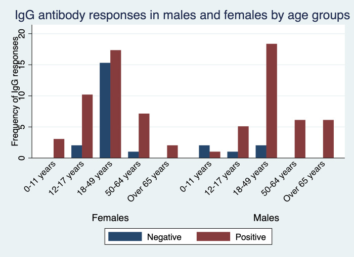
To assess immune response to SARS-CoV-2 among asymptomatic and symptomatic individuals, we tested for their IgG and IgM antibody levels. We further stratified the individuals into several age groups and compared the responses based on gender. Participants aged 18–49 years had the highest levels of detectable IgG antibodies from either gender. While all adult males aged 50–64 years and those over 65 years were all IgG seropositive, all young females aged between 0–11 and adults over 65 years were IgG seropositive (Fig 1).

Fig 1. Seroprevalence of infection-induced SARS-CoV-2 IgG antibodies, by gender and age group—Kisumu and Siaya counties, Kenya, December 2021–March 2022.

Fig 1

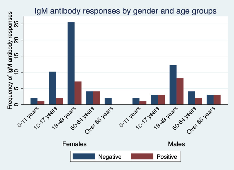
The detectable IgM antibodies were highest in participants aged 18–49 years and lowest in children aged between 0–11 and adults over 65 years though at lower frequency than IgG. Interestingly, all female participants aged 65 years and above were negative for IgM antibodies (Fig 2).

Fig 2. Seroprevalence of infection-induced SARS-CoV-2 IgM antibodies, by gender and age group—Kisumu and Siaya counties, Kenya, December 2021–March 2022.

Fig 2

Seroprevalence and vaccination status

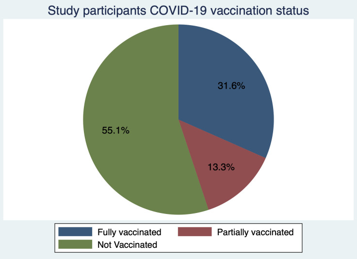
Amidst the ongoing vaccination roll-out during the study period almost one third, (31.6%, 95% CI = 22.6–41.8) of the study participants had been fully vaccinated, with close to a quarter (13.3%, 95% CI = 7.26–21.6) partially vaccinated. In contrast over half of the study participants (55.1%, 95% CI = 44.7–65.2) had not received any vaccine (Fig 3). When considering vaccination status and seroprevalence, out of the 31 fully vaccinated individuals, IgG seropositivity was 87.1% (95% CI = 70.2–96.3) and IgM seropositivity was 35.5% (95% CI = 19.22–54.6). From the partially vaccinated individuals, IgG seropositivity was 76.9% (95% CI = 46.2–95.0) with 53.8% (95% CI = 25.1–80.8) IgM seropositivity. Out of the participants that had not been vaccinated at all, IgG seroprevalence was 70.4% (95% CI 56.4–82.0) with 20.4% (95% CI 10.6–33.5) seropositivity of IgM antibodies.

Fig 3. Vaccination status of the study participants.

Fig 3

PCR positivity and vaccination status

When considering PCR positivity and COVID-19 vaccination status, one third of the individuals were positive (33.7%, 95 CI 24.3–44.1) while two thirds were negative (66.3%, 95% CI 55.9–75.7). The 32 positive individuals include 12(37.5%) fully vaccinated, 8(25%) partially vaccinated and 12(37.5%) unvaccinated. A binomial logistic regression was run to understand the effects of PCR positivity on the IgG and IgM seropositivity. SARs-CoV-2 PCR positivity did not significantly predict IgG (p = 0.469 [95% CI 0.514–4.230]) and IgM (p = 0.964 [95% CI 0.380–2.516]) positivity.

Discussion

With the poor uptake of COVID-19 vaccines in African settings amidst the easing of restrictions on movements and other containment measures, there is need to understand the antibody responses in the population. The present study aimed to describe the anti-SARs-CoV-2 IgG and IgM antibody responses during the COVID-19 pandemic in the period between December 2021 and March 2022 in western Kenya. Generally, we found high levels of IgG and IgM antibody against SARS-CoV-2 in the population corroborating recent and previous findings from population-based surveys in Kenya [11, 15, 16]. As previously observed, this was despite the low vaccination rate with only about one third of the population receiving the full vaccination [17]. This implies that most of the population had been exposed to the COVID-19 virus during this period and had raised antibodies against the infection. This is suggestive that the population may be heading to herd immunity, but this should not lead to vaccine complacency. It is instructive to note, that the Kenyan government has prioritized vaccination of the entire population with first and booster doses readily available in public health facilities.

A large proportion, 26(81.2%) of the 32 PCR positives had SARS-COV-2 IgG antibodies suggestive of a robust immune response. However, both vaccine induced and natural immunity are imperfect with breakthrough infections reported across diverse COVID-19 transmission settings [1821]. While COVID-19 vaccines elicit high levels of protection from symptomatic disease, this wanes over time necessitating booster doses to restore effectiveness [22]. Other studies have reported that immunogenicity may reduce despite of high IgG and neutralizing antibody levels [23]. Its noteworthy that, Kenya recorded its first case of the Omicron variant, besides the Delta variant during the study period. In addition, it’s been suggested that humoral responses generated by vaccination may not be good enough to protect against Omicron infection [24] and that Omicron escapes the vast majority of existing SARS-COV-2 neutralizing antibodies [25].

When considering IgG antibody responses and age groups, seroprevalence peaked in adults aged 18–49 years consistent with other studies in Kenya that reported higher seroprevalence amongst adults [10]. It is plausible that the adults have an expanded immunological memory driven by their catalog of memory B and T cells [26]. Adults older than 65 years of age had a lower antibody response against the SARS-CoV-2 compared to the other age groups in line with existing reports [9, 11, 12]. Increased comorbidities reported in the older adults may lead to the aging immune system not mounting a robust response.

Interestingly, we recorded a higher seroprevalence of IgM antibodies in Siaya County a more rural set up compared to Kisumu County a more urbanized town. This was in line with the higher SARS-CoV-2 RT-qPCR positivity of samples from Siaya county in contrast to Kisumu County during the sample collection period. This finding is consistent with other studies in Kenya that have reported marked geographic variation in seropositivity [10, 15]. Similar observations of variations in seroprevalence have also been documented across Africa [27, 28]. As the IgM antibody responses are short lived and useful in detecting recent infections [29], this suggest an ongoing community transmission in Siaya during the study period. The earlier and robust IgM responses days after onset of symptoms were linked to virus control. The similar profiles of IgG during the same period in the two counties corroborates its role as a more persistent antibody [30].

This study was limited by its recruitment of participants attending the hospitals, as it is possible that seroprevalence was overestimated due to selection bias. Additionally, the results may not be generalizable to the whole population as the samples may not have been representative. Consequently, representative longitudinal studies that follow individuals over a longer time span are needed to fully understand the SARS-CoV-2 antibody profiles and dynamics.

Conclusions

Despite the low number of either fully or partially vaccinated individuals against SARS-CoV-2, the seroprevalence of IgG and IgM antibodies was high. The finding suggests that many study participants were already infected with the virus than what the PCR testing could cover.

Supporting information

S1 File. Raw demographics and laboratory data for Kisumu and Siaya counties, western, Kenya December 2021 to March 2022.

(XLSX)

Acknowledgments

We thank the study team for administrative and technical support. We appreciate the support received from the Kisumu and Siaya Counties health officers during study procedures. We are grateful to the study participants who took part in the study.

Data Availability

All relevant data are within the paper and its Supporting Information files.

Funding Statement

Funding was provided by the National Research Foundation-South Africa under the COVID-19 Africa Rapid Grant Fund (Nr: COV19200616532700). SSA is supported by AREF Research Development Fellowship 2022, (AREF-312-AWAN-F-C0907). The funders had no role in study design, data collection and analysis, decision to publish, preparation of the manuscript.

References

  • 1.Dong E, Du H, Gardner L. An interactive web-based dashboard to track COVID-19 in real time. The Lancet infectious diseases. 2020;20(5):533–4. doi: 10.1016/S1473-3099(20)30120-1 [DOI] [PMC free article] [PubMed] [Google Scholar]
  • 2.Ciotti M, Ciccozzi M, Terrinoni A, Jiang W-C, Wang C-B, Bernardini S. The COVID-19 pandemic. Critical reviews in clinical laboratory sciences. 2020;57(6):365–88. doi: 10.1080/10408363.2020.1783198 [DOI] [PubMed] [Google Scholar]
  • 3.Maeda JM, Nkengasong JN. The puzzle of the COVID-19 pandemic in Africa. Science. 2021;371(6524):27–8. doi: 10.1126/science.abf8832 [DOI] [PubMed] [Google Scholar]
  • 4.Bamgboye EL, Omiye JA, Afolaranmi OJ, Davids MR, Tannor EK, Wadee S, et al. COVID-19 pandemic: is Africa different? Journal of the National Medical Association. 2021;113(3):324–35. doi: 10.1016/j.jnma.2020.10.001 [DOI] [PMC free article] [PubMed] [Google Scholar]
  • 5.Lone SA, Ahmad A. COVID-19 pandemic–an African perspective. Emerging microbes & infections. 2020;9(1):1300–8. doi: 10.1080/22221751.2020.1775132 [DOI] [PMC free article] [PubMed] [Google Scholar]
  • 6.Okba NM, Müller MA, Li W, Wang C, GeurtsvanKessel CH, Corman VM, et al. Severe acute respiratory syndrome coronavirus 2− specific antibody responses in coronavirus disease patients. Emerging infectious diseases. 2020;26(7):1478. doi: 10.3201/eid2607.200841 [DOI] [PMC free article] [PubMed] [Google Scholar]
  • 7.Post N, Eddy D, Huntley C, van Schalkwyk MC, Shrotri M, Leeman D, et al. Antibody response to SARS-CoV-2 infection in humans: a systematic review. PloS one. 2020;15(12):e0244126. doi: 10.1371/journal.pone.0244126 [DOI] [PMC free article] [PubMed] [Google Scholar]
  • 8.Arkhipova-Jenkins I, Helfand M, Armstrong C, Gean E, Anderson J, Paynter RA, et al. Antibody response after SARS-CoV-2 infection and implications for immunity: a rapid living review. Annals of internal medicine. 2021;174(6):811–21. doi: 10.7326/M20-7547 [DOI] [PMC free article] [PubMed] [Google Scholar]
  • 9.Adetifa IM, Uyoga S, Gitonga JN, Mugo D, Otiende M, Nyagwange J, et al. Temporal trends of SARS-CoV-2 seroprevalence during the first wave of the COVID-19 epidemic in Kenya. Nature communications. 2021;12(1):1–6. [DOI] [PMC free article] [PubMed] [Google Scholar]
  • 10.Uyoga S, Adetifa IM, Karanja HK, Nyagwange J, Tuju J, Wanjiku P, et al. Seroprevalence of anti–SARS-CoV-2 IgG antibodies in Kenyan blood donors. Science. 2021;371(6524):79–82. doi: 10.1126/science.abe1916 [DOI] [PMC free article] [PubMed] [Google Scholar]
  • 11.Etyang AO, Lucinde R, Karanja H, Kalu C, Mugo D, Nyagwange J, et al. Seroprevalence of antibodies to severe acute respiratory syndrome coronavirus 2 among healthcare workers in Kenya. Clinical Infectious Diseases. 2022;74(2):288–93. doi: 10.1093/cid/ciab346 [DOI] [PMC free article] [PubMed] [Google Scholar]
  • 12.Ngere I, Dawa J, Hunsperger E, Otieno N, Masika M, Amoth P, et al. High seroprevalence of SARS-CoV-2 but low infection fatality ratio eight months after introduction in Nairobi, Kenya. International Journal of Infectious Diseases. 2021;112:25–34. doi: 10.1016/j.ijid.2021.08.062 [DOI] [PMC free article] [PubMed] [Google Scholar]
  • 13.Frings M, Lakes T, Muller D, Khan MMH, Epprecht M, Kipruto S, et al. Modeling and mapping the burden of disease in Kenya. Sci Rep. 2018;8(1):9826. Epub 2018/07/01. doi: 10.1038/s41598-018-28266-4 ; PubMed Central PMCID: PMC6026135. [DOI] [PMC free article] [PubMed] [Google Scholar]
  • 14.Ministry of Health. COVID-19 Outbreak in Kenya Daily Report SITREP- 22-April-2021. 2021. [Google Scholar]
  • 15.Kagucia EW, Gitonga JN, Kalu C, Ochomo E, Ochieng B, Kuya N, et al. Seroprevalence of anti-SARS-CoV-2 IgG antibodies among truck drivers and assistants in Kenya. medRxiv. 2021. [Google Scholar]
  • 16.Lucinde R, Mugo D, Bottomley C, Aziza R, Gitonga J, Karanja H, et al. Sero-surveillance for IgG to SARS-CoV-2 at antenatal care clinics in two Kenyan referral hospitals. MedRxiv. 2021. [DOI] [PMC free article] [PubMed] [Google Scholar]
  • 17.Stringhini S, Zaballa M-E, Pullen N, Perez-Saez J, de Mestral C, Loizeau AJ, et al. Seroprevalence of anti-SARS-CoV-2 antibodies 6 months into the vaccination campaign in Geneva, Switzerland, 1 June to 7 July 2021. Eurosurveillance. 2021;26(43):2100830. doi: 10.2807/1560-7917.ES.2021.26.43.2100830 [DOI] [PMC free article] [PubMed] [Google Scholar]
  • 18.Bertollini R, Chemaitelly H, Yassine HM, Al-Thani MH, Al-Khal A, Abu-Raddad LJ. Associations of Vaccination and of Prior Infection With Positive PCR Test Results for SARS-CoV-2 in Airline Passengers Arriving in Qatar. JAMA. 2021;326(2):185–8. Epub 2021/06/10. doi: 10.1001/jama.2021.9970 ; PubMed Central PMCID: PMC8190701. [DOI] [PMC free article] [PubMed] [Google Scholar]
  • 19.Bergwerk M, Gonen T, Lustig Y, Amit S, Lipsitch M, Cohen C, et al. Covid-19 breakthrough infections in vaccinated health care workers. New England Journal of Medicine. 2021;385(16):1474–84. doi: 10.1056/NEJMoa2109072 [DOI] [PMC free article] [PubMed] [Google Scholar]
  • 20.Lipsitch M, Krammer F, Regev-Yochay G, Lustig Y, Balicer RD. SARS-CoV-2 breakthrough infections in vaccinated individuals: measurement, causes and impact. Nature Reviews Immunology. 2022;22(1):57–65. doi: 10.1038/s41577-021-00662-4 [DOI] [PMC free article] [PubMed] [Google Scholar]
  • 21.Hacisuleyman E, Hale C, Saito Y, Blachere NE, Bergh M, Conlon EG, et al. Vaccine breakthrough infections with SARS-CoV-2 variants. New England Journal of Medicine. 2021;384(23):2212–8. doi: 10.1056/NEJMoa2105000 [DOI] [PMC free article] [PubMed] [Google Scholar]
  • 22.Gupta RK, Topol EJ. COVID-19 vaccine breakthrough infections. Science. 2021;374(6575):1561–2. doi: 10.1126/science.abl8487 [DOI] [PubMed] [Google Scholar]
  • 23.Chvatal-Medina M, Mendez-Cortina Y, Patiño PJ, Velilla PA, Rugeles MT. Antibody responses in COVID-19: a review. Frontiers in immunology. 2021;12:633184. doi: 10.3389/fimmu.2021.633184 [DOI] [PMC free article] [PubMed] [Google Scholar]
  • 24.Gilboa M, Regev-Yochay G, Mandelboim M, Indenbaum V, Asraf K, Fluss R, et al. Durability of Immune Response After COVID-19 Booster Vaccination and Association With COVID-19 Omicron Infection. JAMA Netw Open. 2022;5(9):e2231778. Epub 2022/09/16. doi: 10.1001/jamanetworkopen.2022.31778 ; PubMed Central PMCID: PMC9478782. [DOI] [PMC free article] [PubMed] [Google Scholar]
  • 25.Cao Y, Wang J, Jian F, Xiao T, Song W, Yisimayi A, et al. Omicron escapes the majority of existing SARS-CoV-2 neutralizing antibodies. Nature. 2022;602(7898):657–63. Epub 2022/01/12. doi: 10.1038/s41586-021-04385-3 ; PubMed Central PMCID: PMC8866119. [DOI] [PMC free article] [PubMed] [Google Scholar]
  • 26.Cox RJ, Brokstad KA. Not just antibodies: B cells and T cells mediate immunity to COVID-19. Nature Reviews Immunology. 2020;20(10):581–2. doi: 10.1038/s41577-020-00436-4 [DOI] [PMC free article] [PubMed] [Google Scholar]
  • 27.Rice BL, Annapragada A, Baker RE, Bruijning M, Dotse-Gborgbortsi W, Mensah K, et al. Variation in SARS-CoV-2 outbreaks across sub-Saharan Africa. Nat Med. 2021;27(3):447–53. Epub 2021/02/04. doi: 10.1038/s41591-021-01234-8 ; PubMed Central PMCID: PMC8590469. [DOI] [PMC free article] [PubMed] [Google Scholar]
  • 28.Chibwana MG, Jere KC, Kamn’gona R, Mandolo J, Katunga-Phiri V, Tembo D, et al. High SARS-CoV-2 seroprevalence in health care workers but relatively low numbers of deaths in urban Malawi. medrxiv. 2020. doi: 10.1101/2020.07.30.20164970 [DOI] [PMC free article] [PubMed] [Google Scholar]
  • 29.Wu J, Liang B, Chen C, Wang H, Fang Y, Shen S, et al. SARS-CoV-2 infection induces sustained humoral immune responses in convalescent patients following symptomatic COVID-19. Nature communications. 2021;12(1):1–9. [DOI] [PMC free article] [PubMed] [Google Scholar]
  • 30.Duysburgh E, Mortgat L, Barbezange C, Dierick K, Fischer N, Heyndrickx L, et al. Persistence of IgG response to SARS-CoV-2. The Lancet Infectious Diseases. 2021;21(2):163–4. doi: 10.1016/S1473-3099(20)30943-9 [DOI] [PMC free article] [PubMed] [Google Scholar]

Decision Letter 0

Adriana Calderaro

20 Sep 2022

PONE-D-22-20904High seroprevalence of immunoglobulin G (IgG) and IgM antibodies to SARS-CoV-2 in asymptomatic and symptomatic individuals amidst vaccination roll-out in western KenyaPLOS ONE

Dear Dr. Awandu,

Thank you for submitting your manuscript to PLOS ONE. After careful consideration, we feel that it has merit but does not fully meet PLOS ONE’s publication criteria as it currently stands. Therefore, we invite you to submit a revised version of the manuscript that addresses the points raised during the review process. The Authors should address to the criticisms detected by the REferees in order to clarify some inconsistencies. Furhtermore, they should check once again the number indicated in the manuscript. 

Please submit your revised manuscript by Nov 04 2022 11:59PM. If you will need more time than this to complete your revisions, please reply to this message or contact the journal office at plosone@plos.org. When you're ready to submit your revision, log on to https://www.editorialmanager.com/pone/ and select the 'Submissions Needing Revision' folder to locate your manuscript file.

Please include the following items when submitting your revised manuscript:

  • A rebuttal letter that responds to each point raised by the academic editor and reviewer(s). You should upload this letter as a separate file labeled 'Response to Reviewers'.

  • A marked-up copy of your manuscript that highlights changes made to the original version. You should upload this as a separate file labeled 'Revised Manuscript with Track Changes'.

  • An unmarked version of your revised paper without tracked changes. You should upload this as a separate file labeled 'Manuscript'.

If you would like to make changes to your financial disclosure, please include your updated statement in your cover letter. Guidelines for resubmitting your figure files are available below the reviewer comments at the end of this letter.

If applicable, we recommend that you deposit your laboratory protocols in protocols.io to enhance the reproducibility of your results. Protocols.io assigns your protocol its own identifier (DOI) so that it can be cited independently in the future. For instructions see: https://journals.plos.org/plosone/s/submission-guidelines#loc-laboratory-protocols. Additionally, PLOS ONE offers an option for publishing peer-reviewed Lab Protocol articles, which describe protocols hosted on protocols.io. Read more information on sharing protocols at https://plos.org/protocols?utm_medium=editorial-email&utm_source=authorletters&utm_campaign=protocols.

We look forward to receiving your revised manuscript.

Kind regards,

Adriana Calderaro

Academic Editor

PLOS ONE

Journal Requirements:

When submitting your revision, we need you to address these additional requirements.

1. Please ensure that your manuscript meets PLOS ONE's style requirements, including those for file naming. The PLOS ONE style templates can be found at 

https://journals.plos.org/plosone/s/file?id=wjVg/PLOSOne_formatting_sample_main_body.pdf and 

https://journals.plos.org/plosone/s/file?id=ba62/PLOSOne_formatting_sample_title_authors_affiliations.pdf

2. We note that the grant information you provided in the ‘Funding Information’ and ‘Financial Disclosure’ sections do not match. 

When you resubmit, please ensure that you provide the correct grant numbers for the awards you received for your study in the ‘Funding Information’ section.

3. Please include your full ethics statement in the ‘Methods’ section of your manuscript file. In your statement, please include the full name of the IRB or ethics committee who approved or waived your study, as well as whether or not you obtained informed written or verbal consent. If consent was waived for your study, please include this information in your statement as well. 

4. We note that Figure 1 in your submission contain [map/satellite] images which may be copyrighted. All PLOS content is published under the Creative Commons Attribution License (CC BY 4.0), which means that the manuscript, images, and Supporting Information files will be freely available online, and any third party is permitted to access, download, copy, distribute, and use these materials in any way, even commercially, with proper attribution. For these reasons, we cannot publish previously copyrighted maps or satellite images created using proprietary data, such as Google software (Google Maps, Street View, and Earth). For more information, see our copyright guidelines: http://journals.plos.org/plosone/s/licenses-and-copyright.

We require you to either (a) present written permission from the copyright holder to publish these figures specifically under the CC BY 4.0 license, or (b) remove the figures from your submission:

a. You may seek permission from the original copyright holder of Figure 1 to publish the content specifically under the CC BY 4.0 license.  

We recommend that you contact the original copyright holder with the Content Permission Form (http://journals.plos.org/plosone/s/file?id=7c09/content-permission-form.pdf) and the following text:

“I request permission for the open-access journal PLOS ONE to publish XXX under the Creative Commons Attribution License (CCAL) CC BY 4.0 (http://creativecommons.org/licenses/by/4.0/). Please be aware that this license allows unrestricted use and distribution, even commercially, by third parties. Please reply and provide explicit written permission to publish XXX under a CC BY license and complete the attached form.”

Please upload the completed Content Permission Form or other proof of granted permissions as an "Other" file with your submission.

In the figure caption of the copyrighted figure, please include the following text: “Reprinted from [ref] under a CC BY license, with permission from [name of publisher], original copyright [original copyright year].”

b. If you are unable to obtain permission from the original copyright holder to publish these figures under the CC BY 4.0 license or if the copyright holder’s requirements are incompatible with the CC BY 4.0 license, please either i) remove the figure or ii) supply a replacement figure that complies with the CC BY 4.0 license. Please check copyright information on all replacement figures and update the figure caption with source information. If applicable, please specify in the figure caption text when a figure is similar but not identical to the original image and is therefore for illustrative purposes only.

The following resources for replacing copyrighted map figures may be helpful:

USGS National Map Viewer (public domain): http://viewer.nationalmap.gov/viewer/

The Gateway to Astronaut Photography of Earth (public domain): http://eol.jsc.nasa.gov/sseop/clickmap/

Maps at the CIA (public domain): https://www.cia.gov/library/publications/the-world-factbook/index.html and https://www.cia.gov/library/publications/cia-maps-publications/index.html

NASA Earth Observatory (public domain): http://earthobservatory.nasa.gov/

Landsat: http://landsat.visibleearth.nasa.gov/

USGS EROS (Earth Resources Observatory and Science (EROS) Center) (public domain): http://eros.usgs.gov/#

Natural Earth (public domain): http://www.naturalearthdata.com/

[Note: HTML markup is below. Please do not edit.]

Reviewers' comments:

Reviewer's Responses to Questions

Comments to the Author

1. Is the manuscript technically sound, and do the data support the conclusions?

The manuscript must describe a technically sound piece of scientific research with data that supports the conclusions. Experiments must have been conducted rigorously, with appropriate controls, replication, and sample sizes. The conclusions must be drawn appropriately based on the data presented.

Reviewer #1: Yes

Reviewer #2: No

**********

2. Has the statistical analysis been performed appropriately and rigorously?

Reviewer #1: Yes

Reviewer #2: Yes

**********

3. Have the authors made all data underlying the findings in their manuscript fully available?

The PLOS Data policy requires authors to make all data underlying the findings described in their manuscript fully available without restriction, with rare exception (please refer to the Data Availability Statement in the manuscript PDF file). The data should be provided as part of the manuscript or its supporting information, or deposited to a public repository. For example, in addition to summary statistics, the data points behind means, medians and variance measures should be available. If there are restrictions on publicly sharing data—e.g. participant privacy or use of data from a third party—those must be specified.

Reviewer #1: Yes

Reviewer #2: Yes

**********

4. Is the manuscript presented in an intelligible fashion and written in standard English?

PLOS ONE does not copyedit accepted manuscripts, so the language in submitted articles must be clear, correct, and unambiguous. Any typographical or grammatical errors should be corrected at revision, so please note any specific errors here.

Reviewer #1: Yes

Reviewer #2: Yes

**********

5. Review Comments to the Author

Please use the space provided to explain your answers to the questions above. You may also include additional comments for the author, including concerns about dual publication, research ethics, or publication ethics. (Please upload your review as an attachment if it exceeds 20,000 characters)

Reviewer #1: I think that this manuscript should be accepted with minor revision in introduction or part of the Demographics in the Results section. I suggest that authors add data on numbers of registered cases from the beginning of the pandemic in these two counties and also data on number of registered cases for the observed period in both counties. Possibly a figure could be drawn on incidence of registered cases and specific incidences by observed age groups as well. This is a minor effort and i belive it would fir well with the rest of the text and give it more clear overall picture

Reviewer #2: In this manuscript, Awandu et al. report the seroprevalence estimate in 98 individuals presenting to Kisumu and Siaya Counties referral hospitals in western Kenya between December 2021-March 2022. The authors measured SARS-CoV2 IgG and IgM antibodies in blood specimens and detected viral RNA in nasopharyngeal samples using commercial kits following manufacturer instructions. Of the 98 subjects, the authors report that 44 were vaccinated fully (n=31) or partially (n=13). They say high IgG seropositivity in both vaccinated (86.7%) and unvaccinated (70.3%) individuals and claim that PCR testing did not predict the seropositivity within the study period. The study has several weaknesses, some of which are listed below.

In the serology data sheet, the authors report a total of 32 PCR positives. This includes 12 (38.7%) PCR positives among 31 fully vaccinated, 8 (61.5%) PCR positives among 13 partially vaccinated, and 11 (20.3%) PCR positives among 54 unvaccinated individuals. Interestingly, 26 (83.8%) of the 31 PCR positives have been reported to have SARS-CoV-2 IgG antibodies. While breakthrough infections are possible, it is unclear why such a large proportion of PCR positives have SARS-CoV-2 IgG Abs. It is also unclear why PCR positives are higher among vaccinated than unvaccinated (47.7% (23 of 44) Vs. 20.3% (11 of 54).

The authors state that they have used commercial kits following manufacturer instructions. It is unclear if the authors have tested pre-pandemic samples to validate the commercial assay specificity.

Authors report the IgG and IgM results as ratios. However, the methods described in line 124 are unclear about how ratios were calculated from optical densities.

The number of vaccinated individuals reported in the abstract (n=30) and the serology data sheet (n=31) do not match.

Authors report that about 26 of 98 were asymptomatic. It will be helpful to clarify how asymptomatic were referred to the hospitals. Are these asymptomatic people exposed to people previously infected with SARS-CoV-2?

**********

6. PLOS authors have the option to publish the peer review history of their article (what does this mean?). If published, this will include your full peer review and any attached files.

If you choose “no”, your identity will remain anonymous but your review may still be made public.

Do you want your identity to be public for this peer review? For information about this choice, including consent withdrawal, please see our Privacy Policy.

Reviewer #1: No

Reviewer #2: No

**********

[NOTE: If reviewer comments were submitted as an attachment file, they will be attached to this email and accessible via the submission site. Please log into your account, locate the manuscript record, and check for the action link "View Attachments". If this link does not appear, there are no attachment files.]

While revising your submission, please upload your figure files to the Preflight Analysis and Conversion Engine (PACE) digital diagnostic tool, https://pacev2.apexcovantage.com/. PACE helps ensure that figures meet PLOS requirements. To use PACE, you must first register as a user. Registration is free. Then, login and navigate to the UPLOAD tab, where you will find detailed instructions on how to use the tool. If you encounter any issues or have any questions when using PACE, please email PLOS at figures@plos.org. Please note that Supporting Information files do not need this step.

PLoS One. 2022 Dec 22;17(12):e0272751. doi: 10.1371/journal.pone.0272751.r002

Author response to Decision Letter 0


24 Oct 2022

Response to Reviewers

Dear Editors and Reviewers,

Thank you for giving me the opportunity to submit a revised draft of our manuscript titled High seroprevalence of Immunoglobulin G (IgG) and IgM antibodies to SARS-CoV-2 in asymptomatic and symptomatic individuals amidst vaccination roll-out in western Kenya to Plos One Journal. We appreciate you and the reviewers for dedicating your precious time in reviewing our manuscript and providing valuable feedback. We have been able to incorporate changes to reflect most of the suggestions provided by the reviewers. We hope that we have satisfactorily addressed them and that the manuscript is now suitable for publication.

Sincerely,

On behalf of all authors,

Shehu Shagari Awandu

Here is a point-by-point response to the reviewers’ comments and concerns.

Comments from the Academic Editor

Comment 1. Please ensure that your manuscript meets PLOS ONE's style requirements, including those for file naming.

Response. Thank you very much for these comments. We have ensured that the manuscript meets PLOS ONE's style requirements, including those for file naming. We hopefully have no divergences from the style requirements now.

Comment 2. We note that the grant information you provided in the ‘Funding Information’ and ‘Financial Disclosure’ sections do not match.

When you resubmit, please ensure that you provide the correct grant numbers for the awards you received for your study in the ‘Funding Information’ section.

Thanks for pointing this out. We have provided the correct grant numbers. The study was supported by the National Research Foundation, South Africa under the COVID-19 Africa Rapid Grant Fund Grant Number COV19200616532700 awarded to Benson Estambale. “

Comment 3. Please include your full ethics statement in the ‘Methods’ section of your manuscript file. In your statement, please include the full name of the IRB or ethics committee who approved or waived your study, as well as whether or not you obtained informed written or verbal consent. If consent was waived for your study, please include this information in your statement as well.

Response: Thanks for the comment. We have included the full ethics statement on and it now reads as follows

“Ethical consideration

Ethical approval for the study was granted by Jaramogi Oginga Odinga University of Science and Technology (ERC/21/5/21-4), and a research license granted from Kenya National Commission of Science and Technolog, (NACOSTI/P/22/17545). Administrative approval was provided by the county governments of Kisumu and Siaya. All participants provided written informed consent or assent before enrollment.”

Comment 4. We note that Figure 1 in your submission contain [map/satellite] images which may be copyrighted. All PLOS content is published under the Creative Commons Attribution License (CC BY 4.0), which means that the manuscript, images, and Supporting Information files will be freely available online, and any third party is permitted to access, download, copy, distribute, and use these materials in any way, even commercially, with proper attribution. For these reasons, we cannot publish previously copyrighted maps or satellite images created using proprietary data, such as Google software (Google Maps, Street View, and Earth).

We require you to either (a) present written permission from the copyright holder to publish these figures specifically under the CC BY 4.0 license, or (b) remove the figures from your submission:

a. You may seek permission from the original copyright holder of Figure 1 to publish the content specifically under the CC BY 4.0 license.

We recommend that you contact the original copyright holder with the Content Permission Form (http://journals.plos.org/plosone/s/file?id=7c09/content-permission-form.pdf) and the following text:

“I request permission for the open-access journal PLOS ONE to publish XXX under the Creative Commons Attribution License (CCAL) CC BY 4.0 (http://creativecommons.org/licenses/by/4.0/). Please be aware that this license allows unrestricted use and distribution, even commercially, by third parties. Please reply and provide explicit written permission to publish XXX under a CC BY license and complete the attached form.”

Please upload the completed Content Permission Form or other proof of granted permissions as an "Other" file with your submission.

In the figure caption of the copyrighted figure, please include the following text: “Reprinted from [ref] under a CC BY license, with permission from [name of publisher], original copyright [original copyright year].”

b. If you are unable to obtain permission from the original copyright holder to publish these figures under the CC BY 4.0 license or if the copyright holder’s requirements are incompatible with the CC BY 4.0 license, please either i) remove the figure or ii) supply a replacement figure that complies with the CC BY 4.0 license. Please check copyright information on all replacement figures and update the figure caption with source information. If applicable, please specify in the figure caption text when a figure is similar but not identical to the original image and is therefore for illustrative purposes only.

The following resources for replacing copyrighted map figures may be helpful:

USGS National Map Viewer (public domain): http://viewer.nationalmap.gov/viewer/

The Gateway to Astronaut Photography of Earth (public domain): http://eol.jsc.nasa.gov/sseop/clickmap/

Maps at the CIA (public domain): https://www.cia.gov/library/publications/the-world-factbook/index.html and https://www.cia.gov/library/publications/cia-maps-publications/index.html

NASA Earth Observatory (public domain): http://earthobservatory.nasa.gov/

Landsat: http://landsat.visibleearth.nasa.gov/

USGS EROS (Earth Resources Observatory and Science (EROS) Center) (public domain): http://eros.usgs.gov/#

Natural Earth (public domain): http://www.naturalearthdata.com/

[Note: HTML markup is below. Please do not edit.]

Response: Thanks for the comment. We have removed the figure

Comments from Reviewer #1

Comments 1: I think that this manuscript should be accepted with minor revision in introduction or part of the Demographics in the Results section.

Response: Thank you very much

Comment 2: I suggest that authors add data on numbers of registered cases from the beginning of the pandemic in these two counties and also data on number of registered cases for the observed period in both counties. Possibly a figure could be drawn on incidence of registered cases and specific incidences by observed age groups as well. This is a minor effort and i belive it would fir well with the rest of the text and give it more clear overall picture.

Response: Thank you very much for this pointing this out. We have updated the manuscript and provided data on the number of registered cases from the beginning of the pandemic, and during our study period. We have included the Kenya Ministry of Health reference that provides a comprehensive COVID-19 data for the whole country.

Response to Reviewer 2

In this manuscript, Awandu et al. report the seroprevalence estimate in 98 individuals presenting to Kisumu and Siaya Counties referral hospitals in western Kenya between December 2021-March 2022. The authors measured SARS-CoV2 IgG and IgM antibodies in blood specimens and detected viral RNA in nasopharyngeal samples using commercial kits following manufacturer instructions. Of the 98 subjects, the authors report that 44 were vaccinated fully (n=31) or partially (n=13). They say high IgG seropositivity in both vaccinated (86.7%) and unvaccinated (70.3%) individuals and claim that PCR testing did not predict the seropositivity within the study period. The study has several weaknesses, some of which are listed below.

Reviewer: In the serology data sheet, the authors report a total of 32 PCR positives. This includes 12 (38.7%) PCR positives among 31 fully vaccinated, 8 (61.5%) PCR positives among 13 partially vaccinated, and 11 (20.3%) PCR positives among 54 unvaccinated individuals. Interestingly, 26 (83.8%) of the 31 PCR positives have been reported to have SARS-CoV-2 IgG antibodies. While breakthrough infections are possible, it is unclear why such a large proportion of PCR positives have SARS-CoV-2 IgG Abs. It is also unclear why PCR positives are higher among vaccinated than unvaccinated (47.7% (23 of 44) Vs. 20.3% (11 of 54).

Response: Thanks for your comments, that have helped us improve the section. We have revised the discussion to include a paragraph providing a possible explanation for the high rate of IgG and PCR positivity.

“A large proportion, 26(81.2%) of the 32 PCR positives had SARS-COV-2 IgG antibodies suggestive of a robust immune response. However, both vaccine induced, and natural immunity are imperfect with breakthrough infections reported across diverse COVID-19 transmission settings (18-21). Other studies have reported that immunogenicity may reduce despite of high IgG and neutralizing antibody levels (22). Its noteworthy that, Kenya recorded its first case of the omicron variant during the study period, and this may explain our findings. In addition, it’s been suggested that humoral responses generated by vaccination may not be good enough to protect against omicron infection (23) and that omicron escapes the vast majority of existing SARS-COV-2 neutralizing antibodies (24).”

Reviewer comments: The authors state that they have used commercial kits following manufacturer instructions. It is unclear if the authors have tested pre-pandemic samples to validate the commercial assay specificity.

Response: Thank you very much for pointing this out. This has been expounded in the manuscript.

Reviewer: Authors report the IgG and IgM results as ratios. However, the methods described in line 124 are unclear about how ratios were calculated from optical densities.

Response: We agree with the reviewer that the methods were unclear. We have included a detailed explanation of how the ratios are computed in the manuscript.

Reviewer: The number of vaccinated individuals reported in the abstract (n=30) and the serology data sheet (n=31) do not match.

Response: Thank you for pointing this out. We admit our error. We have corrected the error(n=31) and now both the abstract and serology datasheet match. We have similarly gone through the entire manuscript and updated the figures based on the data sheet

Reviewer: Authors report that about 26 of 98 were asymptomatic. It will be helpful to clarify how asymptomatic were referred to the hospitals. Are these asymptomatic people exposed to people previously infected with SARS-CoV-2?

Response: Thanks for pointing this out, we have clarified this on the manuscripts and it now reads as follows.

“About 26.5% (26/98) of participants were asymptomatic, however, they were referred to the hospital for surveillance testing as some were contacts of people infected with SARS-CoV-2.”

Decision Letter 1

Adriana Calderaro

24 Nov 2022

High seroprevalence of immunoglobulin G (IgG) and IgM antibodies to SARS-CoV-2 in asymptomatic and symptomatic individuals amidst vaccination roll-out in western Kenya

PONE-D-22-20904R1

Dear Dr. Awandu,

We’re pleased to inform you that your manuscript has been judged scientifically suitable for publication and will be formally accepted for publication once it meets all outstanding technical requirements.

Within one week, you’ll receive an e-mail detailing the required amendments. When these have been addressed, you’ll receive a formal acceptance letter and your manuscript will be scheduled for publication.

An invoice for payment will follow shortly after the formal acceptance. To ensure an efficient process, please log into Editorial Manager at http://www.editorialmanager.com/pone/, click the 'Update My Information' link at the top of the page, and double check that your user information is up-to-date. If you have any billing related questions, please contact our Author Billing department directly at authorbilling@plos.org.

If your institution or institutions have a press office, please notify them about your upcoming paper to help maximize its impact. If they’ll be preparing press materials, please inform our press team as soon as possible -- no later than 48 hours after receiving the formal acceptance. Your manuscript will remain under strict press embargo until 2 pm Eastern Time on the date of publication. For more information, please contact onepress@plos.org.

Kind regards,

Adriana Calderaro

Academic Editor

PLOS ONE

Additional Editor Comments (optional):

Reviewers' comments:

Reviewer's Responses to Questions

Comments to the Author

1. If the authors have adequately addressed your comments raised in a previous round of review and you feel that this manuscript is now acceptable for publication, you may indicate that here to bypass the “Comments to the Author” section, enter your conflict of interest statement in the “Confidential to Editor” section, and submit your "Accept" recommendation.

Reviewer #1: All comments have been addressed

Reviewer #2: All comments have been addressed

**********

2. Is the manuscript technically sound, and do the data support the conclusions?

The manuscript must describe a technically sound piece of scientific research with data that supports the conclusions. Experiments must have been conducted rigorously, with appropriate controls, replication, and sample sizes. The conclusions must be drawn appropriately based on the data presented.

Reviewer #1: Yes

Reviewer #2: Yes

**********

3. Has the statistical analysis been performed appropriately and rigorously?

Reviewer #1: Yes

Reviewer #2: Yes

**********

4. Have the authors made all data underlying the findings in their manuscript fully available?

The PLOS Data policy requires authors to make all data underlying the findings described in their manuscript fully available without restriction, with rare exception (please refer to the Data Availability Statement in the manuscript PDF file). The data should be provided as part of the manuscript or its supporting information, or deposited to a public repository. For example, in addition to summary statistics, the data points behind means, medians and variance measures should be available. If there are restrictions on publicly sharing data—e.g. participant privacy or use of data from a third party—those must be specified.

Reviewer #1: Yes

Reviewer #2: Yes

**********

5. Is the manuscript presented in an intelligible fashion and written in standard English?

PLOS ONE does not copyedit accepted manuscripts, so the language in submitted articles must be clear, correct, and unambiguous. Any typographical or grammatical errors should be corrected at revision, so please note any specific errors here.

Reviewer #1: Yes

Reviewer #2: Yes

**********

6. Review Comments to the Author

Please use the space provided to explain your answers to the questions above. You may also include additional comments for the author, including concerns about dual publication, research ethics, or publication ethics. (Please upload your review as an attachment if it exceeds 20,000 characters)

Reviewer #1: I think all issues have been addressed and paper should be accepted. Paper is well written, all reviewers questions were met. Results are described clearly, Discussion is well referenced and limitations support the conclusion.

Reviewer #2: (No Response)

**********

7. PLOS authors have the option to publish the peer review history of their article (what does this mean?). If published, this will include your full peer review and any attached files.

If you choose “no”, your identity will remain anonymous but your review may still be made public.

Do you want your identity to be public for this peer review? For information about this choice, including consent withdrawal, please see our Privacy Policy.

Reviewer #1: No

Reviewer #2: No

**********

Acceptance letter

Adriana Calderaro

13 Dec 2022

PONE-D-22-20904R1

High seroprevalence of Immunoglobulin G (IgG) and IgM antibodies to SARS-CoV-2 in asymptomatic and symptomatic individuals amidst vaccination roll-out in western Kenya

Dear Dr. Awandu:

I'm pleased to inform you that your manuscript has been deemed suitable for publication in PLOS ONE. Congratulations! Your manuscript is now with our production department.

If your institution or institutions have a press office, please let them know about your upcoming paper now to help maximize its impact. If they'll be preparing press materials, please inform our press team within the next 48 hours. Your manuscript will remain under strict press embargo until 2 pm Eastern Time on the date of publication. For more information please contact onepress@plos.org.

If we can help with anything else, please email us at plosone@plos.org.

Thank you for submitting your work to PLOS ONE and supporting open access.

Kind regards,

PLOS ONE Editorial Office Staff

on behalf of

MD, PhD, Associate Professor Adriana Calderaro

Academic Editor

PLOS ONE

Associated Data

    This section collects any data citations, data availability statements, or supplementary materials included in this article.

    Supplementary Materials

    S1 File. Raw demographics and laboratory data for Kisumu and Siaya counties, western, Kenya December 2021 to March 2022.

    (XLSX)

    Data Availability Statement

    All relevant data are within the paper and its Supporting Information files.


    Articles from PLOS ONE are provided here courtesy of PLOS

    RESOURCES