Abstract
Compared to transcription initiation, much less is known about transcription termination. In particular, large-scale mutagenesis studies have, so far, primarily concentrated on promoter and enhancer, but not terminator sequences. Here, we used a massively parallel reporter assay (MPRA) to systematically analyze the influence of short (8 bp) sequence variants (mutations) located downstream of the polyadenylation signal (PAS) on the steady-state mRNA level of the upstream gene, employing an eGFP reporter and human HEK293T cells as a model system. In total, we evaluated 227,755 mutations located at different overlapping positions within +17..+56 bp downstream of the PAS for their ability to regulate the reporter gene expression. We found that the positions +17..+44 bp downstream of the PAS are more essential for gene upregulation than those located more distal to the PAS, and that the mutation sequences ensuring high levels of eGFP mRNA expression are extremely T-rich. Next, we validated the positive effect of a couple of mutations identified in the MPRA screening on the eGFP and luciferase protein expression. The most promising mutation increased the expression of the reporter proteins 13-fold and sevenfold on average in HEK293T and CHO cells, respectively. Overall, these findings might be useful for further improving the efficiency of production of therapeutic products, e.g., recombinant antibodies.
Keywords: gene expression regulation, transcription termination, massively parallel reporter assay, MPRA, barcode, next-generation sequencing, NGS
1. Introduction
In eukaryotes, the termination of the transcription process can substantially influence the level of gene expression via various mechanisms [1,2,3]. Moreover, gene expression regulation was shown to be primarily controlled by transcription termination in those systems, where background transcription prevails [4,5]. Lesions in the transcription termination were found to be associated with oncological, immunological, neurological, and other diseases [6,7,8,9,10,11,12]. Transcription termination of protein-coding genes relies on the assembly of the functional cleavage and polyadenylation (CPA) complex at the 3′ end of the newly synthesized transcripts [13,14]. This process is tightly coordinated with pre-mRNA synthesis, capping, and splicing [15,16,17,18,19]. The mammalian CPA complex encompasses more than 80 subunits that are grouped into several functional subcomplexes [20,21,22]. Three subcomplexes (cleavage and polyadenylation specificity factor (CPSF), cleavage stimulation factor (CstF), and cleavage factor Im (CF Im)) bind directly to pre-mRNA to form a core complex, which is then accomplished by CF IIm, Symplekin, and poly(A) polymerase (PAP), resulting in a functional CPA complex [20,21]. Assembly of this complex is initiated at a hexamer sequence referred to as the polyadenylation signal (PAS) (AAUAAA and its multiple close variations), during the transcription elongation, and requires the presence of the C-terminal domain of the RNA polymerase II (RNAP II) large subunit that undergoes a set of covalent modifications [23,24,25]. CPSF and CstF subcomplexes recognize the PAS and the downstream sequence element (DSE) in pre-mRNAs, respectively [22,26,27,28]. CPSF is additionally subdivided into two modules by their function: mPSF (mammalian polyadenylation specificity factor) that binds the PAS and recruits PAP, thus catalyzing the polyadenylation process, and mCF that catalyzes the pre-mRNA cleavage reaction [14,29,30]. The CF Im subcomplex is known to bind the actively elongating RNAP II together with CPSF and CstF [31]. CF Im recognizes the UGUA sequence (and its variations), which can be located either upstream or downstream of the mature transcript cleavage site [31,32,33,34], and it stabilizes binding of the CPSF to the primary transcript [35]. The CF Im factor can also bind two UGUA motifs due to the capability of dimerization of its CFI25 subunit, resulting in a loop at the 3′ end of pre-mRNA [36]. This feature, as well as the correlation of increased CF Im levels with alternative polyadenylation (APA), suggests its significant role in the PAS selection during APA [34,37,38]. Several additional subcomplexes play a role in the CPA complex assembly and can influence the efficiency of pre-mRNA processing. The cleavage factor IIm (CF IIm) subcomplex remains one of the less studied factors in the mammalian CPA complex. It is highly homologous to proteins involved in transcription termination in yeast [39,40]. The yeast homolog of one of the CF IIm subunits interacts directly with the C-terminal domain of RNAP II [41,42] and other CPA components, and its knockdown causes impaired transcription termination [43,44]. For another CF IIm subunit homolog, the interaction and functional interrelation with CPSF and CF Im were shown [45,46]. Symplekin protein was also shown to interact directly with several subunits of the CPA complex [20,47]. However, its exact role has not been determined so far. Possibly, Symplekin stabilizes the whole CPA complex, being a multi-binding protein. There is also evidence that Symplekin can act as a regulator of the nuclease activity of the CPSF-73 subunit of mCF by binding additional regulatory factors [48,49,50]. PAP was found in eukaryotes in four variants (PAP, Neo-PAP, Star-PAP, and TPAP) [51,52]. PAP is known to directly interact with subunits of CPSF and CF Im [53], but many studies have indicated that PAP is not stably associated with the CPA complex [54,55], and this may reflect a highly dynamic organization of the core 3′ processing complex. An additional number of proteins that are involved in the PAS recognition and can modulate the activity of the CPA complex were also found [56,57,58,59]. At the same time, it was shown that only CPSF and PAP are necessary and sufficient for polyadenylation of transcripts in vitro [21,60,61]. Thus, the CPA complex is a huge multicomponent machine, which assembly and activity depend on various factors.
The mechanisms and key factors of transcription termination remain much less understood than the processes of transcription initiation and elongation. Two main hypothetical schemes describing the mechanism of transcription termination in mammals have been discussed during the last 30 years. The allosteric model suggests that the CPA complex assembly results in a RNAP II conformation change that leads to the loss of elongation factors and, thus, further synthesis of the RNA chain stops [62,63]. This model is confirmed by the fact that RNAP II ceases to bind elongation factors much earlier than it dissociates from the synthesized pre-RNA and DNA template [64,65]. The torpedo model is based on the assumption that elongating RNAP II meets the exonuclease activity of the CPA complex, resulting in cleavage of pre-mRNA from the 3′ end. The interaction of the CPA complex with RNAP II leads to the dissociation of both complexes [66,67]. The torpedo model better explains the relationship between transcript cleavage and termination and is supported by the experimentally shown “sliding” of RNA synthesis in the absence of the 5′–3′ exoribonuclease 2 (XRN2) activity [68]. Recently, the combined allosteric/torpedo mechanism was proposed, in which termination factor-dependent slowing down of RNAP II within the termination regions facilitates its capture by XRN2 and subsequent 3’ -> 5’ degradation of the 3’ pre-mRNA end [69].
Instability and variations in the CPA complex assembly, as well as the stochastic nature of the cleavage process, result in APA through the selection of a particular one of multiple PASs [9,56,70,71]. Therefore, the length and composition of mRNA 3′ untranslated regions (UTRs) vary, resulting in different sets of targets for RNA-binding proteins and miRNAs, which finally define the mRNA transport, lifespan, and translation efficiency [71,72,73]. In addition, the length of the poly(A) tail is also crucial for the same aspects of mRNA biology [70,74,75].
The particular variant of the CPA complex assembly and the PAS choice depend also on the nucleotide composition of the 3′ region of the immature transcript [76]. Consensus nucleotide motifs that induce and improve the CPA complex assembly are still not well defined. Known motifs are located both upstream and downstream of the pre-mRNA cleavage site, and the latter are, therefore, not included in the mature mRNAs [77,78,79]. Despite numerous attempts, no consensus has yet been found to be associated with the transcript cleavage site during transcription termination. The most well-known, but not the only, transcript cleavage site was found immediately after the CA dinucleotide [77,80,81]. In addition, the DSE is often found ~40 bp downstream of the transcript cleavage site, although about 20% of human mRNAs do not contain such a motif [20,76,77]. To date, it is believed that the transcription termination process is based only on RNA–protein interactions. It was shown that minimal functional PAS requires only a strong DSE and an A-rich upstream sequence in human cells [82]. Mature mRNAs were also found to have AU-rich elements (AREs) in their 3′ UTR regions, although these motifs can also be located near the 5′ end of the transcript. Binding a range of RNA-binding proteins (RBP), the ARE element is known to induce mRNA degradation [83,84]. For instance, early response genes that are sensitive to a wide range of external signals, including oncogenes and cytokines, have a relatively short lifespan of mRNAs due to AREs [85]. However, other RBPs, e.g., HuR, by binding to ARE elements, support mRNA stability by preventing endonuclease access [86]. It is extremely curious that none of the abovementioned motifs is found in all mammalian pre-mRNA molecules. The PAS is also present in only about 70% of mature human mRNAs [3,79,87]. Thus, a detailed functional study of the mechanism(s) of transcription termination with regard to the regulatory capacity of nucleotide motifs at the gene 3’ end is still relevant for understanding the regulation of expression of protein-coding genes in mammals.
Previously, in transient transfection assays, we showed that a single cytosine deletion at the position +32 bp downstream of the single PAS (deltaC) causes a ~2-fold increase in eGFP reporter gene expression in mouse embryonic stem cells (mESCs), cultured mouse 3T3 cells, and human embryonic kidney (HEK293T) cells [88,89], indicating its involvement in a molecular mechanism that is likely to be conserved in mammals. This deltaC appears to be involved in regulating the choice of the cleavage site, as the most proximal site (located 14 bp downstream of the PAS) becomes prevalent upon this single-nucleotide deletion. In addition, the replacement of several 16 bp fragments downstream of the PAS with artificial sequences of the same length showed that regions at +25..+40 and +33..+48 bp relative to the PAS are most sensitive to nucleotide variations and revealed particular sequences (at +25..+40 bp relative to the PAS) that increase eGFP expression up to fourfold [89].
In the present work, we performed a functional massively parallel reporter assay (MPRA) to assess the capability of 8 bp long mutations located within the +17..+56 bp region downstream of the PAS to control the mRNA level of the upstream eGFP reporter in HEK293T cells. As a result, we found that mutations significantly increasing eGFP expression are extremely T-rich. Next, we validated the positive effect of several mutations identified in the MPRA screening on eGFP expression at the protein level in HEK293T and Chinese hamster ovary (CHO) cells. Lastly, for a couple of chosen mutations, we demonstrated that they also increase the expression level of the luciferase reporter both in HEK293T and CHO cells.
2. Results
MPRAs are based on the usage of highly diverse plasmid libraries, which contain two key fragments, a sequence under study (a region of interest, ROI) and a barcode (BC). BCs and ROIs are usually short sequences located within and outside of the transcription unit, respectively. Thus, BCs can be used to quantify the effects of different ROI sequences that are absent in mature transcripts on the abundance of the latter in transfected cells [90,91].
In this study, we aimed to systematically analyze the influence of nucleotide content at different positions downstream of the PAS on the steady-state mRNA levels of the upstream reporter gene in human HEK293T cells. To do that, we constructed nine MPRA plasmid libraries in which BCs and ROIs are located in the 3′ UTR of the eGFP reporter gene and downstream of the PAS, respectively (Figure 1). ROIs were 8 bp sequences located at positions within +17 and +56 bp relative to the PAS. Each ROI was mutagenized using random oligonucleotide primers. BCs were also random sequences but of a substantially longer length (18 bp). For the normalization purpose, an equimolar pool of two reference constructs with the original “wildtype” sequence of the transcription terminator tagged by specific 20 bp BCs was spiked at a ratio of 1/100 in each MPRA plasmid library. HEK293T cells transfected with the MPRA libraries were harvested for the assessment of the BC abundance in the eGFP transcripts 48 h after transfection.
Figure 1.
Structure of MPRA libraries (using the example of the sequence of the wildtype reference construct) used to assess an influence of sequences located downstream of the PAS on the eGFP reporter gene expression. PAS—polyadenylation signal (AATAAA). ROIs are shown as gray boxes. The nucleotide sequence of the 3′ end of the eGFP coding sequence is shown in green. Here, “40 bp” and “37 bp” designate spacers, whose nucleotide sequences are not shown for simplicity reason.
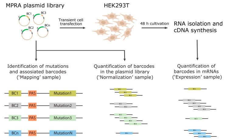
Since we used MPRA libraries with a priori unknown sequences of BCs and ROIs, it was necessary to prepare the “mapping” samples to identify unique BC–ROI-associated sequences (Figure 2). This was achieved by performing two-round conventional PCR amplification of the BC–ROI regions using the plasmid libraries as a template [92]. MPRA “expression” and “normalization” samples were prepared by PCR amplification of the BC sequences using cDNA obtained from the transfected cells and the plasmid library DNA as a template, respectively (Figure 2). The ratio of each BC abundance in the expression and normalization samples allows judging the influence of the corresponding ROI sequence variant (hereafter mutation) on the reporter gene expression. The “mapping”, “normalization”, and “expression” samples were subjected to NGS and, on average, ~1.5 million 151 nt single-end reads were obtained for each sample replicate.
Figure 2.
Schematic showing the experimental steps involved in the preparation of MPRA samples for subsequent NGS analysis. BC—barcode; PAS—polyadenylation signal.
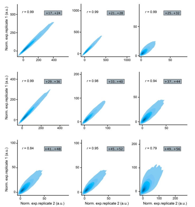
Analysis of the NGS reads was performed using the previously described MPRAdecoder pipeline [93]. It revealed that the MPRA libraries have different numbers of unique mutations (Table 1). On average, 54%, 14%, and 9% of mutations across the libraries were associated with one, two, and three or more BCs, respectively. It should be noted that a high correlation of normalized expression values between the replicates was obtained for all MPRA libraries (Figure 3).
Table 1.
Numbers of unique mutations and associated BCs for each MPRA library.
| MPRA Library ID |
Number of Unique Mutations | Mutations Associated with 1 BC |
Mutations Associated with 2 BCs |
Mutations Associated with ≥3 BCs |
|||
|---|---|---|---|---|---|---|---|
| Number | % | Number | % | Number | % | ||
| +17..+24 | 8380 | 7567 | 90.3 | 731 | 8.7 | 82 | 1.0 |
| +21..+28 | 8266 | 7347 | 88.9 | 797 | 9.6 | 122 | 1.5 |
| +25..+32 | 39,705 | 21,267 | 53.6 | 11,018 | 27.7 | 7420 | 18.7 |
| +29..+36 | 10,084 | 8830 | 87.5 | 944 | 9.4 | 310 | 3.1 |
| +33..+40 | 27,232 | 19,346 | 71.0 | 5807 | 21.3 | 2079 | 7.7 |
| +37..+44 | 33,866 | 19,721 | 58.2 | 8544 | 25.2 | 5601 | 16.6 |
| +41..+48 | 56,470 | 20,208 | 35.8 | 15,077 | 26.7 | 21,185 | 37.5 |
| +45..+52 | 25,814 | 18,373 | 71.2 | 5501 | 21.3 | 1940 | 7.5 |
| +49..+56 | 17,938 | 11,888 | 66.3 | 3489 | 19.5 | 2561 | 14.2 |
Figure 3.
Correlation of normalized expression values between the replicates of the MPRA libraries visualized as density scatterplots; r denotes Pearson’s correlation coefficient.
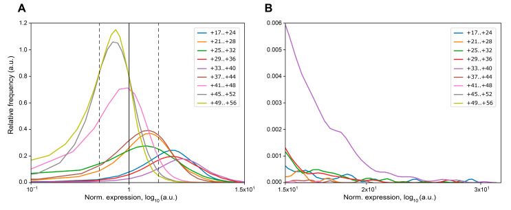
Next, we evaluated the number of mutations in each MPRA library that increase or decrease eGFP expression more than twofold compared to the wildtype reference construct (Table 2, Figure 4). We found that more than 50% of mutations in ROIs +17..+24, +21..+28, +25..+32, +29..+36, and +33..+40 result in more than a twofold increase in eGFP mRNA. At the same time, a proportion of mutations decreasing eGFP expression more than twofold was very low (0.07–3.21%) in these ROIs, as well as in ROI +37..+44. Additionally, these ROIs were characterized by the presence of mutations increasing eGFP expression more than 20-fold, whereas, in the ROIs +41..+48, +45..+52, and +49..+56 about 80% of mutations did not increase the eGFP reporter gene expression (Table 2, Figure 4).
Table 2.
Number of mutations influencing the eGFP expression in the MPRA libraries.
| MPRA Library ID |
Number of Unique Mutations | Number of Mutations with Zero Counts in Expression Samples |
Max Increase of eGFP Expression * |
Max Decrease of eGFP Expression * |
Mutations Leading to the ≥2-fold Increase of eGFP mRNA * |
Mutations Leading to the ≥2-fold Decrease of eGFP mRNA * |
||
|---|---|---|---|---|---|---|---|---|
| Number | % | Number | % | |||||
| +17..+24 | 8380 | 5 | 28.15 | 0.19 | 6961 | 83.07 | 22 | 0.26 |
| +21..+28 | 8266 | 6 | 17.33 | 0.14 | 4474 | 54.13 | 79 | 0.96 |
| +25..+32 | 39,705 | 160 | 27.38 | 0.05 | 23,204 | 58.44 | 1276 | 3.21 |
| +29..+36 | 10,084 | 0 | 26.09 | 0.24 | 8724 | 86.51 | 7 | 0.07 |
| +33..+40 | 27,232 | 11 | 29.45 | 0.23 | 25,323 | 92.99 | 25 | 0.09 |
| +37..+44 | 33,866 | 58 | 19.67 | 0.07 | 16,304 | 48.14 | 746 | 2.20 |
| +41..+48 | 56,470 | 277 | 16.74 | 0.01 | 6216 | 11.01 | 6187 | 10.96 |
| +45..+52 | 25,814 | 25 | 7.59 | 0.01 | 1036 | 4.01 | 3771 | 14.61 |
| +49..+56 | 17,938 | 249 | 6.74 | 0.02 | 518 | 2.89 | 3196 | 17.82 |
* Relative to the wildtype reference construct.
Figure 4.
Kernel density estimation of normalized expression values for the MPRA libraries that were averaged over replicates and normalized to the wildtype reference construct. (A) Density plot with a scale ranging from 1.0 × 10⁻¹ to 1.5 × 101. The expression level of the wildtype reference construct is indicated by a solid black vertical line. The twofold changes in the expression levels are depicted by dotted black vertical lines. (B) Density plot with a scale ranging from 1.5 × 101 to 3.2 × 101.
Then, we analyzed the nucleotide composition of mutations leading to more than a 10-fold increase in eGFP mRNA abundance according to the NGS data. With the exception of three ROIs, it was possible to define consensuses of sequences favorable for gene expression using the pLogo software [94]. These consensus sequences appeared to be extremely T-rich (Figure 5).
Figure 5.
Position weight matrix logos for sequences that dramatically increase (≥10-fold) eGFP mRNA level according to the MPRA screening results. A significance p-value threshold level of 0.05 is indicated by red horizontal lines [94].
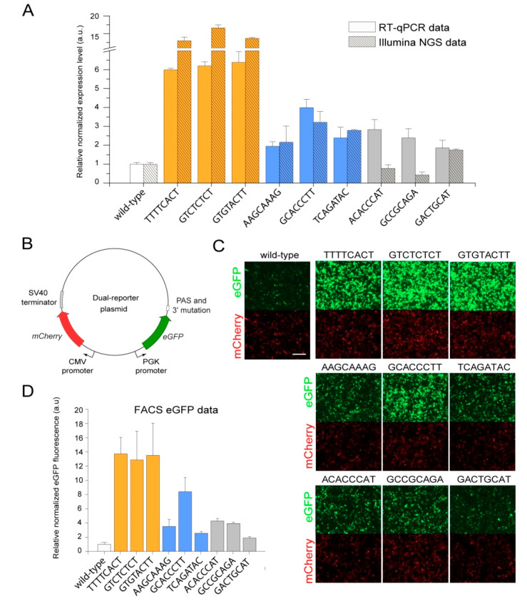
To verify the effects of individual mutations on eGFP expression level detected by the NGS analysis, we individually cloned several particular mutations from the library +29..+36 that change eGFP expression to a different extent downstream of a non-barcoded eGFP reporter. The mutations represent the “high”-, “medium”-, and “low”-expression groups. Each group included three mutations with different read counts in the normalization replicates (Table 3). HEK293T cells were transfected individually by non-barcoded plasmids bearing these mutations and by the non-barcoded wildtype reference construct. Then, 48 h after transfection, we analyzed the levels of eGFP mRNA and protein by RT-qPCR, fluorescence microscopy, and flow cytometry assays (Figure 6). Overall, the results of all approaches used were in good accordance with each other, although, on average, lower and higher fold changes were detected by RT-qPCR and flow cytometry assays compared to the NGS data for mutations from the high and medium/low groups, respectively (Figure 6).
Table 3.
Mutations from the MPRA library +29..+36 selected for individual tests.
| Mutation Sequence |
Normalization Replicate 1, Read Count |
Normalization Replicate 2, Read Count |
Increase of eGFP Expression, Fold * |
Group |
|---|---|---|---|---|
| TTTTCACT | 3 | 15 | 12.99 | High expression |
| GTCTCTCT | 11 | 11 | 16.63 | |
| GTGTACTT | 450 | 382 | 13.68 | |
| AAGCAAAG | 3 | 4 | 2.16 | Medium expression |
| GCACCCTT | 9 | 11 | 3.21 | |
| TCAGATAC | 281 | 264 | 2.78 | |
| ACACCCAT | 3 | 10 | 0.77 | Low expression |
| GCCGCAGA | 11 | 15 | 0.43 | |
| GACTGCAT | 92 | 64 | 1.75 |
* Relative to the wildtype reference construct.
Figure 6.
Validation of the positive effects of several chosen mutations identified in the MPRA screening on eGFP expression at the transcript and protein levels in HEK293T cells. (A) Comparison of eGFP expression data obtained for individual mutations using RT-qPCR with data obtained using Illumina NGS. Both types of data were normalized by the expression level of the wildtype reference construct. (B) The scheme of the dual-reporter plasmid used to test individual mutations. (C) Microscopy detection of eGFP and mCherry fluorescence intensities in HEK293T cells transiently transfected with the plasmids bearing the indicated individual mutations. Scale bar, 50 μm. (D) Flow cytometry analysis of the eGFP protein expression in HEK293T cells transiently transfected with the plasmids bearing the indicated individual mutations.
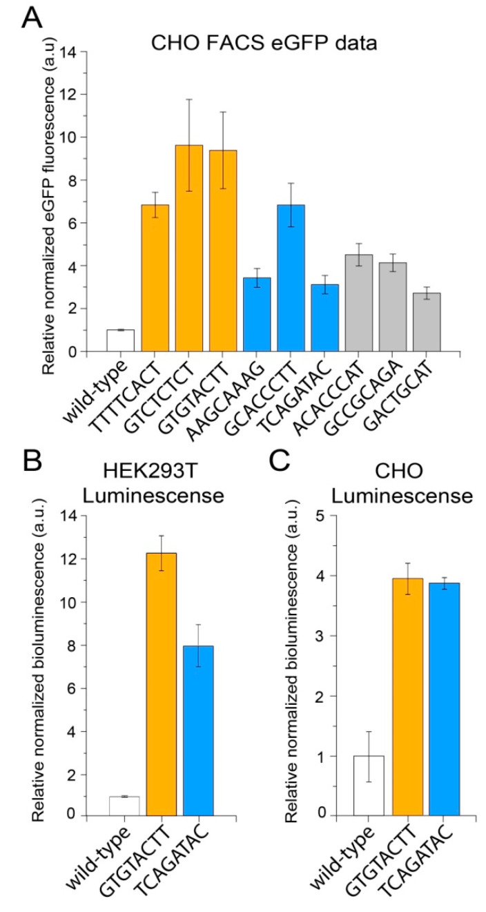
After that, we sought to determine whether the positive effects of mutations on eGFP expression could be reproduced in another mammalian cell line, as previously had been demonstrated for the deltaC mutation [89]. For that, we transiently transfected CHO cells using the same set of non-barcoded plasmids bearing individual mutations and analyzed the eGFP protein levels by flow cytometry (Figure 7A). This revealed a high level of conservation of the found positive effects of mutations on eGFP expression.
Figure 7.
The positive effects of GTGTACTT and TCAGATAC mutations on the expression levels of different upstream reporter genes and in different cell lines. (A) Flow cytometry analysis of the eGFP protein expression in CHO cells transiently transfected with the plasmids bearing the indicated individual mutations. (B,C) The luciferase protein activity in HEK293T (B) and CHO (C) cells transiently transfected with the plasmids bearing the indicated individual mutations.
Lastly, we decided to check whether the effects are limited to the eGFP reporter used in all experiments described above or are a more general phenomenon. With this aim, the two mutations, GTGTACTT from the high-expression group and TCAGATAC from the medium-expression group, which were originally supported by the highest numbers of reads in the NGS data, as well as the wildtype sequence, were individually cloned downstream of a non-barcoded luciferase (NanoLuc) reporter. Flow cytometry analysis of HEK293T and CHO cells transfected by these plasmids revealed that both mutations also significantly increased the luciferase protein activity in both cell lines (Figure 7B,C). Interestingly, both mutations affected the luciferase protein expression to the same extent in CHO cells, suggesting cell-type-specific variations in the transcription termination process. Overall, these findings suggest that the MPRA assays can be effectively used to identify sequence variants in the 3′ end of genes that are likely not included in the mature transcripts but substantially increase gene expression levels.
3. Discussion
Molecular mechanisms of transcription termination in eukaryotes are actively studied. However, the extent to which and particular ways of how the DNA sequence in the 3′ region of a gene downstream the PAS can affect the number of mature transcripts (usually considered as a level of gene expression) remain intriguing questions. In the present study, we used an artificial eGFP reporter gene and the MPRA approach to elucidate the regulatory effects of mutations downstream of the PAS.
We found that the majority of mutations located distal to the PAS have no positive effect on eGFP expression (Figure 4, Table 2). At the same time, the majority of mutations in the ROIs located proximally to the PAS cause a significant increase in eGFP mRNA level (up to 30-fold compared to the wildtype reference construct) (Figure 4, Table 2). For mutations resulting in the decrease in eGFP expression in the ROIs proximal to the PAS, we found very low number of cases causing a twofold or greater impact (or having zero counts in the expression replicates). The proportion of such mutations in the distal ROIs increased up to 11–18% (Table 2).
To verify the NGS data, we also analyzed the effects of individual mutations on eGFP expression using the RT-qPCR assay. As a result, we observed a very high correlation between RT-qPCR and NGS data for mutations from high- and medium-expression groups (Pearson’s correlation coefficient r = 0.94). We did not observe any difference between mutations in these groups depending on the read counts in the normalization replicates (Figure 6A, Table 3). However, the correlation between data obtained using RT-qPCR and NGS for mutations from the low-expression group was very low (Pearson’s correlation coefficient r = −0.75). Despite that, the correlation among these data for mutations from all three groups was high (Pearson’s correlation coefficient r = 0.94) (Figure 6A). It should be noted that, 48 h after transfection, we also performed a microscopic analysis of eGFP fluorescence in HEK293T cells. The level of eGFP signal in cells correlated well with the NGS and RT-qPCR data (Figure 6C). Thus, we can conclude that the NGS data obtained for the mutations leading to the twofold and more increase in eGFP expression level are reliable. At the same time, the NGS data on mutations that just slightly change the eGFP expression should be interpreted with caution. We suppose that this observation does not detract from the merits of the present study, since, in most cases, the need is to increase the expression of the gene of interest not slightly but substantially.
We also found that the consensus of sequences that ensure the highest expression levels of the eGFP reporter is extremely T-rich (Figure 5). Moreover, statistically significant nucleotides were identified only in the ROIs located proximal to the PAS. In the distal ROIs, no statistically significant nucleotides leading to a high increase in eGFP mRNA expression were detected.
As the MPRA assays are based on the usage of BCs to tag sequences absent in mature transcripts, it is important to note that BCs can also affect the measurements by themselves [95]. Typically, this issue is solved by associating each mutant sequence with multiple BCs [91,96]. Although such a strategy was not implemented in the current study, we believe that the strong positive effects on the reporter gene expression were not primarily caused by the BCs. First, the correlation between normalized expression values obtained with different BCs associated with the same mutations was on average 2.79 times higher for mutations increasing eGFP expression ≥2-fold than for the entire pool of analyzed mutations. Second, we identified similar trends in nucleotide composition (which is an enrichment in Ts) for mutations from different ROIs that strongly increase eGFP expression level. This suggests that the sequences of these mutations are more important than the sequences of BCs associated with them. Third, for several mutations from the high- and medium-expression groups, we validated the MPRA NGS data by RT-qPCR, fluorescence microscopy, and flow cytometry approaches using non-barcoded plasmids. Thus, although the effects of BCs cannot be completely ruled out, their influence on the identification of mutations strongly increasing the eGFP reporter expression level seems to be very small.
We assume that the effects of an increase in the reporter expression due to mutations that we detected stemmed from an increase in the efficiency of assembly and/or function of the CPA complex. At the moment, we can only speculate on the exact mechanism of such an increase in the CPA complex efficiency, but further investigation of results presented in this study could aid in the elucidation of specific mechanisms. A large body of evidence suggests the importance of the 3′ gene region composition for the correct completion of transcription termination by the CPA complex action. Previously, a massive analysis of human genomic sequences surrounding frequently and less frequently used poly(A) sites revealed specific cis elements within human 3′ gene regions [32]. Recent transient transcriptome sequencing (TT-seq) studies showed that the termination window of human genes can reach several kbs, and this region is thought to contain motifs crucial for the termination of transcription. Termination windows were found to be highly enriched with the consensus motif (C/G)(2–6)ANx(T/A)(3–6) (where Nx is a short stretch of nucleotides). The (C/G)(2–6) motif was shown to be associated with the paused state of the RNAP II, and (T/A)(3–6) may promote dissociation of the CPA complex due to the low melting temperature of the DNA–RNA hybrids [97,98]. It is also known that RNA forms secondary structures that are extremely important for RNA–protein interactions [99], such as the assembly of the CPA complex. We found a notable difference between predicted secondary structures of mRNA molecules bearing individual mutations from high- and low-expression groups (Supplementary Figure S1). As there are no exact data on which RNA secondary structures correlate with a more stable CPA complex, we hypothesize that, in the case of low-expression mutations, the specific secondary structure prevents the binding or interaction of individual subunits of the CPA complex.
Previously, we showed that deltaC, a single-nucleotide deletion in the 3′ region of the eGFP reporter affects the choice of the cleavage/polyadenylation sites of the transcripts. Thus, differences in (i) the composition of the 3′ UTR, (ii) the proportion of polyadenylated transcripts, and (iii) the length of the polyA tail can be expected in mRNAs synthetized from the mutated templates described in the present study. All these parameters affect the translation efficiency and lifetime of the resulting transcripts (and, therefore, their detectability) [72,73,100]. The whole-transcriptome 3′ UTR-seq of tens of thousands of transcript sequences in zebrafish revealed that stabilizing polyU and UUAG sequences and destabilizing GC-rich signals regulate early-onset rates of mRNA degradation [101].
We also cannot rule out the possibility of other related mechanisms being involved. In particular, the relationship between transcription termination and re-initiation is well established; hence, the efficiencies of these processes are interrelated [18,102]. The RNAP II dissociation from the 3′ end of a gene and the subsequent transcription machinery transition to the active state depend on the rate and success of transcription termination completion. Therefore, some effects observed in this study might also be explained by changes in the transcription re-initiation process. In this respect, it should be noted that a head-to-head arrangement of genes implemented in the dual-reporter plasmids used to verify the results of MPRA screening was reported to be associated with co-regulation of such genes due to simultaneous assembly and initiation of the transcription machinery on their transcription start sites [103]. However, our present and previous data [89] show that the mCherry normalization gene (Figure 6B) expression level is stable and does not depend on different motifs present in the 3′ region of the eGFP reporter. Therefore, we think that the effects of the 8 bp mutations on the upstream reporter gene expression are rather not strongly associated with the transcription re-initiation.
We believe that the results of the present work may be interesting for use in the production of therapeutic proteins and viral vectors [104,105,106], since HEK293T and CHO are the most popular cell lines for these purposes [107,108,109,110,111,112]. Since one of the important tasks in this field is to increase production efficiency, the modification of sequences involved in the transcription termination of transgenes based on the results of the appropriate MPRA screenings may substantially contribute to solving this problem.
4. Materials and Methods
4.1. Gibson Assembly of MPRA Plasmid Libraries
To generate the pTTC-hPGK-eGFP construct, the pTTC-Hsap-WT plasmid [89] was digested with NheI and XbaI and self-ligated. MPRA plasmid libraries were constructed on the basis of the pTTC-hPGK-eGFP plasmid using the Gibson Assembly approach. For each library, the appropriate DNA fragments were amplified from the pTTC-hPGK-eGFP plasmid template using primers indicated in Supplementary Tables S1 and S2. The PCR amplification was conducted in a 50 µL volume containing 1 ng of plasmid DNA, 1× Phusion high-fidelity reaction buffer (Thermo Fisher Scientific, Waltham, MA, USA), 400 µM of each dNTP, 500 nM of each primer, and 1 U of Phusion high-fidelity Hot Start DNA Polymerase (Thermo Fisher Scientific, Waltham, MA, USA) with the following program: 98 °C for 1 min, followed by 35 cycles of 98 °C for 30 s, 60 °C for 30 s, and 72 °C for 3 min, with a final cycle at 72 °C for 5 min. The PCR products were digested with DpnI restriction enzyme (New England Biolabs, Ipswich, MA, USA) at 37 °C for 4 h in a volume of 100 μL in order to destroy the plasmid template and, thus, minimize non-barcoded vector contamination in the libraries. After purification with the PCR Purification Kit (Qiagen, Hilden, Germany), 76 fmol (200 ng) of “vector” and 0.38 pmol (~30 ng) of “insert” fragments were incubated with Gibson Assembly Master Mix (New England Biolabs, Ipswich, MA, USA) at 50 °C for 1 h. Then, the reaction mixture was diluted 10-fold with nuclease-free water and purified using the MinElute PCR Purification Kit (Qiagen, Hilden, Germany); DNA was eluted by 10 µL of prewarmed elution buffer. Then, 5 μL of the purified Gibson cloning reaction was used to transform E. coli TOP10 electrocompetent cells.
4.2. Generation of Reference Barcoded Plasmid Constructs
To generate two reference constructs with the original “wildtype” sequence of the transcription terminator marked by specific 20 bp BCs, the pTTC-hPGK-eGFP plasmid was digested with XhoI and MluI. The barcoded wildtype sequence was amplified from the pTTC-hPGK-eGFP plasmid template using primers 5′–GACACTCGAGGATCGAGTTCCAAGTGCAGGTTAGGCGGAGTTGTGGCCGGCCCTTG–3′ (20 bp BC1) or 5′–GACACTCGAGGATCGAGTGTGTACGGCTTGCTCTCAAGAGTTGTGGCCGGCCCTTG–3′ (20 bp BC2) and 5′–CGCATACGCGTATACTAGATTAACC–3′. The PCR amplification was conducted in 50 µL volume containing 1 ng of plasmid DNA, 1× Phusion high-fidelity reaction buffer (Thermo Fisher Scientific, Waltham, MA, USA), 400 µM of each dNTP, 500 nM of each primer, and 1 U of Phusion high-fidelity Hot Start DNA Polymerase (Thermo Fisher Scientific, Waltham, MA, USA) with the following program: 98 °C for 1 min, followed by 35 cycles of 98 °C for 30 s, 57 °C for 30 s, and 72 °C for 30 s, with a final cycle at 72 °C for 5 min. The PCR products were digested with XhoI and MluI at 37 °C for 1 h. After purification with the PCR Purification Kit (Qiagen, Hilden, Germany), 50 ng of “vector” and ~7 ng of “insert” fragments were ligated at +4 °C overnight with T4 DNA ligase (Evrogen, Moscow, Russia). Then, 1 μL of the ligation mixture was used to transform E. coli TOP10 electrocompetent cells.
4.3. Generation of Non-Barcoded Plasmid Constructs with Individual Mutations
To generate the non-barcoded plasmid constructs with individual mutations, the pTTC-Hsap-WT plasmid [89] was first modified by introducing a point mutation in the SpeI site located within the human PGK promoter driving the expression of the eGFP reporter (cloning details are available upon request). Importantly, the introduced mutation did not affect the activity of the promoter. Next, the modified pTTC-Hsap-WT plasmid was digested at unique restriction sites with SpeI and BsiWI. For each mutation, two single-stranded complementary oligonucleotides (5′–CTAGTCATGCGTCAATTNNNNNNNNGATTATCTTTAAC–3′ and 5′–GTACGTTAAAGATAATCNNNNNNNNAATTGACGCATGA–3′) were annealed to each other by heating their equimolar mixture to 94 °C and then gradually cooling it to room temperature, which resulted in double-stranded DNA fragments with the SpeI and BsiWI sticky ends. After purification of digested vector with the PCR Purification Kit (Qiagen, Hilden, Germany), 50 ng of “vector” and ~1 ng of “insert” fragments were ligated at +4 °C overnight with T4 DNA ligase (Evrogen, Moscow, Russia). Then, 1 μL of the ligation mixture was used to transform E. coli TOP10 electrocompetent cells.
To generate the non-barcoded plasmid constructs with individual mutations and the luciferase (NanoLuc) reporter, the plasmid constructs with wildtype sequence or GTGTACTT and TCAGATAC mutations were digested with SmaI and EcoRI. The NanoLuc coding sequence was amplified from the plasmid template (provided by the laboratory of antibody engineering, IMCB SB RAS) using primers 5′–ATAACCCGGGACCATGGTCTTCACACTCG–3′ and 5′–ATATGAATTCTTACGCCAGAATGCGTTCG–3′. The PCR amplification was conducted in 50 µL volume containing 1 ng of plasmid DNA, 1× Phusion high-fidelity reaction buffer (Thermo Fisher Scientific, Waltham, MA, USA), 400 µM of each dNTP, 500 nM of each primer, and 1 U of Phusion high-fidelity Hot Start DNA Polymerase (Thermo Fisher Scientific, Waltham, MA, USA) with the following program: 98 °C for 1 min, followed by 35 cycles of 98 °C for 30 s, 58 °C for 30 s, and 72 °C for 30 s, with a final cycle at 72 °C for 5 min. The PCR products were digested with SmaI and EcoRI at 37 °C for 1 h. After purification with the PCR Purification Kit (Qiagen, Hilden, Germany), 50 ng of “vector” and ~18 ng of “insert” fragments were ligated at +4 °C overnight with T4 DNA ligase (Evrogen, Moscow, Russia). Then, 1 μL of the ligation mixture was used to transform E. coli TOP10 electrocompetent cells.
All plasmid constructs were verified by Sanger sequencing.
4.4. Cell Lines and Transfections
Human embryonic kidney (HEK293T) cells were obtained from ATCC (USA). The HEK239T cells were cultured at 37 °C in Dulbecco’s modification of Eagle’s medium (DMEM; Gibco) with 10% fetal bovine serum (FBS; Gibco), 7.5% NaHCO3, 100 IU/mL of penicillin, and 100 μg/mL of streptomycin.
Chinese hamster ovary (CHO) cells were obtained from the laboratory of antibody engineering, IMCB SB RAS. The CHO cells were cultured at 37 °C in Iscove’s modified Dulbecco’s medium (IMDM; Gibco) with 10% fetal bovine serum (FBS; Gibco), 100 IU/mL of penicillin and 100 μg/mL of streptomycin.
Transient transfection of HEK293T and CHO cells was performed as described previously [89].
4.5. MPRA Sample Preparation for NGS
The MPRA samples were prepared as described previously [92]. Briefly, the mapping samples were prepared by two-round conventional PCR. The expression samples were prepared by PCR amplification of the BC sequences using 1/20 of cDNA as a template. The normalization samples were prepared by PCR amplification using plasmid libraries as templates. To accurately measure the concentration of the MPRA samples for Illumina NGS, we used quantitative real-time PCR as described previously [92].
4.6. Illumina NGS and Data Analysis
Sequencing of 151 nt-long single-end reads was performed on an Illumina MiSeq platform using the MiSeq Reagent Kit v3 150 cycles (Illumina). Fastq files were processed, and the data were analyzed using previously described MPRAdecoder script [93]. Position weight matrix logos for sequences that dramatically increase (≥10-fold) eGFP mRNA level according to the MPRA screening were defined using the pLogo software [94].
4.7. RNA Isolation and Quantitative Real-Time PCR
RNA isolation from cells was performed 48 h after transfection according to the previously described protocol [89]. The cells were lysed in 1 mL of TRIzol Reagent Solution (Thermo Fisher Scientific, Waltham, MA, USA), and total RNA was isolated according to the manufacturer’s instructions. Then, 10 μg of purified RNA was incubated with 3 U of DNase I (Thermo Fisher Scientific, Waltham, MA, USA) and 40 U of DpnI restriction enzyme (New England Biolabs, Ipswich, MA, USA) at 37 °C for 1 h to remove traces of genomic and plasmid DNA. The RNA was purified using the CleanRNA Standard kit (Evrogen, Moscow, Russia) according to the manufacturer’s instructions. Next, 1 μg of purified total RNA was reverse-transcribed into cDNA using an oligo(dT)20 primer and 100 U of RevertAid Reverse Transcriptase (Thermo Fisher Scientific, Waltham, MA, USA) according to the manufacturer’s instructions.
qPCR was performed using the BioMaster HS-qPCR SYBR Blue (2×) reagent kit (Biolabmix), CFX96 Real-Time PCR Detection System (Bio-Rad), and the following gene-specific primer pairs: 5′–CCCTTCCTGGCCATCCTG–3′ and 5′–TCATCTCATTGACTTTGTCCAGC–3′ for the human PGK1 gene; 5′–CTTCAAGATCCGCCACAACATC–3′ and 5′–GGACCATGTGATCGCGCTTCTC–3′ for the eGFP coding sequence; 5′–ATCAAGGAGTTCATGCGCTTCAAG–3′ and 5′–TCACCTTCAGCTTGGCGGTCT–3′ for the mCherry coding sequence. The thermal cycling conditions were as follows: 5 min at 95 °C, followed by 39 cycles of 15 s at 95 °C, 30 s at 60 °C, and 30 s at 72 °C. No template control and no reverse transcriptase control samples were included in each run. Measurements of gene transcription levels were performed in two independent experiments, each with three technical replicates. The ΔΔCq method was used to calculate relative mRNA abundance; eGFP transcription levels were normalized to those of the mCherry and human PGK1 genes.
4.8. Fluorescence Microscopy
Transfected cells were analyzed immediately before harvesting for RNA isolation. All samples were imaged at the same settings using an Axio Vert.A1 microscope (Zeiss, Jena, Germany) equipped with an AxioCam ICm1 camera (Zeiss, Jena, Germany).
4.9. FACS Analysis
Cells transfected as described above were harvested for FACS analysis 48 h after transfection. To calculate the level of the eGFP expression, the mean fluorescent intensity of eGFP positive cells at 510 nm was multiplied by the number of positive cells and divided by the total number of events in live cells using a FACSCanto II flow cytometer (Becton Dickinson).
4.10. Luciferase Reporter Assay
To measure NanoLuc activity, HEK293T and CHO cells transfected as described above were harvested for a luciferase reporter assay 48 h after transfection. Then, 800 μL of lysis buffer (PBS, 0.5% Triton X-100) was added to each well of a 12-well plate and incubated at room temperature for 15 min. Next, 5 μL of cell lysate was mixed with 95 μL of lysis buffer and 50 μL of coelenterazine h solution (NanoLight Technology, Pinetop, AZ, USA) in wells of a 96-well white plate (SPL Life Sciences, Pocheon-Si, South Korea). The bioluminescence signal of the sample was measured using a Luminoskan Microplate Luminometer (Thermo Fisher Scientific, Waltham, MA, USA).
Acknowledgments
We thank Petr P. Laktionov and Daniil A. Maksimov for assistance with the Illumina DNA sequencing performed at the Institute of Molecular and Cellular Biology of the Siberian Branch of the Russian Academy of Sciences, and Sergey V. Kulemzin and Sergey V. Guselnikov for providing the plasmid with the NanoLuc coding sequence and CHO cells.
Supplementary Materials
The following supporting information can be downloaded at https://www.mdpi.com/article/10.3390/ijms232415485/s1. References [113,114] are cited in the supplementary materials.
Author Contributions
Conceptualization, A.V.P., E.N.A. and L.V.B.; funding acquisition, A.V.P.; investigation, E.S.O., A.E.L., L.A.Y., Y.A.G., A.A.O. and E.N.A.; supervision, A.V.P. and E.N.A.; writing, E.S.O., L.V.B., A.V.P. and A.A.O. All authors have read and agreed to the published version of the manuscript.
Institutional Review Board Statement
Not applicable.
Informed Consent Statement
Not applicable.
Data Availability Statement
Data supporting reported results can be downloaded from GEO (accession number GSE215681).
Conflicts of Interest
The authors declare no conflict of interest.
Funding Statement
This work was financially supported by the Ministry of Science and Higher Education of the Russian Federation (Agreement No. 075-15-2021-1086, contract No. RF----193021X0015, 15.ИП.21.0015).
Footnotes
Publisher’s Note: MDPI stays neutral with regard to jurisdictional claims in published maps and institutional affiliations.
References
- 1.Porrua O., Boudvillain M., Libri D. Transcription termination: Variations on common themes. Trends Genet. 2016;32:508–522. doi: 10.1016/j.tig.2016.05.007. [DOI] [PubMed] [Google Scholar]
- 2.Proudfoot N.J. Transcriptional termination in mammals: Stopping the RNA polymerase II juggernaut. Science. 2016;352:aad9926. doi: 10.1126/science.aad9926. [DOI] [PMC free article] [PubMed] [Google Scholar]
- 3.Neve J., Patel R., Wang Z., Louey A., Furger A.M. Cleavage and polyadenylation: Ending the message expands gene regulation. RNA Biol. 2017;14:865–890. doi: 10.1080/15476286.2017.1306171. [DOI] [PMC free article] [PubMed] [Google Scholar]
- 4.Porrua O., Libri D. Transcription termination and the control of the transcriptome: Why, where and how to stop. Nat. Rev. Mol. Cell Biol. 2015;16:190–202. doi: 10.1038/nrm3943. [DOI] [PubMed] [Google Scholar]
- 5.Turner R.E., Pattison A.D., Beilharz T.H. Alternative polyadenylation in the regulation and dysregulation of gene expression. Semin. Cell Dev. Biol. 2018;75:61–69. doi: 10.1016/j.semcdb.2017.08.056. [DOI] [PubMed] [Google Scholar]
- 6.Masamha C.P., Wagner E.J. The contribution of alternative polyadenylation to the cancer phenotype. Carcinogenesis. 2018;39:2–10. doi: 10.1093/carcin/bgx096. [DOI] [PubMed] [Google Scholar]
- 7.Zhang Y., Liu L., Qiu Q., Zhou Q., Ding J., Lu Y., Liu P. Alternative polyadenylation: Methods, mechanism, function, and role in cancer. J. Exp. Clin. Cancer Res. 2021;40:51. doi: 10.1186/s13046-021-01852-7. [DOI] [PMC free article] [PubMed] [Google Scholar]
- 8.Mayr C., Bartel D.P. Widespread shortening of 3′UTRs by alternative cleavage and polyadenylation activates oncogenes in cancer cells. Cell. 2009;138:673–684. doi: 10.1016/j.cell.2009.06.016. [DOI] [PMC free article] [PubMed] [Google Scholar]
- 9.Gruber A.R., Martin G., Keller W., Zavolan M. Means to an end: Mechanisms of alternative polyadenylation of messenger RNA precursors. Wiley Interdiscip. Rev. RNA. 2014;5:183–196. doi: 10.1002/wrna.1206. [DOI] [PMC free article] [PubMed] [Google Scholar]
- 10.Rehfeld A., Plass M., Krogh A., Friis-Hansen L. Alterations in polyadenylation and its implications for endocrine disease. Front. Endocrinol. 2013;4:53. doi: 10.3389/fendo.2013.00053. [DOI] [PMC free article] [PubMed] [Google Scholar]
- 11.Curinha A., Oliveira Braz S., Pereira-Castro I., Cruz A., Moreira A. Implications of polyadenylation in health and disease. Nucleus. 2014;5:508–519. doi: 10.4161/nucl.36360. [DOI] [PMC free article] [PubMed] [Google Scholar]
- 12.Dharmalingam P., Mahalingam R., Yalamanchili H.K., Weng T., Karmouty-Quintana H., Guha A., A Thandavarayan R.A. Emerging roles of alternative cleavage and polyadenylation (APA) in human disease. J. Cell Physiol. 2022;237:149–160. doi: 10.1002/jcp.30549. [DOI] [PMC free article] [PubMed] [Google Scholar]
- 13.Bentley D.L. Rules of engagement: Co-transcriptional recruitment of pre-mRNA processing factors. Curr. Opin. Cell Biol. 2005;17:251–256. doi: 10.1016/j.ceb.2005.04.006. [DOI] [PubMed] [Google Scholar]
- 14.Zhang Y., Sun Y., Shi Y., Walz T., Tong L. Structural insights into the human pre-mRNA 3′-end processing machinery. Mol. Cell. 2020;77:800–809. doi: 10.1016/j.molcel.2019.11.005. [DOI] [PMC free article] [PubMed] [Google Scholar]
- 15.Andersen P.K., Jensen T.H., Lykke-Andersen S. Making ends meet: Coordination between RNA 3′-end processing and transcription initiation. Wiley Interdiscip. Rev. RNA. 2013;4:233–246. doi: 10.1002/wrna.1156. [DOI] [PubMed] [Google Scholar]
- 16.Lepennetier G., Catania F. Exploring the impact of cleavage and polyadenylation factors on pre-mRNA splicing across eukaryotes. G3. 2017;7:2107–2114. doi: 10.1534/g3.117.041483. [DOI] [PMC free article] [PubMed] [Google Scholar]
- 17.Zhao J., Hyman L., Moore C. Formation of mRNA 3’ ends in eukaryotes: Mechanism, regulation, and interrelationships with other steps in mRNA synthesis. Microbiol. Mol. Biol. Rev. 1999;63:405–445. doi: 10.1128/MMBR.63.2.405-445.1999. [DOI] [PMC free article] [PubMed] [Google Scholar]
- 18.Moore M.J., Proudfoot N.J. Pre-mRNA processing reaches back to transcription and ahead to translation. Cell. 2009;136:688–700. doi: 10.1016/j.cell.2009.02.001. [DOI] [PubMed] [Google Scholar]
- 19.Rigo F., Martinson H.G. Polyadenylation releases mRNA from RNA polymerase II in a process that is licensed by splicing. RNA. 2009;15:823–836. doi: 10.1261/rna.1409209. [DOI] [PMC free article] [PubMed] [Google Scholar]
- 20.Sun Y., Hamilton K., Tong L. Recent molecular insights into canonical pre-mRNA 3′-end processing. Transcription. 2020;11:83–96. doi: 10.1080/21541264.2020.1777047. [DOI] [PMC free article] [PubMed] [Google Scholar]
- 21.Shi Y., Di Giammartino D.C., Taylor D., Sarkeshik A., Rice W.J., Yates J.R., III, Frank J., Manley J.L. Molecular architecture of the human pre-mRNA 3′ processing complex. Mol. Cell. 2009;33:365–376. doi: 10.1016/j.molcel.2008.12.028. [DOI] [PMC free article] [PubMed] [Google Scholar]
- 22.Veraldi K.L., Edwalds-Gilbert G., MacDonald C.C., Wallace A.M., Milcarek C. Isolation and characterization of polyadenylation complexes assembled in vitro. RNA. 2000;6:768–777. doi: 10.1017/S135583820099246X. [DOI] [PMC free article] [PubMed] [Google Scholar]
- 23.McCracken S., Fong N., Yankulov K., Ballantyne S., Pan G., Greenblatt J., Patterson S.D., Wickens M., Bentley D.L. The C-terminal domain of RNA polymerase II couples mRNA processing to transcription. Nature. 1997;385:357–361. doi: 10.1038/385357a0. [DOI] [PubMed] [Google Scholar]
- 24.Hirose Y., Manley J.L. RNA polymerase II is an essential mRNA polyadenylation factor. Nature. 1998;395:93–96. doi: 10.1038/25786. [DOI] [PubMed] [Google Scholar]
- 25.Fusby B., Kim S., Erickson B., Kim H., Peterson M.L., Bentley D.L. Coordination of RNA polymerase II pausing and 3′ end processing factor recruitment with alternative polyadenylation. Mol. Cell Biol. 2016;36:295–303. doi: 10.1128/MCB.00898-15. [DOI] [PMC free article] [PubMed] [Google Scholar]
- 26.Nag A., Narsinh K., Martinson H.G. The poly(A)-dependent transcriptional pause is mediated by CPSF acting on the body of the polymerase. Nat. Struct. Mol. Biol. 2007;14:662–669. doi: 10.1038/nsmb1253. [DOI] [PubMed] [Google Scholar]
- 27.Clerici M., Faini M., Aebersold R., Jinek M. Structural insights into the assembly and polyA signal recognition mechanism of the human CPSF complex. Elife. 2017;6:e33111. doi: 10.7554/eLife.33111. [DOI] [PMC free article] [PubMed] [Google Scholar]
- 28.Salisbury J., Hutchison K.W., Graber J.H. A multispecies comparison of the metazoan 3’-processing downstream elements and the CstF-64 RNA recognition motif. BMC Genomics. 2006;7:55. doi: 10.1186/1471-2164-7-55. [DOI] [PMC free article] [PubMed] [Google Scholar]
- 29.Chan S.L., Huppertz I., Yao C., Weng L., Moresco J.J., Yates J.R., III, Ule J., Manley J.L., Shi Y. CPSF30 and Wdr33 directly bind to AAUAAA in mammalian mRNA 3′ processing. Genes Dev. 2014;28:2370–2380. doi: 10.1101/gad.250993.114. [DOI] [PMC free article] [PubMed] [Google Scholar]
- 30.Mandel C.R., Kaneko S., Zhang H., Gebauer D., Vethantham V., Manley J.L., Tong L. Polyadenylation factor CPSF-73 is the pre-mRNA 3’-end-processing endonuclease. Nature. 2006;444:953–956. doi: 10.1038/nature05363. [DOI] [PMC free article] [PubMed] [Google Scholar]
- 31.Venkataraman K., Brown K.M., Gilmartin G.M. Analysis of a noncanonical poly(A) site reveals a tripartite mechanism for vertebrate poly(A) site recognition. Genes Dev. 2005;19:1315–1327. doi: 10.1101/gad.1298605. [DOI] [PMC free article] [PubMed] [Google Scholar]
- 32.Hu J., Lutz C.S., Wilusz J., Tian B. Bioinformatic identification of candidate cis-regulatory elements involved in human mRNA polyadenylation. RNA. 2005;11:1485–1493. doi: 10.1261/rna.2107305. [DOI] [PMC free article] [PubMed] [Google Scholar]
- 33.Brown K.M., Gilmartin G.M. A mechanism for the regulation of pre-mRNA 3′ processing by human cleavage factor Im. Mol. Cell. 2003;12:1467–1476. doi: 10.1016/S1097-2765(03)00453-2. [DOI] [PubMed] [Google Scholar]
- 34.Yang Q., Gilmartin G.M., Doublié S. The structure of human cleavage factor Im hints at functions beyond UGUA-specific RNA binding: A role in alternative polyadenylation and a potential link to 5′ capping and splicing. RNA Biol. 2011;8:748–753. doi: 10.4161/rna.8.5.16040. [DOI] [PMC free article] [PubMed] [Google Scholar]
- 35.Rüegsegger U., Beyer K., Keller W. Purification and characterization of human cleavage factor Im involved in the 3′ end processing of messenger RNA precursors. J. Biol. Chem. 1996;271:6107–6113. doi: 10.1074/jbc.271.11.6107. [DOI] [PubMed] [Google Scholar]
- 36.Li H., Tong S., Li X., Shi H., Ying Z., Gao Y., Ge H., Niu L., Teng M. Structural basis of pre-mRNA recognition by the human cleavage factor Im complex. Cell Res. 2011;21:1039–1051. doi: 10.1038/cr.2011.67. [DOI] [PMC free article] [PubMed] [Google Scholar]
- 37.Kubo T., Wada T., Yamaguchi Y., Shimizu A., Handa H. Knock-down of 25 kDa subunit of cleavage factor Im in Hela cells alters alternative polyadenylation within 3′-UTRs. Nucleic Acids Res. 2006;34:6264–6271. doi: 10.1093/nar/gkl794. [DOI] [PMC free article] [PubMed] [Google Scholar]
- 38.Gruber A.R., Martin G., Keller W., Zavolan M. Cleavage factor Im is a key regulator of 3′UTR length. RNA Biol. 2012;9:1405–1412. doi: 10.4161/rna.22570. [DOI] [PubMed] [Google Scholar]
- 39.Kyburz A., Sadowski M., Dichtl B., Keller W. The role of the yeast cleavage and polyadenylation factor subunit Ydh1p/Cft2p in pre-mRNA 3′-end formation. Nucleic Acids Res. 2003;31:3936–3945. doi: 10.1093/nar/gkg478. [DOI] [PMC free article] [PubMed] [Google Scholar]
- 40.Minvielle-Sebastia L., Preker P.J., Wiederkehr T., Strahm Y., Keller W. The major yeast poly(A)-binding protein is associated with cleavage factor IA and functions in premessenger RNA 3′-end formation. Proc. Natl. Acad. Sci. USA. 1997;94:7897–7902. doi: 10.1073/pnas.94.15.7897. [DOI] [PMC free article] [PubMed] [Google Scholar]
- 41.Meinhart A., Cramer P. Recognition of RNA polymerase II carboxy-terminal domain by 3′-RNA-processing factors. Nature. 2004;430:223–226. doi: 10.1038/nature02679. [DOI] [PubMed] [Google Scholar]
- 42.Noble C.G., Hollingworth D., Martin S.R., Ennis-Adeniran V., Smerdon S.J., Kelly G., Taylor I.A., Ramos A. Key features of the interaction between Pcf11 CID and RNA polymerase II CTD. Nat. Struct. Mol. Biol. 2005;12:144–151. doi: 10.1038/nsmb887. [DOI] [PubMed] [Google Scholar]
- 43.West S., Proudfoot N.J. Human Pcf11 enhances degradation of RNA polymerase II-associated nascent RNA and transcriptional termination. Nucleic Acids Res. 2008;36:905–914. doi: 10.1093/nar/gkm1112. [DOI] [PMC free article] [PubMed] [Google Scholar]
- 44.Kamieniarz-Gdula K., Gdula M.R., Panser K., Nojima T., Monks J., Wiśniewski J.R., Riepsaame J., Brockdorff N., Pauli A., Proudfoot N.J. Selective roles of vertebrate PCF11 in premature and full-length transcript termination. Mol. Cell. 2019;74:158–172. doi: 10.1016/j.molcel.2019.01.027. [DOI] [PMC free article] [PubMed] [Google Scholar]
- 45.de Vries H., Rüegsegger U., Hübner W., Friedlein A., Langen H., Keller W. Human pre-mRNA cleavage factor IIm contains homologs of yeast proteins and bridges two other cleavage factors. EMBO J. 2000;19:5895–5904. doi: 10.1093/emboj/19.21.5895. [DOI] [PMC free article] [PubMed] [Google Scholar]
- 46.Preker P.J., Ohnacker M., Minvielle-Sebastia L., Keller W. A multisubunit 3’ end processing factor from yeast containing poly(A) polymerase and homologues of the subunits of mammalian cleavage and polyadenylation specificity factor. EMBO J. 1997;16:4727–4737. doi: 10.1093/emboj/16.15.4727. [DOI] [PMC free article] [PubMed] [Google Scholar]
- 47.Mandel C.R., Bai Y., Tong L. Protein factors in pre-mRNA 3’-end processing. Cell. Mol. Life Sci. 2008;65:1099–1122. doi: 10.1007/s00018-007-7474-3. [DOI] [PMC free article] [PubMed] [Google Scholar]
- 48.Dominski Z. The hunt for the 3′ endonuclease. Wiley Interdiscip. Rev. RNA. 2010;1:325–340. doi: 10.1002/wrna.33. [DOI] [PubMed] [Google Scholar]
- 49.Sullivan K.D., Steiniger M., Marzluff W.F. A core complex of CPSF73, CPSF100, and Symplekin may form two different cleavage factors for processing of poly(A) and histone mRNAs. Mol. Cell. 2009;34:322–332. doi: 10.1016/j.molcel.2009.04.024. [DOI] [PMC free article] [PubMed] [Google Scholar]
- 50.Sun Y., Zhang Y., Aik W.S., Yang X.-C., Marzluff W.F., Walz T., Dominski Z., Tong L. Structure of an active human histone pre-mRNA 3′-end processing machinery. Science. 2020;367:700–703. doi: 10.1126/science.aaz7758. [DOI] [PMC free article] [PubMed] [Google Scholar]
- 51.Chan S., Choi E.-A., Shi Y. Pre-mRNA 3′-end processing complex assembly and function. Wiley Interdiscip. Rev. RNA. 2011;2:321–335. doi: 10.1002/wrna.54. [DOI] [PMC free article] [PubMed] [Google Scholar]
- 52.Edmonds M. Polyadenylate polymerases. Methods Enzymol. 1990;181:161–170. doi: 10.1016/0076-6879(90)81118-e. [DOI] [PubMed] [Google Scholar]
- 53.Laishram R.S., Anderson R.A. The poly A polymerase Star-PAP controls 3′-end cleavage by promoting CPSF interaction and specificity toward the pre-mRNA. EMBO J. 2010;29:4132–4145. doi: 10.1038/emboj.2010.287. [DOI] [PMC free article] [PubMed] [Google Scholar]
- 54.Takagaki Y., Ryner L.C., Manley J.L. Separation and characterization of a poly(A) polymerase and a cleavage/specificity factor required for pre-mRNA polyadenylation. Cell. 1988;52:731–742. doi: 10.1016/0092-8674(88)90411-4. [DOI] [PubMed] [Google Scholar]
- 55.Laishram R.S. Poly(A) polymerase (PAP) diversity in gene expression--star-PAP vs canonical PAP. FEBS Lett. 2014;588:2185–2197. doi: 10.1016/j.febslet.2014.05.029. [DOI] [PMC free article] [PubMed] [Google Scholar]
- 56.Di Giammartino D.C., Nishida K., Manley J.L. Mechanisms and consequences of alternative polyadenylation. Mol. Cell. 2011;43:853–866. doi: 10.1016/j.molcel.2011.08.017. [DOI] [PMC free article] [PubMed] [Google Scholar]
- 57.Zheng D., Tian B. RNA-binding proteins in regulation of alternative cleavage and polyadenylation. Adv. Exp. Med. Biol. 2014;825:97–127. doi: 10.1007/978-1-4939-1221-6_3. [DOI] [PubMed] [Google Scholar]
- 58.Shi Y. Alternative polyadenylation: New insights from global analyses. RNA. 2012;18:2105–2117. doi: 10.1261/rna.035899.112. [DOI] [PMC free article] [PubMed] [Google Scholar]
- 59.Xiang K., Tong L., Manley J.L. Delineating the structural blueprint of the pre-mRNA 3′-end processing machinery. Mol. Cell Biol. 2014;34:1894–1910. doi: 10.1128/MCB.00084-14. [DOI] [PMC free article] [PubMed] [Google Scholar]
- 60.Moore C.L., Sharp P.A. Accurate cleavage and polyadenylation of exogenous RNA substrate. Cell. 1985;41:845–855. doi: 10.1016/S0092-8674(85)80065-9. [DOI] [PubMed] [Google Scholar]
- 61.Colgan D.F., Manley J.L. Mechanism and regulation of mRNA polyadenylation. Genes Dev. 1997;11:2755–2766. doi: 10.1101/gad.11.21.2755. [DOI] [PubMed] [Google Scholar]
- 62.Logan J., Falck-Pedersen E., Darnell J.E., Jr., Shenk T. A poly(A) addition site and a downstream termination region are required for efficient cessation of transcription by RNA polymerase II in the mouseβmaj-globin gene. Proc. Natl. Acad. Sci. USA. 1987;84:8306–8310. doi: 10.1073/pnas.84.23.8306. [DOI] [PMC free article] [PubMed] [Google Scholar]
- 63.Richard P., Manley J.L. Transcription termination by nuclear RNA polymerases. Genes Dev. 2009;23:1247–1269. doi: 10.1101/gad.1792809. [DOI] [PMC free article] [PubMed] [Google Scholar]
- 64.Zhang H., Rigo F., Martinson H.G. Poly(A) signal-dependent transcription termination occurs through a conformational change mechanism that does not require cleavage at the poly(A) site. Mol. Cell. 2015;59:437–448. doi: 10.1016/j.molcel.2015.06.008. [DOI] [PubMed] [Google Scholar]
- 65.Zhang Z., Gilmour D.S. Pcf11 is a termination factor in Drosophila that dismantles the elongation complex by bridging the CTD of RNA polymerase II to the nascent transcript. Mol. Cell. 2006;21:65–74. doi: 10.1016/j.molcel.2005.11.002. [DOI] [PubMed] [Google Scholar]
- 66.West S., Gromak N., Proudfoot N.J. Human 5′ --> 3′ exonuclease Xrn2 promotes transcription termination at co-transcriptional cleavage sites. Nature. 2004;432:522–525. doi: 10.1038/nature03035. [DOI] [PubMed] [Google Scholar]
- 67.Tollervey D. Molecular biology: Termination by torpedo. Nature. 2004;432:456–457. doi: 10.1038/432456a. [DOI] [PubMed] [Google Scholar]
- 68.Eaton J.D., Davidson L., Bauer D.L.V., Natsume T., Kanemaki M.T., West S. Xrn2 accelerates termination by RNA polymerase II, which is underpinned by CPSF73 activity. Genes Dev. 2018;32:127–139. doi: 10.1101/gad.308528.117. [DOI] [PMC free article] [PubMed] [Google Scholar]
- 69.Eaton J.D., Francis L., Davidson L., West S. A unified allosteric/torpedo mechanism for transcriptional termination on human protein-coding genes. Genes Dev. 2020;34:132–145. doi: 10.1101/gad.332833.119. [DOI] [PMC free article] [PubMed] [Google Scholar]
- 70.Elkon R., Ugalde A.P., Agami R. Alternative cleavage and polyadenylation: Extent, regulation and function. Nat. Rev. Genet. 2013;14:496–506. doi: 10.1038/nrg3482. [DOI] [PubMed] [Google Scholar]
- 71.Wilton J., Tellier M., Nojima T., Costa A.M., Oliveira M.J., Moreira A. Simultaneous studies of gene expression and alternative polyadenylation in primary human immune cells. Methods Enzymol. 2021;655:349–399. doi: 10.1016/bs.mie.2021.04.004. [DOI] [PubMed] [Google Scholar]
- 72.Manning K.S., Cooper T.A. The roles of RNA processing in translating genotype to phenotype. Nat. Rev. Mol. Cell Biol. 2017;18:102–114. doi: 10.1038/nrm.2016.139. [DOI] [PMC free article] [PubMed] [Google Scholar]
- 73.Tian B., Hu J., Zhang H., Lutz C.S. A large-scale analysis of mRNA polyadenylation of human and mouse genes. Nucleic Acids Res. 2005;33:201–212. doi: 10.1093/nar/gki158. [DOI] [PMC free article] [PubMed] [Google Scholar]
- 74.Eckmann C.R., Rammelt C., Wahle E. Control of poly(A) tail length. Wiley Interdiscip. Rev. RNA. 2011;2:348–361. doi: 10.1002/wrna.56. [DOI] [PubMed] [Google Scholar]
- 75.Zhang X., Virtanen A., Kleiman F.E. To polyadenylate or to deadenylate: That is the question. Cell Cycle. 2010;9:4437–4449. doi: 10.4161/cc.9.22.13887. [DOI] [PMC free article] [PubMed] [Google Scholar]
- 76.Shi Y., Manley J.L. The end of the message: Multiple protein-RNA interactions define the mRNA polyadenylation site. Genes Dev. 2015;29:889–897. doi: 10.1101/gad.261974.115. [DOI] [PMC free article] [PubMed] [Google Scholar]
- 77.Tian B., Graber J.H. Signals for pre-mRNA cleavage and polyadenylation. Wiley Interdiscip. Rev. RNA. 2012;3:385–396. doi: 10.1002/wrna.116. [DOI] [PMC free article] [PubMed] [Google Scholar]
- 78.Ryner L.C., Takagaki Y., Manley J.L. Sequences downstream of AAUAAA signals affect pre-mRNA cleavage and polyadenylation in vitro both directly and indirectly. Mol. Cell. Biol. 1989;9:1759–1771. doi: 10.1128/mcb.9.4.1759-1771.1989. [DOI] [PMC free article] [PubMed] [Google Scholar]
- 79.Gruber A.J., Schmidt R., Gruber A.R., Martin G., Ghosh S., Belmadani M., Keller W., Zavolan M. A comprehensive analysis of 3′ end sequencing data sets reveals novel polyadenylation signals and the repressive role of heterogeneous ribonucleoprotein C on cleavage and polyadenylation. Genome Res. 2016;26:1145–1159. doi: 10.1101/gr.202432.115. [DOI] [PMC free article] [PubMed] [Google Scholar]
- 80.Proudfoot N.J. Ending the message: Poly(A) signals then and now. Genes Dev. 2011;25:1770–1782. doi: 10.1101/gad.17268411. [DOI] [PMC free article] [PubMed] [Google Scholar]
- 81.Wang R., Zheng D., Yehia G., Tian B. A compendium of conserved cleavage and polyadenylation events in mammalian genes. Genome Res. 2018;28:1427–1441. doi: 10.1101/gr.237826.118. [DOI] [PMC free article] [PubMed] [Google Scholar]
- 82.Nunes N.M., Li W., Tian B., Furger A. A functional human poly(A) site requires only a potent DSE and an A-rich upstream sequence. EMBO J. 2010;29:1523–1536. doi: 10.1038/emboj.2010.42. [DOI] [PMC free article] [PubMed] [Google Scholar]
- 83.Beisang D., Bohjanen P.R. Perspectives on the ARE as it turns 25 years old. Wiley Interdiscip. Rev. RNA. 2012;3:719–731. doi: 10.1002/wrna.1125. [DOI] [PMC free article] [PubMed] [Google Scholar]
- 84.Shaw G., Kamen R. A conserved AU sequence from the 3′ untranslated region of GM-CSF mRNA mediates selective mRNA degradation. Cell. 1986;46:659–667. doi: 10.1016/0092-8674(86)90341-7. [DOI] [PubMed] [Google Scholar]
- 85.Chen C.-Y.A., Shyu A.-B. Selective degradation of early-response-gene mRNAs: Functional analyses of sequence features of the AU-rich elements. Mol. Cell. Biol. 1994;14:8471–8482. doi: 10.1128/mcb.14.12.8471-8482.1994. [DOI] [PMC free article] [PubMed] [Google Scholar]
- 86.Xu Y.Z., Di Marco S., Gallouzi I., Rola-Pleszczynski M., Radzioch D. RNA-binding protein HuR is required for stabilization of SLC11A1 mRNA and SLC11A1 protein expression. Mol. Cell. Biol. 2005;25:8139–8149. doi: 10.1128/MCB.25.18.8139-8149.2005. [DOI] [PMC free article] [PubMed] [Google Scholar]
- 87.Beaudoing E., Freier S., Wyatt J.R., Claverie J.-M., Gautheret D. Patterns of variant polyadenylation signal usage in human genes. Genome Res. 2000;10:1001–1010. doi: 10.1101/gr.10.7.1001. [DOI] [PMC free article] [PubMed] [Google Scholar]
- 88.Akhtar W., de Jong J., Pindyurin A.V., Pagie L., Meuleman W., de Ridder J., Berns A., Wessels L.F.A., van Lohuizen M., van Steensel B. Chromatin position effects assayed by thousands of reporters integrated in parallel. Cell. 2013;154:914–927. doi: 10.1016/j.cell.2013.07.018. [DOI] [PubMed] [Google Scholar]
- 89.Boldyreva L.V., Yarinich L.A., Kozhevnikova E.N., Ivankin A.V., Lebedev M.O., Pindyurin A.V. Fine gene expression regulation by minor sequence variations downstream of the polyadenylation signal. Mol. Biol. Rep. 2021;48:1539–1547. doi: 10.1007/s11033-021-06160-z. [DOI] [PubMed] [Google Scholar]
- 90.Komura R., Aoki W., Motone K., Satomura A., Ueda M. High-throughput evaluation of T7 promoter variants using biased randomization and DNA barcoding. PLoS ONE. 2018;13:e0196905. doi: 10.1371/journal.pone.0196905. [DOI] [PMC free article] [PubMed] [Google Scholar]
- 91.Tewhey R., Kotliar D., Park D.S., Liu B., Winnicki S., Reilly S.K., Andersen K.G., Mikkelsen T.S., Lander E.S., Schaffner S.F., et al. Direct identification of hundreds of expression-modulating variants using a multiplexed reporter assay. Cell. 2016;165:1519–1529. doi: 10.1016/j.cell.2016.04.027. [DOI] [PMC free article] [PubMed] [Google Scholar]
- 92.Omelina E.S., Ivankin A.V., Letiagina A.E., Pindyurin A.V. Optimized PCR conditions minimizing the formation of chimeric DNA molecules from MPRA plasmid libraries. BMC Genomics. 2019;20:536. doi: 10.1186/s12864-019-5847-2. [DOI] [PMC free article] [PubMed] [Google Scholar]
- 93.Letiagina A.E., Omelina E.S., Ivankin A.V., Pindyurin A.V. MPRAdecoder: Processing of the raw MPRA data with a priori unknown sequences of the region of interest and associated barcodes. Front. Genet. 2021;12:618189. doi: 10.3389/fgene.2021.618189. [DOI] [PMC free article] [PubMed] [Google Scholar]
- 94.O’Shea J.P., Chou M.F., Quader S.A., Ryan J.K., Church G.M., Schwartz D. pLogo: A probabilistic approach to visualizing sequence motifs. Nat. Methods. 2013;10:1211–1212. doi: 10.1038/nmeth.2646. [DOI] [PubMed] [Google Scholar]
- 95.Lee D., Kapoor A., Lee C., Mudgett M., Beer M.A., Chakravarti A. Sequence-based correction of barcode bias in massively parallel reporter assays. Genome Res. 2021;31:1638–1645. doi: 10.1101/gr.268599.120. [DOI] [PMC free article] [PubMed] [Google Scholar]
- 96.Ashuach T., Fischer D.S., Kreimer A., Ahituv N., Theis F.J., Yosef N. MPRAnalyze: Statistical framework for massively parallel reporter assays. Genome Biol. 2019;20:183. doi: 10.1186/s13059-019-1787-z. [DOI] [PMC free article] [PubMed] [Google Scholar]
- 97.Schwalb B., Michel M., Zacher B., Frühauf K., Demel C., Tresch A., Gagneur J., Cramer P. TT-seq maps the human transient transcriptome. Science. 2016;352:1225–1228. doi: 10.1126/science.aad9841. [DOI] [PubMed] [Google Scholar]
- 98.Baejen C., Andreani J., Torkler P., Battaglia S., Schwalb B., Lidschreiber M., Maier K.C., Boltendahl A., Rus P., Esslinger S., et al. Genome-wide analysis of RNA polymerase II termination at protein-coding genes. Mol. Cell. 2017;66:38–49. doi: 10.1016/j.molcel.2017.02.009. [DOI] [PubMed] [Google Scholar]
- 99.Wan Y., Kertesz M., Spitale R.C., Segal E., Chang H.Y. Understanding the transcriptome through RNA structure. Nat Rev Genet. 2011;12:641–655. doi: 10.1038/nrg3049. [DOI] [PMC free article] [PubMed] [Google Scholar]
- 100.Eisen T.J., Li J.J., Bartel D.P. The interplay between translational efficiency, poly(A) tails, microRNAs, and neuronal activation. RNA. 2022;28:808–831. doi: 10.1261/rna.079046.121. [DOI] [PMC free article] [PubMed] [Google Scholar]
- 101.Rabani M. Massively parallel analysis of regulatory RNA sequences. Methods Mol. Biol. 2021;2218:355–365. doi: 10.1007/978-1-0716-0970-5_28. [DOI] [PubMed] [Google Scholar]
- 102.Lykke-Andersen S., Mapendano C.K., Jensen T.H. An ending is a new beginning: Transcription termination supports re-initiation. Cell Cycle. 2011;10:863–865. doi: 10.4161/cc.10.6.14931. [DOI] [PubMed] [Google Scholar]
- 103.Chen Y., Li Y., Wei J., Li Y.-Y. Transcriptional regulation and spatial interactions of head-to-head genes. BMC Genomics. 2014;15:519. doi: 10.1186/1471-2164-15-519. [DOI] [PMC free article] [PubMed] [Google Scholar]
- 104.Dumont J., Euwart D., Mei B., Estes S., Kshirsagar R. Human cell lines for biopharmaceutical manufacturing: History, status, and future perspectives. Crit. Rev. Biotechnol. 2016;36:1110–1122. doi: 10.3109/07388551.2015.1084266. [DOI] [PMC free article] [PubMed] [Google Scholar]
- 105.Tan E., Chin C.S.H., Lim Z.F.S., Ng S.K. HEK293 cell line as a platform to produce recombinant proteins and viral vectors. Front. Bioeng. Biotechnol. 2021;9:796991. doi: 10.3389/fbioe.2021.796991. [DOI] [PMC free article] [PubMed] [Google Scholar]
- 106.Abaandou L., Quan D., Shiloach J. Affecting HEK293 cell growth and production performance by modifying the expression of specific genes. Cells. 2021;10:1667. doi: 10.3390/cells10071667. [DOI] [PMC free article] [PubMed] [Google Scholar]
- 107.Chin C.L., Goh J.B., Srinivasan H., Liu K.I., Gowher A., Shanmugam R., Lim H.L., Choo M., Tang W.Q., Tan A.H.-M., et al. A human expression system based on HEK293 for the stable production of recombinant erythropoietin. Sci. Rep. 2019;9:16768. doi: 10.1038/s41598-019-53391-z. [DOI] [PMC free article] [PubMed] [Google Scholar]
- 108.König J., Hust M., van den Heuvel J. Validation of the production of antibodies in different formats in the HEK 293 transient gene expression system. Methods Mol. Biol. 2021;2247:59–76. doi: 10.1007/978-1-0716-1126-5_4. [DOI] [PubMed] [Google Scholar]
- 109.Heng Z.S.-L., Yeo J.Y., Koh D.W.-S., Gan S.K.-E., Ling W.-L. Augmenting recombinant antibody production in HEK293E cells: Optimizing transfection and culture parameters. Antib. Ther. 2022;5:30–41. doi: 10.1093/abt/tbac003. [DOI] [PMC free article] [PubMed] [Google Scholar]
- 110.Jäger V., Büssow K., Wagner A., Weber S., Hust M., Frenzel A., Schirrmann T. High level transient production of recombinant antibodies and antibody fusion proteins in HEK293 cells. BMC Biotechnol. 2013;13:52. doi: 10.1186/1472-6750-13-52. [DOI] [PMC free article] [PubMed] [Google Scholar]
- 111.Ahmadi S., Davami F., Davoudi N., Nematpour F., Ahmadi M., Ebadat S., Azadmanesh K., Barkhordari F., Mahboudi F. Monoclonal antibodies expression improvement in CHO cells by PiggyBac transposition regarding vectors ratios and design. PLoS ONE. 2017;12:e0179902. doi: 10.1371/journal.pone.0179902. [DOI] [PMC free article] [PubMed] [Google Scholar]
- 112.Zúñiga R.A., Gutiérrez-González M., Collazo N., Sotelo P.H., Ribeiro C.H., Altamirano C., Lorenzo C., Aguillón J.C., Molina M.C. Development of a new promoter to avoid the silencing of genes in the production of recombinant antibodies in chinese hamster ovary cells. J. Biol. Eng. 2019;13:59. doi: 10.1186/s13036-019-0187-y. [DOI] [PMC free article] [PubMed] [Google Scholar]
- 113.Gruber A.R., Lorenz R., Bernhart S.H., Neuböck R., Hofacker I.L. The Vienna RNA websuite. Nucleic Acids Res. 2008;36:70–74. doi: 10.1093/nar/gkn188. [DOI] [PMC free article] [PubMed] [Google Scholar]
- 114.Lorenz R., Bernhart S.H., Höner Zu Siederdissen C., Tafer H., Flamm C., Stadler P.F., Hofacker I.L. ViennaRNA Package 2.0. Algorithms Mol. Biol. 2011;6:26. doi: 10.1186/1748-7188-6-26. [DOI] [PMC free article] [PubMed] [Google Scholar]
Associated Data
This section collects any data citations, data availability statements, or supplementary materials included in this article.
Supplementary Materials
Data Availability Statement
Data supporting reported results can be downloaded from GEO (accession number GSE215681).


