Abstract
Engineered cyanobacterium Synechococcus elongatus can use light and CO2 to produce sucrose, making it a promising candidate for use in co-cultures with heterotrophic workhorses. However, this process is challenged by the mutual stresses generated from the multispecies microbial culture. Here we demonstrate an ecosystem where S. elongatus is freely grown in a photo-bioreactor (PBR) containing an engineered heterotrophic workhorse (either β-carotene-producing Yarrowia lipolytica or indigoidine-producing Pseudomonas putida) encapsulated in calcium-alginate hydrogel beads. The encapsulation prevents growth interference, allowing the cyanobacterial culture to produce high sucrose concentrations enabling the production of indigoidine and β-carotene in the heterotroph. Our experimental PBRs yielded an indigoidine titer of 7.5 g/L hydrogel and a β-carotene titer of 1.3 g/L hydrogel, amounts 15–22-fold higher than in a comparable co-culture without encapsulation. Moreover, 13C-metabolite analysis and protein overexpression tests indicated that the hydrogel beads provided a favorable microenvironment where the cell metabolism inside the hydrogel was comparable to that in a free culture. Finally, the heterotroph-containing hydrogels were easily harvested and dissolved by EDTA for product recovery, while the cyanobacterial culture itself could be reused for the next batch of immobilized heterotrophs. This co-cultivation and hydrogel encapsulation system is a successful demonstration of bioprocess optimization under photobioreactor conditions.
Subject terms: Symbiosis, Natural products, Gels and hydrogels
Introduction
Photoautotrophs, such as the unicellular cyanobacterium Synechococcus elongatus UTEX 2973, enable conversion of CO2 to renewable products. For example, their high-flux sugar phosphate pathways can generate sucrose from CO2 as high as 2 g/L per day 1. However, genetic tools for cyanobacteria have lagged far behind those for classic heterotrophic hosts. Moreover, cyanobacteria have limited abilities to direct fluxes towards acetyl-CoA and the TCA cycle, which constrains their production of many molecules, such as biofuels and pharmaceutical products 2–5. To avoid this limitation, researchers have proposed microbial co-culture formats of autotrophic and heterotrophic species. These co-cultures can leverage the sucrose-secreting capability of S. elongatus UTEX 2973 to provide substrates for heterotrophic synthesis of high value products 6,7. Such consortia have three advantages: (1) converting CO2 to more diverse and valuable products, (2) easing subpopulation controls and growth optimization, and (3) enabling the division of labor between two hosts (one for CO2 fixation and the other for heterologous pathway engineering) 8. On the other hand, cyanobacterial photosynthesis may produce reactive oxygen species (ROS) that inhibit heterotroph viability, and heterotrophic growth may produce shading effects that reduce algal light harvesting 9. For example, in an artificial consortium of sucrose-secreting S. elongatus UTEX 2973 and 3-hydroxypropionic acid producing Escherichia coli, relatively low light intensity was used to reduce oxidative stresses, but this compromised the sucrose production rate 7. In general, stable and highly productive consortia are difficult to optimize unless the two species form a mutualistic growth relationship 10 with efficient ROS scavenging capability 11.
For this reason, cell immobilization has been attempted to develop stable co-culture systems for bioconversion 12. For cell encapsulation, hydrogel vessels have gained increasing attention owing to their ability to support cell growth 13–15. Particularly, alginate hydrogels are preferred materials because they are non-toxic, cheap, and environmentally benign 13,16. Moreover, the high porosity of alginate hydrogels allows nutrients and O2 to access immobilized cells by simple diffusion 13,17,18. Alginate encapsulation of S. elongatus 7942 was found to enhance its sucrose production and co-culture stability with polyhydroxybutyrate producing Halomona boliviensis over 5 months, and the encapsulated cyanobacteria resisted invasive microbial species during that time 19. Moreover, the same alginate-encapsulated sucrose-secreting cyanobacteria (within a barium–alginate hydrogel) and Pseudomonas putida formed a co-culture that both enhanced the degradation of an environmental pollutant, 2,4-dinitrotoluene, and synthesized the biopolymer polyhydroxyalkanoate 6. As this example demonstrates, hydrogel encapsulation connects individual species that possess different metabolic features, and it supports biological functions over long time periods. In previous studies, the products of engineered heterotrophs were secreted extracellularly, making them easy to separate. In cases where the products remain intracellular, product separation from consortia becomes difficult. Here, hydrogel encapsulation can advantageously trap specific producers and their products for further bio-separations.
Our recent paper 20 characterized Ca-alginate hydrogel for nutrient delivery to support cyanobacterial growth. In this study, Ca-alginate hydrogel was used to encapsulate the heterotrophic workhorses. In general, alginate hydrogels can be formed through crosslinking with a variety of cations. Two of the most used are barium and calcium. Barium produces stronger hydrogel and has a stronger binding affinity to alginate than calcium, and it has been used in making hydrogels to encapsulate cyanobacteria 10,21,22. However, barium in its soluble form is highly toxic 23. While a majority of the barium would be linked within the hydrogel, barium can leak during the microbial cultivation regime, creating a toxicity hazard when handling and disposing of spent medium 24. Additionally, barium strongly cross-links alginate, making it difficult to extract the insoluble product inside the hydrogel. In contrast, Ca-alginate hydrogels have lower binding strength than barium 22, so they are easily dissolved by EDTA for intracellular product recovery. For these reasons, we chose to use calcium as a non-toxic and low cost cross-linker for the alginate hydrogels to maintain hydrogel strength and longevity.
In this study, P. putida was used as a model bacterial factory 25,26 and Y. lipolytica was used as a model yeast factory 27. Complementing these workhorse microbes, S. elongatus UTEX 2973 has the highest sucrose secretion capability (1.9 g/L/day) of all cyanobacterial hosts. In our setup the autotrophic strain in the co-culture provides the sugar feedstock for the heterotrophic workhorses 7,10. However, for fast and efficient photosynthesis, UTEX 2973 requires high light intensity (> 800 μmol photons/m2/s) and high dissolved CO2 (3%) conditions 28,29. To provide sufficient light and CO2 to sustain high sucrose production, a co-culture where cyanobacteria are in free suspension and heterotrophic producers are encapsulated in Ca-alginate hydrogels would be highly beneficial. This approach also makes it convenient to scale up and reuse the photobioreactor (PBR). Additionally, the engineered P. putida and Y. lipolytica strains produce a blue compound (indigoidine, an industrial dye) and an orange compound (β-carotene) intracellularly 30,31. By encapsulating the P. putida or Y. lipolytica inside the Ca-alginate hydrogel, both the biomass and intracellular products will not interfere with cyanobacterial photosynthesis. Also, the hydrogel may protect light sensitive metabolites (e.g., β-carotene). Compared to previously demonstrated hydrogel consortiums using barium 19, the intracellular products inside the Ca-alginate hydrogel can be dissolved by EDTA and facilitate product extraction.
Results and discussion
Hydrogel compartmentalized co-culture (without sucrose utilization)
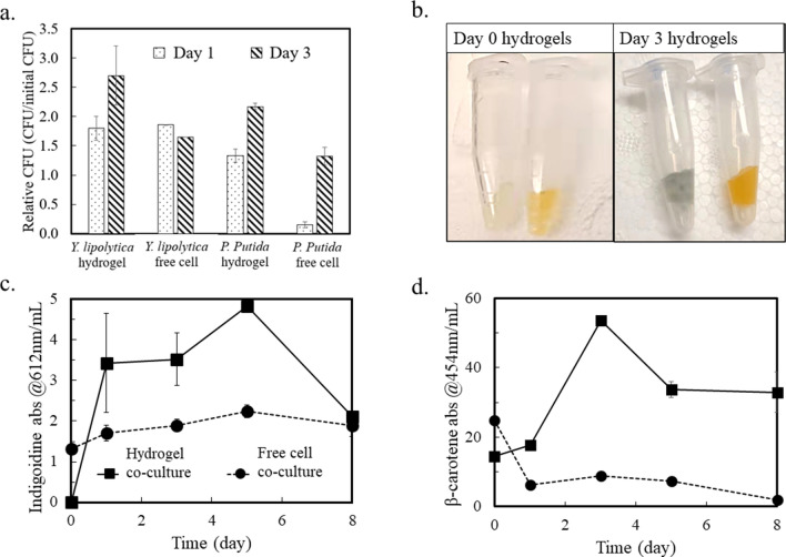
Cyanobacteria naturally supports heterotrophs because its “overflow metabolism” secretes amino acids (e.g., alanine and methionine 11), pyruvate and TCA metabolites under excess light energy input 32–34. Here, we separately co-cultured engineered P. putida and Y. lipolytica strains with S. elongates (Supplementary Table S1) with and without hydrogel compartmentation. Each co-culture system contained one heterotrophic species, either P. putida or Y. lipolytica. Since these heterotrophs cannot utilize sucrose, they survived by using cyanobacterial extracellular metabolites. Figure 1 shows heterotrophic growth and production from P. putida/S. elongatus co-cultures and Y. lipolytica/S. elongatus co-cultures, with and without hydrogel compartmentalization. Under the same initial cultivation condition, controlled by the multi-cultivator, hydrogel compartmentation shows protective effects on the growth of engineered P. putida and Y. lipolytica. Specifically, the heterotrophic strains inside the hydrogel produced much higher concentrations of the colored chemicals than those in free cell co-culture. Moreover, the living population of P. putida in free cell co-culture with S. elongatus drops dramatically from the beginning of co-culture to the end of day 1 (Fig. 1a). In contrast, the P. putida population in the hydrogel co-culture system increases continuously for three days. For Y. lipolytica, the living populations (based on colony forming units, CFU) in free cell co-culture and hydrogel co-culture with S. elongatus are similar in the first 2 days. This contrast indicates that Y. lipolytica are more tolerant to PBR stresses. Later, the Y. lipolytica population in free cell co-culture drops while that in the compartmentalized co-culture kept increasing. To enable two different heterotroph stains to grow and express engineered pathways in a co-culture, the chemical environment of the medium in system must be carefully chosen. As the base medium for cyanobacterial setup and co-culture, BG11 medium is not formulated for optimal growth of either heterotrophic partner (e.g., BG11 contains nitrate as a nitrogen source, whereas the medium for either heterotroph conventionally supplies ammonia 35,36). Additionally, the reactive oxygen species (ROS) generated as byproducts from photosynthesis is another stressor to P. putida and Y. lipolytica. By preventing the P. putida and Y. lipolytica encapsulated in hydrogels from direct contact with S. elongatus, the detrimental effects of cyanobacteria could be minimized so that the viability of the partner heterotroph is dramatically improved.
Figure 1.
Co-culture of sucrose-producing Synechococcus and indigoidine-producing P. putida or β-carotene-producing Y. lipolytica (n = 2). (a) Relative CFUs of heterotrophic strains in co-culture. (b) Pictures of recovered hydrogels from co-culture system at day 0 and day 3. Left tubes, P. putida; right tubes, Y. lipolytica. (c,d) Absorbance of extracted indigoidine and β-carotene from co-culture. By using standard curves, the mass concentration of the two compounds can be estimated from the absorbance measurement (Supplementary Fig. S1). All data are averages of biological duplicates ± standard deviation. Some error bars are too small to be visible.
We further examined engineered heterotrophs’ productivity with cyanobacteria. Figure 1b shows P. putida and Y. lipolytica hydrogels on day 0 and day 3 of co-culturing. The color change indicates the accumulation of blue indigoidine and orange β-carotene. Measured at the respective absorbance wavelengths, the increases of product in the cell-free and hydrogel co-cultures are illustrated in Fig. 1c,d. The engineered heterotrophic microbes in hydrogels obviously generated a much higher concentration of indigoidine and β-carotene than the production in free cell co-cultures. Specifically, the cell density of P. putida in free cell co-culture increased by ~ 9 times from day 1 to day 3, from 2 × 10⁷ to 1.73 × 10⁸ CFUs/mL. However, the indigoidine production did not increase, and free P. putida cells lost their ability to generate the pigment under light and ROS exposure. In contrast, P. putida inside hydrogels maintained their productivity. On the other hand, Y. lipolytica could grow in free cell co-culture. β-carotene extracted from free cell co-cultures followed a descending trend in the 8-day cultivation. This observation was not surprising because the pigments were sensitive to ROS and light conditions (e.g., 50% of β-carotene naturally degrades within 48 h under fluorescent light) 37. In contrast, hydrogel could protect β-carotene from bleaching and increased its half-life (Fig. 1d).
Development of a continuous co-culture platform (with sucrose utilization)
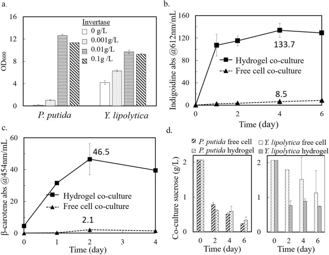
The first round of co-culture experiments was not set up for sucrose feeding, and both heterotrophs had to utilize overflow metabolites from cyanobacteria as nutrient sources. Because our P. putida and Y. lipolytica could not utilize sucrose, invertase, the enzyme hydrolyzing sucrose into fructose and glucose, was used to improve sucrose usage in axenic P. putida and Y. lipolytica cultures. Figure 2a displays the OD600 measurements of engineered P. putida and Y. lipolytica pure cultures after growing for 48 h in M9 and YNB media, respectively, with 10 g/L sucrose and different concentrations of invertase. Here, the addition of 0.001 g/L invertase was essential for bacterial growth, and 0.01 g/L invertase showed the highest growth promotion for both species.
Figure 2.
Axenic and co-culture heterotrophic growth/production with invertase. (a) OD600 of P. putida and Y. lipolytica growing in the defined media and 10 g/L sucrose at different concentrations of invertase (see “Methods” section). (b) Absorbance of extracted indigoidine at 612 nm from engineered P. putida over 6 days of co-culture. The peak production titer of indigoidine, on day 4, was 7.5 ± 0.7 g/L hydrogel. (c) Absorbance of extracted β-carotene from engineered Y. lipolytica over 4 days of co-culture. (d) Sucrose levels in P. putida and Y. lipolytica co-cultures. All data are averages of biological duplicates ± standard deviation. Some error bars are too small to be seen.
After confirming the positive effect of invertase in boosting the growth of heterotrophic strains, biological duplicate co-culture tests with S. elongatus were set up with 0.01 g/L invertase. The measured absorbances of indigoidine and β-carotene are plotted in Fig. 2a,b. The peak P. putida indigoidine production in the hydrogel co-culture system was significantly boosted compared to the no-invertase co-culturing condition. During 6 days of cultivation, the indigoidine that accumulated in the hydrogel beads was protected from bleaching. The peak titer of indigoidine in hydrogel co-culture was 15 times that in free cell co-culture, and it reached over 7.5 ± 0.7 g/L hydrogel volume. Here, because hydrogel beads normally swell in an aqueous environment, the hydrogel volume refers to the initial volume. These results demonstrate the great advantage of our hydrogel co-culture for sensitive pigment production and harvesting. Similarly, the peak titer of β-carotene in hydrogel co-culture was 22 times greater than that in free cell co-culture, reaching 1.3 ± 0.3 g/L hydrogel volume using the sucrose generated by S. elongatus. Although the addition of invertase increased the β-carotene production titer by ~ 50%, the increase in β-carotene inside the hydrogel was not as significant as that of indigoidine (Figs. 1, 2). This observation was expected because the engineered Y. lipolytica strain requires a minimum threshold of exogenously supplied amino acids for β-carotene production 36. Because cyanobacterial culture contained only low levels of free extracellular amino acids, the residual rich nutrients in the medium could quickly become depleted would limit Y. lipolytica β-carotene production. In summary, the presence of invertase enabled the hydrogel co-culture to produce final products sequestered in hydrogels with comparable titers to glucose-based pure cultures 25,31.
The extracellular sucrose concentration from co-cultures was measured using an enzymatic kit and is shown in Fig. 2d. Pre-cultured S. elongatus reached 2.1 g/L sucrose before the heterotrophic species were added to the co-culture. For the S. elongatus/P. putida co-culture, the sucrose concentrations from the hydrogel co-culture and free cell co-culture did not show large differences throughout the 6 days of monitoring. In both co-cultures, the sucrose concentrations (including enzymatically hydrolyzed sucrose) dropped more than 50% within 48 h and then gradually decreased to below 0.4 g/L over 6 days. CFU results of the free cell co-culture showed significant P. putida growth with the help of invertase (data not shown), but the indigoidine accumulation in free P. putida was low, possibly due to ROS stresses and pigment bleaching when P. putida was fully mixed with S. elongatus. On the other hand, the yeast inside the hydrogel consumed moderately more sugar than in the free S. elongatus/Y. lipolytica co-culture. This result further demonstrated the usage of sugars from cyanobacteria by heterotrophic workhorses.
A previous study of compartmentalized co-cultures using hydrogel and sucrose-producing S. elongatus PCC 7942 reported their sucrose production in axenic culture as around 0.2 g/L in 48 h 19, where the cyanobacteria were encapsulated in barium-alginate beads. This study used a free culture of cyanobacteria but kept the heterotrophic workhorses inside the hydrogel, which resulted in 10 times more sucrose production. With better light and mass transfer conditions, we could optimize or scale up PBRs for cyanobacterial sucrose production to sustain workhorse growth and produce high value products inside the hydrogel.
Reconciling incompatible growth conditions between two species
One of the key challenges for co-cultures is incompatible growth conditions for cyanobacteria and heterotrophs. For example, the optimal growth temperature for P. putida and Y. lipolytica is 30 °C, while S. elongatus UTEX 2973 grows best at 41 °C. Moreover, to maintain strain stability, engineered cyanobacteria require antibiotics that may interfere with partner strain growth. Here, engineered S. elongatus UTEX 2973 was grown in PBRs (without invertase), while heterotrophs were placed inside hydrogel beads. We examined heterotroph physiologies under different temperatures (30 °C vs. 35 °C) and antibiotic conditions (with or without 10 µg/mL kanamycin).
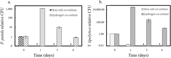
In the stressful environment (35 °C and antibiotics), the hydrogels protected the bacteria and yeast cells capsulated inside (Fig. 3). Based on CFU measurements, free P. putida and Y. lipolytica cells without encapsulation were inviable under phototrophic co-culture conditions within a day. With hydrogel protection, both the P. putida and Y. lipolytica populations increased within the first day of co-culture. Both species survived inside the hydrogel for the 6 monitored days of co-culture. This extended survival demonstrates that hydrogels can protect heterotrophic cells in growth-challenging environments.
Figure 3.
Relative heterotrophic cell population change in a stressful environment with high temperature and antibiotics. CFU values were normalized to the count at day 0. (a) Relative CFU counts of P. putida over 6 co-culture days. (b) Relative CFU of P. putida over six co-culture days. All data are averages of biological duplicates ± standard deviation. Some error bars are too small to see. Asterisks indicate no colony growth on plates.
Maximal growth of bacteria or yeast inside Ca-alginate hydrogels with rich media
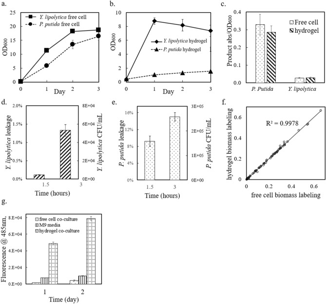
To examine cell growth inside hydrogels, hydrogel beads with encapsulated cells were washed and then added to rich aqueous media (LB or YPD) with inducers as necessary. Figure 4a,b show that the engineered P. putida and Y. lipolytica strains grew inside the hydrogel beads and in the free medium. In the stationary phase, the OD600 of P. putida inside the hydrogels was 1.5 (after EDTA dissolution of the hydrogel), and that of the free P. putida culture was ~ 14. As for Y. lipolytica, the OD600 inside the hydrogel beads reached 7, while the OD600 of free yeast culture was ~ 19. In general, the free monoculture grew faster than the immobilized cells. P. putida inside the hydrogel produced only ~ 10% of the biomass in the free LB culture. In contrast, Y. lipolytica inside the hydrogel reached > 40% of the biomass in the free YPD culture. Inside the hydrogel, the normalized product concentration by OD600 was similar to that in the free monocultures (Fig. 4c). These results confirmed that P. putida and Y. lipolytica cells maintained their productivity inside hydrogel microenvironments. Ca-alginate hydrogels are advantageous for the nutrient diffusion, but they may also have cell leakage. To investigate this problem, hydrogel beads with encapsulated P. putida or Y. lipolytica cells were washed and then incubated in nutrient-free 0.9% NaCl solution. The saline water was then sampled and spread on agar plates to count leaked cells. As illustrated in Fig. 4d,e, the numbers of leaked P. putida and Y. lipolytica cells increased with shaking for 1.5 h to 3 h, showing that the microbial cells were not completely immobilized inside the hydrogel microstructure. P. putida, being smaller than Y. lipolytica, more easily escaped from the hydrogel. Because heterotrophic species survived poorly in free co-culture without hydrogel protection, understanding and optimizing the microstructure of hydrogel beads is important for both restricting cell leakage and maintaining sufficient mass transfer.
Figure 4.
Heterotrophic culture behavior in rich media, with and without hydrogel compartmentation. (a) OD600 measurements of heterotrophic strains growing as free cells over three days. (b) OD600 measurements of heterotrophic strains growing in hydrogel compartmentation beads over 3 days. (c) Production of engineered P. putida (indigoidine) and Y. lipolytica (β-carotene) normalized to cell density. (d) Y. lipolytica cell leakage from hydrogel beads over 3 h, with shaking. (e) P. putida cell leakage from hydrogel beads over 3 h, with shaking. (f) 1,2-13C glucose labeling of Y. lipolytica proteinogenic amino acids from hydrogel and free cell cultures. The data points are from m/z = 57 peaks of GC–MS measurements of labeled amino acids. (g) Yellow fluorescent protein (YFP) fluorescence signal from engineered E. coli strain cultures (free cell co-culture, M9 media pure culture, and hydrogel co-culture). Data from plot (a–e,g) are averages of biological triplicates ± standard deviation. Some error bars are too small to be seen.
Metabolic analyses of spatially distinct microbes present in synthetic consortia
Hydrogel technology provides a convenient way to study individual microbial species’ physiologies in a consortium. First, we investigated the heterotroph’s metabolism inside the hydrogel via isotope tracing 38. Specifically, the metabolic flux distributions in the heterotroph cells could be inferred from 13C-labeling patterns of their proteinogenic amino acids. Using 1,2 position labeled glucose, we performed an isotropic labeling experiment on wild type Y. lipolytica that was grown either in free cell culture or inside the hydrogel. The biomass was harvested during the mid-exponential growth phase, and the labeling data in proteinogenic amino acids (i.e., 13C-fingerprints) were analyzed 39. Figure 4f shows the 13C labeled fraction from the [M-57] MS peak of each amino acid from both the hydrogel-immobilized cells and free cells (numerical results are included in Supplementary Material Table S2). The labeling results in the exponential growth phases are almost the same (R2 > 0.99), showing that during pseudo steady growth phase, the metabolic flux distributions of immobilized Y. lipolytica cells were almost identical to those of the free cell cultures. Therefore, the hydrogel encapsulation process and crosslinking structure did not compromise the metabolic activity of the yeast workhorse.
To further explore the possibility to generalize the Ca-alginate hydrogel co-culture method to other heterotrophic species as well as their heterologous protein expressions, an engineered E. coli strain expressing yellow fluorescent protein (YFP) was tested using the same co-culture step-up (with sucrose utilization). The advantage of hydrogel compartmentation held for this model bacterial species. Comparing the total fluorescent signals among free cell co-culture, hydrogel co-culture, and axenic bacterial culture using M9 media, the hydrogel co-culture shows the highest fluorescence while the axenic bacterial culture showed consistently increasing signal (Fig. 4g). The normalized fluorescence per cell from the hydrogel co-culture is even higher than that from axenic condition. As for growth, the CFU counting results from day 2 showed that the number of living cells from hydrogel co-culture is 10,000 times of that from free cell co-culture. The higher protein expression and healthier growth from hydrogel co-culture E. coli can be explained by the continuous supply of sugar from the autotrophic module and the metabolite overflow, such as amino acids, that promotes the E. coli growth and YFP expression inside hydrogel. Hence, the protein expression level from cells capsulated in hydrogel co-culture is not compromised by the hydrogel fabrication and co-culture stress.
Conclusions
This study demonstrated the production of high-value pigments and chemicals using a hydrogel-enabled phototroph-heterotroph co-culture consortium. Immobilizing the heterotrophic strains inside Ca-alginate hydrogel beads allowed the workhorse strains P. putida and Y. lipolytica to take advantage of the high sucrose production from engineered cyanobacteria in the free medium and produce high product titers inside the beads. The calcium alginate matrix also created a protective micro-environment that protected the heterotrophic species and sensitive pigment products from ROS released by cyanobacteria or from environmental stresses, such as high temperature and antibiotics. This methodology can be applied to more complex consortia involving more than two species. The hydrogel-encapsulated products were easily harvested from the photobioreactor, and the free cyanobacterial culture can be continuously resupplied with new batches of heterotroph-containing hydrogel beads. Furthermore, the hydrogel technology may facilitate metabolic analysis (e.g., 13C-metabolomics or proteomics) from spatially distinct microbes present in synthetic consortia.
While our study is an ideal case study for process optimization to enable the use of photosynthetic organisms, there are still several challenges that remain. First, engineering cyanobacteria or heterotrophs for invertase secretion may be advantageous over the addition of commercially produced invertase to the growth medium. Expression of a sucrose invertase has been demonstrated in P. putida but its secretion is still challenging 40. This strategy will have to be carefully optimized due to the potential tradeoff among final product titers, metabolic burdens for enzyme expressions, and invertase secretion. Second, phototrophic coculture still had lower titer than heterotrophic fermentations using rich medium. Our BG11 medium for cyanobacterial growth was suboptimal for engineered strains that require rich medium to sustain heterologous biosynthesis. In future work, the culture media and cultivation parameters should be further optimized. For example, the cyanobacteria could be engineered to overflow various nutrients to supply workhorse for optimal bioproduction.
Methods
Materials and strains
Sodium alginate (FCC grade) was purchased from Spectrum Chemicals (New Brunswick, NJ, USA). Reagent grade invertase was purchased from Carolina Biological (Burlington, NC, USA). Yeast extract, peptone, and agar for media were purchased from BD Biosciences (Franklin Lakes, NJ, USA). 2000X trace element was purchased from Teknova (Hollister, CA, USA). Amino acid supplements were purchased from US Biological Life Sciences (Salem, MA, USA). All other chemicals were purchased from Sigma-Aldrich (St. Louis, MO, USA). The sucrose producing S. elongatus strain and a β-carotene producing Y. lipolytica strain (provided by Arch Innotek, St. Louis, USA) were described in previous studies 1,36, respectively. The blue pigment producing engineered P. putida strain was engineered at the Lawrence Berkeley National Laboratory and this engineered strain was unable to use arabinose 25,41,42. The yellow fluorescent protein expressing E. coli is constructed from the Pakrasi lab (Supplementary Table S1).
Growth conditions
For sucrose production, engineered S. elongatus UTEX 2973 was first grown in BG11 medium for 48 h at 38 °C (250 μmol photons/m2/s1) in an AlgaeTron growth chamber (Photon Systems Instruments, Czech Republic), and then diluted tenfold in BG11 with 150 mM NaCl and 1 mM IPTG to grow for another 3 days at 38 °C (900 μmol photons/m2/s1) on PBRs (Multi-cultivator MC 1000-OD, Photon Systems Instruments, Czech Republic) before co-culture 1. In brief, BG11 media was modified from the UTEX Culture Collection, containing 17.6 mM NaNO3, 0.23 mM K2HPO4, 0.3 mM MgSO4, 0.24 mM CaCl2, 0.021 mM ferric ammonium citrate, 0.0027 mM Na2EDTA, 0.19 mM Na2CO3, and 10 mM N-[Tris(hydroxymethyl)methyl]-2-aminoethanesulfonic acid (TES) buffer. Around 1% v/v CO2 was maintained in the cyanobacterial media by bubbling CO2 into the sample tubes. The mixing was controlled by bubbling. Y. lipolytica seed cultures were grown in YPD medium (per liter: 10 g yeast extract, 20 g peptone, 20 g of glucose). A single colony (from YPD plates) was used to inoculate seed cultures in YPD and grown overnight at 30 °C in shaking flasks at 250 rpm. P. putida seed cultures (from single colonies picked from LB plates) were grown overnight in LB medium (per liter: 10 g tryptone, 5 g yeast extract, 10 NaCl) in shaking flasks at 30 °C at 250 rpm. E. coli seed cultures (from single colonies picked from LB plates) were grown overnight in LB medium with 20 µg/mL kanamycin in shaking flasks at 37 °C at 250 rpm. For YFP expression in axenic culture, engineered E. coli was grown in M9 media (per liter, 2 g (NH4)2SO4, 6.8 g Na2HPO4, 3 g KH2PO4, 0.5 g NaCl, 1 mL trace elements solution, 100 μL 1 M CaCl2, 2 mL 1 M MgSO4) supplemented with 2 g/L sucrose, 0.01 g/L invertase, and 20 µg/L kanamycin. For leakage test, 3 mL cell encapsulating hydrogels were added into 5 mL 0.9% NaCl solution. The mixtures were shaken in sample tubes at 30 °C at 250 rpm before CFU counting.
For the invertase tests of Y. lipolytica, amino acid was supplemented to BG11 media for β-carotene production. The amino acid supplements added each of 20 amino acids to the medium at 0.04 g/L (except for leucine, 0.2 g/L); uracil and inositol at 0.04 g/L; adenine at 0.01 g/L; and p-Aminobenzoic acid at 0.004 g/L.
Hydrogel fabrication and co-culture
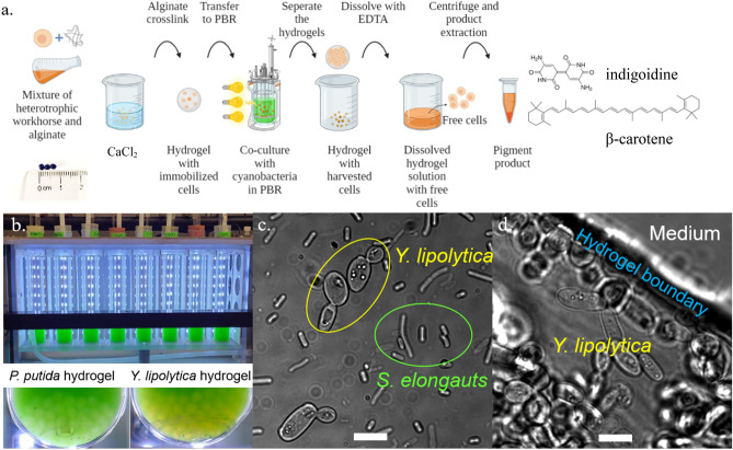
A 0.6% (w/w) sodium alginate solution mixed with P. putida or Y. lipolytica cell culture (OD600 = 2) was dripped into 100 mM calcium chloride gelation baths. The sodium alginate drip was driven by a syringe pump at a rate of 5 mL/min from a height of 10 cm above the bath, with magnetic stirring at 100 rpm. The resulting hydrogel beads were soaked statically in the calcium chloride bath for 2 h and then washed with DI water three times to remove unreacted calcium chloride. The fabricated hydrogels were spherical with diameter around 3 mm. Each bead contains about 10 million P. putida cells or 1 million Y. lipolytica cells. The workhorse (P. putida or Y. lipolytica) embedded hydrogel beads were then soaked in their respective rich media to resume their growth. The immobilized heterotrophs were added to the S. elongatus cultivation cultures to make P. putida/S. elongatus and Y. lipolytica/S. elongatus co-cultures. 3 mL heterotrophic hydrogel beads or cell culture was added to 27 mL cyanobacteria culture in tubes of a multi-cultivator at 30 °C under 800 μmol photons/m2/s1. 5 g/L arabinose was added to the P. putida co-culture to induce the production of indigoidine. For stress study of co-culture, we set the multi-cultivation temperature to 35 °C and added 10 µg/L kanamycin to the co-cultures. The picture of the co-culture system is shown in Fig. 5b. Figure 5c shows the microscope image of free cell co-culture of S. elongatus and Y. lipolytica (× 100 magnification). The microscope image of resulted cell-capsulated hydrogel is shown in Fig. 5d. Fluorescent image of indigoidine producing P. putida images were taken using the 4ʹ,6-diamidino-2-phenylindole (DAPI) filter, due to the fluorescence of indigoidine with 415 nm excitation wavelength and 520 nm emission wavelength 43. The fluorescent filter cube has excitation filter at 375 nm with 28 nm bandwidth and barrier filter at 460 nm with 50 nm bandwidth. Images of free cell co-culture and hydrogel on day 1, and hydrogel on day 4 are shown in Supplementary Fig. S2. The E. coli/S. elongatus co-cultures were constructed in similar way as described earlier in this session, except that the cultivation temperature was set at 38 °C and 20 µg/L kanamycin was added to the co-culture.
Figure 5.
Co-culture illustration diagram, multi-cultivator setup, and microscope images of co-culture and hydrogel. (a) Schematic illustration of co-culture bioproduction process. Created with BioRender.com. (b) Picture of the multi cultivator with P. putida and Y. lipolytica embedded hydrogels. (c) Optical microscope image of free cell S. elongatus/Y. lipolytica co-culture. (d) Optical bright field microscope image of hydrogel free cell S. elongatus/Y. lipolytica co-culture, × 100 magnification. Scale bars are 10 µm.
Growth and production quantification
Growth of the three microbe strains was monitored by measuring the OD600 (P. putida or Y. lipolytica) and OD730 (cyanobacteria) on a Cary 60 UV–Vis Spectrometer. Strain survival was quantified as colony forming units (LB plate for P. putida, YPD plate for Y. lipolytica, and BG11 plate for S. elongatus) from liquid culture or mechanically crushed hydrogels. Sucrose and glucose concentrations in co-culture were measured using colorimetric enzyme kits, and absorption was measured on a Cary 60 UV–Vis Spectrometer. To measure the β-carotene content, 100 mL Y. lipolytica cultures were pelleted at 24,000×g and the supernatant was discarded. The biomass was resuspended in 1 mL hexane and vortexed with glass beads until lysed. β-carotene titer was quantified via absorbance at 454 nm on a Tecan Infinite M200 Pro Microplate Reader 36. Because the measurement of yeast β-carotene from free cell co-culture could be complicated by native cyanobacterial pigments, we measured the background absorbance from pure S. elongatus culture. Then the β-carotene absorbance readings from the free cell co-culture were subtracted by the background readings. The entire hydrogel co-culture production process is illustrated in Fig. 5a.
Indigoidine production and quantification were performed as previously described with slight modifications 25. Briefly, 100 µL of liquid culture was pelleted at 24,000×g for 2 min. The supernatant was discarded, and 500 µL of dimethyl sulfoxide (DMSO) was used to resuspend and extract the indigoidine via vortexing (10 min). Additional DMSO was added if the pellets were not fully dissolved. Absorption intensities at 612 nm were measured with a Tecan Infinite M200 Pro Microplate Reader. Standard curves of β-carotene and indigoidine were obtained by solubilization of the pure color chemicals in their perspective solvents, serial dilution, and absorption measurement with the Tecan microplate reader (Supplementary File).
For extraction of products from hydrogel encapsulated samples, 25 µL of 500 mM EDTA was added to 10 recovered hydrogel beads (total volume ~ 1.5 mL) to dissolve the structure. Once solubilized, the resulting crude or β-carotene or indigoidine was estimated with absorbance after hexane or DMSO solubilization as for a standard cell pellet. Blank samples were prepared using hydrogels without microbes through the same extraction procedure. The blank values were subtracted from raw readings to avoid noise.
Determination of colony forming units (CFUs)
Agar plates were prepared for microbial growth (BG11 plates for S. elongatus, YPD plates for Y. lipolytica, LB plates for P. putida). A serial dilution cultures were done in a 96 well plate using sterilized water as dilutant. For plating, 10 μL aliquot of each dilution was transferred onto the agar plates. Agar plate lids were kept open in laminar flow hood until all of the liquid had dried. The plates were incubated at 30 °C upside down until single colonies were clearly visible. The number of colonies were counted, and original titers were calculated based on total volume plated, and dilution. To determine the CFUs in the hydrogels, the hydrogels were mechanically broken into pieces by pipetting up and down using 1 mL pipette. The CFUs from broken hydrogels were estimated as for a free cell culture.
13C labeling of proteinogenic amino acids
Overnight cultures of wildtype Y. lipolytica were subcultured 0.3% v/v into respective minimal media (yeast nitrogen base without amino acid for Y. lipolytica), provided 5 g/L 13C1,2 glucose. Then, the subcultures were inoculated 1% v/v as free cell culture or hydrogel-immobilized culture in respective minimal media with same labeled glucose. The resulting biomass was harvested in the exponential phase by centrifuge. The biomass protein was washed then hydrolyzed with 6 N HCL at 100 °C for 24 h. A TMDMS (N-(tert-butyldimethylsilyl)-N-methyltrifluoroacetamide) method was used to analyze the proteinogenic amino acid labeling profiles via GC-MS 39.
Supplementary Information
Acknowledgements
The research on the application of hydrogel composites to promote phototrophic biomanufacturing is supported by the DOE Fossil Energy Office (DE-FE0032108). AXT acknowledges support from the National Science Foundation Graduate Research Fellowship Program (Grant #DGE-1745038). TE, JM, and AM are supported by the Joint Bioenergy Institute (http://www.jbei.org) through the US Department of Energy, Office of Science, under contract DE-AC02-05CH11231 between Lawrence Berkeley National Laboratory (LBNL) and the US Department of Energy. AS and HBP was supported by funding from National Science Foundation (MCB-2037887).
Author contributions
Y.J.T., H.P., Y.J. conceived the hydrogel-based co-culture idea and designed experiments. R.Z., T.P. and A.S. performed co-culture studies, A.X.T. and R.W. optimized hydrogel technology, J.M., T.E., A.M. and H.P. provided engineered strains and reagents. All authors wrote, proofread, and approved the manuscript for publication.
Data availability
The datasets used and/or analyzed during the current study available from the corresponding authors on reasonable request.
Competing interests
The authors declare no competing interests.
Footnotes
Publisher's note
Springer Nature remains neutral with regard to jurisdictional claims in published maps and institutional affiliations.
Contributor Information
Young-Shin Jun, Email: ysjun@seas.wustl.edu.
Yinjie J. Tang, Email: yinjie.tang@seas.wustl.edu
Supplementary Information
The online version contains supplementary material available at 10.1038/s41598-022-26437-y.
References
- 1.Lin PC, Zhang F, Pakrasi HB. Enhanced production of sucrose in the fast-growing cyanobacterium Synechococcus elongatus UTEX 2973. Sci. Rep. 2020;10:390. doi: 10.1038/s41598-019-57319-5. [DOI] [PMC free article] [PubMed] [Google Scholar]
- 2.Wang X, et al. Enhanced limonene production in cyanobacteria reveals photosynthesis limitations. Proc. Natl. Acad. Sci. U.S.A. 2016;113:14225–14230. doi: 10.1073/pnas.1613340113. [DOI] [PMC free article] [PubMed] [Google Scholar]
- 3.Wan N, et al. Cyanobacterial carbon metabolism: Fluxome plasticity and oxygen dependence. Biotechnol. Bioeng. 2017;114:1593–1602. doi: 10.1002/bit.26287. [DOI] [PubMed] [Google Scholar]
- 4.Sarnaik A, et al. Metabolic engineering of cyanobacteria for photoautotrophic production of heparosan, a pharmaceutical precursor of heparin. Algal Res. 2019;37:57–63. doi: 10.1016/j.algal.2018.11.010. [DOI] [Google Scholar]
- 5.Sarnaik A, Nambissan V, Pandit R, Lali A. Recombinant Synechococcus elongatus PCC 7942 for improved zeaxanthin production under natural light conditions. Algal Res. 2018;36:139–151. doi: 10.1016/j.algal.2018.10.021. [DOI] [Google Scholar]
- 6.Fedeson DT, Saake P, Calero P, Nikel PI, Ducat DC. Biotransformation of 2,4-dinitrotoluene in a phototrophic co-culture of engineered Synechococcus elongatus and Pseudomonas putida. Microb. Biotechnol. 2020;13:997–1011. doi: 10.1111/1751-7915.13544. [DOI] [PMC free article] [PubMed] [Google Scholar]
- 7.Zhang L, et al. Construction and analysis of an artificial consortium based on the fast-growing cyanobacterium Synechococcus elongatus UTEX 2973 to produce the platform chemical 3-hydroxypropionic acid from CO2. Biotechnol. Biofuels. 2020;13:82. doi: 10.1186/s13068-020-01720-0. [DOI] [PMC free article] [PubMed] [Google Scholar]
- 8.Hays SG, Yan LLW, Silver PA, Ducat DC. Synthetic photosynthetic consortia define interactions leading to robustness and photoproduction. J. Biol. Eng. 2017;11:4. doi: 10.1186/s13036-017-0048-5. [DOI] [PMC free article] [PubMed] [Google Scholar]
- 9.Amin SA, Parker MS, Armbrust EV. Interactions between diatoms and bacteria. Microbiol. Mol. Biol. Rev. 2012;76:667–684. doi: 10.1128/MMBR.00007-12. [DOI] [PMC free article] [PubMed] [Google Scholar]
- 10.Singh AK, Ducat DC. Generation of stable, light-driven co-cultures of cyanobacteria with heterotrophic microbes. Methods Mol. Biol. 2022;2379:277–291. doi: 10.1007/978-1-0716-1791-5_16. [DOI] [PubMed] [Google Scholar]
- 11.Beliaev AS, et al. Inference of interactions in cyanobacterial-heterotrophic co-cultures via transcriptome sequencing. ISME J. 2014;8:2243–2255. doi: 10.1038/ismej.2014.69. [DOI] [PMC free article] [PubMed] [Google Scholar]
- 12.Fu N, Peiris P, Markham J, Bavor J. A novel co-culture process with Zymomonas mobilis and Pichia stipitis for efficient ethanol production on glucose/xylose mixtures. Enzyme Microb. Technol. 2009;45:210–217. doi: 10.1016/j.enzmictec.2009.04.006. [DOI] [Google Scholar]
- 13.Serrano-Aroca Á, Iskandar L, Deb S. Green synthetic routes to alginate-graphene oxide composite hydrogels with enhanced physical properties for bioengineering applications. Eur. Polymer J. 2018;103:198–206. doi: 10.1016/j.eurpolymj.2018.04.015. [DOI] [Google Scholar]
- 14.Huq T, et al. Alginate based nanocomposite for microencapsulation of probiotic: Effect of cellulose nanocrystal (CNC) and lecithin. Carbohyd. Polym. 2017;168:61–69. doi: 10.1016/j.carbpol.2017.03.032. [DOI] [PubMed] [Google Scholar]
- 15.Phillips GO, Williams PA, editors. Handbook of Hydrocolloids. 2. Woodhead Publishing; 2009. pp. 807–828. [Google Scholar]
- 16.Murujew O, et al. Recovery and reuse of alginate in an immobilized algae reactor. Environ. Technol. 2021;42:1521–1530. doi: 10.1080/09593330.2019.1673827. [DOI] [PubMed] [Google Scholar]
- 17.Aneez Ahamad PY, Mohammad Kunhi AA. Enhanced degradation of phenol by Pseudomonas sp. CP4 entrapped in agar and calcium alginate beads in batch and continuous processes. Biodegradation. 2011;22:253–265. doi: 10.1007/s10532-010-9392-6. [DOI] [PubMed] [Google Scholar]
- 18.Zhang S, et al. Bacterial cellulose-alginate composite beads as yarrowia lipolytica cell carriers for lactone production. Molecules. 2020;25:928. doi: 10.3390/molecules25040928. [DOI] [PMC free article] [PubMed] [Google Scholar]
- 19.Weiss TL, Young EJ, Ducat DC. A synthetic, light-driven consortium of cyanobacteria and heterotrophic bacteria enables stable polyhydroxybutyrate production. Metab. Eng. 2017;44:236–245. doi: 10.1016/j.ymben.2017.10.009. [DOI] [PubMed] [Google Scholar]
- 20.Xiao Z, Tan AX, Xu V, Jun YS, Tang YJ. Mineral-hydrogel composites for mitigating harmful algal bloom and supplying phosphorous for photo-biorefineries. Sci. Total Environ. 2022;847:157533. doi: 10.1016/j.scitotenv.2022.157533. [DOI] [PMC free article] [PubMed] [Google Scholar]
- 21.Bajpai SK, Sharma S. Investigation of swelling/degradation behaviour of alginate beads crosslinked with Ca2+ and Ba2+ ions. React. Funct. Polym. 2004;59:129–140. doi: 10.1016/j.reactfunctpolym.2004.01.002. [DOI] [Google Scholar]
- 22.Mørch ÝA, Donati I, Strand BL, Skjåk-Bræk G. Effect of Ca2+, Ba2+, and Sr2+ on alginate microbeads. Biomacromolecules. 2006;7:1471–1480. doi: 10.1021/bm060010d. [DOI] [PubMed] [Google Scholar]
- 23.Moffett D, Smith-Simon C, Stevens YW. Toxicological Profile for Barium and Barium Compounds. Agency for Toxic Substances and Disease Registry; 2007. [PubMed] [Google Scholar]
- 24.Mørch YA, et al. Binding and leakage of barium in alginate microbeads. J. Biomed. Mater. Res. A. 2012;100A:2939–2947. doi: 10.1002/jbm.a.34237. [DOI] [PMC free article] [PubMed] [Google Scholar]
- 25.Banerjee D, et al. Genome-scale metabolic rewiring improves titers rates and yields of the non-native product indigoidine at scale. Nat. Commun. 2020;11:5385. doi: 10.1038/s41467-020-19171-4. [DOI] [PMC free article] [PubMed] [Google Scholar]
- 26.Czajka JJ, et al. Tuning a high performing multiplexed-CRISPRi Pseudomonas putida strain to further enhance indigoidine production. Metabolic Engineering Communications. 2022;15:e00206. doi: 10.1016/j.mec.2022.e00206. [DOI] [PMC free article] [PubMed] [Google Scholar]
- 27.Czajka JJ, Oyetunde T, Tang YJ. Integrated knowledge mining, genome-scale modeling, and machine learning for predicting Yarrowia lipolytica bioproduction. Metab. Eng. 2021;67:227–236. doi: 10.1016/j.ymben.2021.07.003. [DOI] [PubMed] [Google Scholar]
- 28.Abernathy MH, et al. Deciphering cyanobacterial phenotypes for fast photoautotrophic growth via isotopically nonstationary metabolic flux analysis. Biotechnol. Biofuels. 2017;10:273. doi: 10.1186/s13068-017-0958-y. [DOI] [PMC free article] [PubMed] [Google Scholar]
- 29.Srivastava M, Hudson EP, Wangikar PP. Cyanobacteria Biotechnology. Wiley; 2021. pp. 441–476. [Google Scholar]
- 30.Ghiffary MR, et al. High-level production of the natural blue pigment indigoidine from metabolically engineered Corynebacterium glutamicum for sustainable fabric dyes. ACS Sustain. Chem. Eng. 2021;9:6613–6622. doi: 10.1021/acssuschemeng.0c09341. [DOI] [Google Scholar]
- 31.Czajka JJ, et al. Engineering the oleaginous yeast Yarrowia lipolytica to produce the aroma compound beta-ionone. Microb. Cell Fact. 2018;17:136. doi: 10.1186/s12934-018-0984-x. [DOI] [PMC free article] [PubMed] [Google Scholar]
- 32.Abernathy MH, et al. Cyanobacterial carboxysome mutant analysis reveals the influence of enzyme compartmentalization on cellular metabolism and metabolic network rigidity. Metab. Eng. 2019;54:222–231. doi: 10.1016/j.ymben.2019.04.010. [DOI] [PMC free article] [PubMed] [Google Scholar]
- 33.Cano M, et al. Glycogen synthesis and metabolite overflow contribute to energy balancing in cyanobacteria. Cell Rep. 2018;23:667–672. doi: 10.1016/j.celrep.2018.03.083. [DOI] [PubMed] [Google Scholar]
- 34.Huang J, et al. Functionalization of bacterial microcompartment shell proteins with covalently attached heme. Front. Bioeng. Biotechnol. 2019;7:432. doi: 10.3389/fbioe.2019.00432. [DOI] [PMC free article] [PubMed] [Google Scholar]
- 35.Eng T, et al. Engineering Pseudomonas putida for efficient aromatic conversion to bioproduct using high throughput screening in a bioreactor. Metab. Eng. 2021;66:229–238. doi: 10.1016/j.ymben.2021.04.015. [DOI] [PubMed] [Google Scholar]
- 36.Worland AM, et al. Analysis of Yarrowia lipolytica growth, catabolism, and terpenoid biosynthesis during utilization of lipid-derived feedstock. Metab. Eng. Commun. 2020;11:e00130. doi: 10.1016/j.mec.2020.e00130. [DOI] [PMC free article] [PubMed] [Google Scholar]
- 37.Scita G. The stability of β-carotene under different laboratory conditions. J. Nutr. Biochem. 1992;3:124–128. doi: 10.1016/0955-2863(92)90104-Q. [DOI] [PubMed] [Google Scholar]
- 38.Tang YJ, et al. Advances in analysis of microbial metabolic fluxes via (13)C isotopic labeling. Mass Spectrom. Rev. 2009;28:362–375. doi: 10.1002/mas.20191. [DOI] [PubMed] [Google Scholar]
- 39.Au-You L, et al. Metabolic pathway confirmation and discovery through 13C-labeling of proteinogenic amino acids. JoVE. 2012;59:e3583. doi: 10.3791/3583. [DOI] [PMC free article] [PubMed] [Google Scholar]
- 40.Wohlers K, et al. Metabolic engineering of Pseudomonas putida for production of the natural sweetener 5-ketofructose from fructose or sucrose by periplasmic oxidation with a heterologous fructose dehydrogenase. Microb. Biotechnol. 2021;14:2592–2604. doi: 10.1111/1751-7915.13913. [DOI] [PMC free article] [PubMed] [Google Scholar]
- 41.Lim HG, et al. Generation of Pseudomonas putida KT2440 strains with efficient utilization of xylose and galactose via adaptive laboratory evolution. ACS Sustain. Chem. Eng. 2021;9:11512–11523. doi: 10.1021/acssuschemeng.1c03765. [DOI] [Google Scholar]
- 42.Wang Y, et al. Growth of engineered Pseudomonas putida KT2440 on glucose, xylose, and arabinose: Hemicellulose hydrolysates and their major sugars as sustainable carbon sources. GCB Bioenergy. 2019;11:249–259. doi: 10.1111/gcbb.12590. [DOI] [Google Scholar]
- 43.Brown AS, Robins KJ, Ackerley DF. A sensitive single-enzyme assay system using the non-ribosomal peptide synthetase BpsA for measurement of L-glutamine in biological samples. Sci. Rep. 2017;7:41745. doi: 10.1038/srep41745. [DOI] [PMC free article] [PubMed] [Google Scholar]
Associated Data
This section collects any data citations, data availability statements, or supplementary materials included in this article.
Supplementary Materials
Data Availability Statement
The datasets used and/or analyzed during the current study available from the corresponding authors on reasonable request.


