Abstract
Objectives:
Aromatase is an enzyme that catalyzes the conversion of androgens to estrogens. While inhibition of aromatase is a useful approach for treating breast cancer, it may also have toxicological consequences due to its endocrine disrupting/modulating effect. In this study, sensitivity and performance of two in vitro assays -a cell free and a cell-based- for evaluating aromatase activity were investigated by testing known aromatase inhibitors and partial validation of the methods was performed. Advantages and disadvantages of these methods are also discussed.
Materials and Methods:
Aromatase activity was evaluated via two in vitro models; direct measurement with a cell-free assay using a fluorescent substrate and recombinant human enzyme and indirect evaluation with a cell-based assay where cell proliferation was determined in estrogen receptor positive human breast cancer cells (MCF-7 BUS) in the absence of estrogen and the presence of testosterone.
Results:
In the cell-free direct measurement assay, reference compounds ketoconazole and aminoglutethimide have been shown to inhibit the aromatase enzyme with half-maximal inhibitory concentration (IC50) values concordant with literature. In cell-based indirect measurement assay, only ketoconazole dose-dependently inhibited cell proliferation with 3.47 x 10⁻⁷ M IC50. Inter-assay and intra-assay reproducibility of both methods was found to be within acceptable deviation levels.
Conclusion:
Both methods can be successfully applied. However, to evaluate the potential aromatase activity of the novel compounds in vitro, it seems better to perform both the cell-based and the cell-free assays that allows low-moderate biotransformation and eliminate cytotoxicity potential, respectively.
Keywords: Aromatase inhibition, cell-based assay, cell-free assay, in vitro
INTRODUCTION
Endocrine disruptors are exogenous compounds, which cause adverse effects by altering endocrine system functions.1 These compounds have several mechanisms of action, one is to modulate the cytochrome P450 (CYP450) enzymes involved in steroid hormone synthesis/metabolism.2
Aromatase is a member of CYP450 enzyme superfamily, which catalyzes the conversion of androgens to estrogens during the last step of steroidogenesis.3 This conversion by aromatase is a rate-limiting step in estrogen synthesis and the enzyme is responsible for maintaining a homeostatic balance between androgens and estrogens. Aromatase is involved in numerous physiological functions such as reproduction, development, behavior as well as pathologies such as hormone-dependent cancers. Especially in postmenopausal women, local estrogen synthesis via aromatization of androgens plays a crucial role in the development of estrogen-dependent breast cancer.3 Therefore, inhibition of aromatase is a useful approach for treating hormone-dependent breast cancer. On the other side, inhibition of this enzyme may have toxicological consequences because of endocrine disruption/modulation.
In this paper, aromatase activity was measured by two in vitro assays. The first one is a high throughput screening assay, where a fluorescent substrate and recombinant human enzyme are used, and the enzyme activity is detected directly via use of the substrate. In the second assay, enzyme activity is indirectly evaluated via proliferation of the estrogen receptor-positive human breast cancer cells, MCF-7 BUS, in the presence of testosterone in an estrogen-free medium. The sensitivity and performance of both assays were evaluated by testing known aromatase inhibitors and partial validation of the methods was performed. Advantages and disadvantages of cell-based and cell-free assays are discussed.
MATERIALS AND METHODS
MCF-7 BUS cells were kindly provided by Prof. Ana Soto from Tufts Institute and maintained at 37°C in 5% CO2 atmosphere in Dulbecco’s Modified Eagle’s medium (DMEM) supplemented with 10% fetal bovine serum (FBS). Reference compounds (ketoconazole and aminoglutethimide) and other chemicals were purchased from Sigma-Aldrich (St. Louis, MO, USA) and Thermo Fisher Scientific. An aromatase activity assay kit was purchased from Corning Incorporated (New York, USA).
Direct measurement of aromatase activity
Direct measurement of aromatase activity was evaluated by CYP19A/7-methoxy-4-trifluoromethyl coumarin (MFC) screening kit from Corning Incorporated (New York USA). Reaction substrate MFC is converted to 7-hydroxytrifluoromethyl coumarin by aromatase in the presence of nicotinamide adenine dinucleotide phosphate (NADPH) generating system. Thus, reduction of in fluorescence intensity refers to aromatase inhibitor activity.4,5 Enzyme reactions were performed, according to the manufacturer’s protocol as indicated in detailed previously.6 IC50 values of reference materials were obtained using GraphPad Prism5 software.
Intra-assay reproducibility was determined via calculation of the mean and standard deviation (SD) values of the enzyme activity, which were measured in 5 different wells on the same day, while interassay reproducibility was determined via calculation of the values from 3 different days.
Indirect measurement of aromatase activity
If the estrogen-dependent cells are seeded in estrogen-depleted media, cell proliferation occurs via aromatization of androgens. Thus, aromatase activity can be measured indirectly in MCF-7 cells by evaluating cell viability in the medium with testosterone/without estrogen, according to the method7 with minor modifications as previously described.6
Briefly, MCF-7 BUS cells were plated in 96 well plates at a density of 6000 cells/well in DMEM supplemented with 10% FBS and incubated at 37°C in a humid atmosphere containing 5% CO2. After 48 hours of attachment, the medium was replaced with DMEM without phenol red supplemented with 10% charcoal stripped FBS, 1% sodium pyruvate and 1% non-essential amino acid solution containing either testosterone (10 µM) alone or testosterone and the tested compounds together. A control group was also included, in which the cells were grown in estrogen-depleted media without any testosterone or test molecule. Following 5 day incubation period, cell viability was assessed via 3-[4,5-dimethylthiazol-2-yl]-2,5-diphenyltetrazolium bromide (MTT) assay. The medium was removed, cells were washed with phosphate buffered saline and then incubated with MTT (1 mg/mL) for 4 h at 37°C. MTT solution was removed and formazan crystals were dissolved in dimethyl sulfoxide. The absorbance was recorded at 550 nm on a microplate reader. The ratio of the absorbance of treated samples to the absorbance of control (taken as 100%) was expressed as percentage cell viability.
To evaluate performance and the sensitivity of the assay, cells were incubated with 17-β-estradiol (1 nM)-, and testosterone (1 and 10 µM) for 5 days in the presence and absence of aromatase inhibitors.
Statistical analysis
Data were expressed as means ± SD. Statistical analysis was performed using student’s t-test. Differences were considered significant p<0.05. P values are given in figure legends.
RESULTS AND DISCUSSION
In this study, aromatase activity was measured using two different in vitro assays; a cell free, direct measurement assay and a cell-based, indirect measurement assay. Performance and sensitivity of the assays were compared using reference compounds, i.e. ketoconazole, general CYP inhibitor and a well-known aromatase inhibitor, i.e. aminoglutethimide.
In the cell-free aromatase activity assay, human recombinant aromatase enzyme (CYP19) and a fluorescence substrate MFC were used. In NADPH generating system, fluorescence intensity is reduced because of demethylation of MFCs by CYP19 and enzyme activity is calculated fluorometrically. Since this method is performed in 96 well plate format and allows high throughput screening, different groups have previously used it to evaluate novel aromatase inhibitors.8
Sensitivity and performance of the direct measurement assay in our laboratory conditions was evaluated by using a known aromatase inhibitor aminoglutethimide and a general CYP inhibitor ketoconazole. Ketoconazole and aminoglutethimide have been shown to inhibit aromatase in the direct measurement assay with 2.3 x 10⁻⁶ M and 4.7 x 10⁻⁷ M IC50 values, respectively (Table 1). Compared to the IC50 values from the literature, our results were found to be concordant with the literature (Table 1).9
Table 1. IC50 values of ketoconazole and aminoglutethimide that were obtained from literature and from direct aromatase activity assay in the present study9.

Inter-assay and intra-assay reproducibility of the direct measurement assay was also evaluated by measuring enzyme activity in the presence of a fixed amount of recombinant enzyme and substrate (50 µM MFC). Intra-assay and inter-assay coefficients of variation values were 2.8% and 10%, respectively (Table 2). According to these results, direct measurement assay is found to be in an acceptable reproducibility range.
Table 2. Inter-assay and intra-assay reproducibility values of direct aromatase activity assay.

Indirect measurement assay is performed in MCF-7 BUS cells by evaluating proliferation of the cells in estrogen deprived but testosterone-added media. MCF-7 BUS is a well-established estrogen receptor-positive cell line and depends on estrogen for proliferation. Cells possess aromatase activity.10 In this study, we also performed western blotting (data not shown) and confirmed the expression of aromatase in our cell line. Since cell proliferation depends on the presence of estrogens, in the absence of estrogen but in presence of testosterone, cell proliferation depends on the aromatization of testosterone to estrogen via aromatase enzyme,7 that is the principle of this indirect measurement assay.
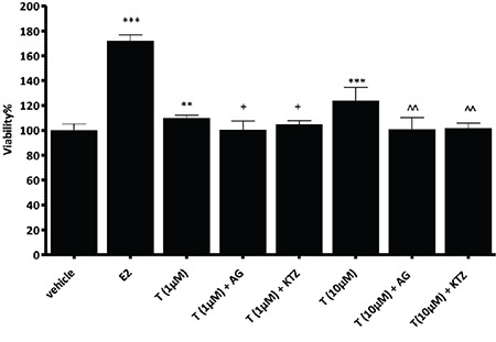
Performance and the sensitivity of the indirect measurement assay was evaluated by using reference compounds (estradiol and testosterone) (Figure 1) in the presence and or absence of aromatase inhibitors. As expected, 17-β-estradiol significantly increased cell proliferation (approximately 2 fold) comparing to the control group. Testosterone also increased cell proliferation in a dose-dependent manner. This effect was reduced by the aromatase inhibitors, i.e. aminoglutethimide (100 µM) and ketoconazole (5 µM), indicating that cell proliferation was estrogen-dependent and catalyzed by aromatase activity of the cells (Figure 1).
Figure 1.

Effect of testosterone, aminoglutethimide (100 μM), and ketoconazole (5 μM) on MCF-7 BUS cell proliferation. Bars show percentage viability values compared to control group (mean ± SD). Statistical analysis was performed by using student’s t-test.
**p<0.005 vs vehicle, ***p<0.001 vs vehicle, +p<0.05 vs T (1 μM); ^^p<0.005 vs T (10 μM)
SD: Standard deviation
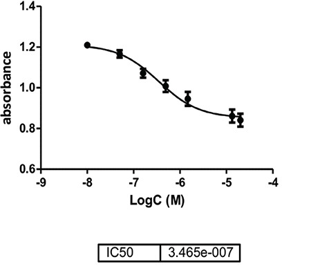
After that, cells were incubated with 10 µM testosterone and varying concentrations of ketoconazole or aminoglutethimide for 5 days to obtain IC50 values in the indirect measurement assay. We found that ketoconazole (0.05-20 µM) inhibited cell proliferation because of aromatization of testosterone to estradiol (Figure 2) with 3.47 x 10⁻⁷ M IC50 value. On the other hand, aminoglutethimide did not inhibit cell proliferation in a dose dependent manner (data not shown). Therefore, IC50 value of aminoglutethimide could not be calculated.
Figure 2.

Inhibitory effect of ketoconazole on indirect aromatase activity. Cells were incubated with testosterone (10 μM) and ketoconazole for 5 days
Inter- and intra-assay reproducibility of the indirect measurement assay was evaluated. Intra-assay reproducibility was calculated using the results of the estradiol and testosterone obtained from four different wells on the same day. % coefficient variation values of testosterone and estradiol were 2.7% and 7.4%, respectively (Table 3). For the inter-assay reproducibility, percent coefficient variation values of testosterone and estradiol were calculated as 2.5% and 2.6%, respectively, which were obtained from the results of the experiments conducted over four different days. According to these results, it was concluded that the indirect measurement assay has a high rate of successful replications and works successfully.
Table 3. Inter-assay and intra-assay reproducibility values for indirect aromatase activity measurement assay.

While the IC50 value of ketoconazole was found to be 2.5 µM in the direct activity measurement method, it was about 10 times lower (0.35 µM) in the indirect aromatase activity. This difference is thought to be because of possible metabolites of ketoconazole. Nevertheless, in a study conducted in a primary culture system of rat hepatocytes, major metabolite of ketoconazole (N-deacetylated ketoconazole) has a more potent cytotoxic effect in an MTT assay.11 Therefore, the reason for this difference may be potential cytotoxic, estrogen receptor antagonists or of aromatase expression modulator effects of the possible metabolites. It was also demonstrated by Yan et al.12 that ketoconazole downregulates aromatase gene expression in goldfish. So, lower IC50 value of ketoconazole in cell-based indirect measurement assay may be the consequence of both inhibition of aromatase enzyme and downregulation of aromatase expression.
It should also be kept in mind that substance concentration interacting with the active site of aromatase enzyme cannot be the same in cell-based and cell-free assay. There are lots of biological steps in cell-based assays, such as passaging through the membranes, entering the cells and metabolism, which can affect the results.
However, the cell-based method has a metabolic capacity compared to the direct measurement assay. Therefore, it is possible to evaluate the potential effects of the active metabolites, which makes it a more advantageous method in reflecting the physiological state in a more realistic way.
CONCLUSION
In conclusion, partial validation results of this study indicate that direct and indirect measurement assays can be used for evaluating aromatase activity, but both of them have some advantages and disadvantages indeed. Therefore, it seems better to perform this cell-based and cell-free assays together to evaluate the potential aromatase activity of the novel compounds to comment on the results correctly. Additional tests like cytotoxicity, effect on enzyme expression levels should also be performed to prevent misinterpretation of the indirect measurement assay results.
Footnotes
Ethics
Ethics Committee Approval: Ethical approval is not required for the study.
Informed Consent: Not necessary.
Peer-review: Externally peer-reviewed.
Authorship Contributions
Concept: E.İ.E., H.G.O., Design: E.İ.E., S.Ö.S., H.G.O., Data Collection or Processing: E.İ.E., S.Ö.S., Analysis or Interpretation: E.İ.E., S.Ö.S., Literature Search: E.İ.E., S.Ö.S., Writing: E.İ.E., H.G.O.
Conflict of Interest: No conflict of interest was declared by the authors.
Financial Disclosure: This work was supported by The Scientific and Technological Research Council of Turkey (TUBITAK) Research and Development Grant 112S375, Ege University Research and Development Grant 13BIL009 and Grant 13ECZ008. Ege University FABAL facilities were used for biological activity assays.
References
- 1.Damstra T, Barlow S, Bergman A, Kavlock R, Van Der Kraak G. World Health Organization: Global assessment of the state-of-the-science of endocrine disruptors. WHO. 2002. Available from: [Internet] https://apps.who.int/iris/bitstream/handle/10665/67357/WHO_PCS_EDC_02.2.pdf?sequence=1&isAllowed=y.
- 2.Sanderson JT. The steroid hormone biosynthesis pathway as a target for endocrine-disrupting chemicals. Toxicol Sci. 2006;94:3–21. doi: 10.1093/toxsci/kfl051. [DOI] [PubMed] [Google Scholar]
- 3.Simpson E, Rubin G, Clyne C, Robertson K, O’Donnell L, Davis S, Jones M. Local estrogen biosynthesis in males and females. Endocr Relat Cancer. 1999;6:131–137. doi: 10.1677/erc.0.0060131. [DOI] [PubMed] [Google Scholar]
- 4.Maiti A, Reddy PV, Sturdy M, Marler L, Pegan SD, Mesecar AD, Pezzuto JM, Cushman M. Synthesis of casimiroin and optimization of its quinone reductase 2 and aromatase inhibitory activities. J Med Chem. 2009;52:1873–1884. doi: 10.1021/jm801335z. [DOI] [PMC free article] [PubMed] [Google Scholar]
- 5.Stresser DM, Turner SD, McNamara J, Stocker P, Miller VP, Crespi CL, Patten CJ. A high-throughput screen to identify inhibitors of aromatase (CYP19) Anal Biochem. 2000;284:427–430. doi: 10.1006/abio.2000.4729. [DOI] [PubMed] [Google Scholar]
- 6.Özcan-Sezer S, İnce E, Akdemir A, Ceylan ÖÖ, Süzen S, Gürer-Orhan H. Aromatase inhibition by 2-methyl indole hydrazone derivatives evaluated via molecular docking and in vitro activity studies. Xenobiotica. 2019;49(5):549–556. doi: 10.1080/00498254.2018.1482029. [DOI] [PubMed] [Google Scholar]
- 7.Cos S, Martínez-Campa C, Mediavilla MD, Sánchez-Barceló EJ. Melatonin modulates aromatase activity in MCF-7 human breast cancer cells. J Pineal Res. 2005;38:136–142. doi: 10.1111/j.1600-079X.2004.00186.x. [DOI] [PubMed] [Google Scholar]
- 8.Bonfield K, Amato E, Bankemper T, Agard H, Steller J, Keeler JM, Roy D, McCallum A, Paula S, Ma L. Development of a new class of aromatase inhibitors: design, synthesis and inhibitory activity of 3-phenylchroman-4-one (isoflavanone) derivatives. Bioorg Med Chem. 2012;20:2603–2613. doi: 10.1016/j.bmc.2012.02.042. [DOI] [PMC free article] [PubMed] [Google Scholar]
- 9.Wouters W, De Coster R, Goeminne N, Beerens D, van Dun J. Aromatase inhibition by the antifungal ketoconazole. J Steroid Biochem. 1988;30:387–389. doi: 10.1016/0022-4731(88)90128-8. [DOI] [PubMed] [Google Scholar]
- 10.Zhou D, Wang J, Chen E, Murai J, Siiteri PK, Chen S. Aromatase gene is amplified in MCF-7 human breast cancer cells. J Steroid Biochem Mol Biol. 1993;46:147–153. doi: 10.1016/0960-0760(93)90289-9. [DOI] [PubMed] [Google Scholar]
- 11.Rodriguez RJ, Acosta D Jr. N-deacetyl ketoconazole-induced hepatotoxicity in a primary culture system of rat hepatocytes. Toxicology. 1997;117:123–131. doi: 10.1016/s0300-483x(96)03560-3. [DOI] [PubMed] [Google Scholar]
- 12.Yan Z, Lu G, Ye Q, Liu J. Modulation of 17β-estradiol induced estrogenic responses in male goldfish (Carassius auratus) by benzo[a]pyrene and ketoconazole. Environ Sci Pollut Res Int. 2016;23:9036–9045. doi: 10.1007/s11356-016-6168-5. [DOI] [PubMed] [Google Scholar]


