Abstract
Psidium guajava (Guava tree) is one of the most widely known species in the family Myrtaceae. The Guava tree has been reported for its potential antioxidant, anti-inflammatory, antimicrobial, and cytotoxic activities. In the current study, the chemical compositions of the n-hexane extract and the essential oil of P. guajava were investigated using the GC/MS analysis, along with an evaluation of their antioxidant potential, and an investigation into the enzyme inhibition of acetylcholinesterase (AChE), butyrylcholinesterase (BchE), tyrosinase, α-amylase, and α-glucosidase. Moreover, molecular docking of the major identified active sites of the target enzymes were investigated. The chemical characterization of the n-hexane extract and essential oil revealed that squalene (9.76%), α-tocopherol (8.53%), and γ-sitosterol (3.90%) are the major compounds in the n-hexane extract. In contrast, the major constituents of the essential oil are D-limonene (36.68%) and viridiflorol (9.68%). The n-hexane extract showed more antioxidant potential in the cupric reducing antioxidant capacity (CUPRAC), the ferric reducing power (FRAP), and the metal chelating ability (MCA) assays, equivalent to 70.80 ± 1.46 mg TE/g, 26.01 ± 0.97 mg TE/g, and 24.83 ± 0.35 mg EDTAE/g, respectively. In the phosphomolybdenum (PM) assay, the essential oil showed more antioxidant activity equivalent to 2.58 ± 0.14 mmol TE/g. The essential oil demonstrated a potent BChE and tyrosinase inhibitory ability at 6.85 ± 0.03 mg GALAE/g and 61.70 ± 3.21 mg KAE/g, respectively. The α-amylase, and α-glucosidase inhibitory activity of the n-hexane extract and the essential oil varied from 0.52 to 1.49 mmol ACAE/g. Additionally, the molecular docking study revealed that the major compounds achieved acceptable binding scores upon docking with the tested enzymes. Consequently, the P. guajava n-hexane extract and oil can be used as a promising candidate for the development of novel treatment strategies for oxidative stress, neurodegeneration, and diabetes mellitus diseases.
Keywords: antioxidants, cholinesterase, enzyme inhibition, GC/MS, Myrtaceae, Psidium guajava, tyrosinase
1. Introduction
Secondary metabolites from natural sources are regarded as a provenance for alleviating and curing a plethora of ailments [1,2,3,4,5]. Neurodegenerative diseases refer to different conditions in the breakdown and damage to the central nervous system (CNS), such as dementia and Alzheimer’s disease (AD) with an impact on more than 20 million people globally. Among the therapeutic approaches followed for the management of AD are the development of therapeutic techniques based on the inhibition of key enzymes involved in the pathogenesis of the disease as antioxidant and anticholinesterase agents (AChE) [6,7,8]. Where, the use of enzyme inhibitors has significant implications for disease prevention and therapy. In addition, the management of diabetes is efficiently based on the inhibition of two enzymes α-amylase and α-glucosidase. The efficient strategy to control blood sugar levels is to delay the breakdown of carbohydrates in the small intestine in order to diminish the postprandial increase in blood glucose [9,10].The use of synthetic drugs have many diverse side effects, such as hypoglycemia, edema, mild anemia, hepatotoxicity, and weight gain [11,12]; therefore, there is a great demand to explore a new agent from natural sources for the management of neurodegenerative diseases, oxidative stress, and diabetes.
The family Myrtaceae is one of the most important commercial families in the world, it has great economical and nutritional values that are linked to the management of different illnesses [13]. It is a diverse botanical family that comprises characteristic genera, including, but not limited to, Syzygium, Eucalyptus, Myricaria, Melaleuca, Eugenia, Myrtus, and Pisidium [14,15,16,17]. It has an economic potential value due to its pleasant sensory properties and bioactive constituents, and it is considered as a continuous source of antioxidant agents [18].
One of the most known genera of the Myrtaceae family is the genus Pisidium, which includes about 150 species [19]. Pisidium guajava L. is the most famous species, having the common name Guava tree, it is an evergreen shrub with curved wide spreading branches and bears opposite green leaves with small petioles [20,21,22]. It is distributed throughout the world’s tropical and subtropical regions [23,24]. It has a long history in traditional medicine all over the world as treatment for diarrhea, diabetes, cough, stomach pain, dysentery, toothache, indigestion, constipation, fever, and wound healing; it is notable that different parts, such as leaves, flowers and barks are involved in traditional uses in the form of decoction and infusions [25,26,27]. Another point is the outstanding pharmacological properties of P. guajava, including, but not limited to, antioxidant, anti-inflammatory, antimicrobial, cytotoxic, analgesic, cardioprotective, hepato-protective, and antidiabetic activities [28,29,30,31].
Regarding the phytochemical composition of P. guajava, it is a rich source of flavonoids, phenolic acids, triterpenoids, vitamins, and minerals [29,32]. Moreover, the volatile components comprise mainly sesquiterpenes and monoterpenes [33,34]. The nutritional value of P. guajava is particularly noticeable as a functional food ingredient [32]. The fruit is the richest part with a high vitamin C content, so it is commonly used for colds and infections. Regarding the essential oil of P. guajava, it has been demonstrated to provide various health benefits, such as antimicrobial, antinociceptive, anti-inflammatory, insect repellent, and insecticidal activities [35].
There have been several reports concerning the biological activities of different Pisidium species [36]. For example, de Souza Cardoso et al. (2018) reported the antidiabetic effects of phenolic compounds, especially anthocyanins in P. cattleyanum fruits [37]. Another study revealed the analgesic activity of the hydroalcoholic extract of the leaves of P. cattleyanum [38]. Another study reported that the methanol extract of P. sartorianum fruit pulp displayed a remarkable antifungal activity [39]. Therefore, we are interested in conducting a comprehensive study to compare the volatile components of the n-hexane extract and the essential oil of P. guajava based on the GC/MS analyzes. Additionally, we want to investigate their antioxidant and enzyme inhibitory activities based on five key enzymes (acetyl/butyryl-cholinesterase, tyrosinase, α-amylase, and α-glucosidase), to assess the efficacy of the oil and the n-hexane extract as enzyme inhibitors. In addition, molecular docking studies were performed to demonstrate the possible mechanism of action of the main compounds identified in n-hexane and essential oils and how they exert their biological activities.
2. Results and Discussion
2.1. GC/MS Analysis of the n-Hexane Extract and Essential Oil of Psidium guajava
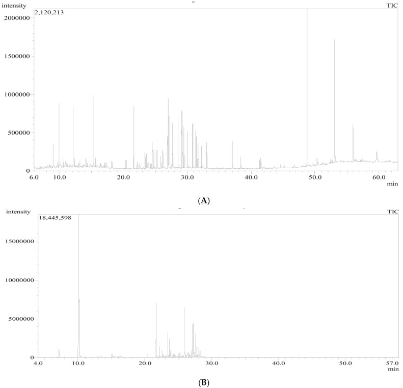
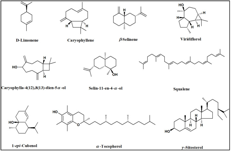
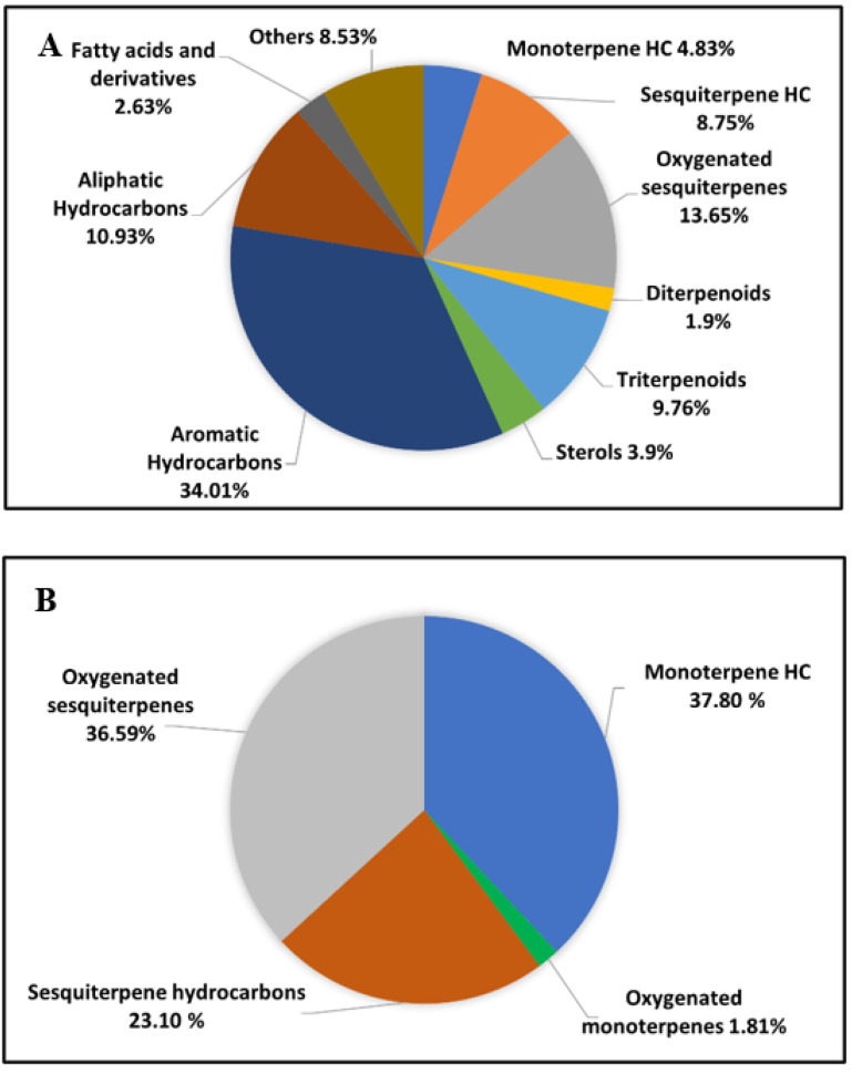
The results of the GC/MS analysis of the n-hexane extract and the essential oil of P. guajava are represented in Figure 1 and Table 1. The chemical characterization of the n-hexane extract and the essential oil revealed the identification of 40 compounds and 39 compounds accounting for (98.89%) and (99.30%), respectively. The n-hexane extract was found to be rich in hydrocarbons; aromatic (34.01%) and aliphatic (10.93%); followed by oxygenated sesquiterpenes (13.65%) and sesquiterpene hydrocarbons (8.75%). On the other hand, the essential oil showed a high percentage of monoterpene hydrocarbons (37.80%) and oxygenated sesquiterpenes (36.59%), followed by sesquiterpene hydrocarbons (23.10%). In the n-hexane extract, squalene and α-tocopherol were the major compounds accounting for (9.76%) and (8.53%), respectively, followed by D-limonene (4.83%), 1-epi-cubenol (4.51%), n-dodecane (4.15%), γ-sitosterol (3.90%), and β-caryophyllene (3.80%). Regarding the essential oil, it showed a high percentage of D-limonene (36.68%), followed by viridiflorol (9.68%), β-caryophyllene (8.41%), caryophylla-4(12),8(13)-dien-5α-ol (6.48%), selin-11-en-4-α-ol (6.35%), and β-selinene (4.10%). It is worth mentioning that D-limonene and β-caryophyllene were common major compounds in both the n-hexane extract and the essential oil of P. guajava. The chemical structures of the major constituents and the distribution of volatile components as a percentage within the n-hexane extract and the essential oil of P. guajava leaves are illustrated in Figure 2 and Figure 3, respectively.
Figure 1.
GC chromatogram of (A) n-hexane extract and (B) essential oil of P. guajava leaves.
Table 1.
Chemical composition (%) of the n-hexane extract (PGH) and essential oil (PGO) isolated from Psidium guajava leaves using GC/MS analysis.
| No. | Rt (min) |
Compound | RIExp. a | RILit b | Molecular Formula |
Content (%) | |
|---|---|---|---|---|---|---|---|
| PGH | PGO | ||||||
| 1 | 7.16 | α-Pinene | 931 | 931 | C10H16 | - | 0.99 |
| 2 | 8.99 | n-Decane | 999 | 1000 | C10H22 | 1.44 | - |
| 3 | 9.94 | p-Cymene | 1024 | 1024 | C10H14 | - | 0.13 |
| 4 | 10.09 | D-Limonene | 1029 | 1029 | C10H16 | 4.83 | 36.68 |
| 5 | 12.15 | n-Undecane | 1099 | 1100 | C11H24 | 3.61 | - |
| 6 | 14.15 | 2-Methylundecane | 1164 | 1165 | C12H26 | 0.70 | - |
| 7 | 14.98 | trans-p-Mentha-1(7),8-dien-2-ol | 1188 | 1185 | C10H16O | - | 0.50 |
| 8 | 15.07 | α-Terpineol | 1191 | 1189 | C10H18O | - | 0.64 |
| 9 | 15.25 | n-Dodecane | 1199 | 1200 | C12H26 | 4.15 | - |
| 10 | 15.67 | 3,6-Dimethylundecane | 1213 | 1210 | C13H28 | 0.58 | - |
| 11 | 15.95 | trans-Carveol | 1221 | 1220 | C10H16O | - | 0.15 |
| 12 | 16.20 | cis-p-Mentha-1(7),8-dien-2-ol | 1229 | 1235 | C10H16O | - | 0.52 |
| 13 | 18.21 | n-Tridecane | 1299 | 1300 | C13H28 | 0.45 | - |
| 14 | 20.43 | α-Copaene | 1378 | 1376 | C15H24 | 0.51 | 0.59 |
| 15 | 21.65 | β-Caryophyllene | 1424 | 1424 | C15H24 | 3.80 | 8.41 |
| 16 | 22.16 | Alloaromadendrene | 1443 | 1442 | C15H24 | 0.42 | 1.60 |
| 17 | 22.54 | Humulene (α-Caryophyllene) | 1458 | 1455 | C15H24 | - | 1.00 |
| 18 | 22.74 | epi-β-Caryophyllene | 1465 | 1466 | C15H24 | - | 0.37 |
| 19 | 23.13 | γ-Muurolene | 1480 | 1479 | C15H24 | - | 0.40 |
| 20 | 23.41 | β-Selinene | 1491 | 1486 | C15H24 | 1.22 | 4.10 |
| 21 | 23.64 | β-Guaiene | 1500 | 1500 | C15H24 | 1.05 | 2.94 |
| 22 | 23.75 | α-Bisabolene | 1504 | 1506 | C15H24 | - | 1.12 |
| 23 | 23.92 | β -Bisabolene | 1511 | 1512 | C15H24 | - | 1.33 |
| 24 | 24.11 | γ-Cadinene | 1519 | 1513 | C15H24 | - | 0.23 |
| 25 | 24.33 | cis-Calamenene | 1528 | 1529 | C15H22 | - | 0.67 |
| 26 | 24.56 | Cubenene | 1537 | 1533 | C15H24 | 1.75 | 0.34 |
| 27 | 24.79 | Ledol | 1547 | 1549 | C15H26O | 1.27 | - |
| 28 | 25.28 | Dodecanoic acid | 1566 | 1566 | C12H24O | 1.20 | 0.88 |
| 29 | 25.54 | Caryophyllene alcohol | 1577 | 1572 | C15H26O | - | 0.35 |
| 30 | 25.68 | Caryophyllene oxide | 1582 | 1583 | C15H24O | - | 0.20 |
| 31 | 25.89 | Viridiflorol | 1591 | 1592 | C15H26O | 0.95 | 9.68 |
| 32 | 26.09 | Globulol | 1598 | 1590 | C15H26O | - | 0.49 |
| 33 | 26.15 | Benzene, (1-methylnonyl)- | 1602 | 1616 | C16H26 | 1.26 | - |
| 34 | 26.42 | β-Atlantol | 1613 | 1608 | C15H24O | - | 1.29 |
| 35 | 26.51 | β-Himachalene oxide | 1616 | 1616 | C15H24O | - | 0.81 |
| 36 | 26.60 | Humulene epoxide II | 1620 | 1620 | C15H24O | - | 0.61 |
| 37 | 26.70 | Alloaromadendrene oxide-(1) | 1624 | 1625 | C15H24O | - | 0.26 |
| 38 | 26.82 | γ-Eudesmol | 1630 | 1632 | C15H26O | 2.08 | 0.39 |
| 39 | 26.94 | 1-epi-Cubenol | 1635 | 1630 | C15H26O | 4.51 | 1.38 |
| 40 | 27.08 | Caryophylla-4(12),8(13)-dien-5ꞵ-ol | 1640 | 1640 | C15H24O | - | 1.87 |
| 41 | 27.17 | Caryophylla-4(12),8(13)-dien-5α-ol | 1645 | 1641 | C15H24O | 3.64 | 6.48 |
| 42 | 27.37 | α-Cadinol | 1653 | 1654 | C15H26O | - | 0.97 |
| 43 | 27.61 | Selin-11-en-4-α-ol | 1663 | 1659 | C15H26O | - | 6.35 |
| 44 | 27.69 | Benzene, (1-ethylnonyl)- | 1668 | 1670 | C17H28 | 2.93 | - |
| 45 | 27.83 | epi-β-Bisabolol | 1672 | 1672 | C15H26O | - | 0.69 |
| 46 | 27.96 | Khusilol | 1678 | 1676 | C14H20O | - | 1.99 |
| 47 | 28.19 | α-Bisabolone oxide A | 1688 | 1686 | C14H22O2 | - | 0.55 |
| 48 | 28.29 | 11αH-Himachal-4-en-1β-ol | 1692 | 1699 | C15H26O | - | 1.35 |
| 49 | 28.55 | Benzene, (1-methyldecyl)- | 1704 | 1715 | C17H28 | 3.46 | - |
| 50 | 29.14 | Benzene, (1-pentylheptyl)- | 1728 | 1718 | C18H30 | 3.60 | - |
| 51 | 29.25 | Benzene, (1-butyloctyl)- | 1733 | 1725 | C18H30 | 3.71 | - |
| 52 | 29.52 | Benzene, (1-propylnonyl)- | 1744 | 1741 | C18H30 | 2.70 | - |
| 53 | 30.01 | Benzene, (1-ethyldecyl)- | 1764 | 1767 | C18H30 | 2.55 | - |
| 54 | 30.85 | Benzene, (1-methylundecyl)- | 1799 | 1797 | C18H30 | 2.94 | - |
| 55 | 31.31 | Benzene, (1-pentyloctyl)- | 1822 | 1819 | C19H32 | 3.19 | - |
| 56 | 31.46 | Benzene, (1-butylnonyl)- | 1830 | 1825 | C19H32 | 2.65 | - |
| 57 | 31.73 | Benzene, (1-propyldecyl)- | 1844 | 1838 | C19H32 | 1.76 | - |
| 58 | 32.23 | Benzene, (1-ethylundecyl)- | 1870 | 1866 | C19H32 | 1.50 | - |
| 59 | 33.05 | Benzene, (1-methyldodecyl)- | 1912 | 1911 | C19H32 | 1.76 | - |
| 60 | 37.04 | Phytol | 2115 | 2114 | C20H40O | 1.90 | - |
| 61 | 38.37 | Palmitic acid, butyl ester | 2187 | 2188 | C20H40O2 | 0.75 | - |
| 62 | 41.38 | Eicosanoic acid, methyl ester | 2358 | 2339 | C21H42O2 | 0.64 | - |
| 63 | 41.52 | Linolenic acid, ethyl ester | 2366 | - | C20H34O2 | 0.83 | - |
| 64 | 48.77 | Squalene | 2834 | 2835 | C30H50 | 9.76 | - |
| 65 | 50.27 | Hexacosanoic acid, methyl ester | 2942 | 2940 | C27H54O2 | 0.41 | - |
| 66 | 53.07 | α-Tocopherol | 3152 | 3149 | C29H50O2 | 8.53 | - |
| 67 | 55.98 | γ-Sitosterol | 3352 | 3351 | C29H50O | 3.90 | - |
| Monoterpene hydrocarbons | 4.83 | 37.80 | |||||
| Oxygenated monoterpenes | - | 1.81 | |||||
| Sesquiterpene hydrocarbons | 8.75 | 23.10 | |||||
| Oxygenated sesquiterpenes | 13.65 | 36.59 | |||||
| Diterpenoids | 1.90 | - | |||||
| Triterpenoids | 9.76 | - | |||||
| Sterols | 3.90 | - | |||||
| Aromatic Hydrocarbons | 34.01 | - | |||||
| Aliphatic Hydrocarbons | 10.93 | - | |||||
| Fatty acids and fatty acids derivatives | 2.63 | - | |||||
| Others | 8.53 | - | |||||
| Total identified compounds | 98.89 | 99.30 | |||||
Compounds listed in order of their elution on DB-5 GC column. Identification was based on comparison of the compounds mass spectral data (MS) and retention indices (RI) with those of NIST Mass Spectral Library (2011), Wiley Registry of Mass Spectral Data 8th edition and the literature [28,30]. a Retention index calculated experimentally on Rtx-1MS column relative to n-alkane series (C8–C28). b Published retention indices.
Figure 2.
Chemical structures of the major constituents identified in the n-hexane extract and essential oil of Psidium guajava leaves using GC/MS analysis.
Figure 3.
Pie charts demonstrate the distribution of volatile components as a percentage within (A) n-Hexane extract and (B) essential oil of P. guajava leaves.
Many reports have been conducted on the essential oil compositions of P. guajava from varied geographical sources [30,33,40]. The major identified compounds in the essential oil isolated from the leaves collected from Brazil were β-caryophyllene, α-humulene, aromadendrene oxide, δ-selinene, and selin-11-en-4α-ol [24]. In contrast, the essential oil from the leaves collected from another source in India showed that β-caryophyllene, L-calamenene, (-)-globulol, and α-copaene were the major constituents [30]. Moreover, the chemical composition of the n-hexane extract of P. guajava, collected from Pakistan, showed a high content of vitamin E, squalene, caryophyllene, and γ-sitosterol [25]. Another study by Arian et al. reported that the essential oil of P. guajava leaves collected from Pakistan was a rich source of β-caryophyllene, globulol, and trans-nerolidol [33]. Regarding, the previous studies into the composition of the essential oil of P. guajava leaves indicated wide variations relative to the different locations of collection.
2.2. Total Phenolic and Flavonoid Content of the n-Hexane Extract of P. guajava Leaves
Phenolics compounds are present in most natural products that induce many biological activities [41,42,43,44]. The total phenolic and flavonoid content in the n-hexane extract of P. guajava leaves, was quantitatively determined, according to Zengin and Aktumsek, 2014 [45]. The Phenolic and flavonoid contents were measured as gallic acid, and rutin equivalents, respectively. The presence of 32.62 ± 0.19 mg GAE/g (gallic acid equivalent) per mg of P. guajava n-hexane extract was recorded for the total phenolics content. While the presence of 2.05 ± 0.14 mg RE/g (rutin equivalent) was recorded for the total flavonoids content. The results established the presence of considerable amounts of phenolics in the n-hexane extract.
2.3. Antioxidant Potential of the n-Hexane Extract and Essential Oil Isolated from P. guajava Leaves
Natural antioxidants, especially polyphenols, are becoming increasingly popular due to their beneficial effects on human health. Consequently, plant polyphenols may be able to mitigate the negative effects of oxidative stress, which has been linked to a variety of pathological processes, such as cancer, kidney disease, cardiovascular disease, neurodegeneration, age-related diseases, and diabetes [46,47,48,49,50,51]. Many reports revealed the potential of different essential oils as antioxidant agents [52], including but not limited to, the essential oil of Cinnamomum zeylanicum, which showed over 78.0% anticholinesterase and radical-scavenging activities [53]. Additionally, the essential oil of Rosmarinus officinalis showed antioxidant activity using the DPPH and FRAP assays [54].
So, there is an increasing demand for the development of natural antioxidants. Several assays were conducted in the current study to examine the in vitro antioxidant potentials of the P. guajava leaves n-hexane extract and the essential oil.
The antioxidant potential of the n-hexane extract and the essential oil was performed using different techniques as 2,2-diphenyl-1-picryl-hydrazyl-hydrate (DPPH), 2,2-azino bis (3-ethylbenzothiazoline-6-sulphonic acid) (ABTS), cupric reducing antioxidant capacity (CUPRAC), ferric reducing power (FRAP), metal chelating ability (MCA), and phosphomolybdenum (PM) assays. The findings represented in Table 2 show that the n-hexane extract and the essential oil have antioxidant properties in the different assays. Concerning the CUPRAC, FRAP, and MCA assays, the n-hexane extract revealed a higher antioxidant potential, equivalent to 70.80 ± 1.46 mg TE/g, 26.01 ± 0.97 mg TE/g, and 24.83 ± 0.35 mg EDTAE/g, respectively. By contrast, the essential oil showed more antioxidant potential in the PM assay equivalent to 2.58 ± 0.14 mmol TE/g, while n-hexane extract showed 2.0 ± 0.07 mmol TE/g. Regarding the DPPH and ABTS assays, none of the n-hexane extract or the essential oil showed any antioxidant activity. Accordingly, it can be concluded that the n-hexane extract and the essential oil from P. guajava leaves have promising antioxidant properties. The higher antioxidant potential of the n-hexane extract could be attributed to the presence of squalene as a major compound (9.76%), which is a well-known triterpenoid hydrocarbon with an antioxidant potential through oxygen scavenging [55]. Furthermore, α-Tocopherol, which is the significant antioxidant isomer of vitamin E [56] through the scavenging of free radicals, cell membrane maintenance, and structural restoration [57], is present as a major constituent in the n-hexane extract (8.53%). Moreover, the presence of phytosterol as γ-sitosterol plays an important role in the antioxidant activity of P. guajava [25,58]. The results were in accordance with the previous studies, where the monoterpene hydrocarbon D-limonene was reported to be a major compound in celery seed oil, which showed a high antioxidant activity using the DPPH assay [59]. Additionally, the antioxidant potential of the essential oil from Wedelia prostrata was attributed to the presence of a high percentage of D-limonene [60]. Regarding squalene, Kraujalis et al. (2013) reported its promising antioxidant activity in the lipophilic fraction of Amaranthus spp. prepared using a supercritical carbon dioxide extraction technique [61]. Moreover, it was reported that D-limonene has the ability to prevent lipidemic-oxidative stress [62,63]. Furthermore, β-caryophyllene was previously reported as a free-radical-scavenging agent [64,65].
Table 2.
Antioxidant potential of the n-hexane extract and essential oil isolated from P. guajava leaves.
| Samples | DPPH | ABTS | CUPRAC | FRAP | MCA | PM |
|---|---|---|---|---|---|---|
| (mg TE/g) | (mg TE/g) | (mg TE/g) | (mg TE/g) | (mg EDTAE/g) | (mmol TE/g) | |
| n-Hexane extract | n.a. | n.a. | 70.80 ± 1.46 | 26.01 ± 0.97 | 24.83 ± 0.35 | 2.0 ± 0.07 |
| Essential oil | n.a. | n.a. | 18.17 ± 0.08 | 12.08 ± 0.17 | 9.02 ± 1.2 | 2.58 ± 0.14 |
Values expressed as means ± S.D. of three parallel measurements. Trolox equivalent (TE); Ethylenediaminetetraacetic acid equivalent (EDTAE); not active (n.a.).
The previous reports found that the antioxidant properties of the n-hexane extract and the essential oil of P. guajava was carried out using different assays, such as DPPH, ABTS, and FRAP assays and our results represented a comprehensive antioxidant profiling of the n-hexane extract and the essential oil of P. guajava available to date, using a standard equivalent way. Ashraf et al. (2016) reported the antioxidant properties of the hexane extract of P. guajava using the DPPH assay and found a low scavenging of free radicals (IC50 value = 426.8 ± 0.19 µg/mL) [25]. In another study, the antioxidant properties of the essential oil of P. guajava were investigated using the DPPH, ABTS, and β-carotene bleaching assays, which showed IC50 values of 17.66 ± 0.07, 19.28 ± 0.03, and 3.17 ± 0.01 μg/mL, respectively [30]. The variability in the results could be attributed to the differences in the harvest times, the maturity stage, and variations in the extraction procedure and the extracting solvent. So, these reports confirmed the antioxidant activity of n-hexane extract and the essential oil of P. guajava.
2.4. Enzyme Inhibitory Activity of the n-Hexane Extract and the Essential Oil Isolated from P. guajava Leaves
The enzyme inhibitory activities of the n-hexane extract and the essential oil were evaluated against different important enzymes, including acetylcholinesterase (AChE), butyrylcholinesterase (BChE), tyrosinase, α-amylase, and α-glucosidase. The results are represented in Table 3. It revealed that the essential oil showed a potent BChE inhibitory ability 6.85 ± 0.03 mg GALAE/g by contrast the n-hexane extract did not display any AChE or BChE inhibitory abilities. The strongest tyrosinase inhibition ability was determined to be the essential oil (61.70 ± 3.21 mg KAE/g) whereas the n-hexane extract was 33.91 ± 2.25 mg KAE/g. Regarding the anti-diabetic enzyme inhibition, the n-hexane extract and essential oil both displayed α-amylase inhibition equivalent to 0.52 ± 0.01 and 0.13 ± 0.01 mmol ACAE/g, respectively. In contrast to the α-amylase inhibition, the essential oil displayed a higher α-glucosidase inhibition equivalent to 1.49 ± 0.01 mmol ACAE/g and the n-hexane extract displayed less α-glucosidase inhibition (0.67 ± 0.03 mmol ACAE/g).
Table 3.
Enzyme inhibitory effects of the n-hexane extract and the essential oil isolated from P. guajava leaves.
| Samples | AChE Inhibition | BChE Inhibition | Tyrosinase Inhibition | α-Amylase Inhibition | α-Glucosidase Inhibition |
|---|---|---|---|---|---|
| (mg GALAE/g) | (mg GALAE/g) | (mg KAE/g) | (mmol ACAE/g) | (mmol ACAE/g) | |
| n-Hexane extract | n.a. | n.a. | 33.91 ± 2.25 | 0.52 ± 0.01 | 0.67 ± 0.03 |
| Essential oil | n.a. | 6.85 ± 0.03 | 61.70 ± 3.21 | 0.13 ± 0.01 | 1.49 ± 0.01 |
Values expressed as means ± S.D. of three parallel measurements. Galanthamine equivalent (GALAE); Kojic acid equivalent (KAE); Acarbose equivalent (ACAE); not active (n.a.).
The significant BChE inhibition by the essential oil could be attributed to the presence of monoterpenes as the major components relative to the previous studies that correlate the presence of several monoterpenes with the anticholinesterase properties [65,66,67,68]. To the best of our knowledge no previous comprehensive studies were available concerning the comparative study on the enzyme inhibition of the Guava essential oil and n-hexane extract. Zhang et al. (2022) reported the significant α-amylase and α-glucosidase inhibitory activities of the essential oil of Guava collected from China with IC50 values of 13.99 ± 0.34 and 5.50 ±1.02 μg/mL, respectively [30]. Bouchoukh et al. (2019) reported the anticholinesterase properties of different extracts of Guava; the chloroform, ethyl-acetate and the n-butanol extracts showed AChE inhibitory activities with IC50 values of 177.11 ± 2.30, 56.11 ± 4.04, and 24.44 ±3.45 μg/mL, respectively; their BChE inhibitory activities were found to have IC50 values of >200, 44.95 ± 2.67 and 21.87 ±10.48 μg/mL, respectively [40].
Bonesi et al. (2010) reported that trans-caryophyllene identified in oil, showed significant BChE inhibitory activity with an IC50 value of 78.6 ± 1.3 μg/mL [6]. Furthermore, it acts as an antagonist to homomeric nicotinic acetylcholine receptors (α7-nAChRs) [69]. Additionally, the AChE inhibitory activity of Artemisia annua oil was attributed to the presence of limonene, β-caryophyllene, and β-caryophyllene oxide as the major components [70]. Zarrad et al. (2015) reported that the AChE inhibitory activity of limonene correlated with its bicyclic monoterpene hydrocarbon containing an allylic methyl group, which has an important role in its insecticidal activity [67]. A previous study by Chear et al. (2016), reported that the combination of sterols and tocopherol played an important role in the cholinesterase inhibitory activity in either AChE or BChE [71]. Recently, it was shown that α-tocopherol has an inhibiting effect on α-glucosidase being beneficial to reduce the risk factors associated with diabetes [72]. In addition, the presence of α-tocopherol in Cosmos caudatus extract revealed the potential α-glucosidase inhibitory activity [73]. You et al. (2011) reported the tyrosinase inhibitory activities of the different parts and different extracts of Guava. The leaves, acetone, ethanol, methanol, and water extracts showed tyrosinase inhibition by 49.67 ± 0.58, 69.56 ± 1.38, 47.33 ± 1.84, and 44.78 ± 1.75%, respectively [74]. Development of tyrosinase inhibitors from natural sources is in great demand due to their low side effects and higher efficacy making natural tyrosinase inhibitors a good candidate for the incorporation in hypopigmenting agents. It is worth mentioning that tyrosinase plays an important role in melanin synthesis [75,76,77].
2.5. Molecular Docking
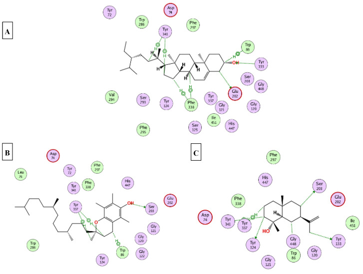
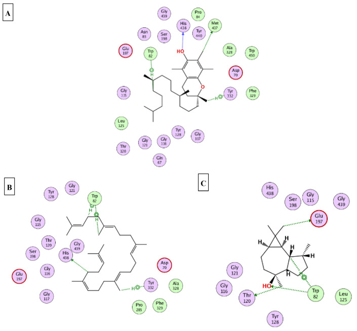
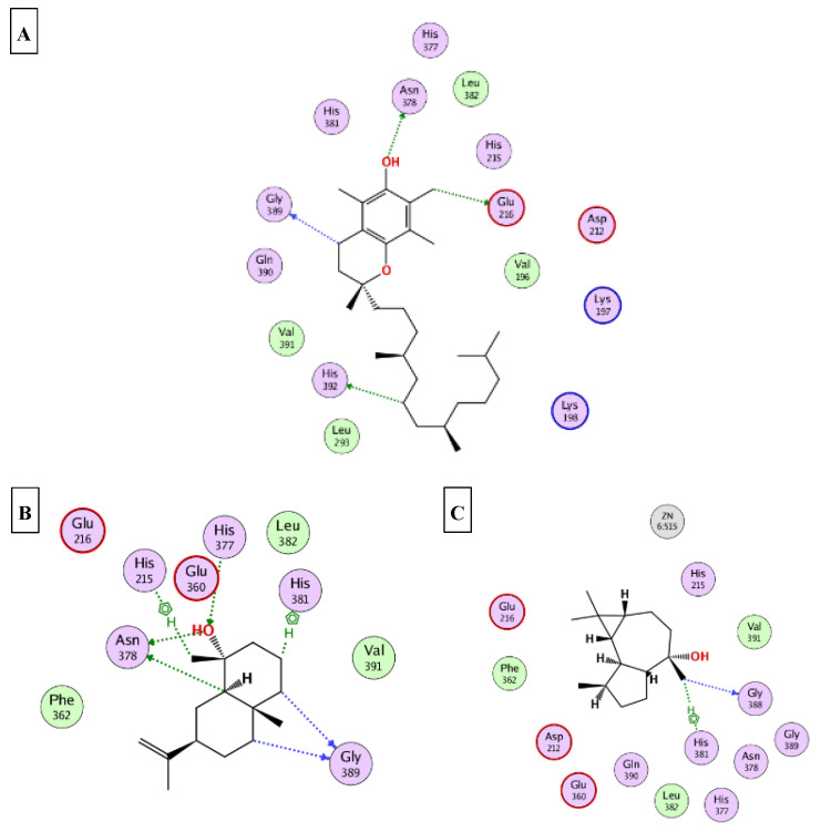
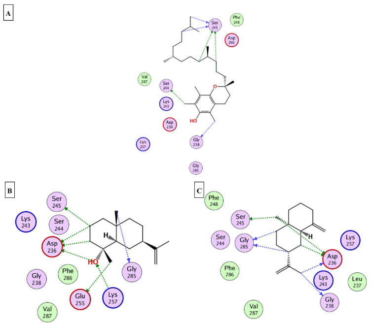
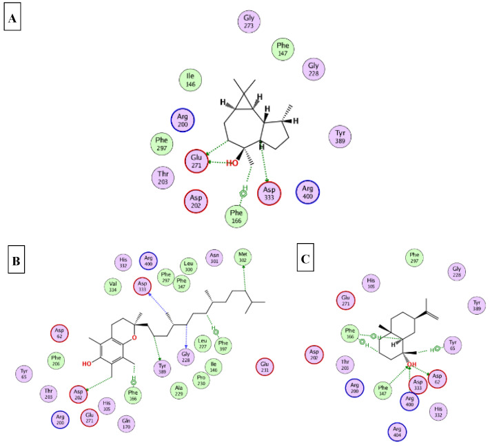
This part was conducted to investigate the possible mechanism of action in which the ten major compounds (D-limonene, β-caryophyllene, β-selinene, viridiflorol, 1-epi-cubenol, caryophylla-4(12),8(13)-dien-5α-ol, selin-11-en-4-α-ol, squalene, α-tocopherol, and γ-sitosterol) exert their biological effects. Accordingly, the 3D structures of AChE, BchE, tyrosinase, α-amylase, and α-glucosidase were downloaded from the protein data bank using the following IDs: 7D9O, 6ESJ, 5M8Q, 4GQQ, and 3WY2, respectively. After that, the ten major compounds were docked into the active site vicinity of the five enzymes. Interestingly, all the compounds achieved acceptable binding scores upon docking with the five targets (Table 4). In the docking of AChE, γ-sitosterol, α-tocopherol, and selin-11-en-4-α-ol the best scores for docking were achieved −15.4, −14.2, and −13.4 Kcal/Mol, respectively. As Figure 4 reveals, γ-sitosterol interacted with AChE through mixed hydrophobic and hydrogen bond interactions with Tyr341, Phe338, Trp86, Tyr133, and Glu203. Moreover, α-tocopherol interacted with Trp86, Ser203 and 337; selin-11-en-4-α-ol interacted with Tyr124, Tyr133, Ser203, Tyr337, and Tyr341. In the docking of BChE, α-tocopherol, squalene, and viridiflorol achieved the best docking scores −13.9, −11.1, and −10.7 Kcal/Mol, respectively. As depicted Figure 5 α-tocopherol bound to BChE through interactions with Trp82, Tyr332, Met437, and His438; squalene interacted with Trp82, Tyr332, and His438; viridiflorol interacted with Trp82, Thr120, and Glu197. In the docking of tyrosinase, α-tocopherol, selin-11-en-4-α-ol, and viridiflorol achieved the best docking scores −9.5, −9.5, and −9.3 Kcal/Mol, respectively. Figure 6 reveals the interaction of the best three compounds with tyrosinase in which, α-tocopherol interacted with Glu216, Asn378, Gly389, and His392, selin-11-en-4-α-ol interacted with His215, His377, Asn378, His381, and Gly389 and viridiflorol interacted with His318 and Gly388. Regarding the docking of α-amylase, α-tocopherol, selin-11-en-4-α-ol, and β-selinene achieved the best docking scores −8.9, −7.8, and −7.7 Kcal/Mol, respectively. Inspecting Figure 7, α-tocopherol was able to interact with the residues of α-amylase through binding with Gly238, Ser244, and Ser245; selin-11-en-4-α-ol interacted with Asp236, Ser245, Glu255, Lys257, and Gly285; β-selinene interacted with Asp236, Ser245, Gly238, and Gly285. In the docking of α-glucosidase, viridiflorol, α-tocopherol, and selin-11-en-4-α-ol achieved the best docking scores −13.6, −12.5, and −12.5 Kcal/Mol, respectively. Figure 8 shows the interaction of the best three compounds with α-glucosidase in which, viridiflorol interacted with Phe166, Glu271, and Asp333; α-tocopherol interacted with Phe166, Asp202, Gly228, Met302, Tyr389, Phe397, and Asp333; selin-11-en-4-α-ol interacted with Phe166, Asp62, Tyr65, Phe147, and Arg400. In conclusion, the docking results supported and justified the biological results giving rise to a synergetic effect for all the components of the n-hexane extract and the essential oil.
Table 4.
The docking scores achieved by the major identified compounds against different enzymes.
| Compound | Docking Scores Kcal/mol | ||||
|---|---|---|---|---|---|
| AChE 7D9O |
BChE 6ESJ |
Tyrosinase 5M8Q |
α-Amylase 4GQQ |
α-Glucosidase 3WY2 |
|
| D-Limonene | −9.2 | −7.2 | −7.3 | −6.1 | −7.9 |
| β-Caryophyllene | −9.3 | −8.7 | −7.2 | −6.3 | −7.9 |
| β-Selinene | −9.5 | −7.7 | −6.8 | −7.7 | −7.6 |
| Viridiflorol | −11.4 | −10.7 | −9.3 | −6.3 | −13.6 |
| 1-epi-Cubenol | −10.2 | −8.8 | −6.9 | −6.4 | −8.3 |
| Caryophylla-4(12),8(13)-dien-5α-ol | −11.4 | −9.1 | −7.6 | −6.8 | −8.9 |
| Selin-11-en-4-α-ol | −13.4 | −9.3 | −9.5 | −7.8 | −12.5 |
| Squalene | −12.3 | −11.1 | −7.9 | −6.4 | −9.1 |
| α-Tocopherol | −14.2 | −13.9 | −9.5 | −8.9 | −12.5 |
| γ-Sitosterol | −15.4 | −9.4 | −8.2 | −7.1 | −9.3 |
Figure 4.
2D binding modes of γ-sitosterol (A); α-tocopherol (B); selin-11-en-4-α-ol (C) to the active binding sites of AChE.
Figure 5.
2D binding modes of α-tocopherol (A); squalene (B); viridiflorol (C) to the active binding sites of BChE.
Figure 6.
2D binding modes of α-tocopherol (A); selin-11-en-4-α-ol (B); viridiflorol (C) to the active binding sites of tyrosinase.
Figure 7.
2D binding modes of α-tocopherol (A); selin-11-en-4-α-ol (B); β-selinene (C) to the active binding sites of α-amylase.
Figure 8.
2D binding modes of viridiflorol (A); α-tocopherol (B); selin-11-en-4-α-ol (C) to the active binding sites of α-glucosidase.
3. Materials and Methods
3.1. Plant Material
Fresh leaves of P. guajava Linn. were collected from the Medicinal Plant Research Station, Pharmacognosy Department, Faculty of Pharmacy, Ain Shams University, Cairo, Egypt, in October 2021. The plant was authenticated by Professor Usama K. Abdel Hameed, Department of Botany, Faculty of Science, Ain Shams University, Cairo, Egypt. A voucher specimen, PHG-P-PG-409, was deposited at the Pharmacognosy Department, Faculty of Pharmacy, Ain Shams University, Cairo, Egypt.
3.2. Isolation of the Essential Oil
The fresh leaves were finely cut and hydrodistilled for 5 h using a Clevenger apparatus. The oil obtained is colorless with a pleasant aroma; the average yield was 0.2% (v/w). It was isolated and kept in a sealed dark glass vial at −4 °C until the GC/MS analysis was performed.
3.3. Preparation of the n-Hexane Extract
The dried leaves of Psidium guajava Linn. (100 g) were extracted with n-hexane three times separately. The filtrate was completely evaporated in vacuo at 40 °C until dryness to obtain the dried residue of the n-hexane extract (3.2 g). The extract was stored in a tight container and stored in a refrigerator for further analysis.
3.4. Gas Chromatography/Mass Spectrometry (GC/MS) Analysis
Gas chromatography/Mass spectrometry (GC/MS) analysis was carried out on a Shimadzu GCMS-QP 2010 chromatograph (Kyoto, Japan) with Rtx-1MS capillary column (30 m × 0.25 mm i.d. × 0.25 μm film thickness; Restek, Bellefonte, PA, USA). The oven temperature was kept at 45 °C for 2 min (isothermal), programmed to 30 °C at a rate of 5 °C/min, and kept constant at 300 °C for 5 min (isothermal); injector temperature was 250 °C. The carrier gas used was helium, with a flow rate set at 1.40 mL/min. The diluted samples (1% v/v) were injected with a split ratio of 15:1 and the injected volume was 1 μL. The MS operating parameters were as follows: interface temperature 280 °C, ion-source temperature 220 °C, EI mode 70 eV, scan range 35–500 amu. Identification of the volatile constituents was made based on their retention indices, matching their fragmentation patterns with the NIST Mass Spectral Library, the Wiley library database, and the published data in the literature [78,79,80,81]. Retention indices (RI) were calculated relative to the homologous series of n-alkanes (C8–C30) and injected under the same conditions.
3.5. Compounds Identification
The identification was accomplished by comparing the Kovats retention index and the mass spectrometric data (molecular ion peaks and fragmentation patterns), to those recorded in the NIST Mass Spectral Library and other published data for the reference compounds under similar conditions [56,78,79,80,81,82,83].
3.6. Total Phenolic and Flavonoid Content
The total phenolic and flavonoid contents were determined using the Folin–Ciocalteu and AlCl3 tests, respectively [45]. Results were presented as gallic acid equivalents (mg GAEs/g dry extract) and rutin equivalents (mg REs/g dry extract) for the assays. All experimental details are given in Supplementary Materials.
3.7. Antioxidant and Enzyme Inhibitory Assays
The antioxidant assays were performed using methods that have been previously reported [84,85]. Trolox and EDTA were used as positive controls in the antioxidant assays. The antioxidant potential was calculated as follows: mg Trolox equivalents (TE)/g extract in the 2,2-diphenyl-1-picrylhydrazyl (DPPH) and 2,2’-azino-bis(3-ethylbenzothiazoline-6-sulfonic acid) (ABTS) radical scavenging tests; cupric reducing antioxidant capacity (CUPRAC) and ferric reducing antioxidant power (FRAP), mmol TE/g extract in phosphomolybdenum assay, and mg ethylenediaminetetraacetic acid equivalents (EDTAE)/g extract in metal chelating assay (MCA). All experimental details for the antioxidant assays are given in Supplementary Materials.
The enzyme inhibition experiments were performed based on previously described procedures [84,85]. Standard inhibitors were used as positive controls (galanthamine for cholinesterases; kojic acid for tyrosinase; acarbose for amylase and glucosidase) in the enzyme inhibitory assays. Amylase and glucosidase inhibition were expressed as mmol acarbose equivalents (ACAE)/g extract, while acetylcholinesterase (AChE) and butyrylcholinesterase (BChE) inhibition was expressed as mg galanthamine equivalents (GALAE)/g extract. Tyrosinase inhibition was expressed as mg kojic acid equivalents (KAE)/g extract. All experimental details for the enzyme inhibitory assays are given in Supplementary Materials.
3.8. Molecular Docking
The X-ray 3D structures of AChE, BChE, tyrosinase, α-amylase, and α-glucosidase were downloaded from the protein data bank “www.pdb.org (accessed on 3 August 2022)” using the following IDs: 7D9O, 6ESJ, 5M8Q, 4GQQ, and 3WY2 [86,87,88,89,90], respectively. All the docking studies were conducted using MOE 2019 [91], which was also used to generate the 2D interaction diagrams between the docked ligands and their potential targets. The ten identified major compounds were prepared using the default parameters and saved in a single MDB file. The active site of each target was determined from the binding of the corresponding co-crystalized ligand. Finally, the docking was finalized through docking the MDB file containing the ten major compounds into the active site of the five enzymes.
4. Conclusions
Chemical investigation of Pisidium guava have proven that this plant species contains a variety of volatile components present in the n-hexane extract and the essential oil, as well as their antioxidant and enzyme inhibitory activities supported with an in-silico study. The GC/MS analysis revealed that the n-hexane extract is a rich source of squalene (9.76%), α-tocopherol (8.53%), and γ-sitosterol (3.90%) that correlates with its antioxidant potential in the CUPRAC, FRAP, and MCA assays. On the other hand, the essential oil was enriched with monoterpenes and sesquiterpenes, especially D-limonene (36.68%) and viridiflorol (9.68%), which correlates with its antioxidant potential in different assays along with its potency as a BChE and a tyrosinase inhibitor. Furthermore, the n-hexane extract and the essential oil showed relevant α-amylase and α-glucosidase inhibitory activities. Furthermore, the major compounds achieved promising docking scores in the active sites of the tested target enzymes. According to these findings and the previous studies, the n-hexane extract and essential oil of P. guajava can be considered a promising candidate for the development of novel therapeutic agents for the management of Alzheimer’s, diabetes mellitus, and oxidative stress disorders. Moreover, their efficacy as tyrosinase inhibitors allows them to be incorporated in the development of hypopigmenting agents. However, further investigations should be conducted concerning the pharmacodynamics as well as the pharmacokinetics pathways accompanied with the in vivo studies.
Supplementary Materials
The following supporting information can be downloaded at: https://www.mdpi.com/article/10.3390/molecules27248979/s1.
Author Contributions
Conceptualization, O.A.E. and G.Z.; methodology, G.Z., S.D., S.H.A., M.A.E.H. and W.M.E.; software, G.Z., S.D., M.A.E.H., W.M.E., F.A.B. and S.T.A.-R.; validation, O.A.E., S.T.A.-R., F.A.B. and G.Z.; investigation, G.Z., S.D., O.A.E., W.M.E., S.H.A. and M.A.E.H.; data curation, O.A.E., G.Z., W.M.E. and S.H.A.; writing—original draft preparation, S.H.A.; writing—review and editing, G.Z., O.A.E. and W.M.E.; funding acquisition, S.T.A.-R., F.A.B. All authors have read and agreed to the published version of the manuscript.
Data Availability Statement
Data are available upon request.
Conflicts of Interest
The authors declare there is no conflict of interest.
Funding Statement
The authors also acknowledge financial support from the Researchers Supporting Project number (RSP-2021/103), King Saud University, Riyadh, Saudi Arabia.
Footnotes
Publisher’s Note: MDPI stays neutral with regard to jurisdictional claims in published maps and institutional affiliations.
References
- 1.Elgindi M.R., El-Nassar Singab A.B., Aly S.H., Mahmoud I.I., Mohamed Elgindi C.R. Phytochemical Investigation and Antioxidant Activity of Hyophorbe verschaffeltii (Arecaceae) J. Pharmacogn. Phytochem. 2016;5:39–46. [Google Scholar]
- 2.Aly S.H., Elissawy A.M., Eldahshan O.A., Elshanawany M.A., Efferth T., Singab A.N.B. The Pharmacology of the Genus Sophora (Fabaceae): An Updated Review. Phytomedicine. 2019;64:153070. doi: 10.1016/j.phymed.2019.153070. [DOI] [PubMed] [Google Scholar]
- 3.Ads E.N., Hassan S.I., Rajendrasozhan S., Hetta M.H., Aly S.H., Ali M.A. Isolation, Structure Elucidation and Antimicrobial Evaluation of Natural Pentacyclic Triterpenoids and Phytochemical Investigation of Different Fractions of Ziziphus spina-christi (L.) Stem Bark Using LCHRMS Analysis. Molecules. 2022;27:1805. doi: 10.3390/molecules27061805. [DOI] [PMC free article] [PubMed] [Google Scholar]
- 4.Chaves-López C., Usai D., Donadu M.G., Serio A., González-Mina R.T., Simeoni M.C., Molicotti P., Zanetti S., Pinna A., Paparella A. Potential of: Borojoa patinoi Cuatrecasas Water Extract to Inhibit Nosocomial Antibiotic Resistant Bacteria and Cancer Cell Proliferation in Vitro. Food Funct. 2018;9:2725–2734. doi: 10.1039/C7FO01542A. [DOI] [PubMed] [Google Scholar]
- 5.Saber F.R., Aly S.H., Khallaf M.A., El-Nashar H.A.S., Fahmy N.M., El-Shazly M., Radha R., Prakash S., Kumar M., Taha D., et al. Hyphaene thebaica (Areceaeae) as a Promising Functional Food: Extraction, Analytical Techniques, Bioactivity, Food, and Industrial Applications. Food Anal. Methods. 2022 doi: 10.1007/s12161-022-02412-1. [DOI] [Google Scholar]
- 6.Bonesi M., Menichini F., Tundis R., Loizzo M.R., Conforti F., Passalacqua N.G., Statti G.A., Menichini F. Acetylcholinesterase and Butyrylcholinesterase Inhibitory Activity of Pinus Species Essential Oils and Their Constituents. J. Enzyme Inhib. Med. Chem. 2010;25:622–628. doi: 10.3109/14756360903389856. [DOI] [PubMed] [Google Scholar]
- 7.Elshafie H.S., Caputo L., De Martino L., Sakr S.H., De Feo V., Camele I. Study of Bio-Pharmaceutical and Antimicrobial Properties of Pomegranate (Punica granatum L.) Leathery Exocarp Extract. Plants. 2021;10:153. doi: 10.3390/plants10010153. [DOI] [PMC free article] [PubMed] [Google Scholar]
- 8.Xiao J., Tundis R. Natural Products for Alzheimer’s Disease Therapy: Basic and Application. J. Pharm. Pharmacol. 2013;65:1679–1680. doi: 10.1111/jphp.12186. [DOI] [PubMed] [Google Scholar]
- 9.Etxeberria U., De La Garza A.L., Campin J., Martnez J.A., Milagro F.I. Antidiabetic Effects of Natural Plant Extracts via Inhibition of Carbohydrate Hydrolysis Enzymes with Emphasis on Pancreatic Alpha Amylase. Expert Opin. Ther. Targets. 2012;16:269–297. doi: 10.1517/14728222.2012.664134. [DOI] [PubMed] [Google Scholar]
- 10.Kazeem M.I., Adamson J.O., Ogunwande I.A. Modes of Inhibition of α-Amylase and α-Glucosidase by Aqueous Extract of Morinda lucida Benth Leaf. Biomed Res. Int. 2013;2013:527570. doi: 10.1155/2013/527570. [DOI] [PMC free article] [PubMed] [Google Scholar]
- 11.Fang L., Kraus B., Lehmann J., Heilmann J., Zhang Y., Decker M. Design and Synthesis of Tacrine-Ferulic Acid Hybrids as Multi-Potent Anti-Alzheimer Drug Candidates. Bioorg. Med. Chem. Lett. 2008;18:2905–2909. doi: 10.1016/j.bmcl.2008.03.073. [DOI] [PubMed] [Google Scholar]
- 12.Mohammed S.A., Yaqub A.G., Sanda K.A., Nicholas A.O., Arastus W., Muhammad M., Abdullahi S. Review on Diabetes, Synthetic Drugs and Glycemic Effects of Medicinal Plants. Sect. Title Pharmacol. 2013;7:2628–2637. doi: 10.5897/JMPR2013.5169. [DOI] [Google Scholar]
- 13.de Paulo Farias D., Neri-Numa I.A., de Araújo F.F., Pastore G.M. A Critical Review of Some Fruit Trees from the Myrtaceae Family as Promising Sources for Food Applications with Functional Claims. Food Chem. 2020;306:125630. doi: 10.1016/j.foodchem.2019.125630. [DOI] [PubMed] [Google Scholar]
- 14.Apel M.A., Sobral M., Schapoval E.E.S., Henriques A.T., Menut C., Bessiere J.-M. Volatile Constituents of Eugenia Mattosii Legr (Myrtaceae) J. Essent. Oil Res. 2005;17:284–285. doi: 10.1080/10412905.2005.9698904. [DOI] [Google Scholar]
- 15.Borotová P., Galovičová L., Vukovic N.L., Vukic M., Tvrdá E., Kačániová M. Chemical and Biological Characterization of Melaleuca Alternifolia Essential Oil. Plants. 2022;11:558. doi: 10.3390/plants11040558. [DOI] [PMC free article] [PubMed] [Google Scholar]
- 16.El-Nashar H.A.S., Eldehna W.M., Al-Rashood S.T., Alharbi A., Eskandrani R.O., Aly S.H. GC/MS Analysis of Essential Oil and Enzyme Inhibitory Activities of Syzygium cumini (Pamposia) Grown in Docking Studies. Molecules. 2021;26:6984. doi: 10.3390/molecules26226984. [DOI] [PMC free article] [PubMed] [Google Scholar]
- 17.Sobeh M., El-Raey M., Rezq S., Abdelfattah M.A.O., Petruk G., Osman S., El-Shazly A.M., El-Beshbishy H.A., Mahmoud M.F., Wink M. Chemical Profiling of Secondary Metabolites of Eugenia Uniflora and Their Antioxidant, Anti-Inflammatory, Pain Killing and Anti-Diabetic Activities: A Comprehensive Approach. J. Ethnopharmacol. 2019;240:111939. doi: 10.1016/j.jep.2019.111939. [DOI] [PubMed] [Google Scholar]
- 18.de Moraes Â.A.B., de Jesus Pereira Franco C., Ferreira O.O., Varela E.L.P., do Nascimento L.D., Cascaes M.M., da Silva D.R.P., Percário S., de Oliveira M.S., de Aguiar Andrade E.H. Myrcia Paivae O.Berg (Myrtaceae) Essential Oil, First Study of the ChemicalComposition and Antioxidant Potential. Molecules. 2022;27:5460. doi: 10.3390/molecules27175460. [DOI] [PMC free article] [PubMed] [Google Scholar]
- 19.Ferreira Macedo J.G., Linhares Rangel J.M., de Oliveira Santos M., Camilo C.J., Martins da Costa J.G., Maria de Almeida Souza M. Therapeutic Indications, Chemical Composition and Biological Activity of Native Brazilian Species from Psidium Genus (Myrtaceae): A Review. J. Ethnopharmacol. 2021;278:114248. doi: 10.1016/j.jep.2021.114248. [DOI] [PubMed] [Google Scholar]
- 20.Arima H., Danno G. Isolation of Antimicrobial Compounds from Guava (Psidium guajava L.) and TheirStructural Elucidation. Biosci. Biotechnol. Biochem. 2002;66:1727–1730. doi: 10.1271/bbb.66.1727. [DOI] [PubMed] [Google Scholar]
- 21.Fan S., Xiong T., Lei Q., Tan Q., Cai J., Song Z., Yang M., Chen W., Li X., Zhu X. Melatonin Treatment Improves Postharvest Preservation and Resistance of Guava Fruit (Psidium guajava L.) Foods. 2022;11:262. doi: 10.3390/foods11030262. [DOI] [PMC free article] [PubMed] [Google Scholar]
- 22.Rouseff R.L., Onagbola E.O., Smoot J.M., Stelinski L.L. Sulfur Volatiles in Guava (Psidium guajava L.) Leaves: Possible Defense Mechanism. J. Agric. Food Chem. 2008;56:8905–8910. doi: 10.1021/jf801735v. [DOI] [PubMed] [Google Scholar]
- 23.Feng C., Feng C., Lin X., Liu S., Li Y., Kang M. A Chromosome-Level Genome Assembly Provides Insights into Ascorbic Acid Accumulation and Fruit Softening in Guava (Psidium guajava) Plant Biotechnol. J. 2021;19:717–730. doi: 10.1111/pbi.13498. [DOI] [PMC free article] [PubMed] [Google Scholar]
- 24.Silva E.A.J., Estevam E.B.B., Silva T.S., Nicolella H.D., Furtado R.A., Alves C.C.F., Souchie E.L., Martins C.H.G., Tavares D.C., Barbosa L.C.A., et al. Antibacterial and Antiproliferative Activities of the Fresh Leaf Essential Oil of Psidium guajava L. (Myrtaceae) Brazilian J. Biol. 2019;79:697–702. doi: 10.1590/1519-6984.189089. [DOI] [PubMed] [Google Scholar]
- 25.Ashraf A., Sarfraz R.A., Rashid M.A., Mahmood A., Shahid M., Noor N. Chemical Composition, Antioxidant, Antitumor, Anticancer and Cytotoxic Effects of Psidium guajava Leaf Extracts. Pharm. Biol. 2016;54:1971–1981. doi: 10.3109/13880209.2015.1137604. [DOI] [PubMed] [Google Scholar]
- 26.Metwally A.M., Omar A.A., Harraz F.M.E.S.S. Phytochemical Investigation and Antimicrobial Activity of Psidium guajava L. Leaves. Pharmacogn. Mag. 2010;6:212. doi: 10.4103/0973-1296.66939. [DOI] [PMC free article] [PubMed] [Google Scholar]
- 27.da Silva I., JR C., G P., Maranho LT P.M. Leaf Extract of Eugenia Uniflora L. Prevents Episodic Memory Impairment Induced by Streptozotocin in Rats. Pharmacognosy Res. 2019;11:24–30. doi: 10.4103/pr.pr_37_19. [DOI] [Google Scholar]
- 28.Hassan E.M., El Gendy A.E.N.G., Abd-ElGawad A.M., Elshamy A.I., Farag M.A., Alamery S.F., Omer E.A. Comparative Chemical Profiles of the Essential Oils from Different Varieties of Psidium guajava L. Molecules. 2020;26:119. doi: 10.3390/molecules26010119. [DOI] [PMC free article] [PubMed] [Google Scholar]
- 29.Kumar M., Tomar M., Amarowicz R., Saurabh V., Nair M.S., Maheshwari C., Sasi M., Prajapati U., Hasan M., Singh S., et al. Guava (Psidium guajava L.) Leaves: Nutritional Composition. Foods. 2021;10:752. doi: 10.3390/foods10040752. [DOI] [PMC free article] [PubMed] [Google Scholar]
- 30.Zhang X., Wang J., Zhu H., Wang J., Zhang H. Chemical Composition, Antibacterial, Antioxidant and Enzyme Inhibitory Activities of the Essential Oil from Leaves of Psidium guajava L. Chem. Biodivers. 2022;19 doi: 10.1002/cbdv.202100951. [DOI] [PubMed] [Google Scholar]
- 31.Zhao M., Li Y., Bai X., Feng J., Xia X., Li F. Inhibitory Effect of Guava Leaf Polyphenols on Advanced Glycation End Products of Frozen Chicken Meatballs (-18C) and Its Mechanism Analysis. Foods. 2022;11:2509. doi: 10.3390/foods11162509. [DOI] [PMC free article] [PubMed] [Google Scholar]
- 32.Naseer S., Hussain S., Naeem N., Pervaiz M., Rahman M. The Phytochemistry and Medicinal Value of Psidium guajava (Guava) Clin. Phytoscience. 2018;4 doi: 10.1186/s40816-018-0093-8. [DOI] [Google Scholar]
- 33.Arain A., Hussain Sherazi S.T., Mahesar S.A. Essential Oil From Psidium guajava Leaves: An Excellent Source of β-Caryophyllene. Nat. Prod. Commun. 2019;14:1934578X19843007. doi: 10.1177/1934578X19843007. [DOI] [Google Scholar]
- 34.E Silva R.C., Da Costa J.S., De Figueiredo R.O., Setzer W.N., Da Silva J.K.R., Maia J.G.S., Figueiredo P.L.B. Monoterpenes and Sesquiterpenes of Essential Oils from Psidium Species and Their Biological Properties. Molecules. 2021;26:965. doi: 10.3390/molecules26040965. [DOI] [PMC free article] [PubMed] [Google Scholar]
- 35.Joseph B., Priya R.M. Phytochemical and Biopharmaceutical Aspects of Psidium guajava (L.) Essential Oil: A Review. Res. J. Med. Plant. 2011;5:432–442. doi: 10.3923/rjmp.2011.432.442. [DOI] [Google Scholar]
- 36.Beltrame B.M., Klein-Junior L.C., Schwanz M., Henriques A.T. Psidium L. Genus: A Review on Its Chemical Characterization, Preclinical and Clinical Studies. Phyther. Res. 2021;35:4795–4803. doi: 10.1002/ptr.7112. [DOI] [PubMed] [Google Scholar]
- 37.de Souza Cardoso J., Oliveira P.S., Bona N.P., Vasconcellos F.A., Baldissarelli J., Vizzotto M., Soares M.S.P., Ramos V.P., Spanevello R.M., Lencina C.L., et al. Antioxidant, Antihyperglycemic, and Antidyslipidemic Effects of Brazilian-NativeFruit Extracts in an Animal Model of Insulin Resistance. Redox Rep. 2018;23:41–46. doi: 10.1080/13510002.2017.1375709. [DOI] [PMC free article] [PubMed] [Google Scholar]
- 38.Alvarenga F.Q., Mota B.C.F., Leite M.N., Fonseca J.M.S., Oliveira D.A., de Andrade Royo V., e Silva M.L.A., Esperandim V., Borges A., Laurentiz R.S. In Vivo Analgesic Activity, Toxicity and Phytochemical Screening of theHydroalcoholic Extract from the Leaves of Psidium Cattleianum Sabine. J. Ethnopharmacol. 2013;150:280–284. doi: 10.1016/j.jep.2013.08.044. [DOI] [PubMed] [Google Scholar]
- 39.Camacho-Hernández I.L., Cisneros-Rodríguez C., Uribe-Beltrán M.J., Ríos-Morgan A., Delgado-Vargas F. Antifungal Activity of Fruit Pulp Extract from Psidium Sartorianum. Fitoterapia. 2004;75:401–404. doi: 10.1016/j.fitote.2004.01.004. [DOI] [PubMed] [Google Scholar]
- 40.Bouchoukh I., Hazmoune T., Boudelaa M., Bensouici C., Zellagui A. Anticholinesterase and Antioxidant Activities of Foliar Extract from a Tropical Species: Psidium guajava L. (Myrtaceae) Grown in Algeria. Curr. Issues Pharm. Med. Sci. 2019;32:160–167. doi: 10.2478/cipms-2019-0029. [DOI] [Google Scholar]
- 41.Al-Madhagy S.A., Mostafa N.M., Youssef F.S., Awad G.E.A., Eldahshan O.A., Singab A.N.B. Metabolic Profiling of a Polyphenolic-Rich Fraction of: Coccinia Grandis Leaves Using LC-ESI-MS/MS and in Vivo Validation of Its Antimicrobial and Wound Healing Activities. Food Funct. 2019;10:6267–6275. doi: 10.1039/C9FO01532A. [DOI] [PubMed] [Google Scholar]
- 42.Mostafa N.M., Ashour M.L., Eldahshan O.A., Singab A.N.B. Cytotoxic Activity and Molecular Docking of a Novel Biflavonoid Isolated from Jacaranda Acutifolia (Bignoniaceae) Nat. Prod. Res. 2016;30:2093–2100. doi: 10.1080/14786419.2015.1114938. [DOI] [PubMed] [Google Scholar]
- 43.El-Nashar H.A.S., Mostafa N.M., Eldahshan O.A., Singab A.N.B. A New Antidiabetic and Anti-Inflammatory Biflavonoid from Schinus Polygama (Cav.) Cabrera Leaves. Nat. Prod. Res. 2022;36:1182–1190. doi: 10.1080/14786419.2020.1864365. [DOI] [PubMed] [Google Scholar]
- 44.Aly S.H., Elissawy A.M., Fayez A.M., Eldahshan O.A., Elshanawany M.A., Singab A.N.B. Neuroprotective Effects of Sophora Secundiflora, Sophora Tomentosa Leaves and Formononetin on Scopolamine-Induced Dementia. Nat. Prod. Res. 2020;35:1–5. doi: 10.1080/14786419.2020.1795853. [DOI] [PubMed] [Google Scholar]
- 45.Zengin G., Aktumsek A. Investigation of Antioxidant Potentials of Solvent Extracts from DifferentAnatomical Parts of Asphodeline Anatolica E. Tuzlaci: An Endemic Plant to Turkey. African J. Tradit. Complement. Altern. Med. AJTCAM. 2014;11:481–488. doi: 10.4314/ajtcam.v11i2.37. [DOI] [PMC free article] [PubMed] [Google Scholar]
- 46.Younis M.M., Ayoub I.M., Mostafa N.M., El Hassab M.A., Eldehna W.M., Al-rashood S.T., Eldahshan O.A. GC/MS Profiling, Anti-Collagenase, Anti-Elastase, Anti-Tyrosinase and Anti-Hyaluronidase Activities of a Stenocarpus Sinuatus Leaves Extract. Plants. 2022;11:918. doi: 10.3390/plants11070918. [DOI] [PMC free article] [PubMed] [Google Scholar]
- 47.El-Nashar H.A.S., Mostafa N.M., El-Shazly M., Eldahshan O.A. The Role of Plant-Derived Compounds in Managing Diabetes Mellitus: A Review of Literature from 2014 To 2019. Curr. Med. Chem. 2020;28:4694–4730. doi: 10.2174/0929867328999201123194510. [DOI] [PubMed] [Google Scholar]
- 48.El-Nashar H.A.S., Aly S.H., Ahmadi A., El-Shazly M. The Impact of Polyphenolics in the Management of Breast Cancer: Mechanistic Aspects and Recent Patents. Recent Pat. Anticancer. Drug Discov. 2021;17:358–379. doi: 10.2174/1574892816666211213090623. [DOI] [PubMed] [Google Scholar]
- 49.Gul M.Z., Bhakshu L.M., Ahmad F., Kondapi A.K., Qureshi I.A., Ghazi I.A. Evaluation of Abelmoschus Moschatus Extracts for Antioxidant, Free Radical Scavenging, Antimicrobial and Antiproliferative Activities Using in Vitro Assays. BMC Complement. Altern. Med. 2011;11:64. doi: 10.1186/1472-6882-11-64. [DOI] [PMC free article] [PubMed] [Google Scholar]
- 50.Liguori I., Russo G., Curcio F., Bulli G., Aran L., Della-Morte D., Gargiulo G., Testa G., Cacciatore F., Bonaduce D., et al. Oxidative Stress, Aging, and Diseases. Clin. Interv. Aging. 2018;13:757–772. doi: 10.2147/CIA.S158513. [DOI] [PMC free article] [PubMed] [Google Scholar]
- 51.Kis B., Pavel I.Z., Avram S., Moaca E.A., Herrero San Juan M., Schwiebs A., Radeke H.H., Muntean D., Diaconeasa Z., Minda D., et al. Antimicrobial Activity, in Vitro Anticancer Effect (MCF-7 Breast Cancer Cell Line), Antiangiogenic and Immunomodulatory Potentials of Populus Nigra L. Buds Extract. BMC Complement. Med. Ther. 2022;22:1–24. doi: 10.1186/s12906-022-03526-z. [DOI] [PMC free article] [PubMed] [Google Scholar]
- 52.Do Nascimento L.D., de Moraes A.A.B., da Costa K.S., Galúcio J.M.P., Taube P.S., Costa C.M.L., Cruz J.N., de Aguiar Andrade E.H., de Faria L.J.G. Bioactive Natural Compounds and Antioxidant Activity of Essential Oils from Spice Plants: New Findings and Potential Applications. Biomolecules. 2020;10:988. doi: 10.3390/biom10070988. [DOI] [PMC free article] [PubMed] [Google Scholar]
- 53.Tepe A.S., Ozaslan M. Anti-Alzheimer, Anti-Diabetic, Skin-Whitening, and Antioxidant Activities of the Essential Oil of Cinnamomum zeylanicum. Ind. Crops Prod. 2020;145:112069. doi: 10.1016/j.indcrop.2019.112069. [DOI] [Google Scholar]
- 54.Bouyahya A., Et-Touys A., Bakri Y., Talbaui A., Fellah H., Abrini J., Dakka N. Chemical Composition of Mentha Pulegium and Rosmarinus officinalis Essential Oilsand Their Antileishmanial, Antibacterial and Antioxidant Activities. Microb. Pathog. 2017;111:41–49. doi: 10.1016/j.micpath.2017.08.015. [DOI] [PubMed] [Google Scholar]
- 55.KO T.-F., Weng Y.-M., Chiou R.Y.Y. Squalene Content and Antioxidant Activity of Terminalia Catappa Leaves and Seeds. J. Agric. Food Chem. 2002;50:5343–5348. doi: 10.1021/jf0203500. [DOI] [PubMed] [Google Scholar]
- 56.Shahat E.A., Bakr R.O., Eldahshan O.A., Ayoub N.A. Chemical Composition and Biological Activities of the Essential Oil from Leaves and Flowers of Pulicaria Incisa Sub. Candolleana (Family Asteraceae) Chem. Biodivers. 2017;14 doi: 10.1002/cbdv.201600156. [DOI] [PubMed] [Google Scholar]
- 57.Sodhi S., Sharma A., Brar A.P.S., Brar R.S. Effect of α Tocopherol and Selenium on Antioxidant Status, Lipid Peroxidation and Hepatopathy Induced by Malathion in Chicks. Pestic. Biochem. Physiol. 2008;90:82–86. doi: 10.1016/j.pestbp.2007.08.002. [DOI] [Google Scholar]
- 58.Weng X.C., Wang W. Antioxidant Activity of Compounds Isolated from Salvia Plebeia. Food Chem. 2000;71:489–493. doi: 10.1016/S0308-8146(00)00191-6. [DOI] [Google Scholar]
- 59.Wei A., Shibamoto T. Antioxidant Activities and Volatile Constituents of Various Essential Oils. J. Agric. Food Chem. 2007;55:1737–1742. doi: 10.1021/jf062959x. [DOI] [PubMed] [Google Scholar]
- 60.Qian H., Zhang W., He Y., Li G., Shen T. Chemical Composition, Antioxidant and Antimicrobial Activities of Essential Oil from Leontopodium Longifolium Ling. J. Essent. Oil-Bearing Plants. 2018;21:175–180. doi: 10.1080/0972060X.2018.1427001. [DOI] [Google Scholar]
- 61.Kraujalis P., Venskutonis P.R. Supercritical Carbon Dioxide Extraction of Squalene and Tocopherols from Amaranth and Assessment of Extracts Antioxidant Activity. J. Supercrit. Fluids. 2013;80:78–85. doi: 10.1016/j.supflu.2013.04.005. [DOI] [Google Scholar]
- 62.Ahmad S., Beg Z.H. Hypolipidemic and Antioxidant Activities of Thymoquinone and Limonene in Atherogenic Suspension Fed Rats. Food Chem. 2013;138:1116–1124. doi: 10.1016/j.foodchem.2012.11.109. [DOI] [PubMed] [Google Scholar]
- 63.Bacanli M., Başaran A.A., Başaran N. The Antioxidant and Antigenotoxic Properties of Citrus Phenolics Limonene and Naringin. Food Chem. Toxicol. 2015;81:160–170. doi: 10.1016/j.fct.2015.04.015. [DOI] [PubMed] [Google Scholar]
- 64.Calleja M.A., Vieites J.M., Montero-Meterdez T., Torres M.I., Faus M.J., Gil A., Suárez A. The Antioxidant Effect of β-Caryophyllene Protects Rat Liver from Carbon Tetrachloride-Induced Fibrosis by Inhibiting Hepatic Stellate Cell Activation. Br. J. Nutr. 2013;109:394–401. doi: 10.1017/S0007114512001298. [DOI] [PubMed] [Google Scholar]
- 65.Dahham S.S., Tabana Y.M., Iqbal M.A., Ahamed M.B.K., Ezzat M.O., Majid A.S.A., Majid A.M.S.A. The Anticancer, Antioxidant and Antimicrobial Properties of the Sesquiterpene β-Caryophyllene from the Essential Oil of Aquilaria Crassna. Molecules. 2015;20:11808–11829. doi: 10.3390/molecules200711808. [DOI] [PMC free article] [PubMed] [Google Scholar]
- 66.López M.D., Pascual-Villalobos M.J. Mode of Inhibition of Acetylcholinesterase by Monoterpenoids and Implications for Pest Control. Ind. Crops Prod. 2010;31:284–288. doi: 10.1016/j.indcrop.2009.11.005. [DOI] [Google Scholar]
- 67.Zarrad K., Hamouda A.B., Chaieb I., Laarif A., Jemâa J.M. Ben Chemical Composition, Fumigant and Anti-Acetylcholinesterase Activity of the Tunisian Citrus Aurantium L. Essential Oils. Ind. Crops Prod. 2015;76:121–127. doi: 10.1016/j.indcrop.2015.06.039. [DOI] [Google Scholar]
- 68.Gad H.A., Mamadalieva N.Z., Böhmdorfer S., Rosenau T., Zengin G., Mamadalieva R.Z., Al Musayeib N.M., Ashour M.L. GC-MS Based Identification of the Volatile Components of Six Astragalus Species from Uzbekistan and Their Biological Activity. Plants. 2021;10:124. doi: 10.3390/plants10010124. [DOI] [PMC free article] [PubMed] [Google Scholar]
- 69.Sharma C., Al Kaabi J.M., Nurulain S.M., Goyal S.N., Amjad Kamal M., Ojha S. Polypharmacological Properties and Therapeutic Potential of β-Caryophyllene: A Dietary Phytocannabinoid of Pharmaceutical Promise. Curr. Pharm. Des. 2016;22:3237–3264. doi: 10.2174/1381612822666160311115226. [DOI] [PubMed] [Google Scholar]
- 70.Yu Z., Wang B., Yang F., Sun Q., Yang Z., Zhu L. Chemical Composition and Anti-Acetylcholinesterase Activity of Flower Essential Oils of Artemisia annua at Different Flowering Stage. Iran. J. Pharm. Res. 2011;10:265–271. [PMC free article] [PubMed] [Google Scholar]
- 71.Chear N.J.Y., Khaw K.Y., Murugaiyah V., Lai C.S. Cholinesterase Inhibitory Activity and Chemical Constituents of Stenochlaena Palustris Fronds at Two Different Stages of Maturity. J. Food Drug Anal. 2016;24:358–366. doi: 10.1016/j.jfda.2015.12.005. [DOI] [PMC free article] [PubMed] [Google Scholar]
- 72.Teng H., Chen L. α-Glucosidase and α-Amylase Inhibitors from Seed Oil: A Review of Liposoluble Substance to Treat Diabetes. Crit. Rev. Food Sci. Nutr. 2017;57:3438–3448. doi: 10.1080/10408398.2015.1129309. [DOI] [PubMed] [Google Scholar]
- 73.Javadi N., Abas F., Hamid A.A., Simoh S., Shaari K., Ismail I.S., Mediani A., Khatib A. GC-MS-Based Metabolite Profiling of Cosmos caudatus Leaves Possessing Alpha-Glucosidase Inhibitory Activity. J. Food Sci. 2014;79:C1130–C1136. doi: 10.1111/1750-3841.12491. [DOI] [PubMed] [Google Scholar]
- 74.You D.H., Park J.W., Yuk H.G., Lee S.C. Antioxidant and Tyrosinase Inhibitory Activities of Different Parts of Guava (Psidium guajava L.) Food Sci. Biotechnol. 2011;20:1095–1100. doi: 10.1007/s10068-011-0148-9. [DOI] [Google Scholar]
- 75.Obaid R.J., Mughal E.U., Naeem N., Sadiq A., Alsantali R.I., Jassas R.S., Moussa Z., Ahmed S.A. Natural and Synthetic Flavonoid Derivatives as New Potential Tyrosinase Inhibitors: A Systematic Review. RSC Adv. 2021;11:22159–22198. doi: 10.1039/D1RA03196A. [DOI] [PMC free article] [PubMed] [Google Scholar]
- 76.Qian W., Liu W., Zhu D., Cao Y., Tang A., Gong G., Su H. Natural Skin-Whitening Compounds for the Treatment of Melanogenesis (Review) Exp. Ther. Med. 2020;20:173–185. doi: 10.3892/etm.2020.8687. [DOI] [PMC free article] [PubMed] [Google Scholar]
- 77.Pillaiyar T., Manickam M., Namasivayam V. Skin Whitening Agents: Medicinal Chemistry Perspective of Tyrosinase Inhibitors. J. Enzyme Inhib. Med. Chem. 2017;32:403–425. doi: 10.1080/14756366.2016.1256882. [DOI] [PMC free article] [PubMed] [Google Scholar]
- 78.Adams R.P. Identification of Essential Oil Components by Gas Chromatography/Mass Spectroscopy. Allured Publishing Corporation; Carol Stream, IL, USA: 2007. [Google Scholar]
- 79.NIST The National Institute of Standards and Technology (NIST) Chemistry WebBook. NIST Standard Reference Database Number 69. [(accessed on 5 July 2022)]; Available online: Http://Webbook.Nist.Gov/Chemistry/
- 80.Aly S.H., Elissawy A.M., Eldahshan O.A., Elshanawany M.A., Singab A.N.B. Variability of the Chemical Composition of the Essential Oils of Flowers and the Alkaloid Contents of Leaves of Sophora Secundiflora and Sophora Tomentosa. J. Essent. Oil-Bearing Plants. 2020:442–452. doi: 10.1080/0972060X.2020.1750489. [DOI] [Google Scholar]
- 81.Aly S.H., Elissawy A.M., Eldahshan O.A., Elshanawany M.A., Singab A.N.B. Phytochemical Investigation Using GC/MS Analysis and Evaluation of Antimicrobial and Cytotoxic Activities of the Lipoidal Matter of Leaves of Sophora Secundiflora and Sophora Tomentosa. Arch. Pharm. Sci. Ain Shams Univ. 2020;4:207–214. doi: 10.21608/aps.2020.38371.1039. [DOI] [Google Scholar]
- 82.Ashmawy A., Mostafa N., Eldahshan O. GC/MS Analysis and Molecular Profiling of Lemon Volatile Oil against Breast Cancer. J. Essent. Oil-Bearing Plants. 2019;22:903–916. doi: 10.1080/0972060X.2019.1667877. [DOI] [Google Scholar]
- 83.Singab A.N.B., Mostafa N.M., Eldahshan O.A., Ashour M.L., Wink M. Profile of Volatile Components of Hydrodistilled and Extracted Leaves of Jacaranda Acutifolia and Their Antimicrobial Activity against Foodborne Pathogens. Nat. Prod. Commun. 2014;9:1007–1010. doi: 10.1177/1934578X1400900731. [DOI] [PubMed] [Google Scholar]
- 84.Grochowski D.M., Uysal S., Aktumsek A., Granica S., Zengin G., Ceylan R., Locatelli M., Tomczyk M. In Vitro Enzyme Inhibitory Properties, Antioxidant Activities, and Phytochemical Profile of Potentilla Thuringiaca. Phytochem. Lett. 2017;20:365–372. doi: 10.1016/j.phytol.2017.03.005. [DOI] [Google Scholar]
- 85.Uysal S., Zengin G., Locatelli M., Bahadori M.B., Mocan A., Bellagamba G., De Luca E., Mollica A., Aktumsek A. Cytotoxic and Enzyme Inhibitory Potential of Two Potentilla Species (P. Speciosa L. and P. Reptans Willd.) and Their Chemical Composition. Front. Pharmacol. 2017;8:1–11. doi: 10.3389/fphar.2017.00290. [DOI] [PMC free article] [PubMed] [Google Scholar]
- 86.Lai X., Wichers H.J., Soler-Lopez M., Dijkstra B.W. Structure of Human Tyrosinase Related Protein 1 Reveals a Binuclear Zinc ActiveSite Important for Melanogenesis. Angew. Chem. Int. Ed. Engl. 2017;56:9812–9815. doi: 10.1002/anie.201704616. [DOI] [PMC free article] [PubMed] [Google Scholar]
- 87.Rosenberry T.L., Brazzolotto X., MacDonald I.R., Wandhammer M., Trovaslet-Leroy M., Darvesh S., Nachon F. Comparison of the Binding of Reversible Inhibitors to Human Butyrylcholinesterase and Acetylcholinesterase: A Crystallographic, Kinetic and Calorimetric Study. Molecules. 2017;22:2098. doi: 10.3390/molecules22122098. [DOI] [PMC free article] [PubMed] [Google Scholar]
- 88.Shen X., Saburi W., Gai Z., Kato K., Ojima-Kato T., Yu J., Komoda K., Kido Y., Matsui H., Mori H., et al. Structural Analysis of the α-Glucosidase HaG Provides New Insights into SubstrateSpecificity and Catalytic Mechanism. Acta Crystallogr. D. Biol. Crystallogr. 2015;71:1382–1391. doi: 10.1107/S139900471500721X. [DOI] [PubMed] [Google Scholar]
- 89.Williams L.K., Li C., Withers S.G., Brayer G.D. Order and Disorder: Differential Structural Impacts of Myricetin and EthylCaffeate on Human Amylase, an Antidiabetic Target. J. Med. Chem. 2012;55:10177–10186. doi: 10.1021/jm301273u. [DOI] [PubMed] [Google Scholar]
- 90.Zhou Y., Fu Y., Yin W., Li J., Wang W., Bai F., Xu S., Gong Q., Peng T., Hong Y., et al. Kinetics-Driven Drug Design Strategy for Next-Generation Acetylcholinesterase Inhibitors to Clinical Candidate. J. Med. Chem. 2021;64:1844–1855. doi: 10.1021/acs.jmedchem.0c01863. [DOI] [PubMed] [Google Scholar]
- 91.Vilar S., Cozza G., Moro S. Medicinal Chemistry and the Molecular Operating Environment (MOE): Application of QSAR and Molecular Docking to Drug Discovery. Curr. Top. Med. Chem. 2008;8:1555–1572. doi: 10.2174/156802608786786624. [DOI] [PubMed] [Google Scholar]
Associated Data
This section collects any data citations, data availability statements, or supplementary materials included in this article.
Supplementary Materials
Data Availability Statement
Data are available upon request.


