



Abstract

The coupling of enzymes and/or intact bacteria with electrodes has been vastly investigated due to the wide range of existing applications. These span from biomedical and biosensing to energy production purposes and bioelectrosynthesis, whether for theoretical research or pure applied industrial processes. Both enzymes and bacteria offer a potential biotechnological alternative to noble/rare metal-dependent catalytic processes. However, when developing these biohybrid electrochemical systems, it is of the utmost importance to investigate how the approaches utilized to couple biocatalysts and electrodes influence the resulting bioelectrocatalytic response. Accordingly, this tutorial review starts by recalling some basic principles and applications of bioelectrochemistry, presenting the electrode and/or biocatalyst modifications that facilitate the interaction between the biotic and abiotic components of bioelectrochemical systems. Focus is then directed toward the methods used to evaluate the effectiveness of enzyme/bacteria–electrode interaction and the insights that they provide. The basic concepts of electrochemical methods widely employed in enzymatic and microbial electrochemistry, such as amperometry and voltammetry, are initially presented to later focus on various complementary methods such as spectroelectrochemistry, fluorescence spectroscopy and microscopy, and surface analytical/characterization techniques such as quartz crystal microbalance and atomic force microscopy. The tutorial review is thus aimed at students and graduate students approaching the field of enzymatic and microbial electrochemistry, while also providing a critical and up-to-date reference for senior researchers working in the field.
Keywords: biocatalysts, bioelectrochemical systems, cyclic voltammetry, hyphenated electrochemical techniques, electrochemical quartz crystal microbalance, fluorescence, mediated electron transfer, spectroelectrochemistry, synthetic biology
1. Introduction
1.1. Enzymatic and Microbial Electrochemistry
Enzymes represent the majority of the natural catalytic machinery enabling several complex reactions in living organisms. Specifically, oxidoreductases are enzymes that catalyze the transfer of electrons between two (or more) substrates, making them arguably the most interesting and commonly utilized enzymes in electrochemical systems on electrode surfaces. The features offered by oxidoreductase enzymes are (i) high selectivity toward their substrates and (ii) good activity under ambient conditions (ambient temperature, near-neutral pH). There is also substantial interest in redox-active proteins (those not catalyzing reactions) at electrode surfaces, although these will not be discussed extensively in this tutorial review as similar principles for their application in bioelectrochemical systems apply. Conversely, intact and viable bacteria provide other unique features: (i) a broad substrate scope, (ii) replication and adaptation capabilities, and (iii) the catalytic machinery for complex multistep reactions. Accordingly, they can be seen as complex biofactories where the presence of several enzymes provides a sophisticated enzymatic machinery that enables critical reactions such as the oxidation of organic substrates coupled to hydrogen production, or water splitting coupled to CO2 reduction.
When coupling enzymes and bacteria with electrodes, an underlying objective is to successfully transfer electrons between the abiotic component (the electrode) and the redox-active center(s) (cofactors) of the enzymes (either isolated or inside the bacterial cells), thus accomplishing a “connection”, also defined as “electrical wiring”, between electrodes and redox-active cofactors.
Accordingly, the tutorial review begins by briefly introducing the various biocatalysts utilized in bioelectrochemical systems and their applications, with a following section dedicated to the electron transfer process at the biotic/abiotic interface. The approaches utilized to accomplish such process are presented, including direct and mediated (extracellular) electron transfer, before discussing the characterization of enzymatic and microbial electrochemical systems. The state-of-the-art techniques utilized in enzymatic and microbial electrochemistry are considered, comparing their advantages, the insights that they provide, and the challenges in their application to study such systems. The tutorial review closes with an outlook on the various approaches and methods for the future development and characterization of bioelectrochemical systems.
1.2. Biocatalysts for Enzymatic and Microbial Electrochemical Systems
Redox proteins and enzymes consist of a polypeptidic protein structure within which one or multiple redox-active cofactors can be housed. These cofactors can be coordinated metallic cofactors such as FeS clusters (ranging from [2Fe-2S] to the [7Fe-9S-C-Mo/V/Fe] cofactor of nitrogenases) or hemes or diffusive/tightly bound organic molecules such as nicotinamide adenine dinucleotide (NAD, E0′ = −0.32 V vs SHE) or pyrroloquinoline quinone (PQQ, E0′ = +0.07 V vs SHE).1,2 The mechanisms of these enzymes include at least one electron transfer event associated with the conversion of the substrate(s) into product(s). One example of a “simple” redox enzyme (in terms of structure and the number of redox-active cofactors) is peroxidase (formally peroxide reductase), which can employ a single heme cofactor for the 2e–/2H+ reduction of peroxides, yielding water (H2O) in the case of hydrogen peroxide (H2O2) reduction. On the other hand, the electron-bifurcating heterodisulfide reductase found in methanogenic archaea contains a total of 28 FeS clusters and two flavin adenine dinucleotide (FAD) cofactors, which together facilitate the thermodynamically uphill (with respect to equilibrium potentials) transfer of electrons for the reduction of carbon dioxide in methanogenesis.3,4 Such large differences in the structures and functions of redox enzymes typically precludes the design of a universal electrode architecture for electron transfer with redox enzymes.
While focusing on bacteria, they contain several enzymes, enabling their variegate and complex metabolisms. Among others, it is possible to distinguish photosynthetic and non-photosynthetic bacteria depending on their capability to utilize sunlight as an energy source by means of a photosynthetic apparatus. Photosynthetic purple bacteria are one of the first species that appeared on Earth (about 3.7 billions years ago) that later led to the evolution of cyanobacteria and the oxygenation of Earth’s atmosphere.5,6 Other bacteria evolved enzymatic machinery, allowing them to utilize complex substrates as electron donors or acceptors, and recent reports have shown that bacteria can even biodegrade chemically stable perfluorinated carboxylic acids.7 One critical point is that bacteria have external membranes (the numbers of membranes varies depending on the type of bacteria, i.e., Gram-negative or Gram-positive) that constitute a physical separation of the enzymes in the cytoplasm or the membrane-bound proteins from the external environment. Accordingly, accomplishing the electrical wiring of intact bacteria with electrode surfaces is one of the major challenges in microbial electrochemistry.8−10
1.3. Applications of Enzymatic and Microbial Electrochemical Systems
Figure 1 shows the three most common applications of enzymatic and microbial electrochemical systems. Perhaps, the most widely investigated use of enzymes on electrodes has been for biomedical applications, with enzyme-based amperometric biosensors providing a current that is related to the concentration of an analyte (the enzymatic substrate). The most famous example has been the development of glucose biosensors, in which a glucose-oxidizing enzyme (immobilized at the surface of an electrode) oxidizes glucose by 2e– to gluconolactone.11 This is the predominant technology employed in commercially available glucometers used by diabetes patients. Expanding this further, it is possible to use these same substrate-oxidizing electrodes within enzymatic fuel cells (EFCs). Such devices depend on redox enzyme catalysis on at least one electrode surface, with traditional metal electrodes typically performing the circuit-closing reaction (i.e., the reduction of O2 to H2O on Pt cathodes). Importantly, redox enzymes can also be utilized on cathodes for reactions such as the 4e– reduction of O2 to H2O. Full EFCs thereby employ enzymes on both the anodes and cathodes, with examples being glucose/O2 and H2/O2 fuel cells.12−15
Figure 1.
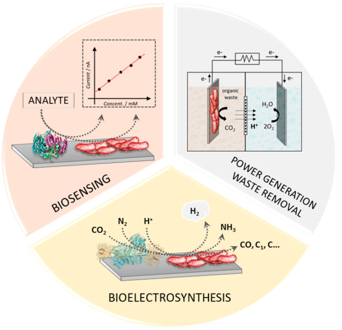
Scheme of different applications of enzymatic and microbial electrochemical systems, including the development of biosensors for the monitoring of medically relevant analytes or toxic compounds, the local generation of micropower with simultaneous waste removal, and the development of bioelectrosynthetic factories.
Given that enzymes are known to be able to activate stable small molecules under mild conditions, there is also significant interest in the electroenzymatic synthesis of value-added products.16−18 For example, hydrogenases are metalloenzymes that catalyze both H2 oxidation and H+ reduction, reversibly; hydrogenase-containing electrodes are therefore able to produce H2 from electrical energy in aqueous solutions.19−21 Similarly, nitrogenases have gained much interest as the only enzyme able to reduce kinetically inert dinitrogen (N2) into ammonia (NH3), a value-added product which is the building block for many nitrogen-based compounds (fertilizers, amino acids, DNA bases, polymers, etc.).22 Carbon dioxide (CO2)-reducing enzymes have also gained interest over the last decades because of their ability to reduce CO2 into C-based value-added fuels such as carbon monoxide (CO) or formate.23,24 Finally, it is also possible to combine these enzymes within EFCs, where electrical energy and value-added products can be produced simultaneously. However, the reductive enzymes outlined here are typically associated with reactions with relatively low E0 (E0′2H+/H2 = −0.41 V vs SHE, E0′CO2/HCOOH = −0.42 V vs SHE, E0′N2/2NH3 = −0.14 V vs SHE), which then requires a low-potential anodic system for the EFC to be truly galvanic. Nevertheless, a hydrogenase anode was combined with a nitrogenase cathode to yield a H2-oxidizing and N2-reducing EFC with an open circuit potential (OCP) of 0.23 ± 0.03 V. 60 mC of charge was drawn from this device, while simultaneously producing NH3 with 26.4% Faradaic efficiency.25
Concerning the use of bacteria on electrodes, the most widely investigated application has been the wiring of electroactive bacteria (species that are naturally capable to transfer electrons across the cellular membrane due to endogenous electron transfer pathways) for power generation and environmental remediation purposes.26,27 Specifically, Shewanella oneidensis (S. oneidensis) and Geobacter sulfurreducens (G. sulfurreducens) are the two species with the most detailed extracellular electron transfer pathways described to date. These two species have been consistently utilized at the anode of microbial fuel cells, achieving relatively high current and power densities (about 4 mA cm–2 and 0.6 mW cm–2, respectively).28 More recently, focus has been posed on the use of microbial electrochemical systems for biosensing29 and bioelectrosynthesis.30 In the case of biosensing applications, the current or power output of the microbial electrochemical system is correlated to the concentration of an analyte of interest, and self-powered biosensing devices have been obtained when utilizing complete microbial fuel cells.31,32 Regarding the possibility to obtain microbial bioelectrofactories, recently, cyanobacterial cells were engineered to express the electron transfer pathway of G. sulfurreducens, which enabled them to utilize a cathode as the electron donor to accomplish N2 reduction to NH3.33
Finally, bacteria and enzymes-based electrodes have been utilized together in the same electrochemical system to obtain enzyme/microbial hybrid systems, where microbial anodes oxidizing various substrates are couple to enzymatic cathodes accomplishing the oxygen reduction reaction for high-performance devices.34,35 These hybrid systems have been further expanded to have microbial fuel cells charging an abiotic electrode that is subsequentially coupled to an enzymatic electrode for CO2 reduction.36
2. Principles of Bioelectrochemistry
Semiclassical Marcus theory for electron transfer in nonadiabatic systems has been extensively applied to the study of electron-transferring proteins and enzymes:37
 |
where the electron transfer rate constant (kET) is dependent on the variables (i) temperature, (ii) thermodynamic driving force of the reaction (ΔrG0), (iii)nuclear reorganization energies (λ), and (iv) matrix coupling element (HDA, also commonly referred to as their electronic coupling). Importantly, HDA exponentially decays with an increase in distance (rDA) between electron transfer partners, and depends on the medium in between (i.e., amino acids, solvent, etc., given as an electron transfer decay parameter “β”):
 |
where HDA0 reflects the electronic coupling of the donor and acceptor when they are in contact (r = 0). Even though electrode-adsorbed proteins are known to deviate from Marcus theory of electron transfer (with conformational reorganizations thought to become rate-limiting to heterogeneous electron transfer), it arguably remains intuitive that the distance between an electrode and the redox-active cofactor of an enzyme should be minimized to maximize heterogeneous kET.38 This is even more intuitive for the case of intact bacteria-electrode systems, where the electron-transferring enzymes responsible for the exchange of electrons are located inside the bacterial cells, thus separated from the electrode surface by several nm (normally more than 30 nm).39
Below, the major electron transfer pathways are briefly presented, both for enzymatic and microbial electrochemical systems. In this tutorial review, we have utilized the classification of electron transfer pathways that is more commonly adapted in bioelectrochemistry, with direct electron transfer (DET) being obtained when an enzyme transfers electrons directly with an electrode. On the contrary, mediated electron transfer (MET) refers to systems requiring an exogenous, artificially added, electron mediator that shuttles electrons between enzymes and electrodes. For the case of intact bacteria, the electron transfer process is more properly defined as “extracellular electron transfer” (EET),40 as electrons need to cross the bacterial membrane, and EET is further divided into direct (E-DET) and mediated EET (E-MET), as shown in Scheme 1.
Scheme 1. Electron Transfer Pathways for Microbial (Left) and Enzymatic Electrodes (Right).
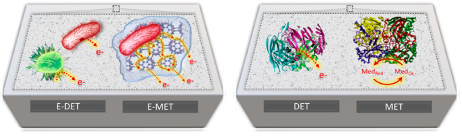
E-DET refers to the EET of electrons through microbial nanowires or membrane-bound proteins. E-MET refers to the EET by means of diffusible or polymer bound-redox mediators. DET refers to the direct transfer from the redox-active sites of an enzyme to the electrode surface, while MET refers to the electron transfer through artificial redox mediators.
2.1. Direct Electron Transfer (DET)
There are both advantages and disadvantages to DET configurations.12,41,42 From an applied perspective, DET can simplify the experimental setup and therefore lower the cost of an enzymatic device if an enzyme can spontaneously adsorb to an electrode surface (electrostatic, π–π stacking, π–cation interactions) and undergo efficient electron transfer.43,44 In this case, an additional electron mediator may also not be necessary, although this approach becomes more costly if tailored electrode/enzyme modifications are necessary to achieve DET. An important benefit that is afforded by DET is the ability to interrogate the redox-active cofactors of an enzyme, paving the way to the possibility of determining their reduction potentials (E0′). Furthermore, DET provides a handle by which relationships between thermodynamic (driving force) and kinetic properties (catalytic rate constants or affinities) of enzymes can be studied.45
One potential disadvantage to DET for practical applications is that only a single monolayer of enzyme is expected to be electronically wired to the electrode, and the quantity of electroactive enzyme (and thus the current) is therefore somewhat limited. One strategy to overcome low current densities is to use conductive, stable and hydrophobic nanoparticles (NPs) and carbon nanotubes (CNTs). For instance, CNTs have been used in the presence of pyrene based anchoring compounds based on π–π stacking interactions.46 Complementary strategies using surfactants, hydrogels or polymers (PEG, chitosan, alginate, etc.) provide a matrix that can also increase stability over time.44,47 In addition, the use of conductive porous materials such as indium–tin oxide (ITO) or metal–organic frameworks (MOFs) permit larger surface areas, and therefore larger enzyme loadings/coverages on electrode surfaces.48,49 Despite adsorption being the simplest way to modify electrode surfaces, this is often not successful for many enzymes because of factors such as (i) rapid denaturation, (ii) low film/enzyme stability over time, and (iii) enzyme leaching/film loss. As discussed above, adsorption should also not inhibit electron transfer between the enzyme and electrode. Covalent anchoring of enzymes enables some of these issues to be overcome, with amide bond formation through coupling carboxylic acid-functionalized surfaces to protein surface lysine residues, or by cross-linking glutamate and aspartate residues from protein surfaces to amine-functionalized surfaces. This coupling can be performed with peptide coupling agents, with EDC/NHS being extensively used due to its stability in water and mild operational conditions.50,51 Another possibility is to modify the electrode surfaces by electrografting diazonium salts, which provides a universal technique to introduce a large diversity of chemical functions on the surface of the electrode.52
One prominent and versatile strategy that has been extensively employed to optimize DET is the use of thiol-containing species, which form self-assembled monolayers on gold electrodes.53,54 The main advantage of this technique is adjustable lengths of chains and the possibility to introduce different terminal end groups. The direct anchoring of enzymes on gold is also possible via the reaction of solvent/surface-exposed cysteine (sulfhydryl-) residues on proteins. Because of the comparatively lower occurrence of cysteine in proteins vs lysine residues, this approach is more specific than amide bond formation (discussed above). Further, cysteine residues can also be inserted in or relocated to strategic positions (i.e., close to a redox-active cofactor) via site-directed mutagenesis. This method will be discussed in depth below. It has also been proposed that nonspecific enzyme adsorption on electrodes leads to a distribution of heterogeneous kET, which can complicate the interpretation of thermodynamic and kinetic parameters.55 Site-specific covalent adsorption (i.e., by exploiting cysteine residues) is considered one potential way to overcome this limitation. It is also possible to exploit surface cysteine chemistry by immobilizing Michael acceptors such as maleimides on electrode surfaces, which then undergo thiol-maleimide Michael additions.56 This ligation method is widely used in the field of chemical biology because of the fast kinetics and high selectivity of this reaction at neutral pH. The value added by this method compared to protein adsorption in self-assembled monolayers (SAMs) or on gold surfaces is the formation of a covalent bond between the thiol group of the cysteine and the maleimide, which is not sensitive to low potential reductive cleavage (and release), such as in the case of Au–thiol bonds.
2.2. Mediated Electron Transfer (MET)
MET is an artificial electron transfer approach that employs redox-active species (either diffusive or bound to polymer backbones) to shuttle electrons between enzymes and electrodes.57 This approach allows overcoming the limitations due to the redox-active cofactor being distant from the surface, and makes specific protein adsorption no longer a requirement. Furthermore, MET extends the bioelectrode design to enzymes in solution, since only the mediator is required to interact with the electrode surface. For applied enzymatic electrochemistry, this configuration provides the ability to exploit flow chemistry techniques, or to have large quantities of biocatalyst present in the cell (compared to a DET monolayer), although the separation of products from the enzyme and mediator is presumably required postprocessing. Important requirements for an appropriate electron mediators include (i) having good electrochemical reversibility (stable in both reduced and oxidized state), (ii) having good solubility, (iii) having fast rates of electron transfer (with the enzyme and the electrode), (iv) and not adversely affecting enzymatic activity.
As a rule of thumb, the electroenzymatic oxidation (e.g., glucose oxidation) of a substrate typically requires an electron mediator with an E0′ that is more positive than the E0′ of the enzyme’s redox-active cofactor and the substrate/product (with the opposite being true for reductive electroenzymatic reactions, e.g., H+ reduction), as dictated by
 |
where n is the number of electrons in the electron transfer process, F is Faraday’s constant (96,485 C mol–1), and E0' is the reduction potential of the reaction. This can be extended to MET by considering the reduction potentials of the substrate/product (the enzymatic reaction, E0'S/P) and the oxidized/reduced electron mediator (E0'O/R) to yield58
 |
and
 |
It is important to keep the relative positions in equilibrium (ΔG) in mind when considering electron mediators: for example, the electron mediator methylviologen (E0′ = −0.45 V vs SHE) can effectively mediate enzymatic H+ reduction and H2 oxidation (E0′ = −0.41 V vs SHE). This principle previously enabled the use of methylviologen in both the anodic and cathodic compartments of the H2/N2 EFC outlined above.25
A breakthrough advance for enzymatic MET was the development of redox polymers.59,60 In such redox mediating systems, suitable electron mediators are first chemically grafted to water-soluble polymeric backbones and the resulting material can following be mixed with a solution of enzyme and drop-casted onto an electrode surface (possibly in the presence of a chemical cross-linker). The result is an enzyme/polymer film that is (i) confined to the electrode surface and (ii) contains a nondiffusing electron mediator, which subsequently permits MET across the entire redox polymer/enzyme film with electrons hopping across the mediator moieties. Redox polymer/enzyme composites have found extensive use in blood glucose monitoring. A recent major advance in redox polymers was the development of a system that simultaneously enabled the electroenzymatic oxidation of H2 by hydrogenase, while simultaneously protecting this enzyme from O2 deactivation (using the same H2 enzymatic oxidation reaction).61
2.3. Extracellular Direct and Mediated Electron Transfer
As previously introduced, interfacing intact bacteria with electrodes is complicated by the presence of the cytoplasmic membrane (often referred to as inner membrane) and other additional membrane layers (i.e., peptidoglycan, outer membrane), which are physical barriers surrounding the bacterial cells acting as insulators. However, some bacterial species were forced to evolve dedicated electron transfer pathways as they found themselves in environments with few or no soluble electron acceptors available, thus having to utilize insoluble electron acceptors located outside of their membranes (i.e., iron oxides).40,62 This E-DET differs from the DET discussed for enzymatic systems due to the various intermediate electron carriers at plays between the redox enzymes and the electrodes. The two better-understood E-DET pathways are the ones of S. oneidensis and G. sulfurreducens. Excellent studies describe in detail the electron hopping mechanism across various cytochromes located in the external membranes of Shewanella species,63 where the electrons are shuttled across more than 25 nm of insulating space.64Geobacter species exhibit a similar electron transfer pathway using porin-cytochrome trans-outer membrane protein complexes,65 while also utilizing conductive nanowires that transfer charge by π–π stacking, in a similar fashion to carbon nanotubes.66 Additionally, Shewanella performs E-MET by self-secreting riboflavin, an endogenous redox mediator that acts as a diffusible shuttle from the redox-active sites to the electrode surface.67Shewanella is not the only species known for producing endogenous mediators, with recent reports available also for other bacteria, including cyanobacteria.68−70 Furthermore, the modification of the electrode surfaces to facilitate E-MET via endogenous mediators have also been investigated, i.e., with the use of carbon nanotubes.71 E-MET is also possible through exogenous diffusible redox mediators, which enabled the use of various photosynthetic bacteria for biophotoelectrodes development.72,73
The use of redox polymers has proven particularly effective for E-MET, with pioneering works employing osmium-based redox polymers to wire several bacterial species.74−76 More recently, bioinspired redox polymers that utilize redox moieties similar to the natural electron mediators found in bacteria, such as quinone molecules, have been employed successfully wiring Gram-positive and Gram-negative bacteria.77 Remarkably, such polymers enabled a consistent enhancement of electron transfer also for the electroactive bacterium S. oneidensis.78 With the aim to provide both artificial redox mediation and adhesive properties to maintain the bacteria on the electrode surface, redox-adhesive polymeric matrices based on polydopamine have been recently developed for E-MET with various bacteria.79−81 As photosynthetic bacteria have intrinsic limitations to the EET due to the location of the photosynthetic apparatus inside the inner membrane, the reader is referred to a recent review discussing this aspect and presenting the various artificial redox-mediating systems specifically developed for this class of bacteria.82
2.4. Synthetic Biology for Electrochemistry
2.4.1. Protein Engineering: Affinity Tags, Cysteine Residue Chemistry, and Unnatural Amino Acids
Given that most enzymes/proteins have not been evolved to transfer electrons with nonproteinogenic surfaces, below are discussed some prominent and emerging protein engineering approaches that have been developed to optimize enzymatic electrochemistry.
One approach utilizes the recombinant insertion of an affinity polypeptide tag within the primary structure of a target protein to either its N- or C-terminus that provides a specific and simplified strategy for protein purification. Perhaps the most-used affinity tag is poly(histidine)(5–8) (His-tag), which then typically displaces two aqua ligands of a coordination complex such as [Ni(NTA)(OH2)]2+ (NTA = nitrilotriacetic acid). The NTA ligand is covalently bound to a solid phase, and alternative first-row d-block metal cations can be used in the place of Ni2+ (Fe2+/Co2+/Zn2+).83 The repeating histidine sequence exploits the chelate effect to provide greater affinity than other individual solvent-exposed histidine residues. The same principle can be exploited to anchor enzymes to electrode surfaces by their His-tags, where gold electrodes have been modified with Ni-NTA SAMs for the immobilization of His-tagged PQQ-dependent aldehyde dehydrogenase (PQQ-AlDH)84 and horse-heart cytochrome c (cyt c).85 By inserting His-tag to either the N- or C-term of each of PQQ-AlDH’s three subunits, six different variations were obtained with one favorably orientated on the electrode showing 6.6-fold higher DET efficiency.84 In the case of cyt c, a C-terminal His-tag containing variant showed the most efficient DET because of minimized electron transfer distances.85 Also, Ganesana et al. functionalized the surface of graphite screen-printed electrodes with nickel oxide nanoparticles for the immobilization of His-tagged acetylcholinesterase obtaining a biosensor with improved sensitivity.86 Recently, Minteer’s research group also introduced another approach for the immobilization of His-tagged proteins on carbon electrodes where a Ni-chelated-Lys3Asp polypeptide linked to a pyrene moiety (for π–π stacking with carbon surfaces) was produced by solid-phase peptide synthesis.87 The His-tagged MoFe protein of nitrogenase was subsequently immobilized for MET with adding methylviologen, without the need to entrap the MoFe protein within a polymeric support layer. However, the use of His-tags has several disadvantages, including the limited modification to either the C- or N-termini of protein subunits.51,88,89
Another approach involves targeting specific amino acids (i.e., lysines, cysteines, aspartates) on the surface of a protein for chemical conjugation. For example, the primary amines of lysines can be targeted for amide bond formation with the use of reagents such as EDC/NHS (as mentioned above). However, a protein’s surface typically has multiple solvent-exposed lysine residues, neutralizing the possibility for oriented protein immobilization. Among all the residues on the proteome, cysteine is one of the most nucleophilic and conserved, making it by far the most reactive residue on any protein, yet the least abundant residue.90 This high reactivity of cysteine has been extensively utilized for various purposes, including drug-development strategies,91−93 bioconjugation strategies, development of various probes for imaging of biological species,94,95 and proteomics.96 Many of these bioconjugation reactions can be extended to enzymatic electrochemistry. For example, these Michael acceptors undergo 1–4 cycloaddition97 (Figure 2a), thus the electrodes can be functionalized with groups that readily react with cysteine thiols, such like maleimide Michael acceptors.98,99
Figure 2.
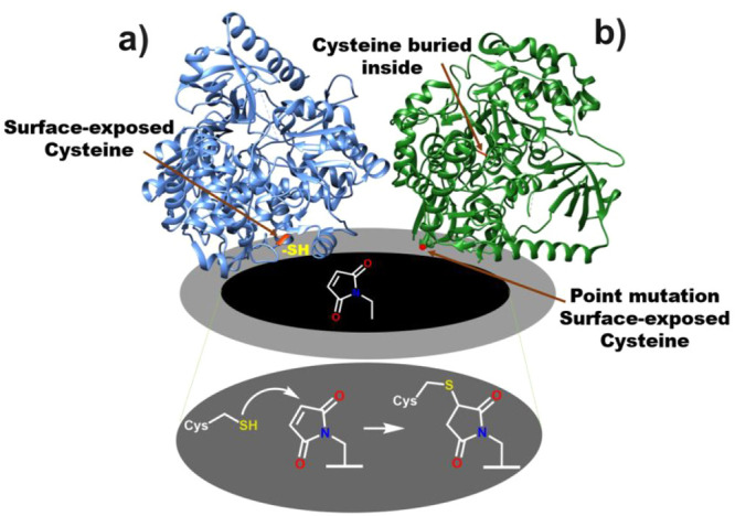
(a) Metalloprotein with a surface-exposed cysteine can readily react with a scaffold incorporated on the electrode, such as maleimide. (b) In case of a protein having a cysteine residue not exposed on the surface, a point mutation can help to introduce a cysteine residue at the suitable position.
In certain proteins, the reactive cysteine residues may be buried inside, i.e., they may not be accessible to the solvent or other reactive molecules, and thus not be readily available on the surface for conjugation (Figure 2b).100 In such cases, using molecular biology techniques, like site-directed mutagenesis, a cysteine residue can be introduced on the protein surface in a suitable position.99 The protein structure or its activity are not particularly affected by point mutations of residues not directly involved in enzymatic processes, making them the ideal way to introduce the cysteine residue in a more suitable position for efficient immobilization on the electrodes. Meneghello et al. exploited site-directed mutagenesis to introduce cysteine residues to different positions on the sugar-oxidizing enzyme cellobiose dehydrogenase, which contains an FAD-dependent dehydrogenase subunit and a heme-containing subunit. Both these subunits are naturally tethered by a flexible peptide linker such that the heme subunit can act as an electron transfer partner to the FAD-dehydrogenase subunit.101 By strategically placing individual cysteine residues for DET investigation, the authors provided convincing evidence that DET takes place exclusively through the heme-containing subunit (the FAD-dehydrogenase does not undergo DET).
The above-mentioned approaches are still limited as they utilize the functionality of the 20 “standard” proteogenic amino acids found in the structure of enzymes/proteins. An emerging approach (genetic code expansion, GCE) allows incorporation of unnatural amino acids (UAAs) into any site of a protein sequence, and thus permits the introduction of functional groups that are not naturally found in the enzymes/proteins. GCE exploits orthogonal tRNA/tRNA synthetase pairs to allow the site-specific incorporation of UAAs by decoding the amber (UAG) stop codon (termed amber codon suppression).102−104 The site-specific incorporation of UAAs can be used to improve enzymatic stability and catalytic properties, and to understand electron transfer mechanisms of metalloproteins/metalloenzymes.105,106 In one example, Schlesinger et al. targeted the orientation of the copper oxidase (CueO) on the electrode surfaces for improved DET, where the alkyne-containing UAA propargyl-l-lysine (PrK) was placed in different positions on the protein’s surface.107 One site was near to the type 1 Cu site and the other two were close to the trinuclear cluster (TNC) Cu site (a control was prepared with PrK located far away from both active sites). Site-specific immobilization of the mutants was performed by the Cu(I)-mediated cycloaddition (CuAAC) “click” reaction on GC electrodes functionalized with different azide-containing pyrene linkers. All of the mutants placing PrK in proximity to either the T1 Cu or the TNC exhibited improved kET with respect to their targeted electron transfer partner, demonstrating that this strategy was indeed effective at improving heterogeneous electron transfer. Further, all three of these mutants displayed improved electrocatalytic performance for O2 reduction by the enzyme.
The same PrK UAA was introduced to the cytochrome c unit that had been artificially fused to glucose dehydrogenase, which was subsequently immobilized on functionalized GC electrodes by click chemistry. A mutation close to the electron-transferring heme domain provided the largest current density on the electrode when compared to the introduction of PrK in proximity to the FAD cofactor of this chimeric enzyme. These site-specific mutations permitted the electron transfer properties of the FAD cofactor and the artificial heme domain to be investigated.108
An alternative UAA was used to improve the DET properties of a “small” laccase multicopper oxidase, where five positions on the surface of the enzyme were targeted for the introduction of an azide-carrying phenylalanine UAA (para-azidophenylalanine, pAzF). Three of the mutation sites were near to the TNC, one addition site was in proximity to the T1 Cu center, and the last position served as a control (in proximity to neither the TNC nor the T1 Cu site). Cyclooctynyloxyethyl-1-pyrenebutyrate (PBCO)-functionalized indium–tin oxide (ITO) electrodes were used for the Cu-free ring-strained “click” immobilization of the enzyme. Contrary to expectations, the mutant prepared as a negative control showed the greatest kET. Interestingly, the structural analysis of laccase revealed the presence a water channel close to the basal site connected with the trinuclear copper cluster. Therefore, the immobilization of laccase from this basal site was attributed to this enhanced kET, and resulted in stable current up to 8 days.109
Although these studies have demonstrated improved DET efficiencies, challenges still remain for the wider deployment of GCE in electrochemistry, such as relatively diminished expression yield (due to protein truncation during RNA translation), the cost of UAAs (when commercially available), and the unavailability of 3D structural information for some proteins/enzymes.110,111 However, recent advances to improve UAA incorporation and protein yields are promising developments in this area.112,113
2.4.2. Bacteria Engineering for Enhanced Extracellular Electron Transfer
In recent years, the possibility to utilize synthetic biology to engineer intact bacteria, introducing non-native properties including the EET pathways of Shewanella or Geobacter in other organisms, has attracted particular attention and enabled enthralling research directions. Specifically, the approach is based on engineering the microorganisms with outer membrane cytochromes, thus overcoming the limitation of insulating membrane layers. The first reports of heterologous gene expression were reported for the Mtr pathway of S. oneidensis in Escherichia coli (E. coli), with the 2010 work of Jensen et al.114 While the engineered strain enabled a 4-fold faster reduction of insoluble Fe(III) compared to the wild-type E. coli, such reduction was still significantly slower compared to wild-type S. oneidensis MR-1, mostly due to the slow rate of the first electron transfer step to reduce MtrA. To tackle this aspect, following works have focused on the expression of cytochrome c CymA as an electron donor to the MtrA,115 as well as the role of exogenous flavins, to further boost electron transfer.116 Recently, Mouhib et al. showed that the complete expression of the Mtr pathway together with periplasmic cytochromes can further enhance electron transfer in engineered E. coli.117 Conversely from the expression of Shewanella EET pathways, considerably less studies investigated the expression of the G. sulfurreducens EET mechanism due to its higher complexity. Notably, Sekar et al. reported the expression of G. sulfurreducens outer membrane cytochrome OmcS in cyanobacteria, enabling a 9-fold current increase.118 Such heterologous expression was recently achieved also by Dong et al. in a previously engineered cyanobacterium for accomplishing NH3 production in a bioelectrochemical system without the need of exogenous diffusible redox mediators.33 Specifically, the engineered bacteria with OmcS allowed a 13-fold higher NH3 production compared to the strain not engineered with the heterologous EET pathway, together with an increased faradaic efficiency of about 23%. We refer the reader to recent reviews on the use of synthetic biology for microbial electrochemical systems for further insights into this topic.119,120
3. Methods to Study Enzymatic and Microbial Electrodes
3.1. Electroanalytical Methods
Protein film voltammetry (PFV) (also known as protein film electrochemistry) is a popular technique to study redox proteins, as the protein or enzyme is wired to an electrode and can be studied via variety of electroanalytical techniques. In PFV, the energy (or driving force) supplied to the enzyme is controlled via the applied potential, while the current provides kinetic and mechanistic information about the enzyme. PFV is attractive as only a small quantity of enzyme/protein is needed, the complex dependence of enzyme diffusion on the electrochemical signal can be eliminated, and a large range of conditions can be tested using a single bioelectrode, including those where the enzyme is only transiently stable (e.g., extreme pH).
During PFV, electrons are transferred between redox centers of the protein/enzyme and the electrode surface (Figure 3a). Page et al. reviewed 31 redox proteins/enzymes and concluded that most redox cofactors undergoing physiologically relevant electron transfer reactions are found within 14 Å of each other.121 Marcus theory was subsequently adapted to enable kET to be predicted for redox proteins with knowledge of driving force and distances between redox cofactors, as well as the nature of the amino acid/solvent environment between cofactors (which is expected to impact their electronic overlap, HDA). Nevertheless, some important points must be kept in-mind: (i) electron transfer does not abruptly stop for distances >14 Å; (ii) X-ray crystal structures do not provide information on dynamic conformational changes of proteins (which could impact kET); (iii) proteins have been known to deviate from Marcus behavior at electrode surfaces.38 Nevertheless, it remains intuitive that the redox cofactors of enzymes/proteins should be oriented for favorable electron transfer.
Figure 3.
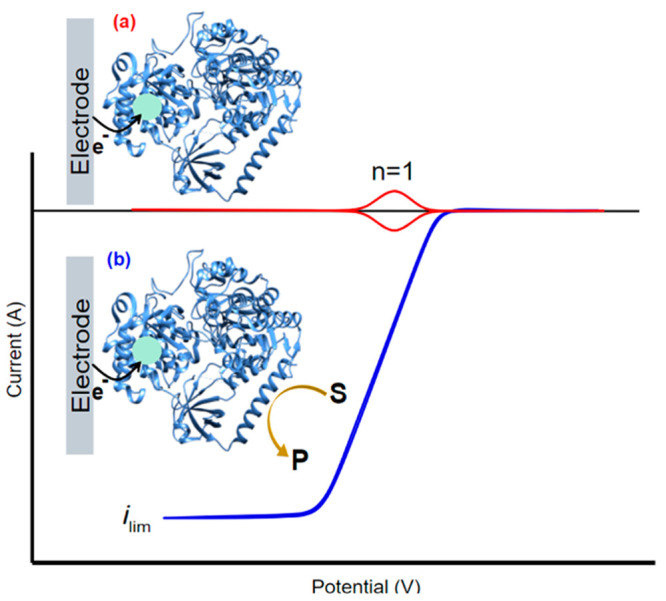
Idealized cyclic voltammetry showing the redox response of an enzyme under reductive conditions with one active redox center (n = 1) under (a) non-turnover conditions where there is only an electron transfer between the redox center of the enzyme and the electrode, and (b) turnover conditions in the presence of a substrate and steady-state conditions. The current is a direct measure of enzyme activity.
As discussed above, redox mediators are often used to overcome sluggish electron transfer in the case of DET.122 This is useful for performance driven applications such as those in biofuel cells, however extracting information solely about the enzyme is challenging as electrochemical responses of the mediator and enzyme are often convoluted.123 DET simplifies the interpretation of electrochemical signals and allows thermodynamic and kinetic parameters to be extracted with reduced inference.
Electroanalytical methods are also a unique tool to study microbial electrodes. The concepts presented for PFV applies also for the case of intact bacterial cells deposited on electrode surfaces, while keeping in mind the constrains introduced by the presence of the bacterial membranes previously introduced. The electrochemical characterization of microbial electrodes allows elucidating the role of redox intermediates in E-DET, as well as unveiling the presence of endogenous diffusible redox mediators produced by the bacterial cells on the electrode. Furthermore, the development of microbial biofilms on the electrode can also be studied by means of long-term potentiostatic polarization. The following sections present the various electrochemical methods employed for PFV and microbial electrodes characterization.
3.1.1. Cyclic Voltammetry
Cyclic voltammetry (CV) is one of the most prevalent electroanalytical techniques employed for both PFV and microbial electrodes, as it is simple to use and the voltammogram provides information in a clear and visual manner. To perform CV, the potential of the working electrode is swept linearly back and forth between two potentials (E in volts (V)) (a single sweep is known as linear sweep voltammetry (LSV)), at a defined scan rate (ν in V s–1) and the current (i in amperes (A)) is recorded.124 For PFV under noncatalytic conditions, thermodynamic information about the redox centers of the protein/enzyme can be extracted if the scan rate is sufficiently low that the protein/enzyme remains under equilibrium conditions. Each peak in a non-turnover CV corresponds to a redox site undergoing oxidation/reduction. The total number of peaks (n) therefore equates to the total number of active redox sites in the protein/enzyme (Figure 2, red). Importantly, the positions of the reduction and oxidation peaks yield E0′, the widths of the peaks at half-height (ΔEp1/2) depends on the number of electrons (n) involved in that electron transfer process (i.e., n = 1, 2, etc.), and the peak current (ip) is proportional to the quantity of adsorbed redox species.125 One of the limiting factors of PFV under DET is the low enzyme surface coverage and unfavorable orientations of the enzyme on the electrode that can greatly reduce electron transfer efficiency. As a result, the response of the enzyme can become convoluted with capacitive charging at the electrode making interpretation of electrochemical signals challenging under non-turnover conditions. To overcome this, graphite is commonly used for PFV under DET conditions as its preparation is facile, the rough surface increases the contact points between the enzyme and electrode, and the oxide rich functionality provides an excellent environment for the electrostatic adsorption of proteins.126 Armstrong and co-workers demonstrated that the immobilization of fumarate reductase on pyrolytic edge plane graphite (PGE) was facile and allowed the enzyme to retain in an extremely electroactive state while maintaining its native structure.127 For this reason detailed kinetic studies have also been performed using PGE.128 Another approach to amplify enzymatic response under non-turnover conditions is Fourier-transformed AC voltammetry (FtacV). A large-amplitude sinusoidal wave is applied to the linear potential sweep during CV, after which the current is Fourier transformed to a power spectrum which allows the separation of faradaic and non-faradaic contributions.129 FtacV is a relatively new technique in PFV; however, over the past 5 years, Parkin and co-workers have been able to elucidate mechanisms of electron transfer in the Mo-containing YedY catalytic subunit,130 the HypD [NiFe]-hydrogenase maturase protein,131 as well as separating the catalytic and electron transfer current contributions in [NiFe] hydrogenase.132
Altering the scan rate under non-turnover conditions provides kinetic information about the protein/enzyme. Fast scan rates have been able to access coupled electron transfer processes between the protein and electrode and the associated rate constant (k0). Hirst et al. demonstrated scan rates up to 3000 V s–1 could be used to probe the electron-exchange characteristics of azurin,133 a copper containing redox protein, where a k0 was determined to be approximately 5000 s–1. Using scan rates up to 3000 V s–1 also allowed coupled electron transfer processes occurring for three different ferredoxin proteins in the millisecond time domain to be resolved.
In the presence of an enzyme’s substrate, the application of a sufficient driving force for electron transfer (Eapplied) can result in “catalytic” currents. In this case, an enzyme that has been oxidized by an electrode can be reduced by the oxidation of its substrate, leading to a second round of oxidation (of the enzyme) by the electrode. The magnitude of this catalytic current is dependent on Eapplied, as well as the product of the enzyme’s turnover frequency and the quantity of enzyme on the electrode surface (strictly, the quantity of enzyme undergoing DET). This second factor is not always easily determined. Further, the confinement of an enzyme to an electrode enables the simple titration of substates within the electrochemical cell; this, in turn, enables the determination of apparent kinetic parameters such as Michaelis constants (KM). If only one cofactor is in communication with the electrode, the formal onset potential for catalysis can be loosely related to the formal potential for substrate reduction (for reduction E0′onset ≈ E0′red), provided the scan rate is sufficiently low that the protein remains at equilibrium. Parameters such as temperature, pH and ionic strength should also be considered as they can also significantly perturb this value. The appropriate potential window should also be chosen to avoid overpotential deactivation of the enzyme. For slower scan rates where the enzyme is maintained in a state of equilibrium a reversible CV may be obtained (Figure 3a, blue).124 Conversely, peak separation or hysteresis observed at elevated scan rates can be indicative of slow reaction kinetics or protein instability/desorption from the electrode.
Hydrodynamic electrochemistry is often used to study enzyme kinetics as mass transport limitations can be removed. The electrode is rotated to induce a controlled flux of fluid to the electrode surface by laminar flow, where the substrate is continually replenished. When a steady-state regime is reached, the rate-limiting step of the system becomes enzyme-dependent (Figure 3, blue line) enabling the investigation of various processes such as intramolecular electron transfer, conformational changes, ion-ligand exchange, or the binding/release of substrates/products within the enzyme.134
For the specific case of microbial electrodes, CV are particularly useful to investigate the mechanism of EET. Specifically, performing CV under noncatalytic and catalytic conditions allows one to study the presence of membrane bound redox proteins responsible for E-DET, as well as the presence of endogenous diffusible mediators responsible for E-MET. The two EET modes can be distinguished by first acclimating the microbial electrode to a specific electrolyte prior to gently washing and transferring the electrode to a fresh electrolyte and immediately performing additional CVs. If the EET is taking place primarily through endogenous diffusible mediators, the CV under turnover should reveal a loss of catalytic response in the new electrolyte as shown by Marsili et al. for the case of endogenous riboflavin produced by S. oneidensis.67 Conversely if E-DET is taking place, the catalytic response should be (at least partially) retained. In the latter case, performing CV under non-turnover conditions would further allow studying the redox potentials and eventually characterizing the involved redox centers responsible for E-DET.
Modeling of experimentally obtained CV can be performed to gain critical insights into the EET process,135 such as unveiling microbial electrodes with dual electron transfer mechanisms (both direct and mediated),136 or studying the rate-determining step in the EET process (i.e., for a microbial anode: (i) mass transport of the substrate in the microbial biofilm formed on the electrode, (ii) microbial turnover of the substrate, (iii) reduction of the redox intermediate, (iv) transfer of electrons throughout the biofilm, and (v) electron transfer at the electrode surface).137
Since several molecules and enzymes can be secreted by bacterial cells into the electrolyte, when E-MET is revealed, the electrolyte can be filtered utilizing membranes with different nominal molecular weight cutoff to gain details on the origin of the redox response. Specifically, performing CV with the filtered electrolytes allows one to estimate the range of molecular weight of the endogenous redox molecules and/or secreted enzymes responsible for the redox response, as recently shown by Saper et al.68 Finally, an extremely interesting aspect of microbial electrodes is that their composition might vary based on the conditions under which the electrodes were formed and/or exposed. If a microbial electrode is formed under electrochemical polarization, CVs could be used to characterize the effect of the utilized potential on the catalytic response,138 revealing for example different onset potentials for the catalytic reaction. This aspect is particularly important for microbial electrodes since bacterial species can over/under express specific genes depending on environmental conditions and external stress. As a general example, it has been reported that G. sulfurreducens can express more than 100 types of cytochromes,139 showing an outstanding flexibility on EET pathways depending on the local environment and physicochemical conditions, which could be characterized based on different CV responses.
3.1.2. Amperometry and Chronoamperometry
Amperometry is an electrochemical technique which measures i as a function of time (t) at a constant Eapplied. For the purposes of this tutorial review, we define chronoamperometry (CA) as a technique involving at least one step in applied potential (Eapplied), whereas amperometric i–t curves (or constant potential chronoamperometry) refers to the measure of i vs t at a single potential. We also acknowledge that CA generally considers experiments in which an analyte/substrate is not added over time (unlike in the case of amperometric i–t).
Amperometry is widely used in biosensors development for the quantitative detection of analytes, starting from the first glucose biosensor developed in 1962 by Clark.140 Today, modern glucose biosensors constitute a large proportion of commercially available amperometric biosensors,141 as well as numerous others employed in the environmental monitoring of hazardous compounds such as peroxides and aromatics,142,143 explosives like TNT,144 and harmful pesticides.145,146 With microbial electrodes, amperometric i–t curves allowed determining the influence on the current response of heavy metals,31,32,147 pesticides,148 organic carbon load149 and other toxic compounds,150 obtaining microbial biosensors for various applications. Amperometry is extensively utilized also for the characterization of biofuel cells and biosupercapacitors,141,151 an important area of research as investment for renewable energies continues to grow. Furthermore, amperometric i–t curves are particularly useful to study biophotocurrent generation during artificial light/dark cycles with photosynthetic apparatus/enzymes152,153 and photosynthetic bacteria73,81,154,155 In this case, the amperometric test is performed while alternating light/dark cycles by means of an artificial light source, and can provide insights into the kinetic of the photoinduced electron transfer reaction between the biophotocatalysts and the electrode.
CA is often chosen for fundamental enzymatic research as kinetic and mechanistic information can be obtained. During CA the applied potential is stepped and the current is recorded. This is particularly helpful since the turnover rate of the biological catalyst can be precisely controlled via the applied potential. Furthermore, the current is a direct measure of enzyme turnover, any changes in current magnitude show how the activity of the enzyme is changing in real time.156 Under non-turnover conditions, CA also permits heterogeneous kET to be determined for surface-confined bioelectrodes.
Amperometric i–t curves can be used in combination with a rotating disk electrode (RDE) to determine the apparent KM of a redox enzyme, an important kinetic parameter to describe the affinity between an enzyme and substrate. The KM value can be easily determined by recording the enzyme activity as a function of substrate concentration and fitting data to the Michaelis–Menten equation. This approach was nicely demonstrated by Léger and co-workers where the KM of periplasmic nitrate reductase from Rhodobacter sphaeroides was determined during nitrate reduction.157 Under the steady-state regime, a constant and large overpotential was applied to drive nitrate reduction, while the substrate concentration in the cell was increased stepwise. The entire experiment lasted only 150 s, and the KM value could be determined directly from the stepwise current profile. This approach is also useful for less stable protein films as the experiment can be completed relatively quickly. Similarly, amperometric i–t curves have been performed with RDE for microbial electrodes, studying mass transport inside the microbial biofilms obtained on electrode surfaces.158 Performing photoamperometric i–t curves with RDE in the presence of Rhodobacter sphaeroides cells and dichlorobenzoquinone as an exogenous diffusible mediator at different concentrations, Kasuno et al. showed that the biophotocurrent production from intact bacteria-based photoanodes follows a Michaelis–Menten-type kinetic model.159
Amperometric i–t curves are also valuable for studying inhibition kinetics under steady-state conditions, as active and inactive states of the biocatalyst can be accessed at different potentials. This is particularly useful for investigating the aerobic inhibition of redox enzymes, such as hydrogenases. The rapid denaturation of these enzymes under O2 presents a major challenge for their implementation into biotechnologies making the understanding of their O2 inhibition mechanisms of critical importance. An additional issue for hydrogenases is that those with the efficient H2 turnover are also particularly oxygen sensitive. Using PFV, Léger et al. investigated the permanent deactivation and reversible inhibition of CrHydA1, a [FeFe]-hydrogenase isolated from Chlamydomonas reinhardtii with CO and O2 during H2 oxidation.160 Rate constants (ki) for reversible inhibition with CO and deactivation with O2 were determined by altering the gas composition in the headspace of the cell, and observing relative changes in current from the amperometric trace. The results can be utilized also to determine the reaction order by plotting ki as a function of inhibitor concentration.
More recently, the reversible inhibition kinetics of [FeFe]-hydrogenase with hydrogen sulfide (H2S) have been studied by Léger and co-workers. Inhibition by sulfide renders the enzyme inactive and protected from O2.161 The electrode was stepped between two potentials (predetermined via CV) where (i) inactivation with sulfide was known to occur and (ii) the enzyme was fully active. The current response immediately following the potential step was fitted to kinetic model and used to determine rate constants of inhibition and reactivation.
3.1.3. Square Wave Voltammetry
Square wave voltammetry (SWV) is among the fastest and most sensitive pulsed electroanalytical techniques, as it allows diminishing the non-faradaic contributions to the current response. This paves the way to the detection of chemical species at nanomolar concentrations, establishing the operating principle behind many electrochemical sensors.162−166 In SWV, the potential is swept linearly as a modulated staircase with a defined step height (Figure 4).
Figure 4.
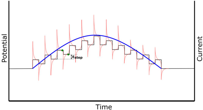
SWV trace where the potential (black) is applied as a staircase potential, the absolute current (red) is sampled twice at the end of each potential step (green dots), and the final current is reported as an average of both values (blue line).
The non-faradaic contribution is mitigated as the current is sampled twice (at the end of each potential step) and reported as a difference of the two values. SWV exploits the relative decay rates of faradic and non-faradic currents as the contribution of capacitive charging is negligible by the time the current is sampled at the end of each potential pulse.162 This discrimination is also useful for when using nanostructured electrodes in PFV, as a large surface area leads to significant capacitive currents, and removes the need for baseline subtraction which may not be accurate. This allows for a more accurate calculation of the electroactive enzymatic surface coverage on an electrode and the value of kET. Leopold and co-workers demonstrated that SWV can be utilized for determination of the kET of the azurin redox protein monolayer on different alkanethiol SAMs (on a gold nanoparticle).167 The values of ΔEp during SWV were used to determine kET by Reeves and co-workers.168 SWV can also be used to assess the activity of certain surface confined proteins. Sun et al. demonstrated SWV could be used in combination with Co(bpy)3 as an electroactive indictor to investigate the antioxidant activities of 4 different flavonoids which were used to probe the oxidative damage of proteins at a graphene-based electrode (protections of bovine serum albumin protein from damage on functionalized graphene-based electrodes by flavonoids).169 The activity of each flavonoid protein could be assessed simply by comparing the relative peak intensity of each SWV peak. In addition, SWV is also a valuable technique in protein monolayer electrochemistry which is used to study the electron transfer properties of immobilized proteins (the reader is directed to a review by Campbell-Rance et al.167).
The application of SWV in PFV remains reasonably limited due to the complex nature of the potential modulation, however it has been utilized for the operation of biosensors due to its high sensitivity.170,171 Recently, Monteiro et al. constructed a disposable biosensor with multiheme cytochrome c nitrate reductase on pencil graphite for the detection of environmental cyanide,172 where SWV was used to determine the sensitivity and response of the bioelectrode under turnover conditions. SWV is also valuable in the case of nanostructured electrodes with a high capacitance. Applications of SWV in PFV for kinetic studies still remains fairly limited, however a number of theoretical studies have modeled the electrochemical signals which may be expected to aid future experimental work.170,173,174 The application of SWV for the characterization of microbial electrodes is also not common, however, the technique provides a powerful tool for the study of the redox intermediates at play in the EET process. Yates et al. utilized SWV to accurately identify the midpoint potential for electron uptake in Marinobacter–Chromatiaceae–Labrenzia-based biocathodes performing oxygen reduction, with the aim to study the long-distance EET transfer process in an electroautotrophic microbial community.175
3.2. Hyphenated Electrochemical Techniques
3.2.1. Spectroelectrochemistry
Spectroelectrochemistry is a hyphenated technique that combines two classical methods, electrochemistry and spectroscopy, to obtain chemical information.125,176,177 Electrochemistry can be coupled with different spectroscopic methods, mainly with absorption spectroscopy in the UV–vis,178 IR,179 X-ray range, as well as Raman scattering spectroscopy,180 and electron paramagnetic resonance.181 Other techniques, such as nuclear magnetic resonance (NMR),182 X-ray absorption spectroscopy (XAS),183 and luminescence184 can be used with electrochemistry but are less common.185 There are several applications for spectroelectrochemical techniques in organic and inorganic chemistry, biochemistry, materials, and nanomaterial science. We focus this section mainly on UV–visible spectroelectrochemistry and its use to investigate proteins/enzymes, and fluorescence spectroelectrochemistry for the study of EET in microbial electrodes.
A few challenges are faced when performing spectroelectrochemical experiments for bioelectrochemical systems, resulting in their limited implementation. Specifically, the experimental setup needs to be designed taking into consideration the light source (in UV/vis and fluorescence spectroscopy), introducing various restrictions such as the use of grid or transparent electrodes. León et al. described in detail the design of electrochemical cells for different spectroelectrochemical analysis.176 While electrochemical analysis of redox proteins/enzymes can be performed with limited quantities (less that μg), this is not always the case with spectroelectrochemistry, since sufficient sample should be provided to be analyzed by the two techniques simultaneously. To this end, a monolayer of proteins adsorbed on an electrode surface often does not provide sufficient signal for the secondary technique; with an example being a redox-dependent absorption with a relatively low molar extinction coefficient (ε). Since an increase in sample concentration should not be possible for the case of electrodes with a monolayer coverage of protein, a possible approach to increase the absorbance signal, considering Beer–Lambert law, is to increase the measurement path length, which could be achieved using proteins in solution (not adsorbed on an electrode). However, this is not compatible with efficient DET and it is often necessary to use MET. Among the drawbacks of the approach we can mention the following: (i) a bulk (at least localized) electrolyte is required, which increases the time required to reach equilibrium; (ii) MET requires either that the mediator oxidation/reduction does not interfere with the feature of interest (or background subtraction would be required), or that the sample is followed at an isosbestic point of the mediator (which is often not at the λmax of the feature of interest).
Spectroelectrochemistry is considered an in situ technique, meaning that it is possible to simultaneously realize spectroscopic measurement within an electrochemical experiment.176 In Figure 5, we give an example of a spectroelectrochemical experiment performed in the Milton group to evaluate the E0′ of the heme-containing protein myoglobin.186 In the study by Guo et al., myoglobin proteins were reconstituted with various artificial cofactors (different metals and different porphyrin ligands) to enhance their natural peroxidase activity. It was proposed that a change in E0′ of the redox cofactor could explain the observed improved activity of one of these myoglobin variants; indeed, spectroelectrochemistry permitted the E0′ of the wild-type and a variant myoglobin to be determined in solution and confirmed a significant increase in E0′ for myoglobin containing the artificial cofactor. The spectroelectrochemical cell contained a Pt grid electrode as the working electrode, a Pt wire as counter electrode (shielded behind a porous-glass junction), and a Ag|AgCl (3 M KCl) reference electrode. A reduction potential titration was performed by stepping the potential from 0.0 to −0.3 V at 50 mV intervals. Each step lasted 1200 s to ensure equilibration of the ox/red states of myoglobin. For each step, an average of absorbance values recorded in the last 30 s was taken (Figure 5A). The Soret band of oxidized myoglobin is observed at 410 nm (ε410 = 157 mM–1 cm–1) in the oxidized form, which shifts to 434 nm in the reduced form.187 Using the Beer–Lambert law, it is then possible to evaluate the ratio between the [Ox] and [Red] form at each applied potential. A graph of absorbance vs E is sigmoidal and can be fit by nonlinear regression to the Nernst equation (Figure 5B):
 |
Thus, fitting the Nernst equation also enables the value of n to be determined for each redox feature (Figure 5C,D).
Figure 5.

(A) Spectroelectrochemical oxidation (blue) and reduction (red) of myoglobin in the presence of methylene blue as an electron mediator. (B) Nonlinear regression and (C) linear fit of data obtained in (A) to the Nernst equation to determine the formal redox potential (E0′) and the number of electrons transferred (n). (D) Changes in the oxidized and reduced features of myoglobin’s heme cofactor at different applied potentials. Adapted with permission under a Creative Commons Attribution License from ref (186). Copyright 2022 The Authors.
In one example, Wohlschlager et al. used UV/vis spectroelectrochemistry to generate the activating radical species for glyoxal oxidase (GLOX, an extracellular source of H2O2 in white-rot secretomes, where it acts in concert with peroxidases to degrade lignin), to continuously measure its concentration, and to simultaneously measure the catalytic activity of GLOX based on its O2 consumption.178 Spectroelectrochemical measurements were carried out in a 3 mL quartz cuvette optically transparent electrochemical cell comprising a three-electrode setup with a machined glassy carbon tile as the working electrode. The electrochemical generation of ABTS+ was controlled by applying electrochemical pulses of 600–700 mV vs Ag|AgCl for 1–10 s (depending on the desired quantity of ABTS+ to be generated, followed by spectroelectrochemistry at 420 nm), while monitoring O2 concentrations using an oxygen microsensor. Chen et al. utilized UV–vis spectroelectrochemistry to investigate riboflavin (RF), the primary redox-active component of flavin cofactors which is involved in many redox processes in biogeochemical systems.184 While the redox behavior and reaction mechanisms of RF in hydrophobic sites remain unclear, it is known that it possesses three accessible oxidation states (similar to quinones), each with protonated and deprotonated forms. In the oxidized form, the flavin shows two peaks at ≈360 and ≈450 nm.188 In their study, Chen et al. integrated spectroelectrochemical analysis and density functional theory calculations to explore the redox behaviors of RF in dimethyl sulfoxide (which was used to create a hydrophobic environment). In situ UV–vis spectra of RF at different Eapplied were obtained to spectroscopically observe the relative change in species during reduction. The absorption peaks at 446, 344, and 271 nm were observed to decrease with decreasing potential (more negative values), along with an increase of absorption peaks at 330 and 262 nm (corresponding to the reduction of RF). Absorption at 373 nm dramatically increased, to a following decrease after the applied potential reached values more negative than −1.0 V vs Ag wire. The slight change in intensity of the two absorption peaks, 373 and 475 nm, indicates that the radicals formed during the reduction were long-lived. A different approach was used by Roy et al. where UV–vis spectroelectrochemistry was used to study a molecular cobalt phthalocyanine catalyst that reduces CO2 to CO. This catalyst has four phosphoric acid functional groups that can be used as anchoring groups to immobilize the catalyst on metal oxide electrodes.189 The reduction of CoPcP/[CoPcP]−/[CoPcP]2– was performed by applying different potentials from 0.21 V to −0.99 V vs SHE while simultaneously recording UV–vis absorbance, revealing a potential-dependent change in normalized absorbance. The feature at 626 nm (corresponding to CoPcP) disappeared, and two new features at 439 and 717 nm appeared, consistent with the formation of [CoPcP]2–. The stability of [CoPcP]2– at more negative potentials was then evaluated under N2 and CO2 by recording the UV–vis spectrum. Under a N2 atmosphere, the spectrum remained unchanged, although in the presence of CO2 (for Eapplied < −0.79 V vs SHE) the [CoPcP]2– state was found to decrease (the two features at 439 and 717 nm decreased in intensity), consistent with the reduction of CO2 by the reduced [CoPcP]2– state of the catalyst. Due to this spectroelectrochemical approach, they were able to study the stability of the catalyst in the presence or absence of CO2, and its behavior at different applied potentials. Finally, they integrated CoPcP to a mesoTiO2 system with a p-type silicon photoelectrode to achieve an improved turnover number with 66% selectivity for CO formation in aqueous conditions.
IR spectroscopy can be coupled with electrochemistry where ideally a change in potential (and the oxidation state of the species) affects the IR absorption spectrum, providing information about the identity of the elements (i.e., a ligand bound to a cofactor), and the structural composition of the molecule. It is also applied to enzymes such as hydrogenases and nitrogenases (and many more), metalloenzymes catalyzing H2 formation and N2 fixation, respectively. IR spectroelectrochemistry has been utilized for the study of [FeFe]-hydrogenase that contains an iron-based catalytic cofactor called the H-cluster, which is composed of two subclusters: a [4Fe-4S]H and a binuclear cluster [2Fe]H. These subclusters are coupled with a cysteine residue, and the diiron cluster has cyanide, carbonyl, and hydride ligands. CO/CN– stretching vibrations are specific for changes in the geometric and electronic configuration when redox and protonation reactions take place, making IR spectroelectrochemistry a valuable technique for the study of this system.190 Duan et al. studied [FeFe]-hydrogenase mutants in which the native aminodithiolate group of the [2Fe]H was replaced with synthetic dithiolates.191 They were able to obtain a quantitative comparison of CO inhibition and reactivation kinetics for cofactor variants and to investigate the geometry of the H-cluster in the reduced form. They proposed an intrinsically flexible diiron site geometry that stabilizes polar ligands at the distal iron ion in catalytic intermediates and inhibited species. IR spectroelectrochemistry was also used to investigate the stability and the catalytic activity of [FeFe]-hydrogenase from Clostridium beijerinckii (CbHydA1) after exposure to oxygen.179 When exposed to oxygen, the H-cluster of the [FeFe]-hydrogenase was observed to form an inactivated state (Hinact). IR spectroelectrochemistry revealed that the transition from Hox to Hinact involves 1e– and takes place at a relatively low potential (E0′ox/inact = −0.38 V vs SHE at pH 8.4). Furthermore, Hinact could be reached by (electro)chemical oxidation in the absence of O2. Recently, the Vincent group demonstrated that this approach could be extended to study individual crystals of [FeFe]-hydrogenase from Clostridium pasteurianum (“CpI”), using a cocktail of electron mediators to shuttle electrons and ultimately control the redox state of the enzyme during FTIR microspectroscopy.192 Importantly, this approach allowed all of the catalytically relevant states of CpI to be probed, in addition to identifying a previously undetected catalytic state (HredH+).
IR spectroelectrochemistry has also been utilized to study Mo-dependent nitrogenase. Paengnakorn et al. employed a range of low-potential Eu complexes to mediate electron transfer to the MoFe protein and the coordination of CO to the FeMo cofactor active site of the MoFe protein was followed by in situ IR spectroscopy.193 Interestingly, the coordination of CO to the FeMo cofactor was demonstrated to be dependent on the potential of the solution (and the redox state of the protein), being triggered at Eapplied more negative than −0.7 V vs SHE. This indicates that the FeMo cofactor requires more reduced levels in order to bind CO, although this reaction is known to take place when using the standard electron-donating Fe protein of nitrogenase (E0′ ≈ −0.43 V vs SHE).194
Fluorescence spectroelectrochemistry is gaining interest to shed light on the complex EET processes taking place in microbial electrodes. Specifically, the technique has been utilized to investigate the redox intermediates at play in the electron transfer and the interaction between exogenous redox mediators and the microorganisms due to the autofluorescence of various components of microbial cells, such as c-type cytochromes of G. sulfurreducens or photosystem II (PSII)-associated chlorophylls in cyanobacteria and algae. Schmidt et al. built a custom-made spectroelectrochemical reactor using transparent ITO electrodes to study the potential-dependent autofluorescence of G. sulfurreducens microbial biofilms.195 The in situ fluorescence emission spectra recorded in multistep spectroelectrochemical experiments allowed identifying the formal potentials of the cytochromes involved in the EET process, and a possible Förster resonance energy transfer (FRET) taking place for the reduced biofilm but not for the oxidized biofilm. Recently, Lemaître and co-workers combined electrochemistry and pulse amplitude modulation fluorescence to monitor photocurrent production from suspended algae (Chlamydomonas reinhardtii) in the presence of a diffusible redox mediator (2,6-dichloro-1,4-benzoquinone) while studying the non-photochemical quenching and photochemical PSII efficiency.196 To achieve this, the authors designed an electrochemical cell using a glass tube with an ITO-coated glass as the working electrode placed at the bottom of the tube. The fiber of a pulse–amplitude–modulation machine was then used to guide the lights used for excitation and fluorescence measurements, pointing at the bottom of the electrochemical cell. Amperometric i-t tests were performed at +0.9 V vs Ag|AgCl while measuring fluorescence under three different light conditions (i.e., dark, white actinic light, and saturating pulse) before and after the addition of the exogenous redox mediator. The developed method enabled the oxidation state of the mediator to be followed (by fluorescence for the oxidized form, and by electrochemistry for the reduced form), as a drop in biophotocurrent production (the photosynthetic apparatus of the algae decreasing the reduction rate of the mediator over time) was mirrored by an increase in non-photochemical quenching performed by the oxidized form of the exogenous quinone. Furthermore, the study revealed a complex evolution over time of the photochemical PSII efficiency. Over a short time, the addition of the exogenous mediator increased the photochemical PSII efficiency, possibly by rerouting “excess electrons” that could otherwise induce photosynthetic damage. However, over longer timeframes the photochemical PSII efficiency decreased, revealing a complex interplay between non-photochemical quenching and toxicity of the exogenous quinone mediator.
3.2.2. Electrochemical Quartz Crystal Microbalance with Dissipation
Quartz crystal microbalance with dissipation monitoring (QCM-D) represents a robust surface-sensitive technique to monitor the adsorption of proteins. The sensor of a QCM-D device consists of a piezoelectric quartz disc. Alternating electric fields are applied between the faces of the disc, which is oscillated at its resonance frequencies (Figure 6a). A QCM-D instrument measures changes in the resonance frequencies (Δfn) and in the dissipation signals (ΔDn) of the piezoelectric quartz crystal. ΔDn values represent the changes of the energy dissipated by the adsorbed film for each resonance frequency and are determined from the characteristic decay time-constants of the corresponding oscillations. The lowest resonance frequency oscillation is called the fundamental mode (n = 1) and the higher frequencies are named overtones (n > 1). Since the changes in resonance frequencies are proportional to the overtone number, most of the instruments display normalized frequency changes Δfn/n. When objects adsorb on the crystal surface, Δfn/n and ΔDn values increase and decrease respectively (Figure 6b). If the film is rigid, the normalized frequency changes can be directly related to the adsorbed amount using the Sauerbrey equation.197 A typical response for a rigid film is characterized by the same Δfn/n values and little or no changes in ΔDn (Figure 6b, left). When the adsorbed layer is “floppy” and dissipative, a split in Δfn/n and ΔDn signals can be observed (Figure 6b, right). The collection of Δfn and ΔDn for the different resonance frequencies allows for quantitative information on the mass, thickness, and viscoelastic properties of the adsorbed layer.198,199
Figure 6.
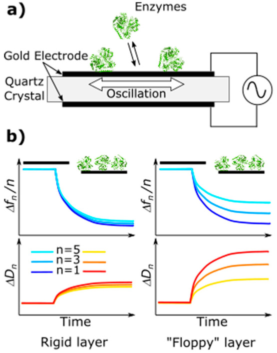
(a) Working principle of the QCM-D. The quartz disk is oscillated at its resonance frequencies with an alternating electric field. (b) During the adsorption of proteins on the surface, the resonance frequencies decrease (Δf) and the dissipations increase (ΔD). Typical responses for a rigid and “floppy” add-layer are shown on the left and right, respectively.
Further, electrochemical and QCM-D measurements can be performed simultaneously permitting correlation between film structures and electrochemical properties. This technique is commonly referred as electrochemical quartz crystal microbalance E-QCM-D. In the case of electroenzymatic catalysis, comparison of the amount of adsorbed enzyme, as measured with QCM-D, with electroenzymatic activity can give insight on the optimal orientation attached enzyme should have to maximize the electron transfer.200−202
Azurin has been found to irreversibly adsorb on a gold electrode modified with a self-assembled monolayer of octanethiol.201 The maximum surface concentration was independent of the protein solution concentration; for all the concentrations studied, the maximum adsorbed amount was 25 ± 1 pmol cm–2, closely corresponding to a monolayer coverage. Only small changes were detected in the dissipation signal suggesting a rigid layer structure. CV measurements performed on the same surface have shown electrochemical activity consistent with a one-electron, surface-confined Cu2+/1+ azurin redox couple. Using the peak area of the reduction peak, the surface coverage of electrochemically active azurin was estimated to be 7 ± 1 pmol cm–2. This discrepancy between QCM-D and cyclic voltammetry data suggested that not all the adsorbed proteins might have been electrochemically active.201 The adsorption of laccase on carboxyl- (Au-R–) and amine (Au-R+)-terminated ethylphenyl layers on gold electrodes has also been studied by the EQCM.200 Even if the amount of adsorbed enzyme was similar for both surfaces, the electrocatalytic current for oxygen reduction was significantly smaller when the proteins were adsorbed on the Au-R– surface compared to the Au-R+ surface. Different orientations of the proteins on the electrode surfaces were hypothesized to be the cause of variation in electrocatalytic activity between Au-R– and Au-R+ functionalization, likely due to the single-entry point of electrons via the “type 1” Cu of laccase (with one orientation favoring DET over the other). Furthermore, E-QCM-D can be used to optimize the enzyme deposition conditions to increase the electrocatalytic activity of the adsorbed species. It has been shown that maximum surface load does not always produce the best electroactive film, suggesting that knowledge of the enzyme-adsorbed amount is an important parameter to maximize the electrode efficiency.203 Bilirubin oxidase has been found to have maximum catalytic activity when adsorbed on unmodified and carboxylate-functionalized gold-coated sensors from a 15 mg mL–1 solution at pH 6.0. These optimal adsorption conditions could originate from a balance between rates of adsorption, reorientation, and unfolding for the protein adsorbed on the electrode surface.203
Hydrogenase (H2ase) and formate dehydrogenase (FDH) adsorption and electroactivity on a range of charged and neutral SAM-modified gold electrodes has been investigated using E-QCM-D.202 Using different types of SAMs, the effects of electrostatic and H-bonding interactions have been resolved, with electrostatic interactions having been found to be important for optimal enzyme orientation and electrocatalytic activity. Positively charged SAMs (SAM+) have shown near quantitative binding of H2ase and FDH in the correct orientation for direct electron transfer. In contrast, enzymes adsorbed on neutral and negatively charged SAMs have shown non-optimal orientation with reduced electroactivities. This behavior has been rationalized with the electrostatic attraction between the negatively charged region surrounding the distal FeS cluster of H2ase, or the FeS cluster of Fdh, and the SAM+. Using the enzyme loading obtained by E-QCM-D, the turnover frequencies (TOFs) of H2ase and FDH adsorbed on SAM+ have been found to be around 2 orders of magnitude smaller than the values found for the enzymes in solution. Since non-optimal orientation could be ruled out, the reason for these discrepancies is not clear, with one possible explanation being that not all the adsorbed enzymes are electroactive due to protein denaturization upon adsorption on the substrate. The stability of the adsorbed layers during electrochemical activity can also be followed using E-QCM-D. For H2ase adsorbed on different types of self-assembled monolayers, desorptive enzyme loss is found when H-bonding is not present at the enzyme–electrode interface.202 For bilirubin oxidase, the catalytic activity decreases even if the adsorbed mass remains stable. However, reduction in activity is associated with an increase in stiffness of the adsorbed layer, suggesting structural rearrangements as the primary mechanism of activity loss.204
One of the limitations of QCM-D consists in its capability on detecting what is called “wet” mass, which includes the adsorbate mass and the water coupled in the layer. Data analysis might be complicated by the fact that the changes in mass observed during one experiment can be attributed to conformational changes of the add-layer and/or desorption/adsorption. For this reason, QCM-D is often coupled with optical techniques, which detect only the dry mass.204,205 A combination of optical and piezoelectric techniques allows for water content determination and can give more detailed information on the structure of the adsorbed layer. Further development in this direction should be addressed to extensively characterize the layer structure of adsorbed enzymes. Conformational changes of adsorbed enzymes during electrocatalysis could be addressed and differentiated from film stability and protein desorption.
QCM-D constitutes a powerful tool also to perform real-time monitoring of bacterial cell deposition on a surface and biofilm growth, and it has been successfully utilized to study these processes on various inorganic materials.206,207 When utilizing E-QCM-D it is further possible to couple the information on biofilm formation with variations in the current–time profile. This approach has been recently utilized by Heidary et al. using G. sulfurreducens and inverse-opal ITO electrodes showing a lag phase between biofilm formation (detected by an increase in dissipation) and current generation (measured by amperometric i–t trace at +0.3 V vs SHE).208 The change in dissipation was utilized to monitor the variations in mass, rather than the change in frequency, due to the thick and viscoelastic nature of G. sulfurreducens biofilms. While the increase in dissipation occurred shortly after inoculation of the bacterial cells, the current remained almost null for more than 12 h, and significantly rose only after 4 days. Accordingly, the E-QCM-D study revealed that significant biofilm formation occurs even in the absence of EET, and that bacterial cells need to adapt to the “electrode respiring metabolism” (transfer of electrons to the electrode rather than to the natural electron acceptor).
3.2.3. Atomic Force Microscopy and Scanning Electrochemical Microscopy
Atomic force microscopy (AFM) represents a well-established technique for topological investigation of surfaces and adsorbed species. Shortly, an AFM image is obtained by scanning a nanometer-sized tip across the sample. The tip is attached to an extreme of a microcantilever, which deflects as the tip interacts with the sample. Bending of the cantilever is detected by a laser beam reflected from the back of the cantilever into a position sensitive four-quadrant photodetector (Figure 7). The cantilever can be either operated in static mode (CM-AFM) or vibrated close to its resonance frequency (AM-AFM). While for CM-AFM the cantilever deflection is detected, in AM-AFM the cantilever oscillation amplitude is recorded. When the AFM tip interacts with a feature on the surface, cantilever deflection or oscillation amplitude changes. A set point value for deflection or oscillation amplitude is chosen and the tip–sample distance is reduced until the set point is reached. The topographic structure of the sample is obtained by scanning the tip over the surface and the tip–sample distance is changed to keep the set point value constant. To this end, AFM has been routinely used to address structures of proteins adsorbed on flat surfaces. High-resolution topographic images of membrane protein porin OmpF have been successfully acquired.209 Cysteine-modified Ompf proteins (Ompf-Cys) have been adsorbed on a flat gold surface and lateral motion of the protein has been hindered with subsequent addition of thio-lipids. High-quality images of isolated Ompf-Cys have been obtained, allowing for the trimers forming the protein to be clearly resolved.209
Figure 7.
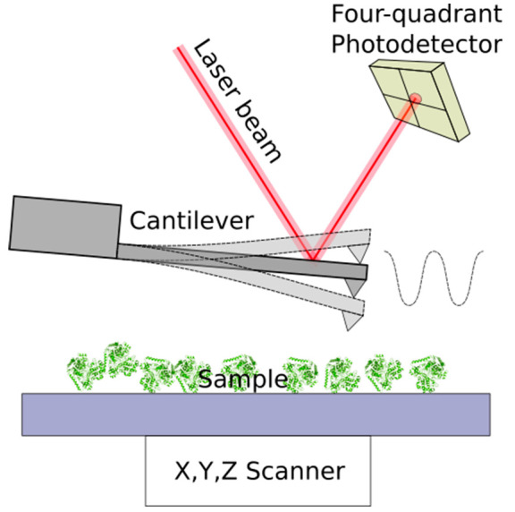
Typical setup of an AFM system. Sample is moved with a nanopositioning scanner. Tip–sample interaction deflects the cantilever, whose deformations are recorded using a laser beam reflected from the back of the cantilever onto a four-quadrant photodetector.
Advanced scanning techniques such as bimodal atomic force microscopy (AM-FM AFM) have enabled the accurate measurement of the elastic modulus of surfaces in liquid with subnanometer spatial resolution.210 AM-FM AFM has been used to simultaneously acquire the topography and the elastic modulus of adsorbed proteins.210,211 High-speed AFM (HS-AFM) has been used to investigate biological processes in physiological conditions with high spatial and temporal resolutions allowing for kinetic studies and real time monitoring of conformational changes. The self-assembly reaction of SAS-6 proteins to form a nine-fold radially symmetric ring structure has been successfully imaged in real-time with HS-AFM.212 Single-molecule kinetics of bacteriorhodopsin (BR) has been studied with HS-AFM. BR conformational changes with millisecond temporal resolution have been observed upon light irradiation activation.213
Extracting single enzyme activity from the macroscopic average could introduce additional understanding on how adsorbed enzymes perform their specific functionalities. For instance, the effect of enzyme conformation, orientation and surface-specific adsorption on its catalytic activity could be addressed at the single-molecule level. Scanning electrochemical microscopy (SECM) is a spatially resolved technique which uses microelectrodes to locally probe or trigger electrochemical processes on surfaces.214,215 Typical spatial resolution of SECM is situated in the micrometer range and, on some occasions, submicron resolution has been demonstrated,216,217 making this technique of particular interest for the study of microbial electrochemical systems. Bard et al. performed SECM of living bacterial cells to investigate the antibacterial effects of Ag+ ions on E. coli by monitoring the variations in oxygen concentration at the tip electrode, positioned at 25 μm from the bacterial cells, polarized at −0.8 V vs a silver paint.218 SECM of microbial electrodes with photosynthetic purple bacteria shed light on the capability of different diffusible redox mediators to cross one or multiple membranes of these organisms, and evidenced that the exogenous mediators react with different redox intermediates depending on the location of the bacterial cell that they can reach.219,220 Specifically, Cai et al. performed current–distance studies by positioning the tip electrode of the SECM on top of the bacterial cells contained in a plastic culture dish, and moving the tip closer to the bacterial cells. By fitting the obtained current–distance curves it was possible to determine the heterogeneous rate constant of electron transfer between various exogenous mediators, both hydrophobic and hydrophilic, and the redox centers of the photosynthetic bacteria. Interestingly, the study evidenced that the exogenous redox mediators react with different redox centers located in the periplasm or inside the cytoplasmic membrane depending on their hydrophilic or hydrophobic properties, respectively.219 Furthermore, Longobardi et al. performed SECM studies with a setup similar to that of Cai et al. but utilizing membrane fragments containing the photosynthetic apparatus of purple bacteria rather than intact bacterial cells to study the influence of the cell walls on electron transfer kinetics.221 The comparisons of an effective heterogeneous rate constant obtained for the same exogenous redox mediator, namely, menadione, with intact bacterial cells and isolated chromatophores were of (2.6 ± 0.2) × 10–3 and (4.8 ± 0.1) × 10–3 cm s–1, respectively, revealing that the diffusion of the mediator across the outer membrane strongly influences the electron transfer kinetics. SECM allowed also the real-time study of quorum sensing in aggregates of bacterial cells.222 Specifically, Whiteley and co-workers utilized SECM to measure the production of pyocyanin, a quorum sensing-controlled secondary metabolite produced by Pseudomonas aeruginosa (P. aeruginosa) that is important for its virulence. In their study, the tip electrode of SECM was polarized at 0 V vs Ag|AgCl to oxidize the reduced form of pyocyanin that is produced by the bacteria, determining the number of cells aggregates required for its production and the distance between different cells aggregates to stimulate the quorum sensing.
SECM studies were recently utilized also to shed light on the microbiologically influenced corrosion process of S. oneidensis MR-1 biofilms on stainless steel. Li et al. utilized riboflavin, an endogenous redox mediator produced by Shewanella, and polarized the ultramicroelectrode tip of SECM to either riboflavin reducing (−0.6 V vs Ag|AgCl) or oxidizing (−0.2 V vs Ag|AgCl) potentials, while biofilms of the bacterial cells were grown on stainless steel electrodes with or without passive layers.223 The study provided in situ evidence of the EET process taking place during the corrosion process, and revealed that the bacterial cells can perform bidirectional extracellular electron transfer using the diffusible mediator depending on the state of the steel surface (active or passive). As highlighted by these works, SECM studies can provide several insights on microbial electrochemical systems, conversely, when focusing on the study of single enzymes, SECM is not capable of resolving such electrocatalytic activity. To address this problem, effort has been undertaken to combine the high resolution of AFM with the electrochemical selectivity of SECM.224,225
AFM combined with scanning electrochemical microscopy (AFM-SECM) has emerged to be a useful technique for simultaneous topographical-electrochemical measurements.224 In AFM-SECM, the tip can function either as primary (WE1) or secondary working electrode (WE2). As the tip is scanned over the sample, the topographic image and the electrochemical signal are recorded simultaneously. Figure 8a shows a typical setup for the probe working as primary electrode. Enzymes are adsorbed on an insulating surface. During the topographical scan, when the tip interacts with the adsorbed molecules, an electrochemical current is detected. When the tip is used as WE2, proteins are adsorbed on a conductive surface which acts as WE1. During the scan over the surface, the tip records the product of the electrochemical reaction (Figure 8b).
Figure 8.
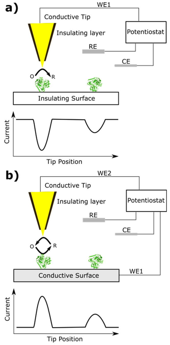
AFM-SECM working sketch. (a) Tip functions as primary working electrode (WE1). The electrochemical reaction takes place when the tip interacts with the adsorbed enzyme. (b) Tip functions as secondary working electrode (WE2). The electrochemical reaction products are detected when the tip is situated in the vicinity of the adsorbed molecule.
Local electrochemical activity sensing with a conductive probe is challenging since the cantilever is covered with a conductive layer and entirely electroactive. In such conditions, the ability to measure processes specifically at the probe-sample interface is lost due to non-faradaic currents produced at the surface of the cantilever hindering the signal originating at the apex of the probe.226 An ideal cantilever for AFM-SECM measurements is insulated and only the apex of the probe is conductive. Different techniques have been developed to build high-resolution AFM-SECM probes that contain well-defined electrode geometries.226,227 High lateral resolution of AFM-SECM has been demonstrated by imaging gold nanoparticles (20 nm) functionalized with redox-labeled PEG chains.228 The PEG corona and the gold core of individual nanoparticles have been simultaneously “visualized” by combining the electrochemical and topological signals. Combining scanning tunneling microscopy with SECM the catalytic turnover of hydrogenase adsorbed on gold SAM have been successfully investigated at single-molecule level.229 The direct observation of the single enzyme activity on the surface joined with macroscopic electrochemical measurements allowed the evaluation of a turnover frequency (TOF) for single hydrogenase molecules. Single-molecule TOF has been determined as a function of number of carbons in the SAM. When extrapolating to zero thickness, the TOF has been estimated to be ∼21,000 s–1 at −0.7 V vs Ag|AgCl.
Individual viral proteins marked by redox antibodies and adsorbed on virus particles have been successfully studied with AFM-SECM.230 Two filamentous plant viruses belonging to the Potyviridae family genus Potyvirus have been studied (PTV). They are rod-shaped particles (∼700–900 nm in length and ∼10–15 nm in diameter) and are made of helical winding coat proteins (CP) packing the viral genomic single-stranded RNA. An additional protein (VPg) is attached at one end of the rod-shaped particles. CP and VPg could be selectively redox immunomarked using ferrocene-functionalized antibodies. When the CP proteins were redox immunomarked, an electrochemical signal has been detected when the AFM tip was scanned over the backbone of the rod-shaped particles. Conversely, when the VPg proteins were redox immunomarked, an electrochemical current was detected only when the tip was located at one extremity of the particle.
Even though AFM-SECM has shown interesting potentials for future applications, it is still considered a highly specialized technique. The cost of production, durability, and reliability of the probes represent major challenges to improve lateral resolution necessary to achieve single enzyme activity detection. Nevertheless, the information at the single-molecule level one might obtain with AFM-SECM could lead to substantial advancements on the understanding of the electroactivity of adsorbed enzymes. For instance, correlation between electrochemical activity of adsorbed molecules and specific binding sites of the surface, such as terraces and imperfections, could drive to the development of a better electrode substrate. Comparisons of topographic heights of adsorbed protein with electrochemical currents could give insight on the preferential molecule orientation for direct electron transfer. Finally, changes in enzyme rigidity during electrochemical and electron transferring processes may provide information on the role of dynamic protein conformations.
3.3. Fluorescence Microscopy and Spectroscopy
Fluorescence microscopy (FM) and fluorescence spectroscopy (FS) are important tools for the characterization of cell physiology and morphology that can be couple to electrochemical evidence to obtain helpful information for the comprehensive study of EET phenomena. Fluorescence spectroscopy, also known as fluorimetry or spectrofluorometry, dates back to 1852 when Stokes observed the emission of light in a lower energy in relation to the wavelength at which the molecule was excited, known as the “Stokes shift”. Fluorescence rapidly turned out as a well-established method allowing the selective recognition of single cells and of specific components comprising biomolecular complex structures.231 FM coupled to FS plays an important role in determination of bacterial activity as it allows the deep investigation of biological processes.232 The development of new fluorescent probes easily adaptable to a wide array of biological applications, coupled to the technical improvements in filters, phase-contrast, and software enhanced the contrast of living microorganisms, strongly supporting the exploitation of fluorescence microscopy as a powerful research tool.233,234 The application of a broad range of fluorophores enables the identification of intact cells and cellular components with a high degree of specificity amidst nonfluorescing material.235 Wide-field fluorescence microscopy (WFM) also referred to as epifluorescence microscopy, is the most common fluorescence microscopy method used in life sciences, and it has permitted bacterial cell density and viability to be studied for the comprehension of dynamics and biogeochemical cycles in aquatic ecosystems. Specifically, in this technique the use of different fluorescent stains enables the identification and counting of cells with intact membranes (viable cells) among those with damaged membranes (nonviable) under the excitation in a specific wavelength.236 In microbial electrochemistry (as in enzymatic electrochemistry), the confirmation of the biotic origin of the current response plays a critical role on the study of bioelectrocatalysis. Similarly, the proper correlation of current generated and viable microbial cell loading on electrodes allows correctly comparing different electron transfer approaches by avoiding misinterpretations due to variations in cells viability. Accordingly, FM and FS provide a power tool to verify and obtain quantitative information on the viability of cells exposed to different environments or entrapped in redox polymers for facilitating EET. This approach was recently utilized by Grattieri and co-workers by combining WFM and FS to study the effects of photosynthetic purple bacteria entrapment in a redox-adhesive polydopamine matrix (PDA).81 Specifically, free Rhodobacter capsulatus (R. capsulatus) cells and cells entrapped in the PDA matrix were incubated with fluorescein diacetate (FDA), a colorless and nonpolar molecule. Intracellularly, or next to membrane associated esterases produced by metabolically active microorganisms, FDA is hydrolyzed to fluorescein, a green fluorescent compound (Figure 9 top), detectable spectroscopically by measuring its fluorescence. Viable and active cells are thus stained with fluorescein, while inactive cells are not stained and do not contribute to the fluorescence response. Following the incubation of R. capsulatus cells, their viability in the different conditions (free or entrapped) was visually confirmed by WFM and quantified by FS (Figure 9 bottom), revealing that cells retain almost 100% viability after entrapment in the PDA matrix. For comparison, cells were heat-treated to obtain dead bacteria that were also incubated with FDA, resulting in the absence of fluorescence (Figure 9, bottom, blue line).
Figure 9.

(Top) Reaction of fluorescein diacetate with esterases present intracellularly or bound to the external membrane of bacterial cells. (Bottom left) FS result obtained for free R. capsulatus cells (red), R. capsulatus cells entrapped in PDA (black), heat-treated R. capsulatus cells at 120 °C for 4 h (blue), and PDA only (purple), adapted with permission under a Creative Commons Attribution License from ref (81). Copyright 2022 The Authors. (Bottom right) Scheme of a microscopy slide covered in bacteria for WFM analysis.
WFM was explored also to evaluate the morphologies of anodic biofilms and their formation. It was possible to correlate the densities of mixed bacterial culture enriched at different temperatures with their power generation performance.237 WFM enabled continuously monitoring the different stages of grow of P. aeruginosa biofilms on the surface of a biosensor chip, by collecting fluorescence images at various cultivation times. Starting from the adhesion of microorganisms to the substrate surface visualized with fluorescent spots, the growth stage is determined with clusters of fluorescent areas that later reach the mature stage where homogeneous fluorescent areas are obtained, and finally the dispersal stage is revealed by a gradual decrease in fluorescence. Specifically, Liu et al. utilized carboxyfluorescein diacetate succinimidyl ester, another colorless nonpolar molecule, that, similarly to FDA, is hydrolyzed to the fluorescent carboxyfluorescein in the presence of esterases to follow biofilm formation on a microelectrode modified with Au nanoparticles, and correlated the WFM images to differential pulse voltammetry studies measuring pyocyanin production.238 Recently, Pirbadian et al. combined WFM with a three-electrode bioelectrochemical reactor using ITO as working electrode modified with S. oneidensis MR-1 cells stained with the fluorescent cationic dye thioflavin T.239 The charged dye worked as a membrane potential indicator, as due to its positive charge it accumulates inside negatively charged membranes. The study unveiled that when the electrode is polarized to positive potentials (+0.3 V vs Ag|AgCl) a more negative membrane potential is obtained, with an increase in thioflavin T fluorescence. Conversely, moving to a low potential polarization (−0.5 V vs Ag|AgCl) resulted in low fluorescence. In addition, the developed method allowed correlating the distance of bacterial cells from the electrode surface and EET activity, by utilizing patterned working electrodes. It was possible to rule out electron transport beyond ∼30 μm from the electrode surface, due to the matching between the cell fluorescence and the electrode pattern.
We previously presented the possibility to utilize SECM to monitor variations in pyocyanin concentration related to the number of P. aeruginosa cells and their distance in the work of Whiteley and co-workers, which utilized confocal scanning microscopy (CSM) to obtain a P. aeruginosa cell count.222 Compared to conventional WFM, CSM provides not only information about cell membrane integrity but also on cellular and tissues’ complex morphology and dynamics with high-quality spatial resolution and contrast. The blurred aspect of images in conventional microscopy is inherent and results from the superimposition of multiple images at the detector, which are formed at different depths of field in optical lens, depending on the focal distance adjusted. Confocal microscopes can generally be classified between noncoherent and laser microscopes. The first type uses mercury or xenon as a light source, while in the second, a single wavelength or multiwavelength laser is used to excite the sample.240 In confocal laser scanning microscopy (CLSM), the acquisition point by point at specific wavelengths using localized laser excitation offers advantages such as minimal background signature, versatility offered by fluorescent stains employed allowing the detection of extracellular DNA and exopolysaccharides in the samples.241 The ability of CLSM to collect serial optical sections from thick specimens enables 3D analysis of biofilm architecture and viability, contributing for the evaluation of efficacy and for implementation of antimicrobial drugs.242 CLSM was used to investigate cell attachment, biofilm formation, and spatial heterogeneity across the entire electrode surface modified with current-producing biofilms of Marinobacter atlanticus on a continuous flow cell. These biofilms engineered to constitutively express a chromosomal copy of the gene for the orange fluorescent protein dTomato were induced to express the yellow fluorescent protein (YFP) under continuous exposure to isopropyl β-d-1-thiogalactopyranoside (IPTG), while monitoring current generation. CLSM made it possible to observe a dynamic spatiotemporal response of YFP expression, since the fluorescence response changed according to the stage of biofilm development.243 A flow cell-based electrochemical reactor coupled to CLSM has been utilized also by Stöckl to simultaneously perform CLSM and electrochemical impedance spectroscopy to study the formation of electroactive biofilms of Shewanella oneidensis MR-1.244
3.4. Bioinformatics Analysis
Bioinformatic analysis has been gaining interest for the study of microbial electrochemical systems.135 This can be easily explained considering the capability of bioinformatic tools to allow studying both the genomic traits in deoxyribonucleic acid (DNA) as well as the transcription rates of ribonucleic acid (RNA). Such analysis paves the way to an unprecedented knowledge, making it possible to unveil the composition of electroactive species in mixed microbial electrodes, identifying the presence of protein-coding sequences responsible for electroactivity (i.e., presence of coding sequences for c-type cytochromes), or revealing how external parameters (i.e., electrode polarization, presence of toxic compounds, physicochemical conditions) influence genes expression.245 Specifically, whole genome sequencing allows the characterization of the entire genome of a microorganism and can be performed for multiple strains simultaneously (metagenomic). This provides quantitative information on the species present in such mixed communities, which is particularly useful to correlate electroactivity to microbial composition and study EET processes in such systems. Ishii et al. combined metagenomic analysis to transcriptomic analysis (the quantification of RNA transcripts, metatranscriptomics for mixed species communities) for electrochemically active mixed microbial communities in microbial fuel cells allowing to identify which microbial groups responded to changing electrochemical stimuli based on gene expression responses.246 Later, the same group utilized the combination of these two approaches to study the effect of variable electrode polarizations and substrates changes on mixed microbial consortia.247 The study enabled identifying the abundance of species depending on the electrochemical conditions utilized (potentials from −200 to +100 mV vs SHE), with abundance of Geobacteraceae microbes. Furthermore, it was shown that the different electrode polarization influenced the expression of genes related to EET, such as multiheme c-type cytochromes and conductive pili, revealing a remarkable capability of Geobacteraceae to adapt to significant changes in electrode potential and substrate availability. Recently, transcriptomic analysis was utilized to unveil the effects of salinity248 and changing substrates249 on photobioelectrocatalysis of purple bacteria. Also for these studies, the combination of bioinformatic analysis and electrochemical experimental evidence allowed to unveil the effects of external stress on the photosynthetic metabolism and resulting photocurrent production.
4. Conclusions
Enzymatic and microbial electrochemical systems have several exciting applications and have the potential to play a critical role in the development of sustainable bioelectrosynthetic approaches, biosensing platforms, and local micropower/decontamination facilities. However, to meet such goals, improving the fundamental understanding of the electron transfer processes at the basis of these technologies will be critical, as a detailed understanding of such processes would enable rationally designing electrodes and cell configurations for biohybrid electrochemical systems with enhanced performances. As discussed in this tutorial review paper, several approaches are now available to tune bioelectrochemical systems, from artificial electron mediation systems to synthetic biology, providing unprecedent research possibilities. At the same time, various methods to characterize in detail enzymatic and microbial electrochemical systems have been successfully applied, shedding light on the mechanisms at the basis of these technologies. It should be noted that some of the presented methods have been only recently applied to the study of bioelectrochemical systems. However, the combination of different techniques (i.e., fluorescence microscopy and SECM or CV) is providing an example of how multidisciplinary approaches are enabling a new level of understanding of these complex systems. We believe that the future research in bioelectrochemistry, with the synergistic implementation of the presented approaches and methods, will pave the way to the development of biohybrid electrochemical systems with improved performance applicable in sustainable industrial processes and everyday life.
Acknowledgments
This project has received funding from the European Union’s Horizon 2020 research and innovation programme under the Marie Skłodowska-Curie Grant Agreement No. 101024443. This publication was created as part of NCCR Catalysis (Grant No. 180544), a National Centre of Competence in Research funded by the Swiss National Science Foundation. G.B., A.K., and R.D.M. thank the Swiss National Science Foundation for support (Grant No. 200021_191985). M.G. would like to acknowledge funding from Fondazione CON IL SUD, project number 2018-PDR-00914.
Author Present Address
# São Paulo State University (UNESP), Institute of Chemistry, Araraquara. 55 Prof. Francisco Degni St, Araraquara, SP, 14800-060, Brazil
Author Contributions
CRediT: Giada Bedendi conceptualization (equal), writing-original draft (equal); Lilian D. De Moura Torquato conceptualization (equal), writing-original draft (equal); Sophie Webb conceptualization (equal), writing-original draft (supporting); Cecile Cadoux conceptualization (equal), writing-original draft (supporting); Amogh Kulkarni conceptualization (equal), writing-original draft (supporting); Selmihan Sahin conceptualization (equal), funding acquisition (supporting), writing-original draft (supporting); Plinio Maroni conceptualization (equal), writing-review & editing (equal); Ross D. Milton funding acquisition (equal), supervision (equal), writing-review & editing (equal); Matteo Grattieri funding acquisition (equal), supervision (equal), writing-review & editing (equal).
The authors declare no competing financial interest.
Notes
In this tutorial review, biological standard reduction potentials (E0′) are given at pH 7, unless stated otherwise.
References
- Unden G.; Bongaerts J. Alternative respiratory pathways of Escherichia coli: energetics and transcriptional regulation in response to electron acceptors. Biochim. Biophys. Acta - Bioenerg. 1997, 1320, 217–234. 10.1016/S0005-2728(97)00034-0. [DOI] [PubMed] [Google Scholar]
- Kano K.; et al. Voltammetric determination of acid dissociation constants of pyrroloquinoline quinone and its reduced form under acidic conditions. Bioelectrochemistry Bioenerg. 1990, 24, 193–201. 10.1016/0302-4598(90)85021-9. [DOI] [Google Scholar]
- Wagner T.; Koch J.; Ermler U.; Shima S. Methanogenic heterodisulfide reductase (HdrABC-MvhAGD) uses two noncubane [4Fe-4S] clusters for reduction. Science (80-.). 2017, 357, 699–703. 10.1126/science.aan0425. [DOI] [PubMed] [Google Scholar]
- Buckel W.; Thauer R. K. Flavin-Based Electron Bifurcation, A New Mechanism of Biological Energy Coupling. Chem. Rev. 2018, 118, 3862–3886. 10.1021/acs.chemrev.7b00707. [DOI] [PubMed] [Google Scholar]
- Blankenship R. E. Early Evolution of Photosynthesis. Plant Physiol. 2010, 154, 434–438. 10.1104/pp.110.161687. [DOI] [PMC free article] [PubMed] [Google Scholar]
- Olson K. R.; Straub K. D. The role of hydrogen sulfide in evolution and the evolution of hydrogen sulfide in metabolism and signaling. Physiology 2016, 31, 60–72. 10.1152/physiol.00024.2015. [DOI] [PubMed] [Google Scholar]
- Huang S.; Jaffé P. R. Defluorination of Perfluorooctanoic Acid (PFOA) and Perfluorooctane Sulfonate (PFOS) by Acidimicrobium sp. Strain A6. Environ. Sci. Technol. 2019, 53, 11410–11419. 10.1021/acs.est.9b04047. [DOI] [PubMed] [Google Scholar]
- Pankratova G.; Gorton L. Electrochemical communication between living cells and conductive surfaces. Curr. Opin. Electrochem. 2017, 5, 193–202. 10.1016/j.coelec.2017.09.013. [DOI] [Google Scholar]
- Liu Y.; Atanassov P. Charge transfer at biotic/abiotic interfaces in biological electrocatalysis. Curr. Opin. Electrochem. 2020, 19, 175–183. 10.1016/j.coelec.2019.09.007. [DOI] [Google Scholar]
- Torquato L. D. de M.; Grattieri M. Photobioelectrochemistry of intact photosynthetic bacteria: Advances and future outlook. Curr. Opin. Electrochem. 2022, 34, 101018. 10.1016/j.coelec.2022.101018. [DOI] [Google Scholar]
- Wang J. Electrochemical glucose biosensors. Chem. Rev. 2008, 108, 814–825. 10.1021/cr068123a. [DOI] [PubMed] [Google Scholar]
- Leech D.; Kavanagh P.; Schuhmann W. Enzymatic fuel cells: Recent progress. Electrochim. Acta 2012, 84, 223–234. 10.1016/j.electacta.2012.02.087. [DOI] [Google Scholar]
- Rasmussen M.; Abdellaoui S.; Minteer S. D. Enzymatic biofuel cells: 30 years of critical advancements. Biosens. Bioelectron. 2016, 76, 91–102. 10.1016/j.bios.2015.06.029. [DOI] [PubMed] [Google Scholar]
- Osman M. H.; Shah A. A.; Walsh F. C. Recent progress and continuing challenges in bio-fuel cells. Part I: Enzymatic cells. Biosens. Bioelectron. 2011, 26, 3087–3102. 10.1016/j.bios.2011.01.004. [DOI] [PubMed] [Google Scholar]
- Mazurenko I.; Wang X.; De Poulpiquet A.; Lojou E. H2/O2 enzymatic fuel cells: from proof-of-concept to powerful devices. Sustain. Energy Fuels 2017, 1, 1475–1501. 10.1039/C7SE00180K. [DOI] [Google Scholar]
- Ruth J. C.; Spormann A. M. Enzyme Electrochemistry for Industrial Energy Applications - A Perspective on Future Areas of Focus. ACS Catal. 2021, 11, 5951–5967. 10.1021/acscatal.1c00708. [DOI] [Google Scholar]
- Cadoux C.; Milton R. D. Recent Enzymatic Electrochemistry for Reductive Reactions. ChemElectroChem. 2020, 7, 1974–1986. 10.1002/celc.202000282. [DOI] [Google Scholar]
- Wu R.; Ma C.; Zhu Z. Enzymatic electrosynthesis as an emerging electrochemical synthesis platform. Curr. Opin. Electrochem. 2020, 19, 1–7. 10.1016/j.coelec.2019.08.004. [DOI] [Google Scholar]
- Lubitz W.; Ogata H.; Rüdiger O.; Reijerse E. Hydrogenases. Chem. Rev. 2014, 114, 4081–4148. 10.1021/cr4005814. [DOI] [PubMed] [Google Scholar]
- Sensi M.; del Barrio M.; Baffert C.; Fourmond V.; Léger C. New perspectives in hydrogenase direct electrochemistry. Curr. Opin. Electrochem. 2017, 5, 135–145. 10.1016/j.coelec.2017.08.005. [DOI] [Google Scholar]
- Armstrong F. A.; et al. Guiding Principles of Hydrogenase Catalysis Instigated and Clarified by Protein Film Electrochemistry. Acc. Chem. Res. 2016, 49, 884–892. 10.1021/acs.accounts.6b00027. [DOI] [PubMed] [Google Scholar]
- Einsle O.; Rees D. C. Structural Enzymology of Nitrogenase Enzymes. Chem. Rev. 2020, 120, 4969–5004. 10.1021/acs.chemrev.0c00067. [DOI] [PMC free article] [PubMed] [Google Scholar]
- Schlager S.; Fuchsbauer A.; Haberbauer M.; Neugebauer H.; Sariciftci N. S. Carbon dioxide conversion to synthetic fuels using biocatalytic electrodes. J. Mater. Chem. A 2017, 5, 2429–2443. 10.1039/C6TA07571A. [DOI] [Google Scholar]
- Jeoung J. H.; Martins B. M.; Dobbek H.. Carbon monoxide dehydrogenases. Methods in Molecular Biology; Humana Press: New York, NY, 2019; Vol. 1876, pp 37–54. [DOI] [PubMed] [Google Scholar]
- Milton R. D.; et al. Bioelectrochemical Haber-Bosch Process: An Ammonia-Producing H2/N2 Fuel Cell. Angew. Chemie - Int. Ed. 2017, 56, 2680–2683. 10.1002/anie.201612500. [DOI] [PubMed] [Google Scholar]
- Santoro C.; Arbizzani C.; Erable B.; Ieropoulos I. Microbial fuel cells: From fundamentals to applications. A review. J. Power Sources 2017, 356, 225–244. 10.1016/j.jpowsour.2017.03.109. [DOI] [PMC free article] [PubMed] [Google Scholar]
- Gajda I.; Greenman J.; Ieropoulos I. A. Recent advancements in real-world microbial fuel cell applications. Curr. Opin. Electrochem. 2018, 11, 78–83. 10.1016/j.coelec.2018.09.006. [DOI] [PMC free article] [PubMed] [Google Scholar]
- Cao B.; et al. Silver nanoparticles boost charge-extraction efficiency in Shewanella microbial fuel cells. Science (80-.). 2021, 373, 1336–1340. 10.1126/science.abf3427. [DOI] [PubMed] [Google Scholar]
- Simoska O.; et al. Recent trends and advances in microbial electrochemical sensing technologies: An overview. Curr. Opin. Electrochem. 2021, 30, 100762. 10.1016/j.coelec.2021.100762. [DOI] [Google Scholar]
- Chu N.; Liang Q.; Jiang Y.; Zeng R. J. Microbial electrochemical platform for the production of renewable fuels and chemicals. Biosens. Bioelectron. 2020, 150, 111922. 10.1016/j.bios.2019.111922. [DOI] [PubMed] [Google Scholar]
- Liu B.; Lei Y.; Li B. A batch-mode cube microbial fuel cell based ‘shock’ biosensor for wastewater quality monitoring. Biosens. Bioelectron. 2014, 62, 308–314. 10.1016/j.bios.2014.06.051. [DOI] [PubMed] [Google Scholar]
- Lazzarini Behrmann I. C.; Grattieri M.; Minteer S. D.; Ramirez S. A.; Vullo D. L. Online self-powered Cr(VI) monitoring with autochthonous Pseudomonas and a bio-inspired redox polymer. Anal. Bioanal. Chem. 2020, 412, 6449–6457. 10.1007/s00216-020-02620-w. [DOI] [PubMed] [Google Scholar]
- Dong F.; Lee Y. S.; Gaffney E. M.; Liou W.; Minteer S. D. Engineering Cyanobacterium with Transmembrane Electron Transfer Ability for Bioelectrochemical Nitrogen Fixation. ACS Catal. 2021, 11, 13169–13179. 10.1021/acscatal.1c03038. [DOI] [Google Scholar]
- Higgins S. R.; Lau C.; Atanassov P.; Minteer S. D.; Cooney M. J. Hybrid biofuel cell: Microbial fuel cell with an enzymatic air-breathing cathode. ACS Catal. 2011, 1, 994–997. 10.1021/cs2003142. [DOI] [Google Scholar]
- Santoro C.; et al. High Power Generation by a Membraneless Single Chamber Microbial Fuel Cell (SCMFC) Using Enzymatic Bilirubin Oxidase (BOx) Air-Breathing Cathode. J. Electrochem. Soc. 2013, 160, H720–H726. 10.1149/2.058310jes. [DOI] [Google Scholar]
- Dubrawski K. L.; et al. Microbial Battery Powered Enzymatic Electrosynthesis for Carbon Capture and Generation of Hydrogen and Formate from Dilute Organics. ACS Energy Lett. 2019, 4, 2929–2936. 10.1021/acsenergylett.9b02203. [DOI] [Google Scholar]
- Marcus R. A.; Sutin N. Electron transfers in chemistry and biology. BBA Rev. Bioenerg. 1985, 811, 265–322. [Google Scholar]
- Jeuken L. J. C. Conformational reorganisation in interfacial protein electron transfer. Biochimica et Biophysica Acta - Bioenergetics 2003, 1604, 67–76. 10.1016/S0005-2728(03)00026-4. [DOI] [PubMed] [Google Scholar]
- Paquete C. M. Electroactivity across the cell wall of Gram-positive bacteria. Comput. Struct. Biotechnol. J. 2020, 18, 3796–3802. 10.1016/j.csbj.2020.11.021. [DOI] [PMC free article] [PubMed] [Google Scholar]
- Shi L.; Dong H.; Reguera G.; Beyenal H.; Lu A.; Liu J.; Yu H.-Q.; Fredrickson J. K. Extracellular electron transfer mechanisms between microorganisms and minerals. Nat. Rev. Microbiol. 2016, 14, 651–662. 10.1038/nrmicro.2016.93. [DOI] [PubMed] [Google Scholar]
- Ghindilis A. L.; Atanasov P.; Wilkins E. Enzyme-catalyzed direct electron transfer: Fundamentals and analytical applications. Electroanalysis 1997, 9, 661–674. 10.1002/elan.1140090902. [DOI] [Google Scholar]
- Milton R. D.; Minteer S. D. Direct enzymatic bioelectrocatalysis: Differentiating between myth and reality. J. R. Soc., Interface 2017, 14, 20170253. 10.1098/rsif.2017.0253. [DOI] [PMC free article] [PubMed] [Google Scholar]
- Mazurenko I.; Hitaishi V. P.; Lojou E. Recent advances in surface chemistry of electrodes to promote direct enzymatic bioelectrocatalysis. Curr. Opin. Electrochem. 2017, 1, 110–120. 10.1016/j.coelec.2019.11.004. [DOI] [Google Scholar]
- Beaufils C.; Man H. M.; de Poulpiquet A.; Mazurenko I.; Lojou E. From Enzyme Stability to Enzymatic Bioelectrode Stabilization Processes. Catalysts 2021, 11, 497. 10.3390/catal11040497. [DOI] [Google Scholar]
- Léger C.; et al. Enzyme electrokinetics: Using protein film voltammetry to investigate redox enzymes and their mechanisms. Biochemistry 2003, 42, 8653–8662. 10.1021/bi034789c. [DOI] [PubMed] [Google Scholar]
- Le Goff A.; Holzinger M. Molecular engineering of the bio/nano-interface for enzymatic electrocatalysis in fuel cells. Sustainable Energy and Fuels 2018, 2, 2555–2566. 10.1039/C8SE00374B. [DOI] [Google Scholar]
- Xiao X.; et al. Tackling the Challenges of Enzymatic (Bio)Fuel Cells. Chem. Rev. 2019, 119, 9509–9558. 10.1021/acs.chemrev.9b00115. [DOI] [PubMed] [Google Scholar]
- Liang W.; et al. Metal-Organic Framework-Based Enzyme Biocomposites. Chem. Rev. 2021, 121, 1077–1129. 10.1021/acs.chemrev.0c01029. [DOI] [PubMed] [Google Scholar]
- Lian X.; et al. Enzyme-MOF (metal-organic framework) composites. Chem. Soc. Rev. 2017, 46, 3386–3401. 10.1039/C7CS00058H. [DOI] [PubMed] [Google Scholar]
- Hermanson G. T.Bioconjugate Techniques, 3rd ed.; Elsevier Inc., 2013. [Google Scholar]
- Steen Redeker E.; et al. Protein engineering for directed immobilization. Bioconjugate Chem. 2013, 24, 1761–1777. 10.1021/bc4002823. [DOI] [PubMed] [Google Scholar]
- Bélanger D.; Pinson J. Electrografting: A powerful method for surface modification. Chem. Soc. Rev. 2011, 40, 3995–4048. 10.1039/c0cs00149j. [DOI] [PubMed] [Google Scholar]
- Love J. C.; Estroff L. A.; Kriebel J. K.; Nuzzo R. G.; Whitesides G. M. Self-assembled monolayers of thiolates on metals as a form of nanotechnology. Chem. Rev. 2005, 105, 1103–1169. 10.1021/cr0300789. [DOI] [PubMed] [Google Scholar]
- Yates N. D. J.; Fascione M. A.; Parkin A. Methodologies for “Wiring” Redox Proteins/Enzymes to Electrode Surfaces. Chem. - A Eur. J. 2018, 24, 12164–12182. 10.1002/chem.201800750. [DOI] [PMC free article] [PubMed] [Google Scholar]
- Léger C.; Jones A. K.; Albracht S. P. J.; Armstrong F. A. Effect of a dispersion of interfacial electron transfer rates on steady state catalytic electron transport in [NiFe]-hydrogenase and other enzymes. J. Phys. Chem. B 2002, 106, 13058–13063. 10.1021/jp0265687. [DOI] [Google Scholar]
- Nair D. P.; et al. The Thiol-Michael addition click reaction: A powerful and widely used tool in materials chemistry. Chem. Mater. 2014, 26, 724–744. 10.1021/cm402180t. [DOI] [Google Scholar]
- Hickey D. P.; et al. Fundamentals and applications of bioelectrocatalysis. In Electrochemistry; Banks C., Mortimer R., McIntosh S., Eds; The Royal Society of Chemistry, 2016; Vol. 13, pp 97–132. [Google Scholar]
- Lyons M. E. G. Mediated electron transfer at redox active monolayers. Part 3: Bimolecular outer-sphere, first order Koutecky-Levich and adduct formation mechanisms. Sensors 2002, 2, 473–506. 10.3390/s21200473. [DOI] [Google Scholar]
- Ruff A. Redox polymers in bioelectrochemistry: Common playgrounds and novel concepts. Curr. Opin. Electrochem. 2017, 5, 66–73. 10.1016/j.coelec.2017.06.007. [DOI] [Google Scholar]
- Yuan M.; Minteer S. D. Redox polymers in electrochemical systems: From methods of mediation to energy storage. Curr. Opin. Electrochem. 2019, 15, 1–6. 10.1016/j.coelec.2019.03.003. [DOI] [Google Scholar]
- Plumeré N.; et al. A redox hydrogel protects hydrogenase from high-potential deactivation and oxygen damage. Nat. Chem. 2014, 6, 822–827. 10.1038/nchem.2022. [DOI] [PubMed] [Google Scholar]
- Bretschger O.; et al. Current production and metal oxide reduction by Shewanella oneidensis MR-1 wild type and mutants. Appl. Environ. Microbiol. 2007, 73, 7003–7012. 10.1128/AEM.01087-07. [DOI] [PMC free article] [PubMed] [Google Scholar]
- Pirbadian S.; El-Naggar M. Y. Multistep hopping and extracellular charge transfer in microbial redox chains. Phys. Chem. Chem. Phys. 2012, 14, 13802–13808. 10.1039/c2cp41185g. [DOI] [PubMed] [Google Scholar]
- Dohnalkova A. C.; et al. Imaging hydrated microbial extracellular polymers: Comparative analysis by electron microscopy. Appl. Environ. Microbiol. 2011, 77, 1254–1262. 10.1128/AEM.02001-10. [DOI] [PMC free article] [PubMed] [Google Scholar]
- Kumar A.; Hsu L. H.-H.; Kavanagh P.; Barriere F.; Lens P. N. L.; Lapinsonniere L.; Lienhard V J. H.; Schroder U.; Jiang X.; Leech D. The ins and outs of microorganism-electrode electron transfer reactions. Nat. Rev. Chem. 2017, 1, 0024. 10.1038/s41570-017-0024. [DOI] [Google Scholar]
- Malvankar N. S.; Yalcin S. E.; Tuominen M. T.; Lovley D. R. Visualization of charge propagation along individual pili proteins using ambient electrostatic force microscopy. Nat. Nanotechnol. 2014, 9, 1012–1017. 10.1038/nnano.2014.236. [DOI] [PubMed] [Google Scholar]
- Marsili E.; et al. Shewanella secretes flavins that mediate extracellular electron transfer. Proc. Natl. Acad. Sci. U. S. A. 2008, 105, 3968–3973. 10.1073/pnas.0710525105. [DOI] [PMC free article] [PubMed] [Google Scholar]
- Saper G.; Kallmann D.; Conzuelo F.; Zhao F.; Toth T. N.; Liveanu V.; Meir S.; Szymanski J.; Aharoni A.; Schuhmann W.; Rothschild A.; Schuster G.; Adir N. Live cyanobacteria produce photocurrent and hydrogen using both the respiratory and photosynthetic systems. Nat. Commun. 2018, 9, 2168. 10.1038/s41467-018-04613-x. [DOI] [PMC free article] [PubMed] [Google Scholar]
- Shlosberg Y.; et al. NADPH performs mediated electron transfer in cyanobacterial-driven bio-photoelectrochemical cells. iScience 2021, 24, 101892. 10.1016/j.isci.2020.101892. [DOI] [PMC free article] [PubMed] [Google Scholar]
- Grattieri M.; et al. Investigating extracellular electron transfer of Rikenella microfusus: a recurring bacterium in mixed-species biofilms. Sustain. Energy Fuels 2017, 1, 1568–1572. 10.1039/C7SE00270J. [DOI] [Google Scholar]
- Sekar N.; Umasankar Y.; Ramasamy R. P. Photocurrent generation by immobilized cyanobacteria via direct electron transport in photo-bioelectrochemical cells. Phys. Chem. Chem. Phys. 2014, 16, 7862–7871. 10.1039/c4cp00494a. [DOI] [PubMed] [Google Scholar]
- Clifford E. R.; et al. Phenazines as model low-midpoint potential electron shuttles for photosynthetic bioelectrochemical systems. Chem. Sci. 2021, 12, 3328–3338. 10.1039/D0SC05655C. [DOI] [PMC free article] [PubMed] [Google Scholar]
- Grattieri M.; Rhodes Z.; Hickey D. P.; Beaver K.; Minteer S. D. Understanding Biophotocurrent Generation in Photosynthetic Purple Bacteria. ACS Catal. 2019, 9, 867–873. 10.1021/acscatal.8b04464. [DOI] [Google Scholar]
- Hasan K.; Patil S. A.; Leech D.; Hägerhäll C.; Gorton L.. Electrochemical communication between microbial cells and electrodes via osmium redox systems. Biochemical Society Transactions; Portland Press, 2012; Vol. 40, pp 1330–1335. [DOI] [PubMed] [Google Scholar]
- Vostiar I.; Ferapontova E. E.; Gorton L. Electrical ‘wiring’ of viable Gluconobacter oxydans cells with a flexible osmium-redox polyelectrolyte. Electrochem. commun. 2004, 6, 621–626. 10.1016/j.elecom.2004.04.017. [DOI] [Google Scholar]
- Coman V.; et al. Electrical wiring of live, metabolically enhanced Bacillus subtilis cells with flexible osmium-redox polymers. J. Am. Chem. Soc. 2009, 131, 16171–16176. 10.1021/ja905442a. [DOI] [PubMed] [Google Scholar]
- Grattieri M.; Patterson S.; Copeland J.; Klunder K.; Minteer S. D. Purple Bacteria and 3D Redox Hydrogels for Bioinspired Photo-bioelectrocatalysis. ChemSusChem 2020, 13, 230–237. 10.1002/cssc.201902116. [DOI] [PubMed] [Google Scholar]
- Hasan K.; Grattieri M.; Wang T.; Milton R. D.; Minteer S. D. Enhanced Bioelectrocatalysis of Shewanella oneidensis MR-1 by a Naphthoquinone Redox Polymer. ACS Energy Lett. 2017, 2, 1947–1951. 10.1021/acsenergylett.7b00585. [DOI] [Google Scholar]
- Liu S. R.; et al. Polydopamine coating on individual cells for enhanced extracellular electron transfer. Chem. Commun. 2019, 55, 10535–10538. 10.1039/C9CC03847G. [DOI] [PubMed] [Google Scholar]
- Li Y.; et al. Enhanced electricity generation and extracellular electron transfer by polydopamine-reduced graphene oxide (PDA-rGO) modification for high-performance anode in microbial fuel cell. Chem. Eng. J. 2020, 387, 123408. 10.1016/j.cej.2019.123408. [DOI] [Google Scholar]
- Buscemi G.; et al. Bio-Inspired Redox-Adhesive Polydopamine Matrix for Intact Bacteria Biohybrid Photoanodes. ACS Appl. Mater. Interfaces 2022, 14, 26631–26641. 10.1021/acsami.2c02410. [DOI] [PMC free article] [PubMed] [Google Scholar]
- Weliwatte N. S.; Grattieri M.; Minteer S. D. Rational design of artificial redox-mediating systems toward upgrading photobioelectrocatalysis. Photochem. Photobiol. Sci. 2021, 20, 1333–1356. 10.1007/s43630-021-00099-7. [DOI] [PMC free article] [PubMed] [Google Scholar]
- Campàs M.; Prieto-Simón B.; Marty J. L. A review of the use of genetically engineered enzymes in electrochemical biosensors. Seminars in Cell and Developmental Biology 2009, 20, 3–9. 10.1016/j.semcdb.2009.01.009. [DOI] [PubMed] [Google Scholar]
- Xu S.; Minteer S. D. Investigating the Impact of Multi-Heme Pyrroloquinoline Quinone-Aldehyde Dehydrogenase Orientation on Direct Bioelectrocatalysis via Site Specific Enzyme Immobilization. ACS Catal. 2013, 3, 1756–1763. 10.1021/cs400316b. [DOI] [Google Scholar]
- Schröper F.; Baumann A.; Offenhäusser A.; Mayer D. Direct electrochemistry of novel affinity-tag immobilized recombinant horse heart cytochrome c. Biosens. Bioelectron. 2012, 34, 171–177. 10.1016/j.bios.2012.01.039. [DOI] [PubMed] [Google Scholar]
- Ganesana M.; Istarnboulie G.; Marty J. L.; Noguer T.; Andreescu S. Site-specific immobilization of a (His)6-tagged acetylcholinesterase on nickel nanoparticles for highly sensitive toxicity biosensors. Biosens. Bioelectron. 2011, 30, 43–48. 10.1016/j.bios.2011.08.024. [DOI] [PubMed] [Google Scholar]
- Patel J.; Cai R.; Milton R.; Chen H.; Minteer S. D. Pyrene-Based Noncovalent Immobilization of Nitrogenase on Carbon Surfaces. ChemBioChem. 2020, 21, 1729–1732. 10.1002/cbic.201900697. [DOI] [PubMed] [Google Scholar]
- Le T. T.; Wilde C. P.; Grossman N.; Cass A. E. G. A simple method for controlled immobilization of proteins on modified SAMs. Phys. Chem. Chem. Phys. 2011, 13, 5271–5278. 10.1039/c0cp02531c. [DOI] [PubMed] [Google Scholar]
- Bello-Gil D.; et al. Specific and reversible immobilization of proteins tagged to the affinity polypeptide C-LytA on functionalized graphite electrodes. PLoS One 2014, 9, e87995 10.1371/journal.pone.0087995. [DOI] [PMC free article] [PubMed] [Google Scholar]
- Go Y.-M.; Chandler J. D.; Jones D. P. The cysteine proteome. Free Radic. Biol. Med. 2015, 84, 227–245. 10.1016/j.freeradbiomed.2015.03.022. [DOI] [PMC free article] [PubMed] [Google Scholar]
- Belete T. M. Novel targets to develop new antibacterial agents and novel alternatives to antibacterial agents. Hum. Microbiome J. 2019, 11, 100052. 10.1016/j.humic.2019.01.001. [DOI] [Google Scholar]
- Hallenbeck K. K.; Turner D. M.; Renslo A. R.; Arkin M. R. Targeting Non-Catalytic Cysteine Residues Through Structure-Guided Drug Discovery. Curr. Top. Med. Chem. 2016, 17, 4–15. 10.2174/1568026616666160719163839. [DOI] [PMC free article] [PubMed] [Google Scholar]
- Visscher M.; Arkin M. R.; Dansen T. B. Covalent targeting of acquired cysteines in cancer. Curr. Opin. Chem. Biol. 2016, 30, 61–67. 10.1016/j.cbpa.2015.11.004. [DOI] [PMC free article] [PubMed] [Google Scholar]
- Wang K.; Peng H.; Wang B. Recent Advances in Thiol and Sulfide Reactive Probes. J. Cell. Biochem. 2014, 115, 1007–1022. 10.1002/jcb.24762. [DOI] [PubMed] [Google Scholar]
- Peng H.; et al. Thiol reactive probes and chemosensors. Sensors (Basel). 2012, 12, 15907–15946. 10.3390/s121115907. [DOI] [PMC free article] [PubMed] [Google Scholar]
- Hoch D. G.; Abegg D.; Adibekian A. Cysteine-reactive probes and their use in chemical proteomics. Chem. Commun. 2018, 54, 4501–4512. 10.1039/C8CC01485J. [DOI] [PubMed] [Google Scholar]
- Yin C.; et al. Thiol-addition reactions and their applications in thiol recognition. Chem. Soc. Rev. 2013, 42, 6032. 10.1039/c3cs60055f. [DOI] [PubMed] [Google Scholar]
- Al-Lolage F. A.; Bartlett P. N.; Gounel S.; Staigre P.; Mano N. Site-Directed Immobilization of Bilirubin Oxidase for Electrocatalytic Oxygen Reduction. ACS Catal. 2019, 9, 2068–2078. 10.1021/acscatal.8b04340. [DOI] [Google Scholar]
- Al-Lolage F. A.; Meneghello M.; Ma S.; Ludwig R.; Bartlett P. N. A Flexible Method for the Stable, Covalent Immobilization of Enzymes at Electrode Surfaces. ChemElectroChem. 2017, 4, 1528–1534. 10.1002/celc.201700135. [DOI] [Google Scholar]
- Marino S. M.; Gladyshev V. N. Cysteine Function Governs Its Conservation and Degeneration and Restricts Its Utilization on Protein Surfaces. J. Mol. Biol. 2010, 404, 902–916. 10.1016/j.jmb.2010.09.027. [DOI] [PMC free article] [PubMed] [Google Scholar]
- Meneghello M.; Al-Lolage F. A.; Ma S.; Ludwig R.; Bartlett P. N. Studying Direct Electron Transfer by Site-Directed Immobilization of Cellobiose Dehydrogenase. ChemElectroChem. 2019, 6, 700–713. 10.1002/celc.201801503. [DOI] [PMC free article] [PubMed] [Google Scholar]
- Pagar A. D.; et al. Recent Advances in Biocatalysis with Chemical Modification and Expanded Amino Acid Alphabet. Chem. Rev. 2021, 121, 6173–6245. 10.1021/acs.chemrev.0c01201. [DOI] [PubMed] [Google Scholar]
- Naowarojna N.; et al. Chemical modifications of proteins and their applications in metalloenzyme studies. Synthetic and Systems Biotechnology 2021, 6, 32–49. 10.1016/j.synbio.2021.01.001. [DOI] [PMC free article] [PubMed] [Google Scholar]
- Wang T.; Liang C.; Xu H.; An Y.; Xiao S.; Zheng M.; Liu L.; Nie L. Incorporation of nonstandard amino acids into proteins: principles and applications. World J. Microbiol. Biotechnol. 2020, 36, 60. 10.1007/s11274-020-02837-y. [DOI] [PubMed] [Google Scholar]
- Zhang W. H.; Otting G.; Jackson C. J. Protein engineering with unnatural amino acids. Curr. Opin. Struct. Biol. 2013, 23, 581–587. 10.1016/j.sbi.2013.06.009. [DOI] [PubMed] [Google Scholar]
- Saab-Rincón G.; Valderrama B. Protein engineering of redox-active enzymes. Antioxidants and Redox Signaling 2009, 11, 167–192. 10.1089/ars.2008.2098. [DOI] [PubMed] [Google Scholar]
- Schlesinger O.; Pasi M.; Dandela R.; Meijler M. M.; Alfonta L. Electron transfer rate analysis of a site-specifically wired copper oxidase. Phys. Chem. Chem. Phys. 2018, 20, 6159–6166. 10.1039/C8CP00041G. [DOI] [PubMed] [Google Scholar]
- Algov I.; Feiertag A.; Alfonta L. Site-specifically wired and oriented glucose dehydrogenase fused to a minimal cytochrome with high glucose sensing sensitivity. Biosens. Bioelectron 2021, 180, 113117. 10.1016/j.bios.2021.113117. [DOI] [PubMed] [Google Scholar]
- Guan D.; Kurra Y.; Liu W.; Chen Z. A click chemistry approach to site-specific immobilization of a small laccase enables efficient direct electron transfer in a biocathode. Chem. Commun. 2015, 51, 2522–2525. 10.1039/C4CC09179E. [DOI] [PubMed] [Google Scholar]
- Algov I.; Alfonta L. Use of Protein Engineering to Elucidate Electron Transfer Pathways between Proteins and Electrodes. ACS Meas. Sci. Au 2022, 2, 78–90. 10.1021/acsmeasuresciau.1c00038. [DOI] [PMC free article] [PubMed] [Google Scholar]
- Smolskaya S.; Andreev Y. A. Site-specific incorporation of unnatural amino acids into escherichia coli recombinant protein: Methodology development and recent achievement. Biomolecules 2019, 9, 255–272. 10.3390/biom9070255. [DOI] [PMC free article] [PubMed] [Google Scholar]
- Chatterjee A.; Sun S. B.; Furman J. L.; Xiao H.; Schultz P. G. A versatile platform for single- and multiple-unnatural amino acid mutagenesis in escherichia coli. Biochemistry 2013, 52, 1828–1837. 10.1021/bi4000244. [DOI] [PMC free article] [PubMed] [Google Scholar]
- Young D. D.; Schultz P. G. Playing with the Molecules of Life. ACS Chem. Biol. 2018, 13, 854–870. 10.1021/acschembio.7b00974. [DOI] [PMC free article] [PubMed] [Google Scholar]
- Jensen H. M.; Albers A. E.; Malley K. R.; Londer Y. Y.; Cohen B. E.; Helms B. A.; Weigele P.; Groves J. T.; Ajo-Franklin C. M. Engineering of a synthetic electron conduit in living cells. Proc. Natl. Acad. Sci. U. S. A. 2010, 107, 19213–19218. 10.1073/pnas.1009645107. [DOI] [PMC free article] [PubMed] [Google Scholar]
- Goldbeck C. P.; et al. Tuning promoter strengths for improved synthesis and function of electron conduits in Escherichia coli. ACS Synth. Biol. 2013, 2, 150–159. 10.1021/sb300119v. [DOI] [PubMed] [Google Scholar]
- Jensen H. M.; TerAvest M. A.; Kokish M. G.; Ajo-Franklin C. M. CymA and Exogenous Flavins Improve Extracellular Electron Transfer and Couple It to Cell Growth in Mtr-Expressing Escherichia coli. ACS Synth. Biol. 2016, 5, 679–688. 10.1021/acssynbio.5b00279. [DOI] [PubMed] [Google Scholar]
- Mouhib M.; Reggente M.; Li L.; Schuergers N.; Boghossian A. A. Tailored extracellular electron transfer pathways enhance the electroactivity of Escherichia coli. bioRxiv 2021, 458029. 10.1101/2021.08.28.458029. [DOI] [Google Scholar]
- Sekar N.; Jain R.; Yan Y.; Ramasamy R. P. Enhanced photo-bioelectrochemical energy conversion by genetically engineered cyanobacteria. Biotechnol. Bioeng. 2016, 113, 675–679. 10.1002/bit.25829. [DOI] [PubMed] [Google Scholar]
- Bird L. J.; et al. Engineering Wired Life: Synthetic Biology for Electroactive Bacteria. ACS Synth. Biol. 2021, 10, 2808–2823. 10.1021/acssynbio.1c00335. [DOI] [PubMed] [Google Scholar]
- Dong F.; Simoska O.; Gaffney E.; Minteer S. D. Applying synthetic biology strategies to bioelectrochemical systems. Electrochem. Sci. Adv. 2021, e2100197 10.1002/elsa.202100197. [DOI] [Google Scholar]
- Page C. C.; Moser C. C.; Chen X.; Dutton P. L. Natural engineering principles of electron tunnelling in biological oxidation-reduction. Nature 1999, 402, 47–52. 10.1038/46972. [DOI] [PubMed] [Google Scholar]
- Chaubey A.; Malhotra B. D. Mediated biosensors. Biosens. Bioelectron. 2002, 17, 441–456. 10.1016/S0956-5663(01)00313-X. [DOI] [PubMed] [Google Scholar]
- Elouarzaki K.; Cheng D.; Fisher A. C.; Lee J. M. Coupling orientation and mediation strategies for efficient electron transfer in hybrid biofuel cells. Nat. Energy 2018, 3, 574–581. 10.1038/s41560-018-0166-4. [DOI] [Google Scholar]
- Hirst J. Elucidating the mechanisms of coupled electron transfer and catalytic reactions by protein film voltammetry. Biochim. Biophys. Acta - Bioenerg. 2006, 1757, 225–239. 10.1016/j.bbabio.2006.04.002. [DOI] [PubMed] [Google Scholar]
- Bard A., Faulkner L. H., Eds. Electochemical Methods: Fundamentals and Application; Wiley, 2000. [Google Scholar]
- Blanford C. F.; Armstrong F. A. The pyrolytic graphite surface as an enzyme substrate: Microscopic and spectroscopic studies. J. Solid State Electrochem. 2006, 10, 826–832. 10.1007/s10008-006-0183-2. [DOI] [Google Scholar]
- Sucheta A.; Cammack R.; Weiner J.; Armstrong F. A. Reversible Electrochemistry of Fumarate Reductase Immobilized on an Electrode Surface. Direct Voltammetric Observations of Redox Centers and Their Participation in Rapid Catalytic Electron Transport. Biochemistry 1993, 32, 5455–5465. 10.1021/bi00071a023. [DOI] [PubMed] [Google Scholar]
- Jones A. K.; Camba R.; Reid G. A.; Chapman S. K.; Armstrong F. A. Interruption and time-resolution of catalysis by a flavoenzyme using fast scan protein film voltammetry [4]. J. Am. Chem. Soc. 2000, 122, 6494–6495. 10.1021/ja000848n. [DOI] [Google Scholar]
- Adamson H.; Bond A. M.; Parkin A. Probing biological redox chemistry with large amplitude Fourier transformed ac voltammetry. Chem. Commun. 2017, 53, 9519–9533. 10.1039/C7CC03870D. [DOI] [PMC free article] [PubMed] [Google Scholar]
- Adamson H.; et al. Electrochemical evidence that pyranopterin redox chemistry controls the catalysis of YedY, a mononuclear Mo enzyme. Proc. Natl. Acad. Sci. U. S. A. 2015, 112, 14506–14511. 10.1073/pnas.1516869112. [DOI] [PMC free article] [PubMed] [Google Scholar]
- Adamson H.; et al. Analysis of HypD Disulfide Redox Chemistry via Optimization of Fourier Transformed ac Voltammetric Data. Anal. Chem. 2017, 89, 1565–1573. 10.1021/acs.analchem.6b03589. [DOI] [PubMed] [Google Scholar]
- Adamson H.; et al. Retuning the Catalytic Bias and Overpotential of a [NiFe]-Hydrogenase via a Single Amino Acid Exchange at the Electron Entry/Exit Site. J. Am. Chem. Soc. 2017, 139, 10677–10686. 10.1021/jacs.7b03611. [DOI] [PMC free article] [PubMed] [Google Scholar]
- Hirst J.; Armstrong F. A. Fast-scan cyclic voltammetry of protein films on pyrolytic graphite edge electrodes: Characteristics of electron exchange. Anal. Chem. 1998, 70, 5062–5071. 10.1021/ac980557l. [DOI] [PubMed] [Google Scholar]
- Armstrong F. A.; et al. Fast voltammetric studies of the kinetics and energetics of coupled electron-transfer reactions in proteins. Faraday Discuss. 2000, 116, 191–203. 10.1039/b002290j. [DOI] [PubMed] [Google Scholar]
- Gaffney E. M.; Grattieri M.; Rhodes Z.; Minteer S. D. Editors’ Choice—Review—Exploration of Computational Approaches for Understanding Microbial Electrochemical Systems: Opportunities and Future Directions. J. Electrochem. Soc. 2020, 167, 065502. 10.1149/1945-7111/ab872e. [DOI] [Google Scholar]
- Renslow R.; et al. Modeling biofilms with dual extracellular electron transfer mechanisms. Phys. Chem. Chem. Phys. 2013, 15, 19262–19283. 10.1039/c3cp53759e. [DOI] [PMC free article] [PubMed] [Google Scholar]
- Strycharz S. M.; et al. Application of cyclic voltammetry to investigate enhanced catalytic current generation by biofilm-modified anodes of Geobacter sulfurreducens strain DL1 vs. variant strain KN400. Energy Environ. Sci. 2011, 4, 896–913. 10.1039/C0EE00260G. [DOI] [Google Scholar]
- Rousseau R.; et al. Halotolerant bioanodes: The applied potential modulates the electrochemical characteristics, the biofilm structure and the ratio of the two dominant genera. Bioelectrochemistry 2016, 112, 24–32. 10.1016/j.bioelechem.2016.06.006. [DOI] [PubMed] [Google Scholar]
- Methé B. A.; et al. Genome of Geobacter sulfurreducens: Metal Reduction in Subsurface Environments. Science (80-.). 2003, 302, 1967–1969. 10.1126/science.1088727. [DOI] [PubMed] [Google Scholar]
- Clark L. C.; Lyons C. Electrode systems for continuous monitoring in cardiovascular surgery. Ann. N.Y. Acad. Sci. 1962, 102, 29–45. 10.1111/j.1749-6632.1962.tb13623.x. [DOI] [PubMed] [Google Scholar]
- Bollella P.; Gorton L. Enzyme based amperometric biosensors. Curr. Opin. Electrochem. 2018, 10, 157–173. 10.1016/j.coelec.2018.06.003. [DOI] [Google Scholar]
- Neumann B.; Wollenberger U. Electrochemical biosensors employing natural and artificial heme peroxidases on semiconductors. Sensors (Switzerland) 2020, 20, 3692. 10.3390/s20133692. [DOI] [PMC free article] [PubMed] [Google Scholar]
- Ruzgas T.; Csöregi E.; Emnéus J.; Gorton L.; Marko-Varga G. Peroxidase-modified electrodes: Fundamentals and application. Anal. Chim. Acta 1996, 330, 123–138. 10.1016/0003-2670(96)00169-9. [DOI] [Google Scholar]
- Smith R. G.; D’Souza N.; Nicklin S. A review of biosensors and biologically-inspired systems for explosives detection. Analyst 2008, 133, 571–584. 10.1039/b717933m. [DOI] [PubMed] [Google Scholar]
- Mulchandani A.; Chen W.; Mulchandani P.; Wang J.; Rogers K. R. Biosensors for direct determination of organophosphate pesticides. Biosens. Bioelectron. 2001, 16, 225–230. 10.1016/S0956-5663(01)00126-9. [DOI] [PubMed] [Google Scholar]
- Štĕpánková Š.; Vorčáková K. Cholinesterase-based biosensors. J. Enzyme Inhib. Med. Chem. 2016, 31, 180–193. 10.1080/14756366.2016.1204609. [DOI] [PubMed] [Google Scholar]
- Xu Z.; et al. Disposable self-support paper-based multi-anode microbial fuel cell (PMMFC) integrated with power management system (PMS) as the real time ‘shock’ biosensor for wastewater. Biosens. Bioelectron. 2016, 85, 232–239. 10.1016/j.bios.2016.05.018. [DOI] [PubMed] [Google Scholar]
- Tucci M.; Grattieri M.; Schievano A.; Cristiani P.; Minteer S. D. Microbial amperometric biosensor for online herbicide detection: Photocurrent inhibition of Anabaena variabilis. Electrochim. Acta 2019, 302, 102–108. 10.1016/j.electacta.2019.02.007. [DOI] [Google Scholar]
- Di Lorenzo M.; Curtis T. P.; Head I. M.; Scott K. A single-chamber microbial fuel cell as a biosensor for wastewaters. Water Res. 2009, 43, 3145–3154. 10.1016/j.watres.2009.01.005. [DOI] [PubMed] [Google Scholar]
- Chouler J.; Cruz-Izquierdo Á.; Rengaraj S.; Scott J. L.; Di Lorenzo M. A screen-printed paper microbial fuel cell biosensor for detection of toxic compounds in water. Biosens. Bioelectron. 2018, 102, 49–56. 10.1016/j.bios.2017.11.018. [DOI] [PubMed] [Google Scholar]
- Shleev S.; González-Arribas E.; Falk M. Biosupercapacitors. Curr. Opin. Electrochem. 2017, 5, 226–233. 10.1016/j.coelec.2017.09.023. [DOI] [Google Scholar]
- Sokol K. P.; et al. Rational wiring of photosystem II to hierarchical indium tin oxide electrodes using redox polymers. Energy Environ. Sci. 2016, 9, 3698–3709. 10.1039/C6EE01363E. [DOI] [Google Scholar]
- Lo Presti M.; et al. Photoelectrodes with Polydopamine Thin Films Incorporating a Bacterial Photoenzyme. Adv. Electron. Mater. 2020, 6, 2000140. 10.1002/aelm.202000140. [DOI] [Google Scholar]
- Hasan K.; et al. Photo-electrochemical communication between cyanobacteria (Leptolyngbia sp.) and osmium redox polymer modified electrodes. Phys. Chem. Chem. Phys. 2014, 16, 24676–24680. 10.1039/C4CP04307C. [DOI] [PubMed] [Google Scholar]
- Zhang J. Z.; et al. Photoelectrochemistry of Photosystem II in Vitro vs in Vivo. J. Am. Chem. Soc. 2018, 140, 6–9. 10.1021/jacs.7b08563. [DOI] [PMC free article] [PubMed] [Google Scholar]
- Léger C.; Bertrand P. Direct Electrochemistry of Redox Enzymes as a Tool for Mechanistic Studies. Chem. Rev. 2008, 108, 2379–2438. 10.1021/cr0680742. [DOI] [PubMed] [Google Scholar]
- Bertrand P.; et al. Effects of slow substrate binding and release in redox enzymes: Theory and application to periplasmic nitrate reductase. J. Phys. Chem. B 2007, 111, 10300–10311. 10.1021/jp074340j. [DOI] [PubMed] [Google Scholar]
- Babauta J. T.; Beyenal H. Mass transfer studies of Geobacter sulfurreducens biofilms on rotating disk electrodes. Biotechnol. Bioeng. 2014, 111, 285–294. 10.1002/bit.25105. [DOI] [PMC free article] [PubMed] [Google Scholar]
- Kasuno M.; et al. Characterization of the photoinduced electron transfer reaction from the photosynthetic system in Rhodobacter sphaeroides to an exogenous electron acceptor. J. Electroanal. Chem. 2009, 636, 101–106. 10.1016/j.jelechem.2009.09.017. [DOI] [Google Scholar]
- Stripp S. T.; et al. How oxygen attacks [FeFe] hydrogenases from photosynthetic organisms. Proc. Natl. Acad. Sci. U. S. A. 2009, 106, 17331–17336. 10.1073/pnas.0905343106. [DOI] [PMC free article] [PubMed] [Google Scholar]
- Felbek C.; et al. Mechanism of Hydrogen Sulfide-Dependent Inhibition of FeFe Hydrogenase. ACS Catal. 2021, 11, 15162–15176. 10.1021/acscatal.1c04838. [DOI] [Google Scholar]
- Chen A.; Shah B. Electrochemical sensing and biosensing based on square wave voltammetry. Anal. Methods 2013, 5, 2158–2173. 10.1039/c3ay40155c. [DOI] [Google Scholar]
- Cheraghi S.; Taher M. A.; Karimi-Maleh H. Highly sensitive square wave voltammetric sensor employing CdO/SWCNTs and room temperature ionic liquid for analysis of vanillin and folic acid in food samples. J. Food Compos. Anal. 2017, 62, 254–259. 10.1016/j.jfca.2017.06.006. [DOI] [Google Scholar]
- Fernandez-Blanco C.; Mugica M.; Rodriguez-Perez M. A.; Garcia-Cabezón C.; Rodriguez-Mendez M. L. Polymeric Foams as the Matrix of Voltammetric Sensors for the Detection of Catechol, Hydroquinone, and Their Mixtures. J. Sensors 2018, 2018, 23–26. 10.1155/2018/2714752. [DOI] [Google Scholar]
- Wang J.; Thongngamdee S. On-line electrochemical monitoring of (TNT) 2,4,6-trinitrotoluene in natural waters. Anal. Chim. Acta 2003, 485, 139–144. 10.1016/S0003-2670(03)00419-7. [DOI] [Google Scholar]
- Trammell S. A.; Zabetakis D.; Moore M.; Verbarg J.; Stenger D. A. Square wave voltammetry of TNT at gold electrodes modified with self-assembled monolayers containing aromatic structures. PLoS One 2014, 9, e115966. 10.1371/journal.pone.0115966. [DOI] [PMC free article] [PubMed] [Google Scholar]
- Campbell-Rance D. S.; Doan T. T.; Leopold M. C. Sweep, step, pulse, and frequency-based techniques applied to protein monolayer electrochemistry at nanoparticle interfaces. J. Electroanal. Chem. 2011, 662, 343–354. 10.1016/j.jelechem.2011.09.005. [DOI] [Google Scholar]
- Reeves J. H.; Song S.; Bowden E. F. Application of Square Wave Voltammetry to Strongly Adsorbed Quasireversible Redox Molecules. Anal. Chem. 1993, 65, 683–688. 10.1021/ac00054a006. [DOI] [Google Scholar]
- Sun B.; et al. Protections of bovine serum albumin protein from damage on functionalized graphene-based electrodes by flavonoids. Mater. Sci. Eng., C 2016, 62, 197–205. 10.1016/j.msec.2016.01.043. [DOI] [PubMed] [Google Scholar]
- Gulaboski R.; Mirceski V.; Lovric M. Square-wave protein-film voltammetry: new insights in the enzymatic electrode processes coupled with chemical reactions. J. Solid State Electrochem. 2019, 23, 2493–2506. 10.1007/s10008-019-04320-7. [DOI] [Google Scholar]
- Martic S.; et al. Electrochemical detection of the Fc-STAT3 phosphorylation and STAT3-Fc-STAT3 dimerization and inhibition. Mol. Biosyst. 2014, 10, 576–580. 10.1039/c3mb70493a. [DOI] [PubMed] [Google Scholar]
- Monteiro T.; Coelho A. R.; Moreira M.; Viana A. S.; Almeida M. G. Interfacing the enzyme multiheme cytochrome c nitrite reductase with pencil lead electrodes: Towards a disposable biosensor for cyanide surveillance in the environment. Biosens. Bioelectron. 2021, 191, 113438. 10.1016/j.bios.2021.113438. [DOI] [PubMed] [Google Scholar]
- Gulaboski R.; Mirceski V. Simple voltammetric approach for characterization of two-step surface electrode mechanism in protein-film voltammetry. J. Solid State Electrochem. 2020, 24, 2723–2732. 10.1007/s10008-020-04563-9. [DOI] [Google Scholar]
- Gulaboski R. Theoretical Contribution towards Understanding Specific Behaviour of “Simple” Protein-film Reactions in Square-wave Voltammetry. Electroanalysis 2019, 31, 545–553. 10.1002/elan.201800739. [DOI] [Google Scholar]
- Yates M. D.; et al. Toward understanding long-distance extracellular electron transport in an electroautotrophic microbial community. Energy Environ. Sci. 2016, 9, 3544–3558. 10.1039/C6EE02106A. [DOI] [Google Scholar]
- León L.; Mozo J. D. Designing spectroelectrochemical cells: A review. TrAC - Trends Anal. Chem. 2018, 102, 147–169. 10.1016/j.trac.2018.02.002. [DOI] [Google Scholar]
- Lozeman J. J. A.; Führer P.; Olthuis W.; Odijk M. Spectroelectrochemistry, the future of visualizing electrode processes by hyphenating electrochemistry with spectroscopic techniques. Analyst 2020, 145, 2482–2509. 10.1039/C9AN02105A. [DOI] [PubMed] [Google Scholar]
- Wohlschlager L.; Kracher D.; Scheiblbrandner S.; Csarman F.; Ludwig R. Spectroelectrochemical investigation of the glyoxal oxidase activation mechanism. Bioelectrochemistry 2021, 141, 107845. 10.1016/j.bioelechem.2021.107845. [DOI] [PubMed] [Google Scholar]
- Corrigan P. S.; Tirsch J. L.; Silakov A. Investigation of the Unusual Ability of the [FeFe] Hydrogenase from Clostridium beijerinckii to Access an O2-Protected State. J. Am. Chem. Soc. 2020, 142, 12409–12419. 10.1021/jacs.0c04964. [DOI] [PubMed] [Google Scholar]
- Larmour I. A.; Faulds K.; Graham D. The past, present and future of enzyme measurements using surface enhanced Raman spectroscopy. Chem. Sci. 2010, 1, 151–160. 10.1039/c0sc00226g. [DOI] [Google Scholar]
- Abdiaziz K.; Salvadori E.; Sokol K. P.; Reisner E.; Roessler M. M. Protein film electrochemical EPR spectroscopy as a technique to investigate redox reactions in biomolecules. Chem. Commun. 2019, 55, 8840–8843. 10.1039/C9CC03212F. [DOI] [PubMed] [Google Scholar]
- Larive C. K.; Larsen S. C. NMR Developments and Applications. Anal. Chem. 2017, 89, 1391. 10.1021/acs.analchem.6b05082. [DOI] [PubMed] [Google Scholar]
- Cza̧stka K.; Oughli A. A.; Rüdiger O.; DeBeer S. Enzymatic X-ray absorption spectroelectrochemistry. Faraday Discuss. 2022, 234, 214–231. 10.1039/D1FD00079A. [DOI] [PubMed] [Google Scholar]
- Chen W.; et al. Redox reaction characteristics of riboflavin: A fluorescence spectroelectrochemical analysis and density functional theory calculation. Bioelectrochemistry 2014, 98, 103–108. 10.1016/j.bioelechem.2014.03.010. [DOI] [PubMed] [Google Scholar]
- Kaim W.; Fiedler J. Spectroelectrochemistry: The best of two worlds. Chem. Soc. Rev. 2009, 38, 3373–3382. 10.1039/b504286k. [DOI] [PubMed] [Google Scholar]
- Guo C.; et al. Peroxidase Activity of Myoglobin Variants Reconstituted with Artificial Cofactors. ChemBioChem. 2022, e202200197 10.1002/cbic.202200197. [DOI] [PMC free article] [PubMed] [Google Scholar]
- De Sanctis G.; et al. A comparative study on axial coordination and ligand binding in ferric mini myoglobin and horse heart myoglobin. Biophys. J. 2007, 93, 2135–2142. 10.1529/biophysj.106.098764. [DOI] [PMC free article] [PubMed] [Google Scholar]
- Chapman S. K.; Reid G. A.. Flavoprotein Protocols; Springer Nature, 1999. [Google Scholar]
- Roy S.; et al. Electrocatalytic and Solar-Driven Reduction of Aqueous CO2with Molecular Cobalt Phthalocyanine-Metal Oxide Hybrid Materials. ACS Catal. 2021, 11, 1868–1876. 10.1021/acscatal.0c04744. [DOI] [Google Scholar]
- Mebs S.; et al. Bridging Hydride at Reduced H-Cluster Species in [FeFe]-Hydrogenases Revealed by Infrared Spectroscopy, Isotope Editing, and Quantum Chemistry. J. Am. Chem. Soc. 2017, 139, 12157–12160. 10.1021/jacs.7b07548. [DOI] [PubMed] [Google Scholar]
- Duan J.; et al. Geometry of the Catalytic Active Site in [FeFe]-Hydrogenase Is Determined by Hydrogen Bonding and Proton Transfer. ACS Catal. 2019, 9, 9140–9149. 10.1021/acscatal.9b02203. [DOI] [Google Scholar]
- Morra S.; et al. Electrochemical control of [FeFe]-hydrogenase single crystals reveals complex redox populations at the catalytic site. Dalt. Trans. 2021, 50, 12655–12663. 10.1039/D1DT02219A. [DOI] [PMC free article] [PubMed] [Google Scholar]
- Paengnakorn P.; et al. Infrared spectroscopy of the nitrogenase MoFe protein under electrochemical control: potential-triggered CO binding. Chem. Sci. 2017, 8, 1500–1505. 10.1039/C6SC02860H. [DOI] [PMC free article] [PubMed] [Google Scholar]
- Rutledge H. L.; Tezcan F. A. Electron Transfer in Nitrogenase. Chem. Rev. 2020, 120, 5158–5193. 10.1021/acs.chemrev.9b00663. [DOI] [PMC free article] [PubMed] [Google Scholar]
- Schmidt I.; et al. In Situ Autofluorescence Spectroelectrochemistry for the Study of Microbial Extracellular Electron Transfer. ChemElectroChem. 2017, 4, 2515–2519. 10.1002/celc.201700675. [DOI] [Google Scholar]
- Beauzamy L.; et al. Mediator-Microorganism Interaction in Microbial Solar Cell: a Fluo-Electrochemical Insight. Anal. Chem. 2020, 92, 7532–7539. 10.1021/acs.analchem.9b05808. [DOI] [PubMed] [Google Scholar]
- Sauerbrey G. Verwendung von Schwingquarzen zur Wägung dünner Schichten und zur Mikrowägung. Zeitschrift für Phys. 1959, 155, 206–222. 10.1007/BF01337937. [DOI] [Google Scholar]
- Höök F.; et al. Variations in Coupled Water, Viscoelastic Properties, and Film Thickness of a Mefp-1 Protein Film during Adsorption and Cross-Linking: A Quartz Crystal Microbalance with Dissipation Monitoring, Ellipsometry, and Surface Plasmon Resonance Study. Anal. Chem. 2001, 73, 5796–5804. 10.1021/ac0106501. [DOI] [PubMed] [Google Scholar]
- Voinova M. V.; Rodahl M.; Jonson M.; Kasemo B. Viscoelastic Acoustic Response of Layered Polymer Films at Fluid-Solid Interfaces: Continuum Mechanics Approach. Phys. Scr. 1999, 59, 391–396. 10.1238/Physica.Regular.059a00391. [DOI] [Google Scholar]
- Olejnik P.; Palys B.; Kowalczyk A.; Nowicka A. M. Orientation of laccase on charged surfaces. Mediatorless oxygen reduction on amino- and carboxyl-ended ethylphenyl groups. J. Phys. Chem. C 2012, 116, 25911–25918. 10.1021/jp3098654. [DOI] [Google Scholar]
- Fleming B. D.; Praporski S.; Bond A. M.; Martin L. L. Electrochemical quartz crystal microbalance study of azurin adsorption onto an alkanethiol self-assembled monolayer on gold. Langmuir 2008, 24, 323–327. 10.1021/la702511w. [DOI] [PubMed] [Google Scholar]
- Badiani V. M.; et al. Elucidating Film Loss and the Role of Hydrogen Bonding of Adsorbed Redox Enzymes by Electrochemical Quartz Crystal Microbalance Analysis. ACS Catal. 2022, 12, 1886–1897. 10.1021/acscatal.1c04317. [DOI] [PMC free article] [PubMed] [Google Scholar]
- McArdle T.; McNamara T. P.; Fei F.; Singh K.; Blanford C. F. Optimizing the Mass-Specific Activity of Bilirubin Oxidase Adlayers through Combined Electrochemical Quartz Crystal Microbalance and Dual Polarization Interferometry Analyses. ACS Appl. Mater. Interfaces 2015, 7, 25270–25280. 10.1021/acsami.5b07290. [DOI] [PubMed] [Google Scholar]
- Singh K.; McArdle T.; Sullivan P. R.; Blanford C. F. Sources of activity loss in the fuel cell enzyme bilirubin oxidase. Energy Environ. Sci. 2013, 6, 2460–2464. 10.1039/c3ee00043e. [DOI] [Google Scholar]
- Porus M.; Maroni P.; Borkovec M. Structure of adsorbed polyelectrolyte monolayers investigated by combining optical reflectometry and piezoelectric techniques. Langmuir 2012, 28, 5642–5651. 10.1021/la204855j. [DOI] [PubMed] [Google Scholar]
- Olsson A. L. J.; Van Der Mei H. C.; Busscher H. J.; Sharma P. K. Influence of cell surface appendages on the bacterium-substratum interface measured real-time using QCM-D. Langmuir 2009, 25, 1627–1632. 10.1021/la803301q. [DOI] [PubMed] [Google Scholar]
- Olsson A. L. J.; Wargenau A.; Tufenkji N. Optimizing Bacteriophage Surface Densities for Bacterial Capture and Sensing in Quartz Crystal Microbalance with Dissipation Monitoring. ACS Appl. Mater. Interfaces 2016, 8, 13698–13706. 10.1021/acsami.6b02227. [DOI] [PubMed] [Google Scholar]
- Heidary N.; et al. Disparity of Cytochrome Utilization in Anodic and Cathodic Extracellular Electron Transfer Pathways of Geobacter sulfurreducens Biofilms. J. Am. Chem. Soc. 2020, 142, 5194–5203. 10.1021/jacs.9b13077. [DOI] [PMC free article] [PubMed] [Google Scholar]
- Cisneros D. A.; Muller D. J.; Daud S. M.; Lakey J. H. An approach to prepare membrane proteins for single-molecule imaging. Angew. Chemie - Int. Ed. 2006, 45, 3252–3256. 10.1002/anie.200504506. [DOI] [PubMed] [Google Scholar]
- Amo C. A.; Perrino A. P.; Payam A. F.; Garcia R. Mapping Elastic Properties of Heterogeneous Materials in Liquid with Angstrom-Scale Resolution. ACS Nano 2017, 11, 8650–8659. 10.1021/acsnano.7b04381. [DOI] [PubMed] [Google Scholar]
- Benaglia S.; Gisbert V. G.; Perrino A. P.; Amo C. A.; Garcia R. Fast and high-resolution mapping of elastic properties of biomolecules and polymers with bimodal AFM. Nat. Protoc. 2018, 13, 2890–2907. 10.1038/s41596-018-0070-1. [DOI] [PubMed] [Google Scholar]
- Nievergelt A. P.; Banterle N.; Andany S. H.; Gönczy P.; Fantner G. E. High-speed photothermal off-resonance atomic force microscopy reveals assembly routes of centriolar scaffold protein SAS-6. Nat. Nanotechnol. 2018, 13, 696–701. 10.1038/s41565-018-0149-4. [DOI] [PubMed] [Google Scholar]
- Perrino A. P.; Miyagi A.; Scheuring S. Single molecule kinetics of bacteriorhodopsin by HS-AFM. Nat. Commun. 2021, 12, 7225. 10.1038/s41467-021-27580-2. [DOI] [PMC free article] [PubMed] [Google Scholar]
- Amemiya S.; Bard A. J.; Fan F. R. F.; Mirkin M. V.; Unwin P. R. Scanning electrochemical microscopy. Annual Review of Analytical Chemistry 2008, 1, 95–131. 10.1146/annurev.anchem.1.031207.112938. [DOI] [PubMed] [Google Scholar]
- Mandler D.Scanning Electrochemical Microscopy (SECM). Encyclopedia of Applied Electrochemistry; Springer: New York, 2014; pp 1826–1836. [Google Scholar]
- Shao Y.; et al. Nanometer-Sized Electrochemical Sensors. Anal. Chem. 1997, 69, 1627–1634. 10.1021/ac960887a. [DOI] [Google Scholar]
- Laforge F. O.; Velmurugan J.; Wang Y.; Mirkin M. V. Nanoscale imaging of surface topography and reactivity with the scanning electrochemical microscope. Anal. Chem. 2009, 81, 3143–3150. 10.1021/ac900335c. [DOI] [PubMed] [Google Scholar]
- Bard A. J.; Li X.; Zhan W. Chemically imaging living cells by scanning electrochemical microscopy. Biosens. Bioelectron. 2006, 22, 461–472. 10.1016/j.bios.2006.04.028. [DOI] [PubMed] [Google Scholar]
- Cai C.; Liu B.; Mirkin M. V.; Frank H. A.; Rusling J. F. Scanning electrochemical microscopy of living cells. 3. Rhodobacter sphaeroides. Anal. Chem. 2002, 74, 114–119. 10.1021/ac010945e. [DOI] [PubMed] [Google Scholar]
- Borghese R.; et al. Structural and electrochemical characterization of lawsone-dependent production of tellurium-metal nanoprecipitates by photosynthetic cells of Rhodobacter capsulatus. Bioelectrochemistry 2020, 133, 107456. 10.1016/j.bioelechem.2020.107456. [DOI] [PubMed] [Google Scholar]
- Longobardi F.; et al. Scanning electrochemical microscopy of the photosynthetic reaction center of Rhodobacter sphaeroides in different environmental systems. Anal. Chem. 2006, 78, 5046–5051. 10.1021/ac060228q. [DOI] [PubMed] [Google Scholar]
- Connell J. L.; Kim J.; Shear J. B.; Bard A. J.; Whiteley M. Real-time monitoring of quorum sensing in 3D-printed bacterial aggregates using scanning electrochemical microscopy. Proc. Natl. Acad. Sci. U. S. A. 2014, 111, 18255–18260. 10.1073/pnas.1421211111. [DOI] [PMC free article] [PubMed] [Google Scholar]
- Li Z.; Chang W.; Cui T.; Xu D.; Zhang D.; Lou Y.; Qian H.; Song H.; Mol A.; Cao F.; Gu T.; Li X. Adaptive bidirectional extracellular electron transfer during accelerated microbiologically influenced corrosion of stainless steel. Commun. Mater. 2021, 2, 67. 10.1038/s43246-021-00173-8. [DOI] [Google Scholar]
- Shi X.; Qing W.; Marhaba T.; Zhang W. Atomic force microscopy - Scanning electrochemical microscopy (AFM-SECM) for nanoscale topographical and electrochemical characterization: Principles, applications and perspectives. Electrochim. Acta 2020, 332, 135472. 10.1016/j.electacta.2019.135472. [DOI] [Google Scholar]
- Kranz C.; Kueng A.; Lugstein A.; Bertagnolli E.; Mizaikoff B. Mapping of enzyme activity by detection of enzymatic products during AFM imaging with integrated SECM-AFM probes. Ultramicroscopy 2004, 100, 127–134. 10.1016/j.ultramic.2003.10.004. [DOI] [PubMed] [Google Scholar]
- Derylo M. A.; Morton K. C.; Baker L. A. Parylene insulated probes for scanning electrochemical-atomic force microscopy. Langmuir 2011, 27, 13925–13930. 10.1021/la203032u. [DOI] [PubMed] [Google Scholar]
- Eifert A.; Mizaikoff B.; Kranz C. Advanced fabrication process for combined atomic force-scanning electrochemical microscopy (AFM-SECM) probes. Micron 2015, 68, 27–35. 10.1016/j.micron.2014.08.008. [DOI] [PubMed] [Google Scholar]
- Huang K.; Anne A.; Bahri M. A.; Demaille C. Probing individual redox PEGylated gold nanoparticles by electrochemical-atomic force microscopy. ACS Nano 2013, 7, 4151–4163. 10.1021/nn400527u. [DOI] [PubMed] [Google Scholar]
- Madden C.; et al. Catalytic turnover of [FeFe]-hydrogenase based on single-molecule imaging. J. Am. Chem. Soc. 2012, 134, 1577–1582. 10.1021/ja207461t. [DOI] [PubMed] [Google Scholar]
- Nault L.; et al. Electrochemical atomic force microscopy imaging of redox-immunomarked proteins on native potyviruses: From subparticle to single-protein resolution. ACS Nano 2015, 9, 4911–4924. 10.1021/acsnano.5b00952. [DOI] [PubMed] [Google Scholar]
- Engelborghs Y.; Visser A. J. W. G.. Fluorescence Spectroscopy and Microscopy Methods and Protocols; Springer Nature, 2014. [Google Scholar]
- Woźniak M.; Połap D.; Kośmider L.; Cłapa T. Automated fluorescence microscopy image analysis of Pseudomonas aeruginosa bacteria in alive and dead stadium. Eng. Appl. Artif. Intell. 2018, 67, 100–110. 10.1016/j.engappai.2017.09.003. [DOI] [Google Scholar]
- Lichtman J. W.; Conchello J. A. Fluorescence microscopy. Nat. Methods 2005, 2, 910–919. 10.1038/nmeth817. [DOI] [PubMed] [Google Scholar]
- Molinari G.Fluorescence Microscopy for Microbiology. In Hydrocarbon and Lipid Microbiology Protocols; McGenity T. J., Timmis K. N., Nogales B., Eds.; Springer: Berlin, 2015; pp 49–69. [Google Scholar]
- Zook C. D.; Busta F. F.. Total Viable Counts | Microscopy. Encyclopedia of Food Microbiology; Elsevier, 1999; pp 2176–2180. [Google Scholar]
- Gamalier J. P.; Silva T. P.; Zarantonello V.; Dias F. F.; Melo R. C. N. Increased production of outer membrane vesicles by cultured freshwater bacteria in response to ultraviolet radiation. Microbiol. Res. 2017, 194, 38–46. 10.1016/j.micres.2016.08.002. [DOI] [PubMed] [Google Scholar]
- Catal T.; Kavanagh P.; O’Flaherty V.; Leech D. Generation of electricity in microbial fuel cells at sub-ambient temperatures. J. Power Sources 2011, 196, 2676–2681. 10.1016/j.jpowsour.2010.11.031. [DOI] [Google Scholar]
- Liu L.; et al. In-situ and continuous monitoring of pyocyanin in the formation process of Pseudomonas aeruginosa biofilms by an electrochemical biosensor chip. Sensors Actuators, B Chem. 2021, 327, 128945. 10.1016/j.snb.2020.128945. [DOI] [Google Scholar]
- Pirbadian S.; Chavez M. S.; El-Naggar M. Y. Spatiotemporal mapping of bacterial membrane potential responses to extracellular electron transfer. Proc. Natl. Acad. Sci. U. S. A. 2020, 117, 20171–20179. 10.1073/pnas.2000802117. [DOI] [PMC free article] [PubMed] [Google Scholar]
- Baliyan A.; Imai H.; Kumar V.. Microscopy. Data Processing Handbook for Complex Biological Data Sources; Academic Press, 2019; pp 97–117. [Google Scholar]
- Mountcastle S. E.; Vyas N.; Villapun V. M.; Cox S. C.; Jabbari S.; Sammons R. L.; Shelton R. M.; Walmsley A. D.; Kuehne S. A. Biofilm viability checker: An open-source tool for automated biofilm viability analysis from confocal microscopy images. npj Biofilms Microbiomes 2021, 7, 44. 10.1038/s41522-021-00214-7. [DOI] [PMC free article] [PubMed] [Google Scholar]
- Rabin N.; et al. Biofilm formation mechanisms and targets for developing antibiofilm agents. Future Med. Chem. 2015, 7, 493–512. 10.4155/fmc.15.6. [DOI] [PubMed] [Google Scholar]
- Phillips D. A.; et al. Activation of Protein Expression in Electroactive Biofilms. ACS Synth. Biol. 2020, 9, 1958–1967. 10.1021/acssynbio.0c00278. [DOI] [PubMed] [Google Scholar]
- Stöckl M.; et al. Membrane Separated Flow Cell for Parallelized Electrochemical Impedance Spectroscopy and Confocal Laser Scanning Microscopy to Characterize Electro-Active Microorganisms. Electrochim. Acta 2016, 220, 444–452. 10.1016/j.electacta.2016.10.057. [DOI] [Google Scholar]
- Kouzuma A.; Ishii S.; Watanabe K. Metagenomic insights into the ecology and physiology of microbes in bioelectrochemical systems. Bioresour. Technol. 2018, 255, 302–307. 10.1016/j.biortech.2018.01.125. [DOI] [PubMed] [Google Scholar]
- Ishii S.; Suzuki S.; Norden-Krichmar T. M.; Tenney A.; Chain P. S. G.; Scholz M. B.; Nealson K. H.; Bretschger O. A novel metatranscriptomic approach to identify gene expression dynamics during extracellular electron transfer. Nat. Commun. 2013, 4, 1601. 10.1038/ncomms2615. [DOI] [PubMed] [Google Scholar]
- Ishii S.; Suzuki S.; Tenney A.; Nealson K. H.; Bretschger O. Comparative metatranscriptomics reveals extracellular electron transfer pathways conferring microbial adaptivity to surface redox potential changes. ISME J. 2018, 12, 2844–2863. 10.1038/s41396-018-0238-2. [DOI] [PMC free article] [PubMed] [Google Scholar]
- Gaffney E. M.; et al. Unveiling salinity effects on photo-bioelectrocatalysis through combination of bioinformatics and electrochemistry. Electrochim. Acta 2020, 337, 135731. 10.1016/j.electacta.2020.135731. [DOI] [PMC free article] [PubMed] [Google Scholar]
- Beaver K.; Gaffney E. M.; Minteer S. D. Understanding metabolic bioelectrocatalysis of the purple bacterium Rhodobacter capsulatus through substrate modulation. Electrochim. Acta 2022, 416, 140291. 10.1016/j.electacta.2022.140291. [DOI] [Google Scholar]