Abstract
Vitamin D is among the increasingly consumed dietary supplements during the COVID-19 pandemic. It plays a regulatory role in the immune system and moderates the renin–angiotensin system, which is implicated in infection pathogenesis. However, the investigation of serum levels of vitamin D3 forms and their relative ratios in COVID-19 patients is worth investigation to understand the impacts of disease severity. Hence, we investigated the serum levels of vitamin D3 (cholecalciferol) and its metabolites (calcifediol and calcitriol), in addition to their relative ratios and correlations with angiotensin-converting enzyme 2 (ACE2), interleukin-6 (Il-6), and neutrophil–lymphocyte ratio (NLR) in COVID-19 patients compared with healthy controls. Oropharyngeal specimens were collected from the study subjects for polymerase chain reaction testing for COVID-19. Whole blood samples were obtained for blood count and NLR testing, and sera were used for the analysis of the levels of the vitamin and its metabolites, ACE2, and IL-6. We enrolled 103 patients and 50 controls. ACE2, Il-6, and NLR were significantly higher in the patients group (72.37 ± 18.67 vs. 32.36 ± 11.27 U/L, 95.84 ± 25.23 vs. 2.76 ± 0.62 pg/mL, and 1.61 ± 0.30 vs. 1.07 ± 0.16, respectively). Cholecalciferol, calcifediol, and calcitriol were significantly lower in patients (18.50 ± 5.36 vs. 29.13 ± 4.94 ng/mL, 14.60 ± 3.30 vs. 23.10 ± 3.02 ng/mL, and 42.90 ± 8.44 vs. 65.15 ± 7.11 pg/mL, respectively). However, their relative ratios were normal in both groups. Levels of the vitamin and metabolites were strongly positively, strongly negatively, and moderately negatively correlated with ACE2, Il-6, and NLR, respectively. COVID-19 infection severity is associated with a significant decrease in vitamin D3 and its metabolites in a parallel pattern, and with a significant increase in ACE2, Il-6, and NLR. Higher levels of vitamin D and its metabolites are potentially protective against severe infection.
Keywords: COVID-19, vitamin D, vitamin D3, cholecalciferol, calcifediol, calcitriol, angiotensin-converting enzyme 2, interleukin-6, NLR
1. Introduction
The COVID-19 outbreak has triggered a global public health crisis [1]. In addition, little is known about the preventive factors for this infection [2,3]. Therefore, preventive health measures that reduce the risk of infection, progression, and severity are highly recommended [4]. The global consumption of dietary supplements, including vitamins, zinc, and several natural products, has increased since the beginning of the pandemic [5]. Vitamin D has been reported to have a regulatory impact on the immune system, as a number of immune-based disorders are associated with vitamin D deficiency [6,7,8]. Moreover, vitamin D was found to play a moderating role in the renin–angiotensin–aldosterone system (RAAS) involved in the pathogenesis of COVID-19 [9].
The SARS-CoV-2 virus can identify human and some animal cells, in which the angiotensin-converting enzyme 2 (ACE2) acts as a functional receptor for the virus [10]. This zinc-metallopeptidase is expressed in several organs such as the heart, gastrointestinal tract, and kidneys, but its high expression in the alveolar epithelium of the lungs is directly linked with the human-to-human and cross-species propagation of SARS-CoV-2 [11,12,13]. ACE2 counteracts the action of ACE in such a way that it promotes the conversion of the vasoconstrictor angiotensin II into the vasodilator angiotensin 1–7, which opposes the action of angiotensin II by stimulating the synthesis of nitric oxide [14]. The binding of the virus to ACE2 decreases the expression of the latter, leading to excessive production of angiotensin II (by ACE), which enhances the formation of free radicals and the proinflammatory cytokine, and hence causes lung injury and pneumonia [9].
Vitamin D was found to inhibit renin production and consequently ACE, angiotensin II, and cytokine, and to enhance ACE2 and angiotensin [1,2,3,4,5,6,7]. This is supposed to protect against acute lung injury and acute respiratory distress syndrome, thus ameliorating the respiratory effects of COVID-19 infection [15,16,17]. Vitamin D exists as a group of fat-soluble secosteroids, of which vitamin D3 (cholecalciferol) and vitamin D2 (ergocalciferol) are the most important variants in humans [18]. Cholecalciferol is metabolized in the liver to calcifediol (25-hydroxycholecalciferol), which is further hydroxylated by the kidneys and liver to calcitriol (1,25-dihydroxycholecalciferol), the active form of the vitamin D [19]. Levels of the latter three forms of the vitamin are currently used as biomarkers for vitamin D status, and their decrease indicates vitamin D deficiency in various disorders such as osteoporosis, hyperparathyroidism, certain autoimmune disorders, and vitamin deficiency states [20].
Serum levels of calcitriol are primarily controlled by enzymes of the cytochrome P450 family. Of these, the 25-hydroxylase (CYP27A1) activity is substrate-dependent and is stimulated by the availability of cholecalciferol but not inhibited by calcifediol concentrations [21]. In contrast, the variable activity of 1-α-hydroxylase (CYP27B1) is substrate-independent and is controlled by several hormones and chemicals, including calcitriol, which induce concentration-dependent negative feedback [22]. Vitamin D levels have been assessed in previous studies using either serum levels of calcifediol or levels of calcitriol. The synthesis route of these metabolites suggests that efficient management of vitamin D hydroxylation may be significant in the prediction of its role in COVID-19 disease treatment [9]. Vitamin D and its metabolites usually exist in relative levels to each other. The vitamin D metabolite ratio (VMR) has been proposed as a better predictor of vitamin D status. VMR was measured in type 2 diabetes patients who were at high risk of vitamin D deficiency and was linked to diabetic complications [23]. In addition, calcitriol/calcifediol ratio was reported as a marker of vitamin D hydroxylation efficiency with an average normal ratio of 2.22 (1.64–3.00 pg/ng) [24]. In contrast to individual assessments of the vitamin and its metabolites, VMR can more accurately represent the effectiveness of vitamin D hydroxylases, which can help with substitutive therapeutic therapy for various individuals [24].
Several studies have suggested a strong association between levels of vitamin D metabolites and the disease severity and/or mortality associated with COVID-19 [25,26]. The risk of COVID-19 infection was found to increase with low vitamin D serum level [27,28]. In addition, the deficiency of the vitamin was linked with poor prognosis of the infection [28]. A recent study reported that vitamin D can directly inhibit viral replication while simultaneously acting as an anti-inflammatory and immunomodulatory agent [29]. Additionally, COVID-19 appears to predominantly take advantage of immune evasion during infection, which is a documented pathogenic mechanism for the development of acute respiratory distress syndrome (ARDS), which is followed in some individuals by hyper-response and cytokine storm [30]. The SARS-CoV-2 virus enters alveolar and intestinal epithelial cells via ACE2 as the host receptor. Following deregulation of the renin–angiotensin system, increased cytokine production may result in potentially deadly ARDS [31]. Moreover, the risk of viral infection and death are decreased by vitamin D through several mechanisms. These include preserving cell and gap junctions, boosting cellular immunity by lowering the cytokine storm with effects on interferon γ and tumor necrosis factor alpha (TNF-α) [32], and control of adaptive immunity by suppression of T helper cell type 1 responses and activation of T cells [33].
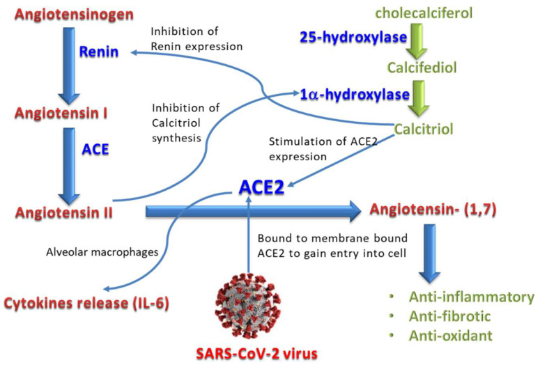
Evidence suggests that the level of interleukin-6 (IL-6) is more accurate than the C-reactive protein (CRP) and other inflammatory indicators in expecting respiratory failure in COVID-19. Immune dysregulation and ARDS in COVID-19 appear to be most significantly influenced by IL-6 [34]. IL-6 secretion in the lung is increased by the proinflammatory cytokines, particularly interleukin-1β (IL-1β) and TNF-α. An important factor in the induction and propagation of the cytokine storm that causes ARDS and lung injury is the overexpression of IL-6 [35]. It is thought that IL-6 increases lung capillary permeability, which has a significant impact on ARDS development, and promotes the coagulation pathway, which results in microthrombi in the circulation of the lungs and raises the chance for thrombotic event [36]. The interaction of Vitamin D3 and its metabolites with the renin–angiotensin System (RAS) and IL-6 in COVID-19 is presented in Figure 1.
Figure 1.
The interaction of Vitamin D3 (cholecalciferol) and its metabolites with the renin–angiotensin System (RAS) and IL-6 in COVID-19 disease.
The neutrophil-to-lymphocyte ratio (NLR) is a valuable measure of systemic inflammation and is being evaluated as a guide for the prognosis of many illnesses, with patients with high NLR being actively followed and controlled [25]. Recently, NLR has been identified as a useful tool in identifying COVID-19 infection [37]. In COVID-19 patients, a relationship between elevated NLR and illness severity has also been demonstrated. Although its usage alone in COVID-19 diagnosis may not be particularly accurate, it should be quite effective in ruling out the infection when combined with a comprehensive physical examination [25]. Additionally, it was revealed that there was a substantial relationship between low serum vitamin D levels and NLR in COVID-19 patients [38].
This study aimed to investigate the levels of cholecalciferol and its main metabolites, calcifediol and calcitriol, in the sera of COVID-19 patients compared with healthy individuals. As far as we know, this is the first study that reports comparative serum levels of all forms of vitamin D3 and their relative ratios in COVID-19 patients. In addition, the ratio of these levels and the correlations between them and the levels of ACE2, Il-6, and NLR were investigated. Finally, the possible effect of COVID-19 infection on cholecalciferol metabolism and activation was assessed.
2. Materials and Methods
This cross-sectional investigation was carried out between October 2020 and December 2021. Study subjects were enrolled at Ohud Hospital, Madinah, Saudi Arabia, where the COVID-19 patients included were followed up and given the treatment according to the protocol approved by the Saudi Ministry of Health.
2.1. Selection of Study Subjects
Patients were chosen based on positive polymerase chain reaction (PCR) testing for COVID-19 as well as the presence of respiratory symptoms such as cough, shortness of breath, and/or lung computed tomography (CT) scans exhibiting ground glass opacity. Controls were selected based on negative PCR and symptoms. Only subjects who agreed to participate in the study were included. The study excluded patients who had comorbid conditions such liver illness, haematological disorders, chronic lung diseases, and those who received radiation and chemotherapy.
2.2. Sample Collection and Handling
Oropharyngeal specimens for PCR testing were collected by trained professionals from the Infection Control Unit under restricted conditions for COVID-19. Virus transport medium was used to convey specimens to the laboratory. On the other hand, blood samples were collected under aseptic safety measures by venipuncture. Parts of the samples were centrifuged on-site at 2500× g for 10 min at 4 °C. The sera were removed from the samples and maintained at −30 °C until the time of the vitamin D3 assay, in addition to the levels of ACE2, IL-6, and glucose. The remaining parts of the whole blood samples were analyzed for complete blood count (CBC).
2.3. Specimen Testing for COVID-19
The oropharyngeal specimens were subjected to viral ribonucleic acid (RNA) extraction followed by complementary deoxyribonucleic acid (cDNA) synthesis. Real-time quantitative PCR (RT-qPCR) was used to test for SARS-CoV-2 using particular probes and primers in accordance with the kit’s instructions. Finally, samples were classified as either positive or negative for COVID-19 based on the PCR findings.
2.4. Blood Sample Analyses
Serum levels of vitamin D3 (cholecalciferol) and its metabolites (calcifediol and calcitriol) were quantified by a novel method using an ultra performance liquid chromatography (UPLC) system with a tandem mass detector system (Waters Quattro Premier XE mass spectrometer, Waters Corporation (Milford, MA, USA) [39]. Detection limits were 50 ng/mL for cholecalciferol, 20 ng/mL for calcifediol, and 20 pg/mL for calcitriol. The level of ACE2 was measured in the sera using a sandwich Enzyme-Linked Immunosorbent Assay (ELISA) (ACE2 Kit, Invitrogen, from (ThermoFisher Scientific, Indianapolis, IN, USA)). In addition, serum level of IL-6 was determined by ELISA (ab46027, Abcam plc, Cambridge, UK) according to the manufacturer’s protocols.
2.5. Statistical Analysis
The Statistical Package for the Social Sciences, 20th version, from International Business Machines Corp. (Armonk, NY, USA), was employed. Chi-square test, students’ unpaired t-tests, and analysis of variance were used to examine the data. Results were presented as mean values ± standard error. Significance was assessed by the p-value, where p ≤ 0.05 and p ≤ 0.001 indicated significant and highly significant differences, respectively.
3. Results
This study included 103 COVID-19 patients and 50 healthy controls. According to Table 1, there were no significant variations in the demographic characteristics of the two groups. Shadows or ground-glass opacities were visible in the CT images of suspected patients. They did, however, go through a confirming diagnostic process that included nucleic acid analysis testing and the collection of oropharyngeal swab samples. As depicted in Table 2, COVID-19 patients had substantially greater levels of ACE2, IL-6, glucose, total white blood cells (WBCs), neutrophils, NLR, platelets, mean corpuscular hemoglobin (MCH), and mean corpuscular hemoglobin concentration (MCHC) than healthy participants. In contrast, lymphocytes, red blood cells (RBCs), hemoglobin, and hematocrit were significantly to highly significantly lower in the patients group. However, the difference was nonsignificant for eosinophils and mean corpuscular volume (MCV).
Table 1.
Demographic features of COVID-19 positive group compared with control group.
| Demographic Features | COVID-19 Group (n = 103) |
Control Group (n = 50) |
p-Value |
|---|---|---|---|
| Age (years) a | 47.32 ± 12.44 | 49.26 ± 9.94 | NS |
| Sex b | |||
| Male | 80 (77.67%) | 37 (74.0%) | - |
| Female | 23 (22.33%) | 13 (26.0%) | - |
| Weight (kg) a | 73.90 ± 12.30 | 72.12 ± 12.89 | NS |
| Height (cm) a | 165.86 ± 9.40 | 165.06 ± 7.52 | NS |
| BMI (kg/m2) a | 26.74 ± 3.38 | 26.47 ± 4.43 | NS |
BMI, body mass index; NS, non-significant. a Mean ± standard error of the mean. b Frequency and ratio.
Table 2.
Laboratory test results for COVID-19 positive group compared with the control group.
| Test | COVID-19 Group * (n = 103) |
Control Group * (n = 50) |
p-Value |
|---|---|---|---|
| ACE2 (U/L) | 72.37 ± 18.67 | 32.36 ± 11.27 | ≤0.001 (HS) |
| IL-6 (pg/mL) | 95.84 ± 25.23 | 2.76 ± 0.62 | ≤0.001 (HS) |
| Glucose (mg/dL) | 188.42 ± 37.62 | 125.75 ± 25.49 | ≤0.001 (HS) |
| Total WBCs (×103/μL) | 7.86 ± 2.59 | 5.18 ± 0.76 | ≤0.05 (S) |
| Neutrophils (%) | 57.22 ± 2.27 | 47.81 ± 6.17 | ≤0.001 (HS) |
| Eosinophils (%) | 5.1 ± 0.16 | 5.08 ± 0.17 | NS |
| Lymphocytes (%) | 36.63 ± 6.14 | 45.02 ± 2.64 | ≤0.001 (HS) |
| Platelets (×103/μL) | 264.42 ± 72.30 | 215.10 ± 25.38 | ≤0.001 (HS) |
| NLR | 1.61 ± 0.30 | 1.07 ± 0.16 | ≤0.001 (HS) |
| RBCs (×106/µL) | 4.54 ± 0.56 | 5.09 ± 0.64 | ≤0.05 (S) |
| Hb (g/dL) | 11.98 ± 1.78 | 13.25 ± 0.9 | ≤0.05 (S) |
| HCT (%) | 36.25 ± 5.44 | 40.92 ± 2.21 | ≤0.05 (S) |
| MCV (fL) | 83.79 ± 5.34 | 81.35 ± 3.47 | NS |
| MCH (pg) | 30.40 ± 3.46 | 27.21 ± 2.82 | ≤0.05 (S) |
| MCHC (g/dL) | 34.00 ± 1.31 | 30.12 ± 1.75 | ≤0.05 (S) |
ACE, angiotensin converting enzyme; Hb, hemoglobin; HCT, haematocrit; HS, highly significant; IL-6, interleukin-6; MCH, mean corpuscular hemoglobin; MCHC, mean corpuscular hemoglobin concentration; MCV, mean corpuscular volume; NLR, neutrophil-to-lymphocyte ratio; NS, nonsignificant; RBCs, red blood cells; S, significant; WBCs, white blood cells. * Mean ± standard error of the mean.
Serum levels of vitamin D3 and its metabolites were highly significantly lower in the COVID-19 patients group compared with the healthy subjects, as demonstrated in Table 3. The calcitriol/calcifediol ratio (VMR1) was 2.97 ± 0.30 pg/ng in COVID-19 patients compared to 2.86 ± 0.46 in healthy subjects, which was within the normal ratio range that was reported to be 1.64–3.00 pg/ng (mean = 2.22) [24]. Similarly, VMR2 (calcifediol/cholecalciferol ratio) was found to be normal (0.82 ± 0.14 ng/ng) in COVID-19 patients compared to 0.81 ± 0.17 in healthy subjects, where the normal VMR2 was reported as 0.8 ± 0.15 ng/ng [40]. The differences between the COVID-19 patients group and healthy group were nonsignificant for both VMR1 and VMR2.
Table 3.
Serum levels of vitamin D3 and its metabolites in COVID-19 positive group compared with control group.
| Vitamin D and Its Metabolites | COVID-19 Group * (n = 103) |
Control Group * (n = 50) |
p-Value |
|---|---|---|---|
| Cholecalciferol (ng/mL) | 18.50 ± 5.36 | 29.13 ± 4.94 | ≤0.001 (HS) |
| Calcifediol (ng/mL) | 14.60 ± 3.30 | 23.10 ± 3.02 | ≤0.001 (HS) |
| Calcitriol (pg/mL) | 42.90 ± 8.44 | 65.15 ± 7.11 | ≤0.001 (HS) |
| Cacitriol/Calcifediol ratio (VMR1, pg/ng) | 2.97 ± 0.30 | 2.86 ± 0.46 | NS |
| Calcifediol/Cholecalciferol ratio (VMR2, ng/ng) | 0.82 ± 0.14 | 0.81 ± 0.17 | NS |
HS, highly significant; NS, Non-significant; VMR, vitamin D metabolite ratio. * Mean ± standard error of the mean.
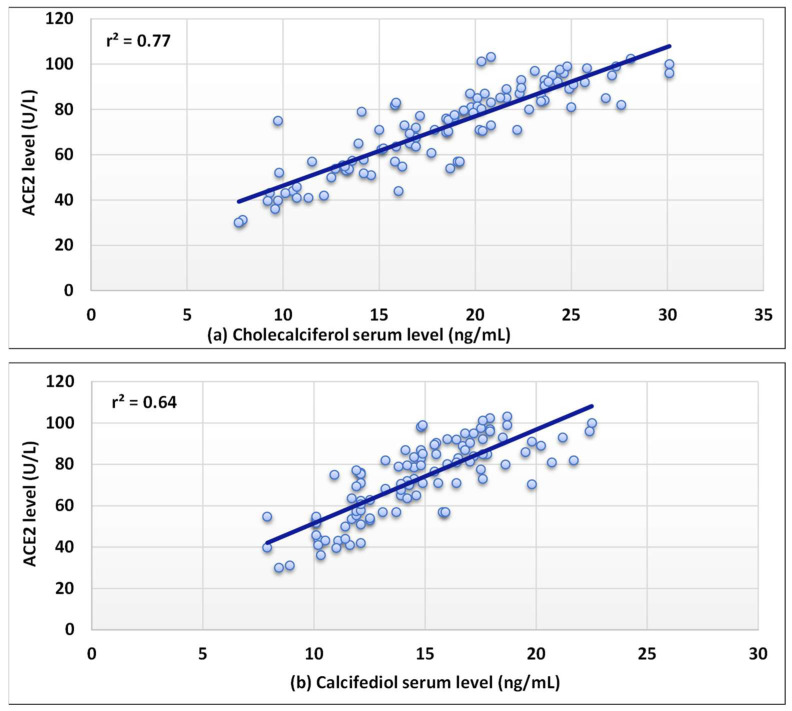
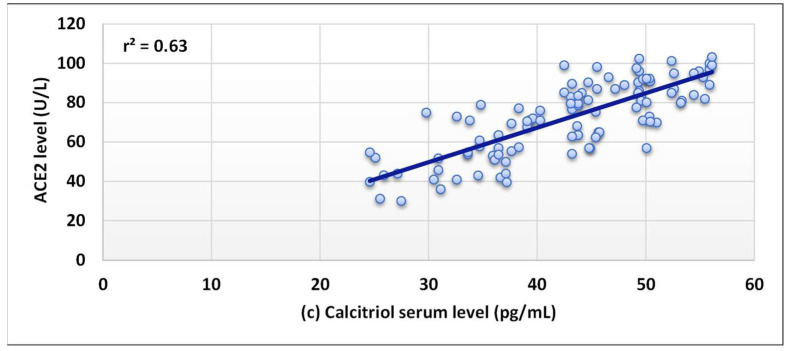
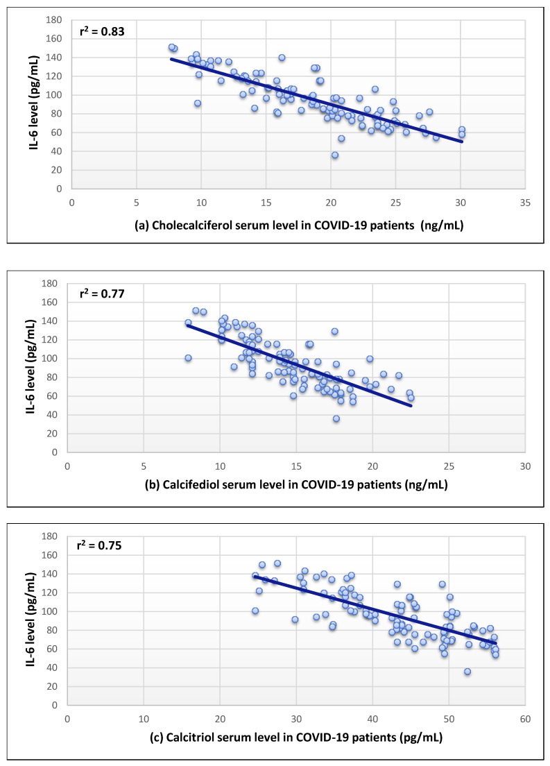
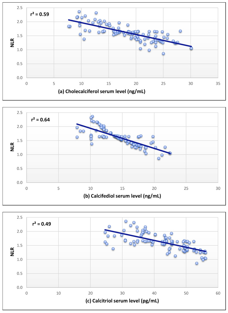
The serum levels of the three forms of vitamin D and serum ACE2 levels were shown to have a substantial positive correlation (r2 = 0.63–0.77) as revealed in Figure 2. In addition, a strong negative correlation was found for the serum level of Il-6 (r2 = 0.75–0.83) (Figure 3). Moreover, a moderate inverse correlation was found between the vitamin forms and the NLR (r2 = 0.49–0.64), as seen in Figure 4.
Figure 2.
Correlation between the levels of angiotensin-converting enzyme 2 (ACE2) and (a–c) in COVID-19 patients.
Figure 3.
Correlation between the levels of interleukin-6 (IL-6) and (a–c) in COVID-19 patients.
Figure 4.
Correlation between the neutrophil-to-lymphocyte ratio (NLR) and (a–c) in COVID-19 patients.
4. Discussion
This study revealed a correlation between vitamin D deficiency, the severity of COVID-19, and specific inflammatory markers in COVID-19 patients. This relationship is based on the fact that vitamin D receptors are widely expressed in the human body and that active vitamin D compounds may have considerable biological effects on a variety of cellular signaling pathways [41]. Several studies have reported a correlation between patients’ history of vitamin D insufficiency and an increased chance of positive COVID-19 test findings [25,26,27,28]. The current research suggests that the low level of vitamin D3 and its metabolites is an indicator of worse COVID-19 illness consequences. The low levels of serum vitamin D metabolites detected during the acute COVID-19 infection, on the other hand, may be a result of persistent inflammation rather than an underlying cause [42].
As reflected by our results, it was noticed that the total WBCs, neutrophils, platelets, MCH, and MCHC were significantly elevated in COVID-19 patients in comparison with healthy subjects, which is consistent with earlier research [43]. This might be related to the virus’s effect on RBC and WBC development or degradation, or the presence of comorbidities. In contrast, lymphocytes, RBCs, and hematocrit were significantly lower in the patients group. The RBCs of COVID-19 patients were reported to have lower amounts of key antioxidant enzymes and higher levels of protein breakdown indicators [44]. Lymphocytopenia and thrombocytopenia were also increasingly found when comparing severe COVID-19 cases with non-severe ones [43].
In addition to a large drop in lymphocyte count, the patients group was noticed to have a considerable rise in neutrophil count. We found that NLR increased in COVID-19 individuals in comparison with the control individuals, with considerably higher levels in patients who died from the disease. The NLR was established as a valuable predictor of systemic inflammation and has been studied as a prognostic indicator for a variety of disorders such as sepsis and cancer [25]. In addition, the association between ARDS and elevated NLR has also been observed [25]. In the current study, moderate inverse correlations were found between serum levels of cholecalciferol, calcifediol, and calcitriol and the NLR. This could be due to type I interferon-dependent transitory lymphopenia, which is seen in a variety of viral infections. The direct viral infection via spike receptors in T lymphocytes may also contributes to lymphopenia [45]. These data may aid in predicting clinical severity in COVID-19 patients.
Our results also showed that serum ACE2 concentrations were significantly increased in the patients compared with the control individuals. Furthermore, they were significantly higher in patients with increased levels of cholecalciferol and its metabolites compared with the other COVID-19 patients. It is reported that increased ACE concentrations lead to increased levels of angiotensin II, which might be responsible for the severe lung injury in COVID-19 patients, while inhibiting the angiotensin II-signaling pathway and/or RAAS, through ACE2, has been shown to protect against lung damage [46]. The strong positive correlation (r2 = 0.63–0.77) between the ACE2 serum levels and levels of cholecalciferol and its metabolites in our COVID-19 individuals confirms the expected role of ACE level in the disease severity. These findings support the belief that vitamin D signaling may cause RAAS to respond and limit vitamin D’s function as a transcription factor in renin gene suppression, hence serving as a negative endocrine regulator on RAAS [47].
Additionally, the levels of IL-6 were observed to be significantly elevated in the COVID-19 patient group compared with the control group. The values were also higher in patients with vitamin D insufficiency. A strong negative correlation (r = 0.75–0.83) was found between cholecalciferol, calcifediol, and calcitriol and IL-6 levels. This can be explained by the significant role of vitamin D3 in reducing immune cell IL-6 synthesis, potentially lowering its pro-inflammatory effects without directly targeting IL-6 receptors, and preventing any negative effect on the IL-6 anti-inflammatory properties [34]. Vitamin D has also been proven to diminish TNF-α levels, implying another treatment approach for combating the COVID-19 cytokine storm [35].
This study showed that vitamin D3 and its metabolites were highly significantly lower in the patients group compared with the healthy controls. Vitamin D status has traditionally been determined using levels of calcifediol or, sometimes, calcitriol. The physiological sophistication of synthesis route of these metabolites implies that, in addition to measuring serum levels of one metabolite, efficient management of vitamin D hydroxylation may be significant [48]. Calcitriol and calcifediol, on the other hand, have distinct biological features and their concentrations are very loosely associated. Determining their ratio to each other is more effective than evaluating their levels separately. This may help estimate how well the vitamin D system is working to keep the picomolar levels of the active metabolite, calcitriol, stable, which is necessary for the predicted biological effects [24]. Two vitamin D metabolite ratios (VMR) were suggested to be suggestive of the effectiveness of vitamin D hydroxylation. VMR1 (calcitriol/calcifediol) theoretically depicts how much calcitriol, in picograms, is produced for every nanogram of circulating calcifediol and can be indicative of vitamin D hydroxylation efficacy at the kidney. On the other hand, VMR2 (calcifediol/cholecalciferol) shows how many nanograms of calcifediol are produced per nanogram of circulating cholecalciferol and can indicate vitamin D hydroxylation effectiveness in the liver. In this study, however, both ratios were found to be within the normal suggested ranges in both patient and control groups. Although both VMRs were higher in the COVID-19 patient group compared with the control group, the differences were nonsignificant. This may indicate that the levels of vitamin D and its metabolites decrease in the same manner in COVID-19 patients without a noticeable effect on the hepatic and renal hydroxylation efficiency.
5. Limitations of the Study
Although this study has shown interesting potential clinical implications, our sample involved only patients from the first wave of COVID-19. In addition, the sample size was relatively small. Therefore, the results cannot be generalized unless further multicenter studies with larger sample sizes are conducted covering patients affected by subsequent waves of the pandemic.
6. Conclusions
This study showed strong correlations between the serum levels of vitamin D3 (cholecalciferol) and its metabolites, calcifediol and calcitriol, with disease severity in COVID-19 patients. Moreover, certain inflammatory markers (especially Il-6 and ACE2) and clinical hematological outcomes of COVID-19 disease were well correlated with the levels of the vitamin and its metabolites. However, the renal and hepatic hydroxylation of the vitamin, represented by VMR, were not significantly affected during the course of the infection. These data may help to clarify logical therapeutic decisions in various patient populations.
Acknowledgments
The authors express their appreciation to the Deputyship for Research and Innovation, Ministry of Education, Saudi Arabia, for funding this research (Project no. 112/442). We also extend our thanks to Taibah University for its supervisory support.
Author Contributions
H.M.J.K.: Conceptualization, project administration, acquisition, writing—review and editing, and supervision. S.A.A.: Conceptualization, funding, methodology, formal analysis, resources writing—original draft, writing—review and editing, and visualization. S.S.A.-T.: Conceptualization, resources, writing—original draft, writing—review and editing, and visualization. Y.M.A.: Conceptualization, project administration, acquisition, writing—review and editing, supervision., data curation, and writing—original draft. Y.A.A.: Conceptualization, methodology, investigation, validation, data curation, and writing—original draft. All authors have read and agreed to the published version of the manuscript.
Institutional Review Board Statement
The study protocol was agreed by the General Directorate of Health Affairs in Madinah (No. H-03-M-085).
Informed Consent Statement
An informed consent was obtained from each participant. Subject identifying data were coded and kept anonymous and confidential.
Conflicts of Interest
The authors declare no conflict of interest.
Funding Statement
This research was funded by Deputyship for Research and Innovation, Ministry of Education, Saudi Arabia, grant number 112/442.
Footnotes
Publisher’s Note: MDPI stays neutral with regard to jurisdictional claims in published maps and institutional affiliations.
References
- 1.Pollard C.A., Morran M.P., Nestor-Kalinoski A.L. The COVID-19 pandemic: A global health crisis. Physiol. Genom. 2020;52:549–557. doi: 10.1152/physiolgenomics.00089.2020. [DOI] [PMC free article] [PubMed] [Google Scholar]
- 2.Bartoszko J.J., Siemieniuk R.A.C., Kum E., Qasim A., Zeraatkar D., Ge L., Han M.A., Sadeghirad B., Agarwal A., Agoritsas T., et al. Prophylaxis against covid-19: Living systematic review and network meta-analysis. BMJ. 2021;373:n949. doi: 10.1136/bmj.n949. [DOI] [PMC free article] [PubMed] [Google Scholar]
- 3.Smit M., Marinosci A., Agoritsas T., Calmy A. Prophylaxis for COVID-19: A systematic review. Clin. Microbiol. Infect. Off. Publ. Eur. Soc. Clin. Microbiol. Infect. Dis. 2021;27:532–537. doi: 10.1016/j.cmi.2021.01.013. [DOI] [PMC free article] [PubMed] [Google Scholar]
- 4.Khojah H.M.J. Community pharmacy services and preparedness during COVID-19 outbreak in Madinah, Saudi Arabia. Saudi Pharm. J. 2020;28:1402–1407. doi: 10.1016/j.jsps.2020.09.004. [DOI] [PMC free article] [PubMed] [Google Scholar]
- 5.Mukattash T.L., Alkhalidy H., Alzu’Bi B., Abu-Farha R., Itani R., Karout S., Khojah H.M.J., Khdour M., El-Dahiyat F., Jarab A. Dietary supplements intake during the second wave of COVID-19 pandemic: A multinational Middle Eastern study. Eur. J. Integr. Med. 2022;49:102102. doi: 10.1016/j.eujim.2022.102102. [DOI] [PMC free article] [PubMed] [Google Scholar]
- 6.Bartley J. Vitamin D: Emerging roles in infection and immunity. Expert Rev. Anti-Infect. Ther. 2010;8:1359–1369. doi: 10.1586/eri.10.102. [DOI] [PubMed] [Google Scholar]
- 7.Vanherwegen A.-S., Gysemans C., Mathieu C. Regulation of Immune Function by Vitamin D and Its Use in Diseases of Immunity. Endocrinol. Metab. Clin. N. Am. 2017;46:1061–1094. doi: 10.1016/j.ecl.2017.07.010. [DOI] [PubMed] [Google Scholar]
- 8.Ismailova A., White J.H. Vitamin D, infections and immunity. Rev. Endocr. Metab. Disord. 2021;23:265–277. doi: 10.1007/s11154-021-09679-5. [DOI] [PMC free article] [PubMed] [Google Scholar]
- 9.Malek Mahdavi A. A brief review of interplay between vitamin D and angiotensin-converting enzyme 2: Implications for a potential treatment for COVID-19. Rev. Med. Virol. 2020;30:e2119. doi: 10.1002/rmv.2119. [DOI] [PMC free article] [PubMed] [Google Scholar]
- 10.Beyerstedt S., Casaro E.B., Rangel É.B. COVID-19: Angiotensin-converting enzyme 2 (ACE2) expression and tissue susceptibility to SARS-CoV-2 infection. Eur. J. Clin. Microbiol. Infect. Dis. Off. Publ. Eur. Soc. Clin. Microbiol. 2021;40:905–919. doi: 10.1007/s10096-020-04138-6. [DOI] [PMC free article] [PubMed] [Google Scholar]
- 11.Kuhn J.H., Li W., Choe H., Farzan M. Angiotensin-Converting Enzyme 2: A Functional Receptor for SARS Coronavirus. Cell Mol. Life Sci. 2004;61:2738–2743. doi: 10.1007/s00018-004-4242-5. [DOI] [PMC free article] [PubMed] [Google Scholar]
- 12.Warner F.J., Rajapaksha H., Shackel N., Herath C.B. ACE2: From protection of liver disease to propagation of COVID-19. Clin. Sci. 2020;134:3137–3158. doi: 10.1042/CS20201268. [DOI] [PubMed] [Google Scholar]
- 13.Turner A.J., Hiscox J.A., Hooper N.M. ACE2: From vasopeptidase to SARS virus receptor. Trends Pharmacol. Sci. 2004;25:291–294. doi: 10.1016/j.tips.2004.04.001. [DOI] [PMC free article] [PubMed] [Google Scholar]
- 14.Scialo F., Daniele A., Amato F., Pastore L., Matera M.G., Cazzola M., Castaldo G., Bianco A. ACE2: The Major Cell Entry Receptor for SARS-CoV-2. Lung. 2021;198:867–877. doi: 10.1007/s00408-020-00408-4. [DOI] [PMC free article] [PubMed] [Google Scholar]
- 15.Xiao D., Li X., Su X., Mu D., Qu Y. Could SARS-CoV-2-induced lung injury be attenuated by vitamin D? Int. J. Infect. Dis. 2020;102:196–202. doi: 10.1016/j.ijid.2020.10.059. [DOI] [PMC free article] [PubMed] [Google Scholar]
- 16.Rafiullah M. Can a Combination of AT1R Antagonist and Vitamin D Treat the Lung Complication of COVID-19? Am. J. Med. Sci. 2020;360:338–341. doi: 10.1016/j.amjms.2020.07.018. [DOI] [PMC free article] [PubMed] [Google Scholar]
- 17.Honardoost M., Ghavideldarestani M., Khamseh M.E. Role of vitamin D in pathogenesis and severity of COVID-19 infection. Arch. Physiol. Biochem. 2020;126:1–7. doi: 10.1080/13813455.2020.1792505. [DOI] [PubMed] [Google Scholar]
- 18.Urena-Torres P., Souberbielle J.C. Pharmacologic role of vitamin D natural products. Curr. Vasc. Pharmacol. 2014;12:278–285. doi: 10.2174/15701611113119990020. [DOI] [PubMed] [Google Scholar]
- 19.Kulda V. Vitamin D metabolism. Vnitr. Lek. 2012;58:400–404. [PubMed] [Google Scholar]
- 20.Brown A.J. Therapeutic uses of vitamin D analogues. Am. J. Kidney Dis. Off. J. Natl. Kidney Found. 2001;38:S3–S19. doi: 10.1053/ajkd.2001.28111. [DOI] [PubMed] [Google Scholar]
- 21.Jones G., Prosser D.E. In: Chapter 3-The Activating Enzymes of Vitamin D Metabolism (25- and 1α-Hydroxylases) Feldman D., Pike J.W., Adams J.S.B.T.-V.D., Third E., editors. Academic Press; San Diego, CA, USA: 2011. pp. 23–42. [Google Scholar]
- 22.Takayama K., Kato S. The Vitamin D3 1alpha-Hydroxylase Gene and Its Regulation by Active Vitamin D3. Biosci. Biotechnol. Biochem. 2011;75:208–213. doi: 10.1271/bbb.100684. [DOI] [PubMed] [Google Scholar]
- 23.Ahmed L.H.M., Butler A.E., Dargham S.R., Latif A., Chidiac O.M., Atkin S.L., Abi Khalil C. Vitamin D3 metabolite ratio as an indicator of vitamin D status and its association with diabetes complications. BMC Endocr. Disord. 2020;20:161. doi: 10.1186/s12902-020-00641-1. [DOI] [PMC free article] [PubMed] [Google Scholar]
- 24.Pasquali M., Tartaglione L., Rotondi S., Muci M.L., Mandanici G., Farcomeni A., Marangella M., Mazzaferro S. Calcitriol/calcifediol ratio: An indicator of vitamin D hydroxylation efficiency? BBA Clin. 2015;3:251–256. doi: 10.1016/j.bbacli.2015.03.004. [DOI] [PMC free article] [PubMed] [Google Scholar]
- 25.Mardani R., Alamdary A., Nasab S.M., Gholami R., Ahmadi N., Gholami A. Association of vitamin D with the modulation of the disease severity in COVID-19. Virus Res. 2020;289:198148. doi: 10.1016/j.virusres.2020.198148. [DOI] [PMC free article] [PubMed] [Google Scholar]
- 26.Raisi-Estabragh Z., Martineau A.R., Curtis E.M., Moon R.J., Darling A., Lanham-New S., Ward K.A., Cooper C., Munroe P.B., Petersen S.E., et al. Vitamin D and coronavirus disease 2019 (COVID-19): Rapid evidence review. Aging Clin. Exp. Res. 2021;33:2031–2041. doi: 10.1007/s40520-021-01894-z. [DOI] [PMC free article] [PubMed] [Google Scholar]
- 27.Szarpak L., Rafique Z., Gasecka A., Chirico F., Gawel W., Hernik J., Kaminska H., Filipiak K.J., Jaguszewski M.J., Szarpak L. A systematic review and meta-analysis of effect of vitamin D levels on the incidence of COVID-19. Cardiol. J. 2021;28:647–654. doi: 10.5603/CJ.a2021.0072. [DOI] [PMC free article] [PubMed] [Google Scholar]
- 28.Munshi R., Hussein M.H., Toraih E.A., Elshazli R.M., Jardak C., Sultana N., Youssef M.R., Omar M., Attia A.S., Fawzy M.S., et al. Vitamin D insufficiency as a potential culprit in critical COVID-19 patients. J. Med. Virol. 2021;93:733–740. doi: 10.1002/jmv.26360. [DOI] [PubMed] [Google Scholar]
- 29.Teymoori-Rad M., Shokri F., Salimi V., Marashi S.M. The interplay between vitamin D and viral infections. Rev. Med. Virol. 2019;29:e2032. doi: 10.1002/rmv.2032. [DOI] [PubMed] [Google Scholar]
- 30.Guo Y.-R., Cao Q.-D., Hong Z.-S., Tan Y.-Y., Chen S.-D., Jin H.-J., Tan K.-S., Wang D.-Y., Yan Y. The origin, transmission and clinical therapies on coronavirus disease 2019 (COVID-19) outbreak—An update on the status. Mil. Med. Res. 2020;7:11. doi: 10.1186/s40779-020-00240-0. [DOI] [PMC free article] [PubMed] [Google Scholar]
- 31.Ali N. Role of vitamin D in preventing of COVID-19 infection, progression and severity. J. Infect. Public Health. 2020;13:1373–1380. doi: 10.1016/j.jiph.2020.06.021. [DOI] [PMC free article] [PubMed] [Google Scholar]
- 32.Grant W.B., Lahore H., McDonnell S.L., Baggerly C.A., French C.B., Aliano J.L., Bhattoa H.P. Evidence that Vitamin D Supplementation Could Reduce Risk of Influenza and COVID-19 Infections and Deaths. Nutrients. 2020;12:988. doi: 10.3390/nu12040988. [DOI] [PMC free article] [PubMed] [Google Scholar]
- 33.Cantorna M.T., Snyder L., Lin Y.-D., Yang L. Vitamin D and 1,25(OH)2D Regulation of T cells. Nutrients. 2015;7:3011–3021. doi: 10.3390/nu7043011. [DOI] [PMC free article] [PubMed] [Google Scholar]
- 34.Sabaka P., Koščálová A., Straka I., Hodosy J., Lipták R., Kmotorková B., Kachlíková M., Kušnírová A. Role of interleukin 6 as a predictive factor for a severe course of Covid-19: Retrospective data analysis of patients from a long-term care facility during Covid-19 outbreak. BMC Infect. Dis. 2021;21:308. doi: 10.1186/s12879-021-05945-8. [DOI] [PMC free article] [PubMed] [Google Scholar]
- 35.Magro G. SARS-CoV-2 and COVID-19: Is interleukin-6 (IL-6) the ‘culprit lesion’ of ARDS onset? What is there besides Tocilizumab? SGP130Fc. Cytokine X. 2020;2:100029. doi: 10.1016/j.cytox.2020.100029. [DOI] [PMC free article] [PubMed] [Google Scholar]
- 36.Hunter C.A., Jones S.A. IL-6 as a keystone cytokine in health and disease. Nat. Immunol. 2015;16:448–457. doi: 10.1038/ni.3153. [DOI] [PubMed] [Google Scholar]
- 37.Lagunas-Rangel F.A. Neutrophil-to-lymphocyte ratio and lymphocyte-to-C-reactive protein ratio in patients with severe coronavirus disease 2019 (COVID-19): A meta-analysis. J. Med. Virol. 2020;92:1733–1734. doi: 10.1002/jmv.25819. [DOI] [PMC free article] [PubMed] [Google Scholar]
- 38.Demir A.D., Durmaz Z.H. A Comparison of vitamin D deficiency with neutrophil lymphocyte ratio and CRP levels in Covid-19 patients. Med. Sci. Discov. 2021;8:306–309. doi: 10.36472/msd.v8i5.537. [DOI] [Google Scholar]
- 39.Ahmed S.A., Khojah H.M.J., Al-Thagfan S.S., Alahmadi Y.M., Mohammed Y.A. An ultrasensitive UHPLC-ESI-MS/MS method augmented with a controlled microwave derivatization reaction for quantitation of vitamin D3 and its major metabolites in COVID-19 patients. Talanta. 2022;246:123497. doi: 10.1016/j.talanta.2022.123497. [DOI] [PMC free article] [PubMed] [Google Scholar]
- 40.Al-Thagfan S.S., Alolayan S.O., Ahmed S., Emara M.M., Awadallah M.F. Impacts of deficiency in vitamin D derivatives on disease severity in adult bronchial asthma patients. Pulm. Pharmacol. Ther. 2021;70:102073. doi: 10.1016/j.pupt.2021.102073. [DOI] [PubMed] [Google Scholar]
- 41.Wacker M., Holick M.F. Sunlight and Vitamin D. Dermato-Endocrinol. 2013;5:51–108. doi: 10.4161/derm.24494. [DOI] [PMC free article] [PubMed] [Google Scholar]
- 42.Smolders J., van den Ouweland J., Geven C., Pickkers P., Kox M. Letter to the Editor: Vitamin D deficiency in COVID-19: Mixing up cause and consequence. Metabolism. 2021;115:154434. doi: 10.1016/j.metabol.2020.154434. [DOI] [PMC free article] [PubMed] [Google Scholar]
- 43.Słomka A., Kowalewski M., Żekanowska E. Coronavirus Disease 2019 (COVID-19): A Short Review on Hematological Manifestations. Pathogens. 2020;9:493. doi: 10.3390/pathogens9060493. [DOI] [PMC free article] [PubMed] [Google Scholar]
- 44.Thomas T., Stefanoni D., Dzieciatkowska M., Issaian A., Nemkov T., Hill R.C., Francis R.O., Hudson K.E., Buehler P.W., Zimring J.C., et al. Evidence of Structural Protein Damage and Membrane Lipid Remodeling in Red Blood Cells from COVID-19 Patients. J. Proteome Res. 2020;19:4455–4469. doi: 10.1021/acs.jproteome.0c00606. [DOI] [PMC free article] [PubMed] [Google Scholar]
- 45.Kamphuis E., Junt T., Waibler Z., Forster R., Kalinke U. Type I interferons directly regulate lymphocyte recirculation and cause transient blood lymphopenia. Blood. 2006;108:3253–3261. doi: 10.1182/blood-2006-06-027599. [DOI] [PubMed] [Google Scholar]
- 46.Shenoy V., Ferreira A.J., Qi Y., Fraga-Silva R.A., Díez-Freire C., Dooies A., Jun J.Y., Sriramula S., Mariappan N., Pourang D., et al. The Angiotensin-Converting Enzyme 2/Angiogenesis-(1–7)/Mas Axis Confers Cardiopulmonary Protection against Lung Fibrosis and Pulmonary Hypertension. Am. J. Respir. Crit. Care Med. 2010;182:1065–1072. doi: 10.1164/rccm.200912-1840OC. [DOI] [PMC free article] [PubMed] [Google Scholar]
- 47.Shroff R., Wan M., Rees L. Can vitamin D slow down the progression of chronic kidney disease? Pediatr. Nephrol. 2012;27:2167–2173. doi: 10.1007/s00467-011-2071-y. [DOI] [PubMed] [Google Scholar]
- 48.Holick M.F. Vitamin D in Health and Disease: Vitamin D for Health and in Chronic Kidney Disease. Semin. Dial. 2005;18:266–275. doi: 10.1111/j.1525-139X.2005.18402.x. [DOI] [PubMed] [Google Scholar]


