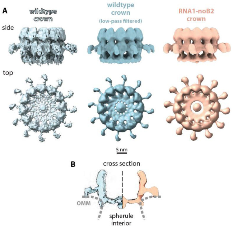
Figure 6.
Comparison of nodavirus RNA replication complex crowns formed in nodavirus-infected insect cells to those formed in mammalian cells in the absence of Protein B2 and genomic RNA2. (A) On the right, light orange side and top view presentations of RNA1-noB2 replication-induced crowns in mammalian cells display all of the hallmark 12-fold symmetrical features of wildtype FHV infection-induced RC crowns shown in the published high-resolution crown image [24] on the left and the matched low-pass gaussian-filtered, slightly lower resolution version in the center. As shown, these common features include the apical and basal lobes of the central turret, inner floors with similar diameter central openings and central densities, and leg-like radial extensions. (B) Cross-sectioned comparison to further illustrate the matching shapes and membrane interactions of the high-resolution wildtype crown (leftmost images in panel (A)) and the RNA1-noB2 crown structures. OMM = outer mitochondrial membrane.

