Abstract
Pseudomonas aeruginosa plays a major role in respiratory tract infections or sepsis in patients with cystic fibrosis or upon suppression of the immune system. Several P. aeruginosa strains have been shown to be internalized by human epithelial cells; however, the molecular mechanisms of the invasion process are poorly characterized. Here, we show that the internalization of P. aeruginosa into human epithelial cells results in and requires activation of the Src-like tyrosine kinases p59Fyn and p60Src and the consequent tyrosine phosphorylation of several eukaryotic proteins. The significance of Src-like tyrosine kinase activation is shown by an almost complete blockade of P. aeruginosa internalization, but not adhesion, upon inhibition of Src-like tyrosine kinases. Likewise, inhibition of P. aeruginosa binding to CFTR, which has been shown to block P. aeruginosa internalization, prevents Src and Fyn activation, supporting a pivotal role of Src-like tyrosine kinases for invasion by P. aeruginosa.
Many pathogens are internalized by mammalian cells, constituting an essential step in the infection process (2, 8). Invasion may protect the bacteria from the immune response or enable bacteria to penetrate the epithelial cell layer to reach the mucosa, the bloodstream, and finally several organs, causing severe disease symptoms and sepsis. On the other hand, uptake of pathogens in lysosomes may represent an important element of the host defense system against the bacteria. Several mechanisms for invasion involving the signaling machinery of the host cells, have been identified. In particular, it has been suggested that tyrosine kinases and, consequently, induction of tyrosine phosphorylation in the target cells play an important role in bacterial internalization. Thus, a variety of pathogens, including Shigella flexneri (5), Salmonella enterica serovar Typhimurium (12, 21, 28), Yersinia spp. (2, 27), Listeria monocytogenes (33), and several enteropathogenic Escherichia coli strains (27, 29), have been demonstrated to trigger tyrosine phosphorylation of various eukaryotic proteins during invasion. Among several tyrosine kinases, the family of Src-like tyrosine kinases seems to be particularly important for the induction of cellular tyrosine phosphorylation by several bacteria (3, 6, 17, 23, 30, 35). All Src-like tyrosine kinases consist of an N-terminal SH3 and SH2 domain, the kinase domain, and a regulatory C-terminal tyrosine residue (Tyr-527 for Src). The SH2 domain contains a second regulatory tyrosine residue (Tyr-192 for Src). In general, Src-like tyrosine kinases are kept in an inactive state by tyrosine phosphorylation of the C-terminal regulatory tyrosine. Dephosphorylation of that tyrosine residue alters the conformation of the protein and opens the kinase domain, resulting in autophosphorylation of a stimulatory tyrosine in the kinase domain (Tyr-416 for Src) that is required for full activation of the kinase (30).
In the present study we investigated internalization mechanisms of Pseudomonas aeruginosa, a gram-negative facultative pathogen that causes infections of the respiratory and urinary tracts, skin, and eye, predominantly in immune compromised patients. The bacterium also plays a major role in pulmonary infections of patients with cystic fibrosis. One of the initial steps in the infection process seems to be the uptake of P. aeruginosa by host cells (3, 9, 16); however, the molecular mechanisms of the bacterial internalization are largely unknown. It has been reported that the bacteria adhere to epithelial cells via a variety of adhesins, e.g., pili (3, 6), lipopolysaccharides (LPS) (13), several exoenzymes (1), and exopolysaccharides (26). Following adhesion, the internalization of P. aeruginosa into epithelial cells seems to be mediated by binding of the bacterial LPS to the eukaryotic cystic fibrosis transmembrane conductance regulator protein (CFTR) (23, 24). The CFTR molecule has been shown to be a chloride channel, but the exact function of this protein in the internalization process is unknown. Further, it has been recently shown that tyrosine kinases play a role in invasion by P. aeruginosa, since inhibitors of tyrosine kinases prevent the uptake of P. aeruginosa into rabbit corneal epithelial cells (7). The kinases involved in cellular tyrosine phosphorylation induced by P. aeruginosa are unknown.
In the present study we provide evidence for a pivotal role of nonreceptor Src-like tyrosine kinases, in particular p60Src and p59Fyn, in the internalization of P. aeruginosa into different human epithelial cells. Stimulation of the two Src-like tyrosine kinases results in tyrosine phosphorylation of several cellular proteins, which seems to be required for the invasion of human epithelial cells by P. aeruginosa.
MATERIALS AND METHODS
Materials and cell culture.
All reagents were purchased from Sigma (Deisenhofen, Germany), except as otherwise indicated. The human conjunctiva epithelial cell line Chang (ATCC CCL 20.2; American Type Culture Collection, Manassas, Va.) was cultured in RPMI 1640 (Life Technologies, Eggenstein, Germany) supplemented with 5% fetal calf serum (FCS) and 2 mM l-glutamine (Life Technologies) at 37°C and 5% CO2. The human pulmonary epithelial cell line WI-38 (ATCC CCL-75) was cultured in minimal essential medium (Life Technologies) supplemented with 10% FCS, 2 mM l-glutamine, 1% nonessential amino acids, 1% sodium pyruvate, and 1% penicillin-streptomycin (10,000 IU/ml) (all from Life Technologies). Cells were labeled with 1 μCi of [3H]choline chloride (75 Ci/mmol; DuPont, NEN)/ml for 48 h prior to any experiment determining tyrosine phosphorylation or kinase activity. This enabled us to normalize all samples for equal amounts of cell equivalents by liquid scintillation counting of an aliquot of the cell lysates.
The Src-like tyrosine kinase inhibitor herbimycin A (Sigma) was added to the cells 12 h prior to infection at a concentration of 1 μM. PP1 (New England Biolabs, Frankfurt am Main, Germany), a second Src-like tyrosine kinase inhibitor, was added for 15 min at 50 nM. Cells were then washed twice in phosphate-buffered saline (PBS) to deplete the inhibitors prior to infection with P. aeruginosa.
Infection experiments.
Infection experiments were performed with a P. aeruginosa strain, designated 696, isolated from the sputum of a hospitalized patient or with a laboratory strain, ATCC 27853. Bacteria were grown overnight on tryptic soy agar plates at 37°C, resuspended in tryptic soy broth (TSB) (Difco Laboratories, Detroit, Mich.) to an optical density at 550 nm (OD550) of 0.25, shaken at 130 rpm for 1 h at 37°C to reach the mid-logarithmic phase, pelleted, and resuspended in fresh TSB. Cells were washed twice with RPMI 1640 supplemented with 2 mM l-glutamine prior to any infection and maintained in the same medium during the infection. Infection was initiated by inoculating subconfluent cell layers at a bacterium/host cell ratio of 1,000:1. Synchronous infection conditions and an enhanced bacterium-host cell interaction were achieved by a 2-min centrifugation (35 × g) of the bacteria onto the cells. The end of the centrifugation was defined as the start point in all experiments. The infection was terminated by cell lysis or fixation in the indicated buffer as described below.
To exclude possible effects of the inhibitors used on the viability of P. aeruginosa, bacteria were incubated with each inhibitor in the absence of human cells and then washed, and the ability to infect untreated epithelial cells was determined based on crystal violet and polymyxin assays. In addition, treated or untreated bacteria were washed and plate cultured to determine any effect on bacterial viability.
To distinguish the effects of bacterial adhesion from those of invasion of Chang epithelial cells by P. aeruginosa, we incubated P. aeruginosa 696 or ATCC 27853 for 10 min with 200 nM CFTR peptide GRIIASYDPDNKEER in PBS and used those bacteria to infect Chang epithelial cells for 30 min. This treatment has previously been shown to prevent internalization, but not adhesion, of the bacteria (24).
Internalization and survival assays.
The invasion of host cells by P. aeruginosa was determined by polymyxin survival assays and light microscopy (34, 35). The polymyxin survival assay (18) was performed analogously to gentamicin assays to measure the number of live intracellular bacteria. Briefly, Chang cells (105/well) were infected for 10 min as above and washed three times with RPMI 1640 supplemented with 2 mM l-glutamine to remove nonadherent bacteria. Samples were then incubated for 2 h in medium as above with 100 μg of polymyxin/ml to kill extracellular and adherent bacteria (adhesion control was performed without polymyxin). The cells were washed twice with PBS (140 mM NaCl, 2.5 mM KCl, 8.1 mM NaHPO4, 1.5 mM KH2PO4, 10 mM HEPES, 1 mM CaCl2 [pH 7.4]) and lysed with 5 mg of saponin/ml in PBS for 10 min at 37°C. Since human cells are impermeable to polymyxin (18), intracellular bacteria will survive this procedure. Those colonies were plated, and growing bacteria were counted after an incubation time of 24 h at 37°C. Experiments were performed in duplicate and repeated three times. Values are presented as means ± standard deviations (SD) from the three independent experiments.
Invasion of P. aeruginosa was confirmed by crystal violet staining of epithelial cells. To this end, cells (105/well) were seeded onto 12-mm circular glass coverslips in a 24-well tissue culture plate, infected, fixed with 1% paraformaldehyde in PBS for 15 min at room temperature, and stained overnight with 0.07% crystal violet in H2O at 4°C. Intracellular bacteria were light microscopically counted from at least 50 cells as previously described (34, 35). Adherence was scored by estimating the number of binding bacteria per cell. Intracellular bacteria were identified by two criteria. First, many bacteria are inside a clearly visible vacuole, proving intracellular location. Second, focusing on intracellular structures with the microscope enables differentiation between intracellular and adherent (out-of-focus) bacteria. All values are given as means ± SD from at least three independent experiments.
To determine the significance of Src-like tyrosine kinases for invasion by P. aeruginosa, epithelial cells were preincubated for 12 h with 1 μM herbimycin A or for 15 min with 50 nM PP1, washed, and infected with the indicated P. aeruginosa strain. Samples were then subjected to crystal violet staining or polymyxin survival assays.
Immunoblotting.
Infection was terminated by lysis of the cells in a solution containing 25 mM HEPES (pH 7.4), 0.1% sodium dodecyl sulfate (SDS), 0.5% sodium deoxycholate, 1% Triton X-100, 125 mM NaCl, 10 mM each sodium fluoride, Na3 VO4, and sodium pyrophosphate, 10 μg of aprotinin/ml, 10 μg of leupeptin/ml, and 10 μM arsine oxide for total cell lysates. Samples were incubated for 5 min on ice to achieve complete lysis and centrifuged at 20,000 × g for 15 min. The supernatants were added to 5× reducing SDS sample buffer consisting of 60 mM Tris-HCl (pH 6.8), 2.3% SDS, 10% glycerol, and 5% β-mercaptoethanol. Proteins were separated by SDS–10% polyacrylamide gel electrophoresis (PAGE), followed by electrophoretic transfer to nitrocellulose membranes (Bio-Rad, Munich, Germany). Blots were analyzed for tyrosine phosphorylation by incubation with 1 μg of 4 G10 antibody/ml for 4 h at 4°C and were developed by a further 60-min incubation with horseradish peroxidase (HRP)-conjugated protein G (Bio-Rad). A possible tyrosine phosphorylation of bacterial proteins was excluded by lysis of bacteria only and analysis of proteins for tyrosine phosphorylation by the same procedure.
Src-like tyrosine kinase activity.
The activity of Src-like-tyrosine kinases was determined by lysis of infected or uninfected cells in a solution containing 25 mM HEPES (pH 7.4), 3% NP-40, 1% Triton X-100, 125 mM NaCl, 10 mM each sodium fluoride, Na3 VO4, and sodium pyrophosphate, 10 μg of aprotinin/ml, 10 μg of leupeptin/ml, and 10 μM arsine oxide. Debris was pelleted by a 15-min centrifugation at 20,000 × g, and the supernatants were subjected to immunoprecipitation of the Src-like tyrosine kinases p59Fyn and p60Src by addition of polyclonal goat anti-Fyn or anti-Src antibodies (Santa Cruz Biotechnology, Santa Cruz, Calif.), respectively, at 2 μg/sample. Control immunoprecipitates were formed using irrelevant polyclonal sera from the same species. Immune complexes were immobilized with 30 μl of protein A/G-coupled agarose and further incubation for 60 min. Immune complexes were then washed three times each in lysis buffer and in kinase buffer (25 mM HEPES [pH 7.0], 150 mM NaCl, 10 mM MnCl2, 1 mM Na3 VO4, 5 mM dithiothreitol [DTT], and 0.5% NP-40). The kinase reaction was initiated by resuspension of the samples in 30 μl of kinase buffer supplemented with 10 μg of acid-denatured enolase/ml, 10 μCi of [32P]γ-ATP (6,000 Ci/mmol; NEN), and ATP (10 μM). Enolase was acid denatured by 5 min of incubation in 50 mM acetic acid at 37°C and was neutralized by 25 mM HEPES (pH 7.3). Acid-denatured enolase functions as a preferred substrate for Src-like tyrosine kinases, and phosphorylation of enolase by the immunoprecipitated kinase reflects the activity of the kinase. The samples were incubated at 30°C for 15 min, the reaction was stopped with 5 μl of reducing 5× SDS sample buffer, samples were boiled for 5 min, and SDS–10% PAGE was performed followed by autoradiography. Samples were normalized for equal cell equivalents as described above by labeling the cells with [3H]choline chloride prior to infection and by liquid scintillation counting of the supernatants from the agarose-immobilized immune complexes. In addition, an aliquot of the immunoprecipitates was blotted with anti-Src or anti-Fyn and developed with HRP-coupled protein L (Pierce). To exclude a possible activity of cross-reacting tyrosine kinases from P. aeruginosa, bacterial lysates were also subjected to immunoprecipitation with anti-Src or anti-Fyn antibodies and tested for the presence of kinase activity. The blots were scanned in order to quantify the increase in activity. All experiments were performed three times. For the scanning data we provide the means ± SD of the three experiments; for the Western blots we show a result representative of the three independent experiments.
Statistical analysis.
Invasion of control cells or cells treated with inhibitors by P. aeruginosa was analyzed by t test for two observations and by analysis of variance (ANOVA) comparing three observations. Post hoc analysis was done using the method of Bonferroni and Dunn.
RESULTS
The present study aimed to identify signaling events initiated by invasion of human epithelial cells by P. aeruginosa. Since tyrosine phosphorylation seems to be a key event in many receptor or bacterial internalization processes (2), we investigated whether entry of P. aeruginosa into human Chang conjunctiva or WI-38 pulmonary epithelial cells results in tyrosine phosphorylation of cellular proteins and whether eukaryotic tyrosine kinases are required for bacterial uptake.
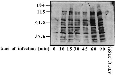
To this end, Chang epithelial cells were infected for the indicated times with the invasive P. aeruginosa strain ATCC 27853. Tyrosine phosphorylation was determined by immunoblotting total cell lysates with the monoclonal anti-phosphotyrosine antibody 4G10. The results, depicted in Fig. 1, show that infection of Chang cells with P. aeruginosa ATCC 27853 induces an increase in the tyrosine phosphorylation of several cellular proteins after 10 min, whereas blotting of bacterial lysates only does not reveal any significant tyrosine phosphorylation (Fig. 1). In particular, bacterial infection induced tyrosine phosphorylation of proteins with molecular sizes of approximately 140, 95, 70, 58, and 42 kDa. Similar results were obtained for infection with another invasive P. aeruginosa strain, 696 (data not shown).
FIG. 1.
Internalization of P. aeruginosa into human epithelial cells induces cellular tyrosine phosphorylation. Chang conjunctiva epithelial cells were infected with the invasive P. aeruginosa strain ATCC 27853; then the cells were lysed, and proteins were separated by SDS–10% PAGE and analyzed for tyrosine phosphorylation by Western blotting using the monoclonal anti-phosphotyrosine antibody 4G10. Results reveal a rapid and marked tyrosine phosphorylation of several cellular proteins, in particular those with molecular sizes of 140, 95, 70, 58, and 42 kDa, as soon as 10 min after infection with P. aeruginosa ATCC 27853. Samples were normalized by in vivo labeling of mammalian cells prior to infection and counting aliquots of the lysates.
To identify molecular mechanisms mediating cellular tyrosine phosphorylation, we investigated whether Src-like tyrosine kinases, which have been shown to be involved in many aspects of mammalian cell activation (30), participate in the host response to P. aeruginosa.
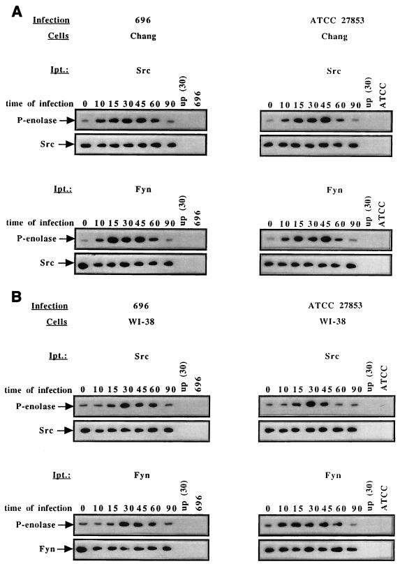
First, we investigated the activities of p59Fyn and p60Src in human epithelial cells upon infection with P. aeruginosa (Fig. 2). The results reveal that infection of Chang cells with P. aeruginosa 696 or ATCC 27853 induces a 5 ± 1.3-fold stimulation of p60Src and a 4 ± 1.1-fold stimulation of p59Fyn kinase activity as early as 10 min after initiation of the infection (Fig. 2A). The activities of p60Src and p59Fyn peaked 15 to 45 min after initiation of the infection (Fig. 2A). Additional experiments with WI-38 cells infected with P. aeruginosa 696 or ATCC 27853 revealed very similar results, confirming an activation of p60Src and p59Fyn after infection (Fig. 2B). This indicates that activation of p60Src and p59Fyn upon P. aeruginosa uptake is not restricted to a certain epithelial cell line or bacterial strain but seems to be a general response upon invasion by P. aeruginosa.
FIG. 2.
Infection of Chang epithelial cells with P. aeruginosa activates p60Src and p59Fyn. Infection of Chang or WI-38 epithelial cells with P. aeruginosa 696 or ATCC 27853 for the indicated times (in minutes) results in rapid activation of p60Src or p59Fyn. Control immunoprecipitates (up) from infected cells with irrelevant goat sera did not show phosphorylation of the substrate enolase. Likewise, immunoprecipitates (Ipt.) obtained from P. aeruginosa 696 or ATCC 27853 only did not reveal any cross-reactivity. Chang epithelial cells (A) or WI-38 (B) cells were infected with P. aeruginosa 696 or ATCC 27853 for the indicated times in minutes, lysed, and subjected to immunoprecipitation with goat polyclonal anti-Src or anti-Fyn antibodies. Cells were labeled with [3H]choline chloride prior to infection, permitting us to use the supernatants of the agarose-immobilized immunoprecipitates for normalization of the samples prior to the kinase assay. The kinase reaction was initiated by resuspending the immunoprecipitates in kinase buffer supplemented with 10 μg of enolase/ml and 10 μCi of [32P]γ-ATP. The samples were separated by SDS–10% PAGE and analyzed by autoradiography. The activities of p60Src and p59Fyn were determined by phosphorylation of the substrate enolase (P-enolase). Aliquots of the immunoprecipitates were blotted with anti-Fyn or anti-Src, followed by HRP-coupled protein L and enhanced chemiluminescence development, demonstrating similar amounts of Src or Fyn in each experiment. Control experiments were performed by incubation of bacterial lysates from P. aeruginosa 696 or ATCC 27853 with anti-Src or anti-Fyn antisera and reveal that the bacteria do not contain a cross-reacting kinase. Further, lysates from infected cells were subjected to precipitation with an irrelevant antiserum, demonstrating the specificity of the kinase assay.
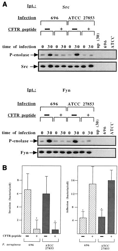
To determine whether it is the adhesion of P. aeruginosa to human cells or its uptake into cells that triggers the activation of p60Src and p59Fyn, we inhibited bacterial internalization into epithelial cells with a CFTR peptide containing amino acids 103 to 117 of mature CFTR. This peptide has been recently shown by Pier et al. (24) to block the uptake of P. aeruginosa into the lung cells of BALB/c mice by competing with CFTR for binding to P. aeruginosa LPS. However, this peptide does not alter the attachment of P. aeruginosa to mammalian cells (24). P. aeruginosa was incubated for 10 min with 200 nM CFTR peptide and then used for 30-min infection assays. The results (Fig. 3A) reveal that activation of p60Src and p59Fyn upon infection with P. aeruginosa 696 or ATCC 27853 strictly depends on the invasion of these strains, since preincubation with the CFTR peptide almost completely blocked the stimulation of the two kinases upon infection of Chang cells with P. aeruginosa 696 or ATCC 27853. To confirm the blocking effect of the peptide on the internalization of P. aeruginosa into Chang epithelial cells, uptake was determined with crystal violet, and intracellular versus adherent bacteria were counted using light microscopy (Fig. 3B). The rate of invasion of Chang epithelial cells by P. aeruginosa 696 or ATCC 27853 incubated with 200 nM CFTR peptide was 0.7 ± 1.9 (696) or 0.6 ± 1 (ATCC 27853) bacteria per cell, and the adhesion rate was 14.9 ± 5.6 (696) or 16 ± 3 (ATCC 27853) bacteria per cell, respectively (Fig. 3B). Control experiments with cells which were infected with bacteria that had not been preincubated with the CFTR peptide showed an invasion rate of 6.6 ± 4.3 (696) or 7 ± 2 (ATCC 27853) bacteria per cell and an adhesion rate of 6 ± 3.5 (696) or 5 ± 2 (ATCC 27853) bacteria per cell, respectively. These experiments indicate that internalization of P. aeruginosa into Chang epithelial cells, but not adhesion, is blocked (P = 0.034 for 696; P = 0.027 for ATCC 27853) after incubation of the bacteria with the CFTR peptide.
FIG. 3.
Inhibition of P. aeruginosa LPS binding to CFTR prevents activation of p60Src and p59Fyn. Prevention of P. aeruginosa binding to CFTR by incubation with a CFTR peptide encompassing amino acids 103 to 117 blocks activation of Src-like tyrosine kinases (A) and invasion (B) in Chang cells upon infection with the indicated strains. (A) Chang epithelial cells were infected with P. aeruginosa 696 or ATCC 27583 for the indicated times (in minutes) and lysed; then p60Src or p59Fyn was immunoprecipitated and subjected to kinase assays. Chang cells were labeled as above with [3H]choline chloride, the supernatants of the immobilized immunoprecipitates were counted, and the samples were normalized accordingly. P. aeruginosa 696 or ATCC 27853 was incubated with the CFTR peptide 10 min prior to the infection assay. Activities of p60Src and p59Fyn were determined by phosphorylation of enolase as above. An aliquot of the immunoprecipitates was blotted with anti-Src or anti-Fyn, displaying similar amounts of protein in all lanes. Control experiments were performed as above. (B) Chang epithelial cells were infected for 30 min as above with CFTR peptide-treated or untreated P. aeruginosa 696 or ATCC 27583, and invasion was determined by crystal violet staining. Results shown are means ± SD from three independent experiments. Significance was determined by t test, and significant differences are indicated by asterisks.
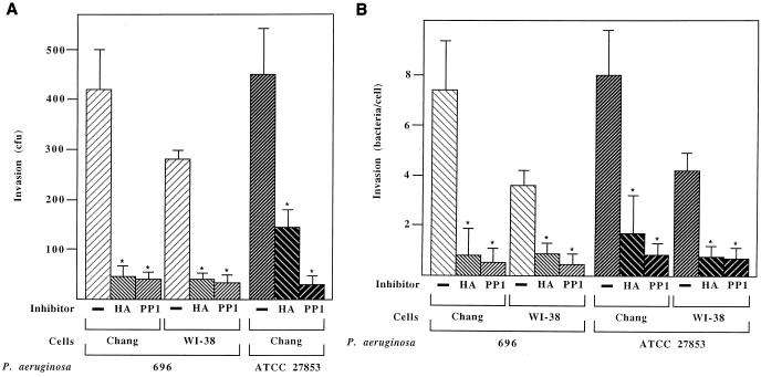
In order to determine the significance of p60Src and p59Fyn activation for bacterial invasion of human epithelial cells, we preincubated Chang cells with 1 μM herbimycin A or 50 nM PP1. Both herbimycin A and PP1 are inhibitors of Src-like tyrosine kinases (19, 32), and both significantly prevented invasion by P. aeruginosa, as judged by polymyxin survival assays 30 min after initiation of the infection (Fig. 4A). Specifically, herbimycin A reduced the invasion of Chang cells by P. aeruginosa 696 or ATCC 27853 by 89.2% or 68.3% (P < 0.0001), respectively. Likewise, the invasion of WI-38 cells by P. aeruginosa 696 was blocked by 64.4% (P < 0.0001). Similar results were obtained for PP1, with inhibition of 696 or ATCC 27853 invasion of Chang cells by 91.5 or 85%, respectively (P < 0.0001). Adhesion of these P. aeruginosa strains was not significantly affected and was only slightly increased by the tyrosine kinase inhibitors used. Control experiments demonstrated that incubation of the host cells with the inhibitors was not toxic, as judged by trypan blue staining of the cells. Further, neither herbimycin A nor PP1 affected the viability of the bacteria, as indicated by the fact that incubation of the bacteria alone for 30 min (i.e., the infection time) with the indicated inhibitor did not alter the ability of the pathogens to invade untreated mammalian cells. Furthermore, plating of bacteria treated with herbimycin A or PP1 or left untreated revealed no differences in bacterial growth.
FIG. 4.
Blockade of Src-like tyrosine kinases by herbimycin A or PP1 prevents P. aeruginosa uptake into different human epithelial cells. Chang and WI-38 epithelial cells were either pretreated for 12 h with 1 μM herbimycin A (HA) or for 15 min with 50 nM PP1 or left untreated. Cells were infected for 30 min, and invasion was scored by polymyxin survival (A) or crystal violet assay (B). Shown is the invasion of untreated Chang epithelial cells by two P. aeruginosa strains, (control), compared with the uptake of the bacteria into the indicated cells pretreated with HA or PP1. Means ± SD from three independent experiments are shown. Significance was determined by post hoc ANOVA done by the Bonferroni and Dunn method. Asterisks indicate significant differences.
To exclude the possibility that herbimycin A or PP1 affected the viability of intracellular bacteria and therefore altered the outcome of the polymyxin invasion assays, we light microscopically counted intracellular bacteria by crystal violet assays (Fig. 4B). The results confirm those of the polymyxin assays and demonstrate that herbimycin A inhibited P. aeruginosa 696 invasion of Chang cells by 90.5% (P = 0.009) and ATCC 27853 invasion by 79% (P = 0.005). Invasion of WI-38 epithelial cells was inhibited by 75.6% for 696 (P = 0.003) and 81.2% for ATCC 27853 (P = 0.002). PP1 reduced the uptake of 696 or ATCC 27853 into Chang cells by 92.2% (P = 0.007) and 89.5% (P = 0.003), respectively. Invasion of WI-38 cells was blocked by 85.5% (P = 0.0004) for 696 and 81.5% (P = 0.001) for ATCC 27853. The potencies of the two inhibitors to block P. aeruginosa invasion were not significantly different.
DISCUSSION
The activation of host signal transduction pathways seems to be an essential prerequisite for many bacteria to invade mammalian cells (2, 8). Our results indicate that invasion by P. aeruginosa of several human epithelial cells requires the activation of the Src-like tyrosine kinases p60Src and p59Fyn. These results identify a novel group of signaling molecules crucially involved in the internalization of P. aeruginosa into human epithelial cells.
The pivotal role of Src-like tyrosine kinases for P. aeruginosa internalization by mammalian cells is indicated by several lines of evidence. First, infection of several mammalian cells with various P. aeruginosa strains results in a very rapid and marked activation of the kinases. Src-like tyrosine kinase activation is observed as early as 5 to 10 min after infection and thus precedes internalization of the bacteria, which is detected as early as 15 min after initiation of the infection. Second, the addition of the CFTR peptide, which competes with the LPS of P. aeruginosa for binding to the endogenous CFTR (24), prevents Src-like tyrosine kinase activation as well as bacterial internalization, but actually increases bacterial adhesion. This indicates that adhesion does not require Src-like tyrosine kinases. These data also suggest that binding of P. aeruginosa LPS to the CFTR peptide constitutes a prerequisite for Src-like tyrosine kinase activation. Since CFTR functions as an ion channel (31), it is unlikely that the activation of Src-like tyrosine kinases is directly mediated by CFTR. Therefore, binding of the bacterial LPS to CFTR might result in an association and activation of other mammalian receptor molecules stimulating Src-like tyrosine kinases. Alternatively, the binding of LPS might enable P. aeruginosa to introduce into the mammalian target cell, via its type III secretion system, proteins or factors, which activate Src-like tyrosine kinases and thus trigger bacterial internalization.
Third and most important, inhibition of the kinases by two independent Src-like tyrosine kinase inhibitors prevents internalization, but not adhesion, of P. aeruginosa. If Src-like tyrosine kinase activation were a consequence of internalization, the two Src-like tyrosine kinase inhibitors should not block invasion. This suggests that activation of Src-like tyrosine kinases is a requirement for internalization and not a consequence of the uptake process.
In summary, the data with herbimycin A, PP1, and the CFTR peptide suggest that binding of P. aeruginosa LPS to CFTR results via unknown intermediates in activation of the Src-like tyrosine kinases p60Src and p59Fyn, which then mediate the internalization of the bacteria. Herbimycin A and PP1 have been shown in several studies to block Src-like tyrosine kinases relatively specifically (11, 14). However, even if they are not absolutely specific for Src-like tyrosine kinases, the use of two independent inhibitors with different effects on other kinases strongly suggests that the blockade of P. aeruginosa internalization by these inhibitors is due to the inhibition of Src-like tyrosine kinases.
Activation of Src-like tyrosine kinases results in tyrosine phosphorylation of several cellular proteins. Identification of these phosphorylated substrates remains to be elucidated. Considering the size of the tyrosine-phosphorylated proteins, Vav, phosphatidylinositol 3′ kinase (PI-3-K), and p42 mitogen-activated protein (MAP) kinase might be good candidates as substrates for Src-like tyrosine kinases upon infection (4, 22, 25). These proteins have been shown to be directly or indirectly involved in the regulation of cytoskeleton changes. Since the invasion of P. aeruginosa requires a reorganization of the cytoskeleton, as shown by the inhibition of P. aeruginosa internalization upon incubation with cytochalasin D (10), which destroys microfilaments, cytoskeletal proteins might play a role in the uptake of P. aeruginosa. Vav and PI-3-K might be involved in reorganization of the actin cytoskeleton via the activation of the small G protein Rac1, which has been shown to be one of the key regulators of the actin cytoskeleton (4, 20, 22, 25). Likewise, MAP kinases are good candidates to be involved in a reorganization of microtubuli.
An involvement of tyrosine kinases in the internalization of P. aeruginosa into epithelial cells has been recently implied by a study by Evans et al. (7). These authors showed that incubation of rabbit corneal cells with herbimycin A or the broad-spectrum kinase inhibitor genistein prevents uptake of P. aeruginosa, supporting the notion of the present report.
The finding that P. aeruginosa invades epithelial cells via activation of Src-like tyrosine kinases correlates with the invasion mechanisms of several other bacteria, including Yersinia enterocolitica, S. flexneri, L. monocytogenes, and Neisseria gonorrhoeae (5, 15, 17, 33). Uptake of these bacteria into mammalian cells has been shown to require the activation of Src-like tyrosine kinases and several protein tyrosine kinase-linked pathways. Thus, it might be possible that bacterial internalization processes into cells are conserved, and different bacteria may utilize similar or even the same signaling pathways for internalization.
In summary, we show that invasion of human epithelial cells by P. aeruginosa induces activation of the Src-like tyrosine kinases p60Src and p59Fyn, resulting in tyrosine phosphorylation of various cellular proteins. The activation of p60Src and p59Fyn seems to be crucial for P. aeruginosa internalization but not for P. aeruginosa adherence.
ACKNOWLEDGMENTS
We thank B. Mordmüller for critical reading of the manuscript.
This study was supported by DFG grant Gu 335/10-1/2, the Fortune program of the University of Tuebingen, the IZKF Tuebingen, and ALSAC (to E.G.).
REFERENCES
- 1.Baker N R, Minor V, Deal C, Shahrabadi M S, Simpson D A, Woods D E. Pseudomonas aeruginosa exoenzyme S is an adhesin. Infect Immun. 1991;59:2859–2863. doi: 10.1128/iai.59.9.2859-2863.1991. [DOI] [PMC free article] [PubMed] [Google Scholar]
- 2.Bliska J B, Galán J E, Falkow S. Signal transduction in the mammalian cell during bacterial attachment and entry. Cell. 1993;73:903–920. doi: 10.1016/0092-8674(93)90270-z. [DOI] [PubMed] [Google Scholar]
- 3.Chi E, Mehl T, Nunn D, Lory S. Interaction of Pseudomonas aeruginosa with A549 pneumocyte cells. Infect Immun. 1991;59:822–828. doi: 10.1128/iai.59.3.822-828.1991. [DOI] [PMC free article] [PubMed] [Google Scholar]
- 4.Crespo P, Schuebel K E, Ostrom A A, Gutkind J S, Bustelo X R. Phosphotyrosine-dependent activation of Rac-1 GDP/GTP exchange by the vav proto-oncogene product. Nature. 1997;385:169–172. doi: 10.1038/385169a0. [DOI] [PubMed] [Google Scholar]
- 5.Dehio C, Prévost M C, Sansonetti P J. Invasion of epithelial cells by Shigella flexneri induces tyrosine phosphorylation of cortactin by a pp60c-src-mediated signalling pathway. EMBO J. 1995;14:2471–2482. doi: 10.1002/j.1460-2075.1995.tb07244.x. [DOI] [PMC free article] [PubMed] [Google Scholar]
- 6.Doig P, Sastry P A, Hodges R S, Lee K K, Paranchych W, Irvin R T. Inhibition of pilus-mediated adhesion of Pseudomonas aeruginosa to human buccal epithelial cells by monoclonal antibodies directed against pili. Infect Immun. 1990;58:124–130. doi: 10.1128/iai.58.1.124-130.1990. [DOI] [PMC free article] [PubMed] [Google Scholar]
- 7.Evans D J, Frank D W, Finck-Barbancon V, Wu C, Fleiszig S M. Pseudomonas aeruginosa invasion and cytotoxicity are independent events, both of which involve protein tyrosine kinase activity. Infect Immun. 1998;66:1453–1459. doi: 10.1128/iai.66.4.1453-1459.1998. [DOI] [PMC free article] [PubMed] [Google Scholar]
- 8.Finlay B B, Cossart P. Exploitation of mammalian host cell functions by bacterial pathogens. Science. 1997;276:718–725. doi: 10.1126/science.276.5313.718. [DOI] [PubMed] [Google Scholar]
- 9.Fleiszig S M, Zaidi T S, Fletcher E L, Preston M J, Pier G B. Pseudomonas aeruginosa invades corneal epithelial cells during experimental infection. Infect Immun. 1994;62:3485–3493. doi: 10.1128/iai.62.8.3485-3493.1994. [DOI] [PMC free article] [PubMed] [Google Scholar]
- 10.Fleiszig S M, Zaidi T S, Pier G B. Pseudomonas aeruginosa invasion of and multiplication within corneal epithelial cells in vitro. Infect Immun. 1995;63:4072–4077. doi: 10.1128/iai.63.10.4072-4077.1995. [DOI] [PMC free article] [PubMed] [Google Scholar]
- 11.Fukuzawa H, Li P M, Yamamoto C, Murakami Y, Mizuno S, Uehara Y. Specific inhibition of cytoplasmic protein tyrosine kinases by herbimycin A in vitro. Biochem Pharmacol. 1991;42:1661–1671. doi: 10.1016/0006-2952(91)90500-5. [DOI] [PubMed] [Google Scholar]
- 12.Galán J E, Pace J, Hayman M J. Involvement of the epidermal growth factor receptor in the invasion of cultured mammalian cells by Salmonella typhimurium. Nature. 1992;357:588–589. doi: 10.1038/357588a0. [DOI] [PubMed] [Google Scholar]
- 13.Hancock R E, Mutharia L M, Chan L, Darveau R P, Speert D P, Pier G B. Pseudomonas aeruginosa isolates from patients with cystic fibrosis: a class of serum-sensitive, nontypeable strains deficient in lipopolysaccharide O side chains. Infect Immun. 1983;42:170–177. doi: 10.1128/iai.42.1.170-177.1983. [DOI] [PMC free article] [PubMed] [Google Scholar]
- 14.Hanke J H, Gardner J P, Dow R L, Changelian P S, Brissette W H, Weringer E J, Pollok B A, Connelly P A. Discovery of a novel, potent, and Src-family-selective tyrosine kinase inhibitor. J Biol Chem. 1996;271:695–701. doi: 10.1074/jbc.271.2.695. [DOI] [PubMed] [Google Scholar]
- 15.Hauck C R, Meyer T F, Lang F, Gulbins E. CD66-mediated phagocytosis of Opa52Neisseria gonorrhoeae requires a Src-like tyrosine kinase- and Rac1-dependent signalling pathway. EMBO J. 1998;17:443–454. doi: 10.1093/emboj/17.2.443. [DOI] [PMC free article] [PubMed] [Google Scholar]
- 16.Hirakata Y, Izumikawa K, Yamaguchi T, Igimi S, Furuya N, Maesaki S, Tomono K, Yamada Y, Kohno S, Yamaguchi K, Kamihira S. Adherence to and penetration of human intestinal Caco-2 epithelial cell monolayers by Pseudomonas aeruginosa. Infect Immun. 1998;66:1748–1751. doi: 10.1128/iai.66.4.1748-1751.1998. [DOI] [PMC free article] [PubMed] [Google Scholar]
- 17.Isberg R R, Leong J M. Multiple β1 chain integrins are receptors for invasin, a protein that promotes bacterial penetration into mammalian cells. Cell. 1990;60:861–871. doi: 10.1016/0092-8674(90)90099-z. [DOI] [PubMed] [Google Scholar]
- 18.Lee C K, Roberts A L, Finn T M, Knapp S, Mekalanos J J. A new assay for invasion of HeLa 229 cells by Bordetella pertussis: effects of inhibitors, phenotypic modulation, and genetic alterations. Infect Immun. 1990;58:2516–2522. doi: 10.1128/iai.58.8.2516-2522.1990. [DOI] [PMC free article] [PubMed] [Google Scholar]
- 19.Melford S K, Turner M, Briddon S J, Tybulewicz V L, Watson S P. Syk and Fyn are required by mouse megakaryocytes for the rise in intracellular calcium induced by a collagen-related peptide. J Biol Chem. 1997;272:27539–27542. doi: 10.1074/jbc.272.44.27539. [DOI] [PubMed] [Google Scholar]
- 20.Nobes C D, Hall A. Rho, rac and cdc 42 GTPases regulate the assembly of multimolecular focal complexes associated with actin stress fibers, lamellipodia, and filopodia. Cell. 1995;81:53–62. doi: 10.1016/0092-8674(95)90370-4. [DOI] [PubMed] [Google Scholar]
- 21.Pace J, Hayman M J, Galán J E. Signal transduction and invasion of epithelial cells by S. typhimurium. Cell. 1993;72:505–514. doi: 10.1016/0092-8674(93)90070-7. [DOI] [PubMed] [Google Scholar]
- 22.Pelech S L, Sanghera J S. MAP kinases: charting the regulatory pathways. Science. 1992;257:1355–1356. doi: 10.1126/science.1382311. [DOI] [PubMed] [Google Scholar]
- 23.Pier G B, Grout M, Zaidi T S, Olsen J C, Johnson L G, Yankaskas J R, Goldberg J B. Role of mutant CFTR in hypersusceptibility of cystic fibrosis patients to lung infections. Science. 1996;271:64–67. doi: 10.1126/science.271.5245.64. [DOI] [PMC free article] [PubMed] [Google Scholar]
- 24.Pier G B, Grout M, Zaidi T S. Cystic fibrosis transmembrane conductance regulator is an epithelial cell receptor for clearance of Pseudomonas aeruginosa from the lung. Proc Natl Acad Sci USA. 1997;94:12088–12093. doi: 10.1073/pnas.94.22.12088. [DOI] [PMC free article] [PubMed] [Google Scholar]
- 25.Pleiman C M, Hertz W M, Cambier J C. Activation of phosphatidylinositol-3′ kinase by Src-family kinase SH3 binding to the p85 subunit. Science. 1994;263:1609–1612. doi: 10.1126/science.8128248. [DOI] [PubMed] [Google Scholar]
- 26.Ramphal R, Pier G B. Role of Pseudomonas aeruginosa mucoid exopolysaccharide in adherence to tracheal cells. Infect Immun. 1985;47:1–4. doi: 10.1128/iai.47.1.1-4.1985. [DOI] [PMC free article] [PubMed] [Google Scholar]
- 27.Rosenshine I, Duronio V, Finlay B B. Tyrosine protein kinase inhibitors block invasin-promoted bacterial uptake by epithelial cells. Infect Immun. 1992;60:2211–2217. doi: 10.1128/iai.60.6.2211-2217.1992. [DOI] [PMC free article] [PubMed] [Google Scholar]
- 28.Rosenshine I, Ruschkowski S, Foubister V, Finlay B B. Salmonella typhimurium invasion of epithelial cells: role of induced host cell tyrosine protein phosphorylation. Infect Immun. 1994;62:4969–4974. doi: 10.1128/iai.62.11.4969-4974.1994. [DOI] [PMC free article] [PubMed] [Google Scholar]
- 29.Rosenshine I, Ruschkowski S, Stein M, Reinscheid D J, Mills S D, Finlay B B. A pathogenic bacterium triggers epithelial signals to form a functional bacterial receptor that mediates actin pseudopod formation. EMBO J. 1996;15:2613–2624. [PMC free article] [PubMed] [Google Scholar]
- 30.Schwartzberg P L. The many faces of Src: multiple functions of a prototypical tyrosine kinase. Oncogene. 1998;17:1463–1468. doi: 10.1038/sj.onc.1202176. [DOI] [PubMed] [Google Scholar]
- 31.Sheppard D N, Ostedgaard L S, Rich D P, Welsh M J. The amino-terminal portion of CFTR forms a regulated Cl− channel. Cell. 1994;76:1091–1098. doi: 10.1016/0092-8674(94)90385-9. [DOI] [PubMed] [Google Scholar]
- 32.Uehara Y, Hori M, Takeuchi T, Umezawa H. Phenotypic change from transformed to normal induced by benzoquinonoid ansamycins accompanies inactivation of p60src in rat kidney cells infected with Rous sarcoma virus. Mol Cell Biol. 1986;6:2198–2206. doi: 10.1128/mcb.6.6.2198. [DOI] [PMC free article] [PubMed] [Google Scholar]
- 33.Van Langendonck N, Velge P, Bottreau E. Host cell protein tyrosine kinases are activated during the entry of Listeria monocytogenes. Possible role of pp60c-src family protein kinases. FEMS Microbiol Lett. 1998;162:169–176. doi: 10.1111/j.1574-6968.1998.tb12995.x. [DOI] [PubMed] [Google Scholar]
- 34.Van Putten J P M, Hopman C T P, Weel J F L. The use of immunogold-silver staining to study antigen variation and bacterial entry into eukaryotic cells by conventional light microscopy. J Med Microbiol. 1990;33:35–41. doi: 10.1099/00222615-33-1-35. [DOI] [PubMed] [Google Scholar]
- 35.Van Putten J P M, Weel J F L, Grassmé H U C. Measurements of invasion by antibody labelling and electron microscopy. Methods Enzymol. 1994;236:420–437. doi: 10.1016/0076-6879(94)36031-6. [DOI] [PubMed] [Google Scholar]


