Abstract
At least five proteins are secreted extracellularly by enteropathogenic Escherichia coli (EPEC), a leading cause of infant diarrhea in developing countries. However only one, EspC, is known to be secreted independently of the type III secretion apparatus encoded by genes located within the 35.6-kb locus of enterocyte effacement pathogenicity island. EspC is a member of the autotransporter family of proteins, and the secreted portion of the molecule is 110 kDa. Here we determine that the espC gene is located within a second EPEC pathogenicity island at 60 min on the chromosome of E. coli. We also show that EspC is an enterotoxin, indicated by rises in short-circuit current and potential difference in rat jejunal tissue mounted in Ussing chambers. In addition, preincubation with antiserum against the homologous Pet enterotoxin of enteroaggregative E. coli eliminated EspC enterotoxin activity. Like the EAF plasmid, the espC pathogenicity island was found only in a subset of EPEC, suggesting that EspC may play a role as an accessory virulence factor in some but not all EPEC strains.
Secretion of effector molecules allows pathogenic bacteria to interact with their host and cause disease. Enteropathogenic Escherichia coli (EPEC), a leading cause of infantile diarrhea in developing countries, secretes at least six proteins (24, 28). Four of these proteins, EspA (29), EspD (31), EspB (46), and EspF (36) (E. coli secreted protein), are secreted by a type III secretion system, and the Esp molecules as well as the secretion apparatus are encoded within a 35.6-kb pathogenicity island termed the locus of enterocyte effacement (LEE) (11, 35). Recently, it was demonstrated that the EspA, EspD, and EspB proteins form a translocon for delivering effector molecules into the host cytoskeleton (15, 30). Another secreted molecule, Tir (translocated intimin receptor) (27), is hypothesized to pass through this structure en route to translocation into the host cell membrane, where it serves as a receptor for the EPEC adhesin intimin, also encoded within the LEE (25). Tir is involved in host cell signaling and disruption of the cytoskeleton (27). These secreted effector molecules are involved in the formation of attaching and effacing (AE) intestinal lesions, a hallmark of EPEC disease.
EPEC also secretes a 110-kDa protein which does not require the type III secretion system for delivery into the extracellular milieu (24, 45). This protein, EspC, shows amino acid homology to members of the immunoglobulin A (IgA) protease family of autotransporters which include, among others, the IgA protease of Neisseria gonorrhoeae (38), Hap of Haemophilus influenzae (23), Tsh of avian-pathogenic E. coli (39), the SepA and ShMu proteins of Shigella flexneri (4, 40), Pic of enteroaggregative E. coli (EAEC) and Shigella flexneri 2457T (22), Pet of EAEC (12), and EspP of enterohemorrhagic E. coli (EHEC) (7). For a review, see reference 21.
The mechanism of autotransport was first described for the IgA protease of N. gonorrhoeae (38). The precursor protein is exported beyond the cytoplasmic membrane in a Sec-dependent manner, coupled to the cleavage of a signal peptide. The mature protein possesses a C-terminal domain that is thought to form a β-barrel in the outer membrane, and the N-terminal passenger domain is exported beyond the outer membrane through the β-barrel. The passenger domain is then clipped autocatalytically from the β-barrel and released. In the case of EspC of EPEC, the molecular mass of the exported passenger protein is 110 kDa (45).
Several members of the autotransporter family of proteins, including Tsh, SepA, ShMu, EspP, Pet, Pic, and EspC, have a conserved serine protease motif. None of these proteins, however, cleave IgA. This subfamily of autotransporters have thus been termed SPATE, for serine protease autotransporters of Enterobacteriaceae (21). It was recently demonstrated that Pet of EAEC is an enterotoxin that induces loss of actin microfilaments (12, 37). The function of EspC of EPEC, however, is unknown. A mutation in espC did not affect the ability of EPEC to disrupt cytoskeletal rearrangement, phosphorylate a 90-kDa host protein (now known to be the bacterium-derived Tir protein), or adhere to or invade three different tissue culture cell lines (45).
Virulence genes of pathogenic bacteria are often located on transmissible genetic elements such as plasmids, transposons, or bacteriophages, and many of these proteins are encoded within specific regions of the chromosome termed pathogenicity islands (17, 20, 32); the SPATE proteins adhere to this observation. EspP of E. coli O157:H7 (7) and Pet of EAEC (12) are encoded on virulence plasmids, pO157 and pAA2, respectively, and ShMu of Shigella flexneri and Pic of EAEC are located within a pathogenicity island (22, 40). The location of espC in EPEC, however, is yet to be determined.
The gene encoding EspC has been cloned and sequenced. Previous studies indicated that espC was not located within the LEE pathogenicity island of EPEC strain E2348/69 (11, 35) or on the EAF plasmid (45), which contains genes necessary for bundle-forming pilus biogenesis and the Per regulator. We therefore hypothesized that espC was chromosomally located, perhaps associated with other loci found only in pathogenic strains of E. coli. In light of an earlier report that EspC was not involved in the AE phenotype (45), we wanted to further investigate a potential function for this protein. In this study, we determine that espC is encoded within a pathogenicity island located at 60 min on the chromosome of EPEC and that the EspC protein has enterotoxic activity.
MATERIALS AND METHODS
Bacterial strains and growth conditions.
The EPEC strain containing espC used in this study was the well-characterized, prototype AE pathogen E2348/69 (O127:H6) (33). JPN15 is an EAF plasmid-cured derivative of strain E2348/69 (25). Strains used in DNA colony hybridization studies are listed in Table 1. For isolating secreted proteins, recombinant plasmids were transformed into laboratory strain HB101. All strains were grown in Luria-Burtani (LB) broth aerobically at 37°C unless otherwise stated. Where appropriate, culture media were supplemented with ampicillin (100 μg/ml).
TABLE 1.
Colony hybridization studiesa
| Strain | Category | Serotype | DECb | Hybridizationc with probe:
|
Source or reference | ||||||
|---|---|---|---|---|---|---|---|---|---|---|---|
| A | B | C | D | E | F | G | |||||
| HB101 | K-12 | + | − | − | − | − | + | + | Lab stock | ||
| DH5α | K-12 | + | − | − | − | − | − | + | Lab stock | ||
| MG1655 | K-12 | + | − | − | − | − | − | + | 6 | ||
| E2348/69 | EPEC1 | O127:H6 | 1 | + | + | + | + | + | + | + | 33 |
| JPN15 | EPEC1 | O127:H6 | 1 | + | + | + | + | + | + | + | 25 |
| #19 | EPEC1 | O142:H6 | + | + | + | + | + | + | + | 44 | |
| #15 | EPEC1 | O142:H6 | + | + | + | + | + | + | + | 44 | |
| 572-56 | EPEC1 | O55:H6 | 1 | + | w | + | w | + | + | + | 49 |
| C54-58 | EPEC1 | O55:H6 | 1 | − | + | + | + | + | + | + | 49 |
| 5513-56 | EPEC1 | O55:NM | 2 | + | + | + | + | + | + | + | 49 |
| 3299-85 | EHEC1 | O157:H7 | 3 | + | − | − | − | + | − | + | 49 |
| 3104-88 | EHEC1 | O157:H7 | 3 | + | − | − | − | + | − | + | 49 |
| C999-87 | EHEC1 | O157:H7 | 4 | + | − | − | − | + | − | + | 49 |
| 5338-66 | EPEC2 | 0111:H21 | 6 | + | − | − | − | + | − | + | 49 |
| C142-54 | EPEC | O111:H12 | 6 | + | − | − | − | − | + | + | 49 |
| 2277-67 | EPEC | O111:H12 | 6 | + | − | − | − | + | − | + | 49 |
| 184-83 | EPEC2 | O111:NM | 6 | + | − | − | − | + | + | + | 49 |
| 2198-77 | EPEC2 | O111:NM | 8 | + | − | − | − | + | − | + | 49 |
| C194-65 | EHEC2 | O111:H8 | 8 | + | − | − | − | + | − | + | 49 |
| 3233-61 | EHEC2 | O26:H11 | 9 | + | − | − | − | − | + | + | 49 |
| 2262-79 | EHEC | O26:HN | 9 | w | − | − | − | − | − | + | 49 |
| C814-67 | EHEC2 | O26:H11 | 9 | + | − | − | − | − | + | + | 49 |
| 2254-75 | EPEC2 | O128:H2 | 11 | w | − | − | − | − | − | + | 49 |
| 3733-71 | EPEC2 | O128:H2 | 11 | − | − | − | − | − | − | + | 49 |
| A9619-c2 | EPEC2 | O45:H2 | 11 | + | − | − | − | − | − | + | 49 |
| WM-63 | EPEC2 | O128:H2 | 11 | + | − | w | w | − | + | + | 49 |
| F1-50 | EPEC2 | O111:H2 | 12 | + | − | − | − | − | + | + | 49 |
| 2966-56 | EPEC2 | O111:H2 | 12 | + | − | − | − | + | + | + | 49 |
| 3350-73 | ETEC | O128:H7 | 13 | + | − | − | − | − | + | + | 49 |
| 5024-71 | ETEC | O128:H7 | 13 | + | − | − | − | − | + | + | 49 |
| C500-74 | ETEC | O128:H7 | 13 | + | − | − | − | − | + | + | 49 |
| C916-70 | ETEC | O128:H21 | 14 | + | − | − | − | − | + | + | 49 |
| C691-71 | ETEC | O128:H21 | 14 | + | − | − | − | − | − | + | 49 |
| #6 | EPEC | O119:H6 | + | − | − | − | + | + | + | 16 | |
| #15 | EPEC | O119:H6 | + | − | − | − | − | + | + | 16 | |
| RDEC1 | RDEC1 | + | − | − | − | + | − | + | Lab stock | ||
| B171 | EPEC | O111:NM | + | − | − | − | + | + | + | Lab stock | |
| BG6119 | EHEC2 | O26:H11 | + | − | − | − | + | − | + | Lab stock | |
| EDL933 | EHEC1 | O157:H7 | + | − | − | − | + | − | + | Lab stock | |
| 86-24 | EHEC1 | O157:H7 | + | − | − | − | + | − | + | Lab stock | |
| DK38 | EPEC | O55:H7 | + | − | − | − | − | + | + | Lab stock | |
| DK154 | EHEC | O103 | + | − | − | − | + | − | + | Lab stock | |
| Citrobacter rodentium 4280 | − | − | − | − | − | − | + | Lab stock | |||
All strains tested were E. coli except for Citrobacter rodentium. All strains except K-12 and enterotoxigenic E. coli (ETEC) strains possessed the eae gene and presumably the LEE pathogenicity island. Hybridizations were performed at 65°C under standard conditions and repeated under lower stringency conditions, 50°C, with identical results.
Diarrheagenic E. coli clones according to Whittam et al. 49.
w, weak hybridization.
Recombinant DNA techniques.
All genetic manipulations were performed using DH5α as a host strain. Unless otherwise stated, enzymes were purchased from BRL and procedures were performed as described previously (41). A previously constructed cosmid library containing EPEC DNA in pHC79 (34) was screened using an internal espC SalI fragment (see Fig. 1) and the colony hybridization procedure described below. For electrophysiological measurements in rat jejunum tissue, a minimal DNA fragment containing espC was directionally ligated into the pBAD30 vector (19) after PCR amplification with Pwo enzyme (Boehringer Mannheim) using the oligonucleotide primers K1314B (5′-CCGGAATTCTGGACTTCAGATCTGGTGATA-3′) and 1315B (5′-CTACGAAGCTTGCGAGCTTCTTGTGAGAAAGA-3′) containing EcoRI and HindIII restriction sites (underlined), respectively. Cosmid DNA was used as the template for PCR. The plasmid containing the 4-kb espC minimal fragment was designated pJLM174.
FIG. 1.
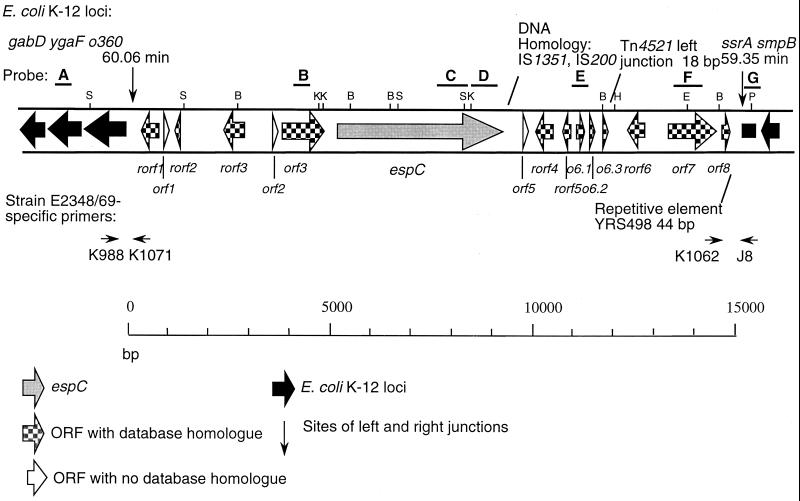
Genetic map of espC pathogenicity island. The 15,195 bp of unique DNA at approximately 60 min of the EPEC chromosome, including the espC gene and identified ORFs, is presented. Sequences corresponding to probes A through G used in the hybridization studies are noted. ORFs found in E. coli K-12 strain MG1655 are in black. Restriction enzyme cleavage sites: S, SalI; K, KpnI; B, BglII; E, EcoRI; H, HindIII; P, PvuII.
Primers K988 (5′-ACATCATCGATACCCACCGC-3′), K1071 (5′-GTCATTTTGCAGTGAAGCCAT-3′), and K1072 (5′-CGGTTCAACACATCAGGTAAG-3′) (5′ position 275 in the espC pathogenicity island) were used in PCR analysis to confirm the site of the left junction. Primers J8 (5′-TCCGCAGCTGGCAAGCGAAT-3′), J10 (5′-ACGGCAGGATAGGCACCGAA-3′), and K1062 (5′-CGCTGGCTGCACGATGTTGC-3′) (5′ position 14013 in the espC pathogenicity island) were used in PCR analysis to confirm the site of the right junction.
For DNA hybridization, colonies were transferred to Whatman 541 filters after overnight incubation on LB agar and lysed. Filters were neutralized, DNA probes (see Fig. 1 and Table 2) were amplified by PCR and labeled with [α-32P]dCTP using Ready-To-Go DNA Labeling beads, and unincorporated nucleotides were removed using ProbeQuant G-50 Micro columns following the manufacturer's instructions. Both kits were purchased from Pharmacia Biotech. After hybridization, approximately 106 cpm of each radiolabeled probe was added to filters, and incubation proceeded at 65°C overnight with shaking. Filters were washed twice for 30 min each at 65°C and exposed to autoradiograph film.
TABLE 2.
Oligonucleotides used for constructing DNA probes
| Probe | Primer | Sequence (5′ to 3′) | Strand | Site | S′ enda |
|---|---|---|---|---|---|
| A | K1108 | TCGCCGCCTCTGGCATGGAA | + | ygaF | — |
| K1077 | CGGCAGCCTGAAGGCGCAGT | − | ygaF | — | |
| B | K929 | ATGAGGTTTCCATATTTCCT | + | orf3 | 3920 |
| K912 | TAGAGACCAAGATAACAGC | − | orf3 | 4333 | |
| C | K948 | CGTTGGCCGTAATGCTA | + | espC | 7229 |
| K947 | GGTCTGATACCCTGTCA | − | espC | 8315 | |
| D | K994 | CATGAGCTGGACGGTGTGGA | + | espC | 8535 |
| K1315Bb | CTACGAAGCTTGCGATCTTCTTGTGAGAAAGA | − | espC | 9299 | |
| E | K1396 | GGAAATTCAGCAGCGGCTA | + | orf6.1 | 11021 |
| K1395 | GGCAAAGGACTTTGTGGATT | − | orf6.1 | 11243 | |
| F | K1032 | GCCGTTCGGTTCGAAACTG | + | orf7 | 13543 |
| K1154 | TTTTCACCGCCGGGAAGGC | − | orf7 | 14428 | |
| G | K2119 | TGGATTCGACGGGATTTGCGA | + | ssrA | — |
| K2118 | GTGCGGCTCTTAGGACTTCA | − | ssrA | — |
5′ position in the espC pathogenicity island.
HindIII site used for cloning is underlined.
DNA sequencing.
DNA sequence analysis was performed by the University of Maryland Biopolymer Facility on an ABI automated sequencer with cosmid DNA templates purified using Qiagen midicolumns. Double-stranded sequence was aligned and analyzed using Sequencher software.
Preparation of concentrated supernatants.
Wild-type E2348/69, HB101, HB101 (p2R) harboring espC in a cosmid clone, and the cosmid vector-only control HB101 (pHC79) were grown overnight at 37°C in 100 ml of LB broth. HB101(pJLM174) harboring the 4-kb espC minimal fragment in pBAD30 was also grown overnight at 37°C in 100 ml of LB broth supplemented with 0.2% glycerol and 0.2% glucose (repression) or 0.2% arabinose (induction). The bacterial cultures were centrifuged at 12,000 × g for 10 min and filtered through a 0.22-μm cellulose acetate filter, and the supernatants were concentrated 100 × and size fractionated (100-kDa cut off) using Ultrafilters (Biomax-100; Ultrafree; Millipore, Bedford, Mass.) according to the manufacturer's instructions.
Western immunoblot.
Proteins present in concentrated supernatants from HB101, E2348/69, HB101(p2R), and the espC minimal clone in HB101(pJLM174), grown with glycerol and glucose or arabinose, were analyzed by sodium dodecyl sulfate-polyacrylamide gel electrophoresis (SDS-PAGE) under reducing conditions (boiling for 5 min in the presence of β-mercaptoethanol). Proteins separated by SDS-PAGE were transferred to nitrocellulose BA85 membranes (Schleicher and Schuell, Keene, N.H.) by standard methods. The membranes were incubated with rabbit antibodies against Pet protein (37) diluted 1:100. Immunostaining was performed with alkaline phosphatase-labeled polyclonal antibodies against rabbit immunoglobulins (dilution, 1:2,000) and developed with 5-bromo-4-chloro-3-indolylphosphate and Nitro Blue Tetrazolium by standard methods.
Electrophysiological measurements in rat jejunum.
Ussing chamber experiments were performed as described previously (37). Jejunal segments removed from adult male Sprague-Dawley rats under sodium pentobarbital anesthesia were placed in ice-cold Ringer's solution for mammals and gassed with an O2-CO2 (95%–5%) mixture. The excised segments were cut open along their mesenteric border, washed with cold Ringer's solution, divided into six fragments, and mounted between the circular openings of two adjacent Ussing hemichambers. Each hemichamber was filled with 10 ml of Ringer's solution and kept at 37°C under constant O2-CO2 bubbling. Supernatants (100 μl) from HB101, HB101(p2R), HB101(pHC79), and HB101(pJLM174) were added to the mucosal hemichamber of rat jejunum preparations after 30 min of equilibration. Transepithelial electrical potential difference (PD) and total tissue conductance (Isc) were measured at 10-min intervals using a voltage clamp apparatus. Short-circuit current was calculated using Ohm's law (18). Statistical analyses were performed with Student's t test on data recorded from at least four experiments.
Ussing chamber experiments in which enterotoxic activity was inhibited by antibodies against Pet protein were performed by methods described previously (37).
Concentrated supernatants (100 μl) were incubated for 30 min at room temperature with rabbit polyclonal antibodies directed against the Pet protein (diluted 1:25) prior to addition to the luminal hemichamber.
Nucleotide sequence accession number.
The nucleotide sequence of the EPEC pathogenicity island containing espC will appear in the EMBL/GenBank/DDBJ data library under accession number AF297061.
RESULTS
EspC is encoded within a region of unique DNA on the EPEC chromosome.
An internal 1.6-kb SalI espC fragment (Fig. 1) was used to identify cosmids containing espC and flanking sequences from a previously constructed cosmid library of EPEC E2348/69 DNA (34). Four independent, nonidentical cosmid clones (p2R, p2L, p9R, and p10R) were obtained by colony hybridization; cosmids were isolated, and restriction digests demonstrated that all contained the 1.6-kb SalI fragment, a 4-kb KpnI fragment, and other identical restriction fragments (Fig. 1 and data not shown). Therefore, all four cosmids contained espC and surrounding DNA. Two of the cosmids, p2R and p10R, were subjected to sequence analysis.
Oligonucleotide primers were designed to sequence upstream and downstream from 5′ and 3′ termini of espC. Comparison of the initial sequence data to the E. coli K-12 genome sequence indicated that the DNA flanking espC was not found in E. coli K-12 strains. Continued sequencing revealed 15,195 bp of unique DNA, including approximately 5 kb upstream and 6 kb downstream of espC (Fig. 1). Predicted restriction sites (Fig. 1) were confirmed by restriction enzyme digestion of two independent plasmids, p9R and p10R, and Southern hybridization using these plasmids and chromosomal DNA isolated from strains E2348/69 and JPN15 (data not shown). The G+C content of the 15.2 kb of unique DNA was 40.5%.
Gapped BLAST analysis (1) of the unique DNA sequences flanking espC revealed one predicted open reading frame (ORF) with similarity to a known virulence factor, and several predicted ORFs with similarity to mobile genetic elements (Table 3). ORF3 showed predicted amino acid similarity with VirA of Shigella flexneri (19% identity and 37% similarity), which is involved in cellular invasion and intracellular spreading (47). ORF3 also showed predicted amino acid similarity with rORF2 of the EPEC LEE (43% identity and 63% similarity) (11). Predicted amino acid sequences from ORF6.1 were homologous to a putative transposase, BfpM, encoded on the EAF plasmid of EPEC (43). Though there was a high degree of amino acid identity, 93%, we presumed that the ORF6.1 product is not functional because of a large deletion relative to BfpM. ORF6.1, ORF6.2, and ORF6.3 all showed predicted amino acid similarity with a related putative transposase found in the Vibrio cholerae pathogenicity island (26). ORF7 showed predicted amino acid homology (95% identity) with the IS100 putative transposase found on the pesticin plasmid of Yersinia pestis. Sequences identical to the 44-bp repetitive element YRS498 also found in Y. pestis were located adjacent to the 3′ end of this ORF (13). DNA sequences directly downstream of the 3′ end of the espC structural gene were similar to IS1351 and IS200 elements, and 18 bp located downstream of orf6.3 were identical to sequences within the left junction of Tn4521.
TABLE 3.
Similarities of ORFs within the espC pathogenicity island
| Locus | Coordinates | No. of amino acids | Similar protein or locus | % Identity | % Similarity |
|---|---|---|---|---|---|
| rorf1 | 179–559 | 126 | o90a adjacent to o360 in E. coli K-12 | 51 | 65 |
| orf1 | 728–928 | 66 | None | ||
| rorf2 | 935–1105 | 56 | Nitrogen assimilation regulatory protein of Synechocystis sp. | 29 | 55 |
| rorf3 | 2258–2744 | 161 | Neutral endopeptidase of Lactococcus lactis subsp. cremoris | 23 | 40 |
| orf2 | 3324–3488 | 54 | None | ||
| orf3 | 3567–4739 | 390 | VirA of Shigella flexneri | 19 | 37 |
| rOrf2 of the EPEC LEE | 43 | 63 | |||
| espC | 5309–9226 | 1,305 | Pet of EAEC | 52 | 67 |
| orf5 | 9493–9666 | 57 | None | ||
| rorf4 | 10096–10476 | 126 | Cytochrome b of Anthropoides virgo | 26 | 44 |
| rorf5 | 10612–10800 | 62 | NahR activator protein of Pseudomonas putida | 31 | 53 |
| orf6.1 | 11013–11255 | 80 | BfpM of E. coli (putative transposase) | 93 | 95 |
| V. cholerae putative transposase xtn | 67 | 74 | |||
| orf6.2 | 11329–11484 | 51 | V. cholerae putative transposase xtn | 77 | 89 |
| orf6.3 | 11746–11886 | 46 | V. cholerae putative transposase xtn | 42 | 66 |
| rorf6 | 12208–12549 | 113 | Prophage CP4-57 integrase | 91 | 93 |
| orf7 | 13209–14600 | 463 | Probable transposase a of Yersinia pestis plasmid pMT1 | 95 | 95 |
| orf8 | 14715–14966 | 83 | Probable transposase b of Yersinia pestis plasmid pMT1 | 93 | 96 |
Additional predicted amino acid sequences showed similarities with known proteins (Fig. 1, Table 3). The predicted rORF2 amino acid sequences were similar to a nitrogen assimilation regulatory protein found in Lactococcus lactis, those from rORF4 were similar to a cytochrome b of eukaryotic origin, and those from rORF5 were similar to NahR, which is an activator protein of Pseudomonas putida.
Junctions of unique DNA sequences.
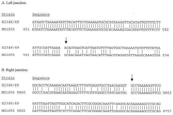
The left junction of the unique DNA sequences was defined by Blast search and PCR analysis. Blast search identified DNA sequences within cosmid p9R identical to those located adjacent to the orf360 gene at 60.06 min of the E. coli strain MG1655 chromosome (Fig. 2A).
FIG. 2.
Left and right junction sequences. (A) Comparison of DNA sequences immediately upstream of the left junction of the espC pathogenicity island (see arrows in Fig. 1) with section 241 of 400 of the complete E. coli K-12 strain MG1655 (accession no. AE000351) revealed 94% identity (6). (B) Comparison of DNA sequences downstream of the right junction of the espC pathogenicity island with section 237 of 400 of the complete E. coli K-12 strain MG1655 (accession no. AE000347) revealed 96% identity with the complete genome sequence of E. coli K-12 strain MG1655 (6). Position 1 of the espC pathogenicity island begins at the left arrow.
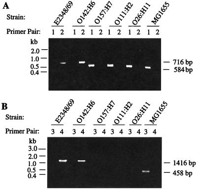
In order to confirm the site of the left junction of the unique DNA sequences, we performed PCR analysis. Primer K988 is specific to sequences within orf360 (b2659) of K-12 strain MG1655, and primer K1071 is specific to sequences located within the unique region in EPEC strain E2348/69 (Fig. 1). When these primers (designated primer pair 2 in Fig. 3A) were used in PCR with E2348/69 DNA as the template, a predicted amplicon of 716 bp was visualized (Fig. 3A), confirming the chromosomal location of the left junction as being adjacent to orf360 at 60.06 min of the K-12 genome. Some differences in the flanking sequence were observed between K-12 and EPEC. Whereas gene b2656 is adjacent to orf360 in K-12 strain MG1655, sequence analysis of the region adjacent to orf360 in EPEC E2348/69 did not detect b2656 sequences. The absence of b2656 in this region was confirmed using primers K988 (specific for orf360) and K1072 (specific for b2656). With MG1655 DNA as the template, these primers (primer pair 1 in Fig. 3A) yielded the expected 584-bp amplicon, whereas with E2348/69 DNA, no PCR product was detected (Fig. 3A).
FIG. 3.
PCR analysis of the left and right junctions. (A) PCR analysis identified the left junction at 60.03 min adjacent to orf360 of the E. coli chromosome in strain E2348/69 and EPEC serotype O142:H6, both members of the EPEC1 category (48). PCR with primer pair 1 (K988 and K1072) resulted in an amplicon of 584 bp using K-12 strain MG1655 DNA as the template, whereas primer pair 2 (K988 and K1071) resulted in an amplicon of 716 bp using E2349/69 DNA as the template. (B) Similarly, the right junction was at 59.35 min within the ssrA gene of the the E. coli chromosome in strain E2348/69 and EPEC serotype O142:H6. PCR with primer pair 3 (J8 and J10) amplified a K-12-specific amplicon of 458 bp, whereas primer pair 4 (J8 and K1062) amplified an E2348/69-specific amplicon of 1,416 bp.
Similarly, the right junction of the unique DNA was defined by sequence analysis and PCR. Using a Blast search (1), we identified DNA sequences within cosmid p10R identical to those located adjacent to the ssrA gene at 59.35 min of the E. coli strain MG1655 chromosome (6) (Fig. 2B).
To confirm the junction by PCR analysis, we designed primer J8, which is specific to the ssrA gene, and primer K1062, which corresponds to sequences located with the unique EPEC DNA. When these primers (designated primer pair 4 in Fig. 3B) were used in PCR with E2348/69 DNA as the template, a predicted amplicon of 1,416 bp was visualized (Fig. 3B), confirming the chromosomal location of the right junction as being adjacent to ssrA at 59.35 min of the K-12 genome. This location was also confirmed using primer K1062 and primer J9, which is specific to the smpB gene adjacent to ssrA in MG1655. The use of these primers and E2348/69 template DNA gave the predicted amplicon of 2.5 kb (data not shown). As with the left junction, differences were seen in the regions flanking the unique EPEC DNA. In MG1655, the intA gene is adjacent to ssrA, and primers J8 (specific for ssrA) and J10 (specific for intA) gave the expected amplicon of 458 bp using the MG1655 template (Fig. 3B). However, sequence analysis of the right-hand junction of E2348/69 revealed no intA sequences, and PCR analysis using primers J8 and J10 (primer pair 3) and the E2348/69 template gave no PCR product (Fig. 3B). Together, these results confirm that the unique EPEC DNA is located in the K-12 genome at the orf360 and ssrA genes on the left and right side, respectively, and that differences exist in the flanking regions relative to the K-12 MG1655 genome.
Distribution of unique DNA sequences.
Colony hybridization studies were used to determine the distribution in 42 E. coli strains of espC and other DNA elements found within the unique 15.2-kb region, as well as the E. coli K-12 genes ygaF and ssrA, located near the left and right junctions, respectively. The majority of these strains were previously categorized into diarrheagenic E. coli clones (DEC) groups according to the phylogenetic analyses of Whittam et al. (48, 49). Probes B, C, and D (Fig. 1), directed to orf3 and espC, only hybridized to DNA from pathogenic strains of the EPEC1 category of DEC clones, which includes DEC1 and DEC2 (Table 1) (49). DNA sequences homologous to orf3 and espC were found only in EPEC E2348/69 (O127:H6), the EAF plasmid-cured derivative of this strain, JPN15, and serotypes O142:H6, O55:H6, and O55:NM. Probes B, C, and D did not hybridize to serotypes of the DEC collection belonging to the EPEC2, EHEC1, or EHEC2 categories. We looked at other AE pathogens, specifically Citrobacter rodentium biotype 4280, RDEC1, and other strains of both EHEC and EPEC categories, and found that probes B, C, and D did not hybridize to DNA from these strains.
PCR was performed to determine whether other EPEC or EHEC strains related to E2348/69 possessed insertions at the left and right junctions. Identical to the PCR results obtained with E2348/69, the DNA template from another EPEC strain, serotype O142:H6, generated a 716-bp amplicon with primer pair 2 and no product with primer pair 1 (Fig. 3A), a 1,416-bp amplicon with primer pair 4, and no product using primer pair 3 (Fig. 3B). EPEC E2348/69 and O142:H6 are both members of the EPEC1 diarrheagenic E. coli category (49). Serotypes O157:H7, O111:H2, and O26:H11 from diarrheagenic E. coli categories EHEC1, EPEC2, and EHEC2, respectively, were also subjected to this analysis. DNA templates from these strains generated K-12-specific amplicons of 584 bp using primer pair 1 and no product using primer pair 2. These results indicated that there were no insertions located at 60.06 min on the chromosomes of these strains (Fig. 3A), consistent with the results of the DNA hybridization studies (Table 1). PCR using EHEC1, EPEC2, and EHEC2 DNA as templates generated no products with either primer pair 3 or 4 (Fig. 3B). In addition, no PCR products were visualized using the same DNA templates and primers J9, which is specific to smpB, and K1062 or the primer pair J9 and J10. These data suggested that DNA sequences located at 59.35 min in the EHEC1, EPEC2, and EHEC2 strains differed from the DNA sequences at the same location of the chromosome in E. coli K-12 strain MG1655.
The 15.2-kb region of unique DNA contained several mobile genetic elements, most of which appeared to be nonfunctional due to deletions or frameshift mutations (Fig. 1). Hybridization probes E and F corresponded to orfs with predicted amino acid homology to putative transposases found in E. coli and Y. pestis, respectively (13, 43). DNA sequences homologous to probes E and F, compared to those homologous to probes B, C, and D, were more commonly found in pathogenic strains of E. coli, including several of the AE pathogens (Table 1). In addition, probe F hybridized to DNA from laboratory strain HB101. We were unable, however, to observe any correlation between the existence of sequences homologous to genes encoding the putative transposases and a specific category of E. coli pathogen.
The E. coli K-12 gene ygaF located at 60.08 min is near the left junction at 60.06 min of the chromosome. Probe A, corresponding to ygaF, also hybridized to all strains of E. coli tested, including K-12 laboratory strains and enteric pathogens (Table 1). The only exception was that probe A did not hybridize with one strain of DEC clone 11; however, this probe did hybridize to three out of four strains of the DEC clone 11 tested. Probe G, corresponding to ssrA, located at 59.35 min of the chromosome, also hybridized to all strains of E. coli tested (Table 1).
Phenotypic analysis of the EspC protein.
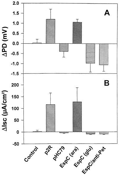
Comparison of the deduced EspC amino acid sequence with those available in GenBank databases revealed 52% identity (67% similarity) with the Pet protein of enteroaggregative E. coli (EAEC) (12), a toxin which produces enterotoxic activity on intestinal tissue mounted in Ussing chambers (12, 37). To determine the possible enterotoxic activity of EspC protein, the > 100-kDa fraction of supernatants from HB101(p2R) harboring the cloned espC gene and HB101(pHC79) containing the vector only were added to the luminal side of rat jejunal tissue mounted in the Ussing chamber. Concentrated HB101(p2R) supernatant fractions containing EspC produced an increase in jejunal PD and Isc (Fig. 4A and B), whereas concentrated supernatants from HB101 (pHC79) failed to induce increases in PD and Isc.
FIG. 4.
Enterotoxic activity of concentrated supernatant containing EspC protein. Changes in PD (A) and Isc (B) were measured in the presence of concentrated supernatants from HB101 (control), HB101(p2R), HB101(pHC79), HB101(pJLM174) grown with arabinose (ara) or glycerol and glucose (glu), and HB101(pJLM174) preincubated with antibodies against Pet protein. Concentrated supernatant protein (100 μg) was added to the mucosal hemichambers of rat jejunum preparations. The average values of experiments performed with preparations from different animals (n = 4) are presented.
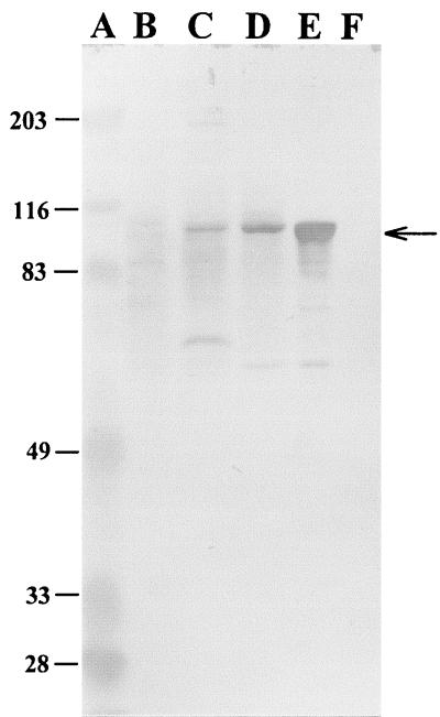
Since p2R is a large cosmid clone containing espC, a minimal espC fragment generated by PCR was ligated into the cloning vector pBAD30, which contains the PBAD promoter of the araBAD (arabinose) operon and the gene encoding the positive and negative regulator of this promoter, AraC. Thus, EspC expression can be modulated over a wide range of inducer (arabinose) concentrations and reduced to extremely low levels by the presence of glycerol and glucose, which represses expression (Fig. 5, lanes E and F, respectively). The addition of concentrated supernatant from HB101 (pJLM174), grown in the presence of arabinose, to the luminal side of jejunal tissue also induced rises in PD and Isc (Fig. 4A and B). Concentrated supernatant from the same clone grown in the presence of glucose did not induce changes in jejunal PD and Isc (Fig. 4A and B).
FIG. 5.
Immunoblot analysis for detection of EspC protein. Concentrated supernatants from HB101 (B), E2348/69 (C), HB101(p2R) (D), and HB101(pJLM174) grown in the presence of arabinose (E) or glycerol and glucose (F) were subjected to electrophoresis in SDS–10% polyacrylamide gels and transferred to nitrocellulose membranes. Detection of the EspC protein was performed by incubation of the membranes with rabbit serum against Pet protein followed by immunostaining with alkaline phosphatase-labeled polyclonal antibodies to rabbit immunoglobulins (1:2,000 dilution). Positions of the molecular size markers in lane A are indicated at the left (in kilodaltons). The arrow at the right side indicates EspC protein.
The rises in jejunal PD and Isc by EspC were similar to those produced by the Pet protein. Antibodies against Pet, which can neutralize the Pet enterotoxic activity (37), were found to cross-react with EspC from the wild-type EPEC strain E2348/69, HB101(p2R), and HB101(pJLM174) containing the espC minimal clone grown in the presence of arabinose (Fig. 5, lanes C, D, and E, respectively) but not in the presence of glucose (Fig. 5, lane F). In view of the sequence and antigenic similarity between Pet and EspC, we used polyclonal anti-Pet antibodies in an attempt to inhibit the enterotoxic effects of EspC in Ussing chambers. Preincubation with antiserum directed against Pet inhibited rises in jejunal PD and Isc produced by EspC (Fig. 4A and B).
DISCUSSION
In this study, we have demonstrated that the espC gene of the prototype EPEC strain E2348/69 is located within a unique region of DNA not found in commensal or laboratory strains of E. coli. Based on previously presented definitions (20), we have designated this region the espC pathogenicity island because (i) the region contains at least two loci associated with virulence, the previously described espC gene (45) and ORF3, which shows predicted amino acid similarity with VirA of Shigella flexneri (47), (ii) sequences homologous to espC and orf3 were present in pathogenic but not laboratory strains of E. coli, (iii) the G+C content of the unique 15, 195-bp region is 40.5%, substantially lower than that of E. coli K-12 (50.8%) (6), suggesting that this region was acquired by horizontal transfer (2), and (iv) several ORFs with predicted protein similarity to a variety of mobile genetic elements are contained in this region. The espC pathogenicity island inserted at a chromosomal site adjacent to a tRNA-like gene, ssrA, which is also the site of insertion of the pathogenicity island of V. cholerae (26).
By DNA sequence and PCR analysis, the left junction of the espC pathogenicity island was located at 60.06 min of the complete sequence of the E. coli strain MG1655 chromosome, adjacent to orf360. Similarly, by DNA sequence and PCR analysis, the right junction of the espC pathogenicity island of EPEC1 strains was located at 59.35 min, adjacent to the ssrA gene (6). It was recently reported that one of the so-called loops or genomic regions that differ between Salmonella enterica serovar Typhimurium and E. coli is located at 60 min, between the smpB and nrdE genes (3). There is approximately 91 kb of DNA between smpB and nrdE in S. enterica compared to 46 kb at this site in E. coli strain MG1655. Therefore, within this region of S. enterica lies 45 kb of DNA not found in E. coli, and it has been proposed that this region may contain loci that distinguish Salmonella from E. coli (3). Similarly, the espC pathogenicity island may distinguish the EPEC1 category from commensal strains and other categories of E. coli intestinal pathogens.
The observation that the left and right junctions were at 60.06 and 59.35 min, respectively, of the E. coli chromosome suggests that approximately 33 kb of DNA found between these locations in the K-12 strain MG1655 chromosome are missing from the EPEC1 strains or that chromosomal rearrangements have occurred in this region. Rearrangements or sequence differences may also be present in the EPEC2, EHEC1, and EHEC2 pathogens at these locations, because oligonucleotide primers directed to neither the ssrA nor smpB sequences of strain MG1655 amplified predicted fragments by PCR when EPEC2, EHEC1, or EHEC2 DNA was used as the template. However, a 407-bp DNA probe directed to the ssrA sequences of strain MG1655 hybridized to DNA from all three of these pathotypes.
DNA probes directed to espC hybridized to DNA from strains of the EPEC1 category (E2348/69 and DEC clones 1 and 2 (48, 49), but not to other AE pathogens, including representatives of the EPEC2, EHEC1, and EHEC2 categories of diarrheagenic E. coli, RDEC1, and Citrobacter rodentium biotype 4280. This observation and the fact that the cloned LEE from strain E2348/69 (EPEC1) in the E. coli laboratory strain HB101 is sufficient to form AE lesions in vitro (35) are consistent with the conclusion that EspC is not associated with the intestinal AE phenotype (45).
espC and adjacent non-K-12 sequences may have been acquired by EPEC1 in a single horizontal transfer event. Alternatively, espC and the flanking sequences may have been acquired via multiple events. Evidence supporting the latter hypothesis comes from analysis of the G+C content of the espC pathogenicity island. The G+C content of the espC structural gene is 42.5%, whereas that of the sequences spanning from rorf1 to orf3 is approximately 35% and of those including orf5 to orf8 is 46%. These data might suggest that the island was acquired by multiple events of horizontal transfer, since bacteria develop characteristic G+C contents during evolution. These data are also consistent with the PCR analyses performed on a distribution of pathogenic E. coli serotypes. We observed a decrease in the G+C percentage from the right to left junction that likely signifies that the DNA closer to the left junction was horizontally acquired earlier than the DNA towards the right junction. A similar mosaic structure, with DNA sequences acquired from distinct sources, has been described for the smpB-nrdE intergenic region of S. enterica (3).
Downstream of espC we identified DNA sequences similar to IS1351 and IS200. Remnants of IS elements flank the pet gene on the pAA2 plasmid of EAEC, and IS1203-like and iso-IS1-like elements flank espP on the pO157 plasmid of E. coli serotype O157:H7 (7, 9, 12). The 102-kb high pathogenicity island of Y. pestis can be deleted by a recombination event occurring between two flanking IS100 element (14), and it was proposed that the IS629 and IS911 elements may have played a role in the insertion of pet onto plasmid pAA2 (12). Similarly, IS911 and IS629 elements are found upstream of the chromosome-located pic gene of EAEC (22). From these observations, we hypothesize that the IS1351 or IS200 elements or perhaps the IS100 putative transposase and associated repetitive sequence may have played a role in the acquisition of espC by the EPEC1 strains.
Members of the autotransporter family have been identified in a number of pathotypes of E. coli. Surprisingly, DNA probes directed to the conserved C terminus of the espC structural gene did not hybridize to DNA from EAEC, which contain the gene encoding the homologous Pet autotransported enterotoxin, and a pet probe did not hybridize to EPEC DNA (data not shown). Similar to these results, an espP DNA probe encoding the homologous EspP protein in EHEC O157:H7 did not hybridize to EPEC strain E2348/69 in a Southern analysis, though it did hybridize to DNA from several isolates of serotype O157:H7 and one isolate of serotype O26 (7). However, Brunder et al. (7) visualized secreted proteins from two EPEC strains, including E2348/69, that cross-reacted with EspP antiserum. These data might suggest that, due to divergent DNA sequences, hybridization studies are not appropriate for identifying genes encoding related autotransporters of the SPATE subfamily, whereas antibody cross-reactivity in immunoblot procedures more readily detects the structural relatedness of these proteins.
We have determined that EspC possesses enterotoxic activity, finding that concentrated supernatants from a laboratory strain of E. coli expressing EspC increased tissue PD and Isc using rat jejunum mounted in Ussing chambers. Antibodies directed against the homologous Pet protein of EAEC cross-reacted with EspC in Western blots, and preincubation with Pet antiserum eliminated EspC's ability to increase PD and Isc in Ussing chambers. A previous study examined the possible role of EspC in interactions with epithelial cells (45). These investigators found that EspC was not necessary for mediating EPEC-induced signal transduction necessary for formation of AE lesions in HeLa cells and did not play a role in either adherence or invasion of tissue culture cells. Enterotoxin activity was not tested in this previous study.
In addition to EspC, the orf3 gene product of the espC pathogenicity island may also encode a virulence factor. The predicted ORF3 protein shows similarity with VirA of Shigella and rORF2 of EPEC. Both cloned ORF3 and rORF2 were able to rescue a Shigella ΔvirA mutation that eliminates this bacterium's ability to invade epithelial cells in culture (S. Elliott, E. O. Krejany, J. L. Mellies, R. M. Robins-Browne, C. Sasakawa, and J. B. Kaper, submitted for publication). An orf3 and rorf2 double mutation did not affect EPEC invasion, though it was demonstrated that the protein product of rorf2 of EPEC was secreted by the type III secretion system and translocated into host epithelial cells (Elliott et al., submitted). Because of its homology with the product of rorf2, the protein product of orf3 of the espC pathogenicity island may also be secreted by the type III system, but the function of these proteins encoded by orf3 and rorf2 remains obscure.
Identification of autotransporters in at least 30 gram-negative pathogens (21) which cause a variety of diseases suggests that these proteins play an important role in pathogenesis. The C-terminal β-domains of autotransporters show a high degree of amino acid homology and appear to perform a common function, whereas the passenger domains are more divergent. Many functions have been attributed to the passenger domains, including adhesion, protease and toxin activity, and cellular invasion (5, 12, 22, 23, 39, 42). Of the SPATE autotransporters most closely related to EspC, Pet of EAEC is also an enterotoxin (12, 37), whereas the EspP protein of EHEC serotype O157:H7 cleaves human coaggulation factor V and has been proposed to be a contributing factor in mucosal hemorrhaging in patients with hemorrhagic colitis (7). Pic of EAEC was implicated in mucinase activity, serum resistance, and hemagglutination (22). Like the EAF plasmid of EPEC, the espC pathogenicity island was found only in a subset of these pathogens, though other strains may possess genes encoding comparable function. EspC most likely plays an accessory role in EPEC pathogenesis, presumably as an enterotoxin, and the location of the gene encoding EspC within a pathogenicity island supports this hypothesis.
ACKNOWLEDGMENTS
We thank Lisa Sadzewicz and Nick Ambulos of the University of Maryland at Baltimore Biopolymer Laboratory for DNA sequencing and analysis and Maria Dubois for plasmid isolations, PCR, and other molecular procedures. We also thank Tom Whittam, Vanessa Sperandio, Simon Elliott, and David Karaolis for providing strains.
This work was supported by NIH grants AI21657 (J.B.K.) and AI43615 and AI33069 (J.P.N.). Work completed by J. F. was funded in part by an NSF AIRE Fellowship.
REFERENCES
- 1.Altschul S F, Madden T L, Schaffer A A, Zhang J, Zhang Z, Miller W, Lipman D J. Gapped BLAST and PSI-BLAST: a new generation of protein database search programs. Nucleic Acids Res. 1997;25:3389–3402. doi: 10.1093/nar/25.17.3389. [DOI] [PMC free article] [PubMed] [Google Scholar]
- 2.Aoyama K, Haase A M, Reeves P. Evidence for effect of random genetic drift on G+C content after lateral transfer of fucose pathway genes to Escherichia coli. Mol Biol Evol. 1994;11:829–838. doi: 10.1093/oxfordjournals.molbev.a040166. [DOI] [PubMed] [Google Scholar]
- 3.Bäumler A J, Heffron F. Mosaic structure of the smpB-nrdE intergenic region of Salmonella enterica. J Bacteriol. 1998;180:2220–2223. doi: 10.1128/jb.180.8.2220-2223.1998. [DOI] [PMC free article] [PubMed] [Google Scholar]
- 4.Benjelloun-Touimi Z, Sansonetti P J, Parsot C. SepA, the major extracellular protein of Shigella flexneri: autonomous secretion and involvement in tissue invasion. Mol Microbiol. 1995;17:123–135. doi: 10.1111/j.1365-2958.1995.mmi_17010123.x. [DOI] [PubMed] [Google Scholar]
- 5.Benz I, Schmidt M A. Cloning and expression of an adhesin (AIDA-I) involved in diffuse adherence of enteropathogenic Escherichia coli. Infect Immun. 1989;57:1506–1511. doi: 10.1128/iai.57.5.1506-1511.1989. [DOI] [PMC free article] [PubMed] [Google Scholar]
- 6.Blattner F R, Plunkett G I, Bloch C A, Perna N T, Burland V, Riley M, Collado-Vides J, Glasner J D, Rode C K, Mayhew G F, Gregor J, Davis N W, Kirkpatrick H A, Goeden M, Rose D J, Mau B, Shao Y. The complete genome sequence of Escherichia coli K-12. Science. 1997;277:1453–1462. doi: 10.1126/science.277.5331.1453. [DOI] [PubMed] [Google Scholar]
- 7.Brunder W, Schmidt H, Karch H. EspP, a novel extracellular serine protease of enterohaemorrhagic Escherichia coli O157:H7 cleaves human coagulation factor V. Mol Microbiol. 1997;24:767–778. doi: 10.1046/j.1365-2958.1997.3871751.x. [DOI] [PubMed] [Google Scholar]
- 8.Burland V, Shao Y, Perna N T, Plunkett G, Sofia H J, Blattner F R. The complete DNA sequence and analysis of the large virulence plasmid of Escherichia coli O157:H7. Nucleic Acids Res. 1998;26:4196–4204. doi: 10.1093/nar/26.18.4196. [DOI] [PMC free article] [PubMed] [Google Scholar]
- 9.Djafari S, Ebel F, Deibel C, Krämer S, Hudel M, Chakraborty T. Characterization of an exported protease from Shiga toxin-producing Escherichia coli. Mol Microbiol. 1997;25:771–784. doi: 10.1046/j.1365-2958.1997.5141874.x. [DOI] [PubMed] [Google Scholar]
- 10.Donnenberg M S, Yu J, Kaper J B. A second chromosomal gene necessary for intimate attachment of enteropathogenic Escherichia coli to epithelial cells. J Bacteriol. 1993;175:4670–4680. doi: 10.1128/jb.175.15.4670-4680.1993. [DOI] [PMC free article] [PubMed] [Google Scholar]
- 11.Elliott S, Wainwright L A, McDaniel T, MacNamara B, Donnenberg M, Kaper J B. The complete sequence of the locus of enterocyte effacement (LEE) from enteropathogenic Escherichia coli E2348/69. Mol Microbiol. 1998;28:1–4. doi: 10.1046/j.1365-2958.1998.00783.x. [DOI] [PubMed] [Google Scholar]
- 12.Eslava C, Navarro-Garcia F, Czeczulin J R, Henderson I R, Cravioto A, Nataro J P. Pet, an autotransporter enterotoxin from enteroaggregative Escherichia coli. Infect Immun. 1998;66:3155–3163. doi: 10.1128/iai.66.7.3155-3163.1998. [DOI] [PMC free article] [PubMed] [Google Scholar]
- 13.Fetherston J D, Perry R D. The pigmentation locus of Yersinia pestis KIM6+ is flanked by an insertion sequence and includes the structural genes for pesticin sensitivity and HMWP2. Mol Microbiol. 1994;13:697–708. doi: 10.1111/j.1365-2958.1994.tb00463.x. [DOI] [PubMed] [Google Scholar]
- 14.Fetherston J D, Schuetze P, Perry R D. Loss of the pigmentation phenotype in Yersinia pestis is due to the spontaneous deletion of 102 kb of chromosomal DNA which is flanked by a repetitive element. Mol Microbiol. 1992;6:2693–2704. doi: 10.1111/j.1365-2958.1992.tb01446.x. [DOI] [PubMed] [Google Scholar]
- 15.Frankel G, Phillips A D, Rosenshine I, Dougan G, Kaper J B, Knutton S. Enteropathogenic and enterohaemorrhagic E. coli: more subversive elements. Mol Microbiol. 1998;30:911–921. doi: 10.1046/j.1365-2958.1998.01144.x. [DOI] [PubMed] [Google Scholar]
- 16.Gonçalves A G, Campos L C, Gomes T A T, Rodrigues J, Sperandio V, Whittam T S, Trabulsi L R. Virulence properties and clonal structures of strains of Escherichia coli O119 serotypes. Infect Immun. 1997;65:2034–2040. doi: 10.1128/iai.65.6.2034-2040.1997. [DOI] [PMC free article] [PubMed] [Google Scholar]
- 17.Groisman E A, Ochman H. Pathogenicity islands: bacterial evolution in quantum leaps. Cell. 1996;87:791–794. doi: 10.1016/s0092-8674(00)81985-6. [DOI] [PubMed] [Google Scholar]
- 18.Guandalini S, Fasano A, Migliavacca M, Marchesano G. Effects of Berberine on basal secretagogue-modified ion transport in the rabbit ileum in vitro. J Pediatr Gastroenterol Nutr. 1987;64:4751–4760. doi: 10.1097/00005176-198711000-00023. [DOI] [PubMed] [Google Scholar]
- 19.Guzman L- M, Belin D, Carson M, Beckwith J. Tight regulation, modulation, and high-level expression by vectors containing the arabinose PBAD promoter. J Bacteriol. 1995;177:4121–4130. doi: 10.1128/jb.177.14.4121-4130.1995. [DOI] [PMC free article] [PubMed] [Google Scholar]
- 20.Hacker J, Blum-Oehler G, Mühldorfer I, Tschäpe H. Pathogenicity islands of virulent bacteria: structure, function, and impact on microbial evolution. Mol Microbiol. 1997;23:1089–1097. doi: 10.1046/j.1365-2958.1997.3101672.x. [DOI] [PubMed] [Google Scholar]
- 21.Henderson I R, Navarro-Garcia F, Nataro J P. The great escape: structure and function of the autotransporter proteins. Trends Microbiol. 1998;6:370–378. doi: 10.1016/s0966-842x(98)01318-3. [DOI] [PubMed] [Google Scholar]
- 22.Henderson I R, Czeczulin J, Eslava C, Noriega F, Nataro J P. Characterization of Pic, a secreted protease of Shigella flexneri and enteroaggregative Escherichia coli Infect. Immun. 1999;67:5587–5596. doi: 10.1128/iai.67.11.5587-5596.1999. [DOI] [PMC free article] [PubMed] [Google Scholar]
- 23.Hendrixson D R, De la Morena M L, Stathopoulos C, St. Geme J W. Structural determinants of processing and secretion of the Haemophilus influenzae Hap protein. Mol Microbiol. 1997;26:505–518. doi: 10.1046/j.1365-2958.1997.5921965.x. [DOI] [PubMed] [Google Scholar]
- 24.Jarvis K G, Girón J A, Jerse A E, McDaniel T K, Donnenberg M S, Kaper J B. Enteropathogenic Escherichia coli contains a specialized secretion system necessary for the export of proteins involved in attaching and effacing lesion formation. Proc Natl Acad Sci USA. 1995;92:7996–8000. doi: 10.1073/pnas.92.17.7996. [DOI] [PMC free article] [PubMed] [Google Scholar]
- 25.Jerse A E, Yu J, Tall B D, Kaper J B. A genetic locus of enteropathogenic Escherichia coli necessary for the production of attaching and effacing lesions on tissue culture cells. Proc Natl Acad Sci USA. 1990;87:7839–7843. doi: 10.1073/pnas.87.20.7839. [DOI] [PMC free article] [PubMed] [Google Scholar]
- 26.Karaolis D K, Johnson J A, Bailey C C, Boedeker E C, Kaper J B, Reeves P R. A Vibrio cholerae pathogenicity island associated with epidemic and pandemic strains. Proc Natl Acad Sci USA. 1998;95:3134–3139. doi: 10.1073/pnas.95.6.3134. [DOI] [PMC free article] [PubMed] [Google Scholar]
- 27.Kenny B, DeVinney R, Stein M, Reinscheid D J, Frey E A, Finlay B B. Enteropathogenic E. coli (EPEC) transfers its receptor for intimate adherence into mammalian cells. Cell. 1997;91:511–520. doi: 10.1016/s0092-8674(00)80437-7. [DOI] [PubMed] [Google Scholar]
- 28.Kenny B, Finlay B B. Protein secretion by enteropathogenic Escherichia coli is essential for transducing signals to epithelial cells. Proc Natl Acad Sci USA. 1995;92:7991–7995. doi: 10.1073/pnas.92.17.7991. [DOI] [PMC free article] [PubMed] [Google Scholar]
- 29.Kenny B, Lai L C, Finlay B B, Donnenberg M S. EspA, a protein secreted by enteropathogenic Escherichia coli, is required to induce signals in epithelial cells. Mol Microbiol. 1996;20:313–323. doi: 10.1111/j.1365-2958.1996.tb02619.x. [DOI] [PubMed] [Google Scholar]
- 30.Knutton S, Rosenshine I, Pallen M J, Nisan I, Neves B C, Bain C, Wolff C, Dougan G, Frankel G. A novel EspA-associated surface organelle of enteropathogenic Escherichia coli involved in protein translocation into epithelial cells. EMBO J. 1998;17:2166–2176. doi: 10.1093/emboj/17.8.2166. [DOI] [PMC free article] [PubMed] [Google Scholar]
- 31.Lai L-C, Wainwright L A, Stone K D, Donnenberg M S. A third secreted protein that is encoded by the enteropathogenic Escherichia coli pathogenicity island is required for transduction of signals and for attaching and effacing activities in host cells. Infect Immun. 1997;65:2211–2217. doi: 10.1128/iai.65.6.2211-2217.1997. [DOI] [PMC free article] [PubMed] [Google Scholar]
- 32.Lee C A. Pathogenicity islands and the evolution of bacterial virulence. Infect Agents Dis. 1996;5:1–7. [PubMed] [Google Scholar]
- 33.Levine M M, Bergquist E J, Nalin D R, Waterman D H, Hornick R B, Young C R, Sotman S, Rowe B. Escherichia coli strains that cause diarrhoea but do not produce heat-labile or heat-stable enterotoxins and are non-invasive. Lancet. 1978;i:1119–1122. doi: 10.1016/s0140-6736(78)90299-4. [DOI] [PubMed] [Google Scholar]
- 34.McDaniel T K. A large genetic locus of enteropathogenic Escherichia coli sufficient to confer the attaching and effacing phenotype in vitro Ph. D. thesis. Baltimore: University of Maryland; 1996. [Google Scholar]
- 35.McDaniel T K, Kaper J B. A cloned pathogenicity island from enteropathogenic Escherichia coli confers the attaching and effacing phenotype on E. coli K-12. Mol Microbiol. 1997;23:399–407. doi: 10.1046/j.1365-2958.1997.2311591.x. [DOI] [PubMed] [Google Scholar]
- 36.McNamara B P, Donnenberg M S. A novel proline-rich protein, EspF, is secreted from enteropathogenic Escherichia coli via the type III export pathway. FEMS Microbiol Lett. 1998;166:71–78. doi: 10.1111/j.1574-6968.1998.tb13185.x. [DOI] [PubMed] [Google Scholar]
- 37.Navarro-Garcia F, Eslava C, Villaseca J M, Lopez-Revilla R, Czeczulin J R, Srinivas S, Nataro J P, Cravioto A. In vitro effects of a high-molecular-weight heat-labile enterotoxin from enteroaggregative Escherichia coli. Infect Immun. 1998;66:3149–3154. doi: 10.1128/iai.66.7.3149-3154.1998. [DOI] [PMC free article] [PubMed] [Google Scholar]
- 38.Pohlner J, Halter K, Beyreuther K, Meyer T F. Gene structure and extracellular secretion of Neisseria gonorrhoeae IgA protease. Nature. 1987;325:458–462. doi: 10.1038/325458a0. [DOI] [PubMed] [Google Scholar]
- 39.Provence D L, Curtiss R., III Isolation and characterization of a gene involved in hemagglutination by an avian pathogenic Escherichia coli strain. Infect Immun. 1994;62:1369–1380. doi: 10.1128/iai.62.4.1369-1380.1994. [DOI] [PMC free article] [PubMed] [Google Scholar]
- 40.Rajakumar K, Sasakawa C, Adler B. Use of a novel approach, termed island probing, identifies the Shigella flexneri she pathogenicity island which encodes a homolog of the immunoglobulin A protease-like family of proteins. Infect Immun. 1997;65:4606–4614. doi: 10.1128/iai.65.11.4606-4614.1997. [DOI] [PMC free article] [PubMed] [Google Scholar]
- 41.Sambrook J, Fritsch E F, Maniatis T. Molecular cloning: a laboratory manual. Cold Spring Harbor, N.Y: Cold Spring Harbor Laboratory Press; 1989. [Google Scholar]
- 42.Schmitt W, Haas R. Genetic analysis of the Helicobacter pylori vacuolating cytotoxin: structural similarities with the IgA protease type of exported protein. Mol Microbiol. 1994;12:307–319. doi: 10.1111/j.1365-2958.1994.tb01019.x. [DOI] [PubMed] [Google Scholar]
- 43.Sohel I, Puente J L, Ramer S W, Bieber D, Wu C, Schoolnik G K. Enteropathogenic Escherichia coli: identification of a gene cluster coding for bundle-forming pilus morphogenesis. J Bacteriol. 1996;178:2613–2628. doi: 10.1128/jb.178.9.2613-2628.1996. [DOI] [PMC free article] [PubMed] [Google Scholar]
- 44.Sperandio V, Kaper J B, Bortolini M R, Neves B C, Keller R, Trabulsi L R. Characterization of the locus of enterocyte effacement (LEE) in different enteropathogenic Escherichia coli (EPEC) and Shiga-toxin producing Escherichia coli (STEC) serotypes. FEMS Microbiol Lett. 1998;164:133–139. doi: 10.1111/j.1574-6968.1998.tb13078.x. [DOI] [PubMed] [Google Scholar]
- 45.Stein M, Kenny B, Stein M A, Finlay B B. Characterization of EspC, a 110-kilodalton protein secreted by enteropathogenic Escherichia coli which is homologous to members of the immunoglobulin A protease-like family of secreted proteins. J Bacteriol. 1996;178:6546–6554. doi: 10.1128/jb.178.22.6546-6554.1996. [DOI] [PMC free article] [PubMed] [Google Scholar]
- 46.Taylor K A, O'Connell C B, Luther P W, Donnenberg M S. The EspB protein of enteropathogenic Escherichia coli is targeted to the cytoplasm of infected HeLa cells. Infect Immun. 1998;66:5501–5507. doi: 10.1128/iai.66.11.5501-5507.1998. [DOI] [PMC free article] [PubMed] [Google Scholar]
- 47.Uchiya K I, Tobe T, Komatsu K, Suzuki T, Watarai M, Fukuda I, Yoshikawa M, Sasakawa C. Identification of a novel virulence gene, virA, on the large plasmid of Shigella, involved in invasion and intercellular spreading. Mol Microbiol. 1995;17:241–250. doi: 10.1111/j.1365-2958.1995.mmi_17020241.x. [DOI] [PubMed] [Google Scholar]
- 48.Whittam T S, McGraw E A. Clonal analysis of EPEC serogroups. Rev Microbiol Sao Paulo. 1996;27(Suppl. 1):7–16. [Google Scholar]
- 49.Whittam T S, Wolfe M L, Wachsmuth I K, Ørskov F, Ørskov I, Wilson R A. Clonal relationships among Escherichia coli strains that cause hemorrhagic colitis and infantile diarrhea. Infect Immun. 1993;61:1619–1629. doi: 10.1128/iai.61.5.1619-1629.1993. [DOI] [PMC free article] [PubMed] [Google Scholar]


