Abstract
Introduction:
Viola plant has been used traditionally to treat neurological disorders. We aimed at determining whether pretreatment with Viola spathulata extract can alleviate the severity of ischemic-reperfusion damages and exert its protective effects through the regulation of a sodium/calcium exchanger (NCX3) gene expression in a rat brain.
Methods:
Male Wistar rats were divided into two main groups: one main group for evaluating Neurologic Deficit Score (NDS) and Infarct Volume (IV) and the other group for the evaluation of NCX3 gene expression in the brain tissue. The latter group was subdivided into the intact, control (vehicle), sham, V5, and V10. The vehicle (control) subgroup received Dimethyl Sulfoxide (DMSO), and V5 and V10 subgroups received V. spathulata extract at the doses of 5 and 10 mg/kg (IP), respectively, for 7 days. After pretreatment, we carried out Middle Cerebral Artery Occlusion (MCAO) for 60 min.
Results:
In the V5 and V10 subgroups, NDS and IV significantly decreased. MCAO upregulated NCX3 gene expression in the core, penumbra, and subcortical regions compared with the intact subgroup. The V5 subgroup significantly downregulated the NCX3 gene expression level in the core compared with the control subgroup. The V10 subgroup showed downregulation of the NCX3 gene expression level in the core, penumbra, and subcortex compared with the control subgroup.
Conclusion:
V. spathulata extract may have a neuroprotective role against MCAO-induced ischemic brain damage, possibly by preventing the alteration of NCX3 gene expression level.
Highlights
MCAO results Infarct Volume (IV) and Neurologic Deficit Score (NDS);
MCAO upregulated NCX3 gene expression in brain tissues;
Viola spathulata extract pretreatment decreased IV and NDS in brain ischemia;
Viola spathulata pretreatment downregulated NCX3 gene expression in brain tissues.
Plain Language Summary
Stroke is the second leading cause of death and long term disability. Recently it has been reported that herbal extracts have protective role in ischemia injury. In Iranian traditional medicine Viola plant has a long history to treat disorders such as cancer. So we designed an animal study to investigate Viola plant extract in brain ischemia injury. Viola spathulata extract was administrated to rats for seven days, then animal model of brain ischemia was operated on them and some behavioral, histological and molecular factors were analyzed. Our findings showed that Viola spathulata extract improved behavioral disability, decreased infarct volume in brain tissue, and modulate Sodium/Calcium exchanger 3 gene expression. It could be concluded that Viola spathulata has the neuroprotective effect in animal stroke model and is a good candidate for nutritional supplements, although further studies are needed.
Keywords: Viola spathulata, Viola extract, Brain ischemia, NCX3 gene, Stroke, Neuroprotective
1. Introduction
Cerebrovascular attack or stroke is a sudden disruption of blood supply to the brain which triggers serious consequences such as cerebral infarction and neurological deficits. In developing countries such as Iran, stroke is the second leading cause of death and long-term disability ( Bacigaluppi, Pluchino, Martino, Kilic, & Hermann, 2008). Ischemia is a major type of stroke ( Mergenthaler, Dirnagl, & Meisel, 2004). Several changes are initiated by ischemia and reperfusion, such as inhibition of electron transport, decreasing ATP and pH, increasing cell Ca2+, releasing glutamate, and increasing arachidonic acid. Also, gene activation leads to the synthesis of the cytokines and enzymes involved in free radical production ( Lipton, 1999).
Na+/Ca2+ exchanger (NCX) has nine transmembrane segments widely distributed in the brain ( Blaustein & Lederer, 1999). This protein couples transmembrane movement of Ca2+ to reciprocal movements of Na+ in a bidirectional way in the Central Nervous System (CNS) ( Meyer, 1989). In the CNS, NCX plays a fundamental role in controlling changes in the intracellular concentrations of Na+ and Ca2+ ions under physiologic conditions ( Canitano et al., 2002). NCX1, NCX2, and NCX3 are three different genes of the NCX family that are differentially expressed in distinct regions of the CNS ( Papa et al., 2003). Depending on the intracellular Ca2+ and Na+ concentrations ([Ca2+]i and [Na+]i), NCX can act either in the forward mode, coupling the uphill extrusion of Ca2+ to the influx of Na+ ions, or in the reverse mode, mediating the extrusion of Na+ and the influx of the Ca2+ ions ( Blaustein & Lederer, 1999). NCX activation reduces the brain infarct volume extension after permanent Middle Cerebral Artery Occlusion (MCAO). Also, the selective pharmacological blockade of NCX worsens the brain lesion, suggesting a protective role played by the exchanger during the events leading to brain ischemia ( Pignataro et al., 2004). NCX3, unlike the other NCXs family members (NCX1 and NCX2), has a peculiar capability to maintain [Ca2+]i homeostasis even when ATP levels are reduced considerably. Therefore, it has a major role in neuronal preservation during hypoxic conditions ( Mohammadi & Bigdeli, 2014; Secondo et al., 2007).
Recently, it has been demonstrated that herbal extracts and oils such as olive oil ( Mohagheghi, Bigdeli, Rasoulian, Zeinanloo, & Khoshbaten, 2010), Coriandrum sativum (Linn) ( Vekaria, Patel, Bhalodiya, Patel, V., Desai, & Tirgar, 2012) and Ocimum basilicum ( Bora, Arora, & Shri, 2011) have a protective role in ischemia-reperfusion injury. It has also been shown that Viola odorata and Viola tricolor have antioxidant activities and protect neuronal cells against serum glucose deprivation ( Mousavi, Naghizade, Pourgonabadi, & Ghorbani, 2016).
Viola has a long remedial history in Iranian traditional medicine to treat disorders such as cancer ( Koochek, Pipelzadeh, & Mardani, 2003). Previous studies demonstrated that viola tissue contains melatonin ( Ansari et al., 2010; Kim, Yoon, & Park, 2011). Plants with a high level of melatonin have been used traditionally to treat neurological disorders ( Murch, Simmons, & Saxena, 1997) and diseases caused by free radicals generation ( Chen, Huo, Tan, Liang, Zhang, & Zhang, 2003). It has also been found that melatonin reduces hypoxia-ischemia damages, improves sleep, ameliorates cardiac ischemia/reperfusion injuries, and inhibits oxidative stress-mediated endothelial cell death ( Lin et al., 2016; Manchester et al., 2015; Mao et al., 2016; Nduhirabandi, Lamont, Albertyn, Opie, & Lecour, 2016). Treatment with melatonin improves calcium handling by preserving SERCA gene expression in hypoxic rats and performs as a cardioprotective factor against myocardial injury ( Yeung, Hung, & Fung, 2008).
Because of using viola in traditional medicine to treat neurodegenerative diseases, we designed a study to evaluate whether the administration of Viola spathulata can alleviate brain injuries by using an in vivo model of transient focal cerebral ischemia in rats. In the second part, because calcium overload is a major mechanism in ischemic injury ( Dirnagl, Iadecola, & Moskowitz, 1999), we sought to identify whether such effects on brain ischemia might be associated with changes in the expression of the NCX3 gene.
2. Methods
Experimental procedures
Viola extraction
Aerial parts of V. spathulata were collected from Gadouk neck, Firoozkouh road, Savadkouh City (Mazandaran Province, Iran) in May 2017, and the species were authenticated at the herbarium of the Department of Biology, School of Medical Sciences (Sari Payame Noor University; Herbarium No.: SPNH-4727).
The aerial parts of V. spathulata were separately dried, powdered, and extracted with 70% ethanol in a Soxhlet apparatus for 48 h. The hydroalcoholic extracts were then concentrated in a water bath and kept at −20°C until use.
Finally, the extract was dissolved in Dimethyl Sulfoxide (DMSO) to be used in this study.
Animals and group assignment
All experimental animal procedures were approved and conducted under the Animal Research and Ethics Committee (IR.GUMS.REC.1394.323) of Guilan University of Medical Sciences. Every attempt was made to minimize the number of animal use and their suffering. The rats were housed under controlled temperature (24°C) with food and water ad libitum with lights on from 07:00 to 19:00 (light cycle) and off from 19:00 to 07:00 (dark cycle).
Male Wistar rats (200–250 g) were divided into two main groups, and each group was divided into the control (vehicle), sham, intact, V5 (V. spathulata 5 mg/kg/d) ( Liu et al., 2014; Robertson et al., 2012) and V10 (V. spathulata 10 mg/kg/d) ( Letechipía-Vallejo, González-Burgos, & Cervantes, 2001; Robertson et al., 2012) subgroups. One main group was used for evaluating Neurologic Deficit Score (NDS) and Infarct Volume (IV), Viola extracts (5 and 10 mg/kg) was administrated by i.p. injection for 7 days before MCAO operation and control group received DMSO. ( Yanpallewar, Rai, Kumar, & Acharya, 2004). After pretreatment, a 60-min MCAO was carried out in the control, V5, and V10 subgroups. In the sham subgroup, all steps were similar to the control group, except MCAO. Data from the sham and intact subgroups were pooled together, as there was no significant difference between them.
Focal cerebral ischemia
The rats were weighed and anesthetized with chloral hydrate (400 mg/kg bodyweight; Merck, Germany). MCAO was applied as described previously by Longa et al. (Longa, Weinstein, Carlson, & Cummins, 1989). Briefly, under microscopic surgery, a 3-0 silicone-coated nylon filament was introduced through the external carotid artery stump. The occluder was advanced into the internal carotid artery 20 to 22 mm past the carotid bifurcation until mild resistance indicated that the tip was lodged in the anterior cerebral artery and blocked the blood flow to the middle cerebral artery. After 60 min of ischemia, reperfusion was started by withdrawing the filament. During the surgery, the animal’s body temperature was monitored and maintained at around 37°C by using a heating and cooling surface.
Neurologic Deficit Score (NDS)
After the filament was withdrawn, the rats were returned to their separate cages. The rats were assessed neurologically 24 h later by an observer blinded to the animal groups. The neurobehavioral scoring was performed using the 6-point scale previously described by Longa et al. (Longa et al., 1989) as follows: normal motor function=0; flexion of contralateral forelimb on suspension vertically by the tail, failure to extend forepaw=1; circling to the contralateral side but normal posture at rest=2; loss of righting reflex=3; and no spontaneous motor activity=4. Death was considered for scoring 5 only when a large infarct volume was present without subarachnoid hemorrhage. The rats were excluded from the study when they died due to subarachnoid hemorrhage or pulmonary insufficiency and asphyxia.
Infarct Volume (IV) assessment
After killing animals with chloral hydrate (800 mg/kg), they were decapitated, and their brains were rapidly removed and cooled in saline (4°C) for 15 min. Eight 2-mm thick coronal sections were cut (Brain Matrix, Tehran, Iran) through the brain, starting at the olfactory bulb. The slides were immersed in 2% 2, 3, 5-triphenyl tetrazolium chloride solution (Merck, Germany) and kept at 37°C in a water bath for 15 min. The slices were then digitally photographed by a camera (iPhone 5s) connected to a computer. Unstained areas were defined as infarct and measured using the image analysis software (Image Tools, National Institutes of Health). The infarct volume was calculated by measuring the unstained and stained area in each hemisphere slice in three defined regions (core, penumbra, and subcortex), multiplying by slice thickness (2 mm), followed by summiting all eight slices according to the method by Swanson et al. (Swanson et al., 1990): corrected infarct volume=left hemisphere volume – (right hemisphere volume - infarct volume).
Brain sampling
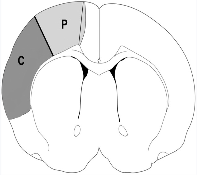
The intact (I) (no surgery), control, sham-operated, V5+MCAO, and V10+ MCAO animals were killed by chloral hydrate (800 mg/kg) 24 h after MCAO operation and decapitated for measurement of NCX3 gene expression. Core, penumbra, and subcortex of the brain tissues were isolated as previously described by Lei et al. (Lei, Popp, Capuano-Waters, Cottrell, & Kass, 2004) (Figure 1).
Figure 1.

Schematic drawing showing the brain regions 5-mm from the frontal pole of the brain
Shaded areas indicate the ischemic areas. P: ischemic penumbra; C: ischemic core; S: subcortex.
NCX3 gene measurement
RNA extraction
After excision of the brain tissues, tissue sections (10 mg) were prepared, turned into small pieces using a grinder, and were transferred to the 1 mL YTzol Pure RNA solution (YEKTA TAJHIZ AZMA, Iran). RNA was extracted according to the manufacturer’s instructions. Briefly homogenized samples were incubated for 5 min at 15°C to 30°C. Then, 200 μL of chloroform was added to the sample tube and vigorously shook for 15 s. The sample tube was incubated at room temperature for 2–3 min. The specimens were centrifuged at 12000 × g for 10 min at 4°C (Sigma 3-18KS, Germany). The transparent supernatant was transferred to a 1.5 mL nuclease-free Eppendorf tube, and 500 μL isopropanol was added. The tubes were mixed well by inverting several times. The samples were incubated at 15°C–30°C for 10 min and centrifuged at 12000 × g for 10 min at 4°C. Then, RNA washing was done using 1 mL of 75% ethanol (in nuclease-free water) (CinnaGen, Iran). The precipitant was dissolved in 20 μL of nuclease-free water, and the purity of the extracted RNA was determined through the A260/A280 ratio using the NanoDrop system (Thermo Fisher Scientific Inc., USA).
DNase I treatment
DNase I is an endonuclease that digests single- and double-stranded DNA. To prepare DNA-free RNA before real-time Polymerase Chain Reaction (PCR), we treated the extracted RNA with DNase I, an RNase-free enzyme, according to the manufacturer’s protocol (Thermo Fisher Scientific Inc., USA). Briefly, 1 μg/μL RNA, 1 μL 10X reaction buffer with MgCl2, and 1 μL DNase I, RNase-free, were added into a 1.5 mL nuclease-free microtube and then reached 10 μL using nuclease-free water and mixed mildly. The contents were transferred to a 37°C water bath for 30 min, and 1 μL of EDTA (50 mM) was added and incubated at 65°C for 10 min.
Complementary DNA synthesis
cDNA was synthesized from 1 μg/μL total RNA with reverse transcriptase (HyperScriptTM First-strand Synthesis Kit, Gene All, South Korea). Briefly, 1 μg/μL RNA, 1 μL oligo(dT) primer (50 μM), and 1 μL dNTPs (10 mM) were added into a 0.2 mL nuclease-free microtube and then reached 14 μL by nuclease-free water and mildly mixed. The contents were transferred to a 65°C water bath for 5 min and immediately placed on ice for at least 1 min. Following a small spinning, the mixture was pi-petted into the reaction tube containing 2 μL of RTase reaction buffer (10x), 2 μL of 0.1 M DTT, 1 μL of HyperScriptTM reverse transcriptase (200 U/μL), and 1 μL of ZymAllTM RNase inhibitor. After brief centrifugation, the microtubes were incubated at 55°C for 60 min. The termination of the reaction was done by incubating at 85°C for 5 min. Finally, the samples were placed on ice for a while and then preserved in a −20°C freezer until needed.
Real-Time PCR
Following the extraction of total RNA from the treated and untreated groups and the synthesis of cDNA, the real-time PCR technique was exploited to determine the changes in the expression of NCX3 (NCBI Reference Sequence: 017593995.1_). The expression of NCX3 mRNAs was quantified compared to the β-actin gene as a reference gene and represented relative gene expression. The PCR primers were designed by Primer3web (version 4.0.0), and the sequences and product sizes are given in Table 1. The specificity of the designed primers was checked for each interest gene using the Primer-BLAST system available at the National Center for Biotechnology Information (NCBI).
Table 1.
The sequences of the gene-specific Polymerase Chain Reaction (PCR) primers for real-time PCR
| Gene | NCBI Code | Forward | Reverse |
|---|---|---|---|
| β2 μg | NM_012512.2 | TACATGTCTCGGTCCCAGGT | AATTCACACCCACCGAGACC |
| NCX3 | NM_078620.2 | CGACGGTACAAGAGCACACT | TTCCATGTGTCCGCTGGTAC |
β2 μg: β2 microglobulin; NCX3: Na+/Ca2+ exchanger; NCBI: National Center for Biotechnology Information.
We employed a reaction buffer, including 1 μL of each primer, 10 μL SYBR Green reagent (YEKTA TAJHIZ AZMA, Iran), 4 μL diluted cDNA, and 4 μL nuclease-free water using a real-time PCR method (Applied Biosystems, StepOneTM, USA). The thermal program was initially planned at 95°C for 10 min and then continued with 40 tow-step cycles, at 95°C for 10 s and 60°C for 60 s, and finally terminated with a cycle at 72°C for 5 minutes. Each amplification product was analyzed by a dissociation curve certifying that for each gene, the amplified product showed all nonspecific bands or primer dimer formation. The differences between mRNA expression of the reference and test samples were calculated, and the relative mRNA expressions of NCX3 were calculated using the Ct method (2−ΔDDt) ( Livak & Schmittgen, 2001). All reactions were run in triplicate.
Statistical analysis
NCX gene expression and IV were compared using a 1-way Analysis of Variance (ANOVA). NDS was analyzed using the Mann-Whitney U test. Data were expressed as Mean±SEM, and a P-value of less than 0.05 was considered significant. SPSS v. 16 software was used for data analysis.
3. Results
Effects of V. spathulata pretreatment on NDS and IV
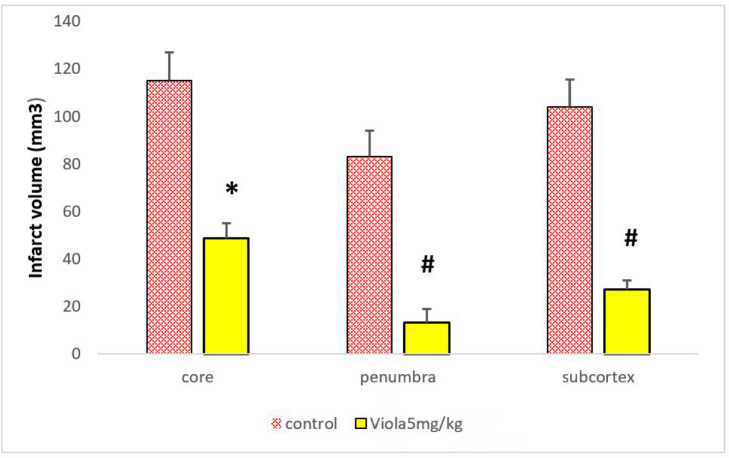
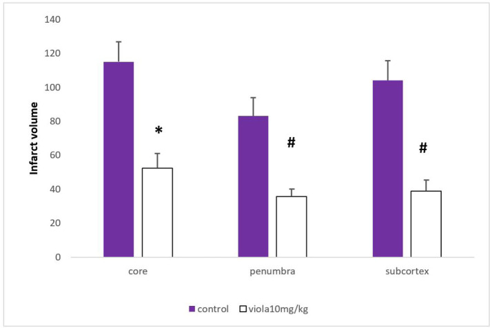
Median NDS significantly reduced in the V5 and V10 groups compared with the control group (P=0.004 and P=0.02, respectively) (Table 2). The putative beneficial effects of 7 days of pretreatment with 5 mg/kg of V. spathulata extract were confirmed by reducing the IV of the core, penumbra, and subcortex regions (P=0.02, P=0.0001, and P=0.0001, respectively; Figure 2). Seven days of pretreatment with 10 mg/kg of V. spathulata extract also reduced IV in the core, penumbra, and subcortex regions significantly (P=0.01, P=0.0001, and P=0.0001, respectively; Figure 3). There was no significant difference in NDS and IV between the rats treated with 5 and 10 mg/kg of V. spathulata extract.
Table 2.
The distribution of neurologic deficit score in each experimental group
| Experimental Groups | Median | Neurologic Deficit Score | Total (n) | P-Value | |||||
|---|---|---|---|---|---|---|---|---|---|
|
| |||||||||
| 0 | 1 | 2 | 3 | 4 | 5 | ||||
| Control | 3 | 0 | 0 | 1 | 3 | 2 | 0 | 6 | - |
| V5 | 1 | 1 | 4 | 3 | 1 | 0 | 0 | 6 | ** P<0.01 |
| V10 | 2 | 3 | 3 | 2 | 1 | 0 | 0 | 6 |
*
P<0.05 # P<0.05 |
P<0.01 significant differences between the V5 (Viola spathulata, 5 mg/kg/d) and control groups;
P<0.05 significant difference between the V10 (Viola spathulata, 10 mg/kg/d) and control groups;
P<0.05 significant difference between the V5 and V10 groups.
Figure 2.
The effect of Viola spathulata (5 mg/kg) pretreatment on infarct volume in the core, penumbra and subcortex areas compared with the control group (n=7).
Values are presented as Mean±SEM obtained from two independent experiments in the core, penumbra and subcortex of the brain.
*P<0.05, #P<0.001
Figure 3.
The effect of Viola spathulata (10 mg/kg) pretreatment on infarct volume in the core, penumbra and subcortex areas compared with the control group (n=7).
Values are presented as Mean±SEM obtained From two independent experiments in the core, penumbra and subcortex of the brain.
*P<0.05, #P<0.001.
Effects of V. spathulata Pretreatment on NCX3 gene expression
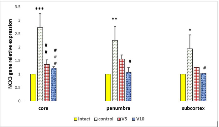
Brain Ischemia (MCAO) upregulated NCX3 gene expression in the core (P=0.0001), penumbra (P=0.007), and subcortex (P=0.38) regions significantly compared with the intact subgroup (Figure 4).
Figure 4.
Relative expression of NCX3 in the intact (Healthy Rats, n=4), control (MCAO, n=4), V5 (Viola spathulata, 5 mg/kg/d pretreatment with MCAO operation (n=4), and V10 (Viola spathulata, 10 mg/kg/d; pretreatment with MCAO operation (n=4) groups detected by Real-Time Polymerase Chain Reaction (RT-PCR)
NCX3 transcript was upregulated in the control group compared with the intact groups. NCX3 transcript was down-regulated in the V5 and V10 groups compared with the control group.
Values are presented as Means±SEM obtained from two independent experiments in the core, penumbra and subcortex areas of the brain.
*P<0.05, **P<0.01, ***P<0.001 compared with the intact group.
#P<0.05, ##P<0.01, and ###P<0.001 compared with the control group.
Pretreatment with 5 mg/kg V. spathulata down-regulated NCX3 gene expression level in the core (P=0.001) region significantly compared with the control group (Figure 4).
Pretreatment with 10 mg/kg V. spathulata down-regulated NCX3 gene expression level in core (P=0.0001), penumbra (P=0.011), and subcortex (P=0.047) regions compared with the control group (Figure 4).
4. Discussion
NDS and IV are considered indicators of neurologic deficits in cerebral ischemia/reperfusion damage. Our results demonstrated that preconditioning with V. spathulata at the doses of 5 and 10 mg/kg reduced IV and NDS in three regions of the brain (core, penumbra, and subcortex). In the present study, administration of V. spathulata was done for the first time in the animal model of cerebral ischemia. Our results confirmed that V. spathulata had a protective role in ischemia-reperfusion injury. Similar previous studies have shown the protective role of olive oil ( Mohagheghi et al., 2010), Coriandrum sativum (Linn.) ( Vekaria et al., 2012), and Ocimum basilicum ( Bora et al., 2011) in ischemia-reperfusion injury. Ischemic-reperfusion leads to the generation of excessive Reactive oxygen Species (ROS) ( Amantea et al., 2009), which causes oxidative damage to the cellular and mitochondrial structures. These changes ultimately result in the initiation of some pathways that lead to apoptotic and necrotic cell death ( Manzanero, Santro, & Arumugam, 2013). Viola tricolor and Viola odorata extracts protected the neuronal cell against adverse effects of intracellular ROS, which resulted in serum glucose deprivation ( Mousavi et al., 2016). Therefore, V. spathulata might protect the neural cells from ischemia-reperfusion damage, probably alleviating ROS effects. However, in our study, intracellular ROS was not evaluated.
The NCX3 is a bi-directional membrane ion transporter that exchanges Ca2+ and Na+ ions across the cell membrane in the CNS and contributes significantly to maintaining intracellular Ca2+ homeostasis during experimental conditions mimicking ischemia ( Secondo et al., 2007). It has been suggested that during ischemia, the calcium entry mode of NCX may account for a major portion of the calcium influx and induced excitotoxicity in cerebellar granule cells (Czyż & Kiedrowski, 2002). However, to confirm the hypothesis that NCX acts as an exchanger contributing to calcium homeostasis during ischemia, it is necessary to measure calcium levels in the early hours after the stroke. NCX1 mRNA was upregulated in the peri-infarct area with the induction of permanent MCAO in rats ( Boscia et al., 2006). Bano et al. demonstrated that NCX3 is cleaved and inactivated during ischemia in a rat model of focal ischemia ( Bano et al., 2005). In our study, NCX3 gene transcription was upregulated after 24 h reperfusion in the brain of MCAO rats; thus, a possible mechanism of NCX3 gene upregulation is a compensatory response to cleavage of NCX3 isoform during one-hour ischemia.
This work is the first study to exhibit the effect of V. spathulata on NCX3 gene transcription in neurological impairment induced by MCA occlusion. Our findings showed that pretreatment with V. spathulata downregulated NCX3 transcription in the brain of MCAO rats. NCX activity causes calcium entry, which leads to lethal calcium load, and the inhibition of NCX may be protective in excitotoxicity when ATP depletion occurs (Czyż & Kiedrowski, 2002; Jeffs, Meloni, Bakker, & Knuckey, 2007). The mechanism by which V. spathulata pretreatment reduced ischemic damage could be partly due to downregulating NCX3 gene transcription and protecting the cells from lethal calcium load in ischemia conditions. However, the NCX3 protein level was not evaluated by the western blot technique in our study, which is recommended to be addressed in further studies.
5. Conclusion
Regarding the effects of V. spathulata pretreatment on IV, NDS, and NCX3 gene transcription, our data suggested that V. spathulata extract has a neuroprotective role. V. spathulata extract reversed the deleterious effects of ischemia-reperfusion on cells, possibly by overcoming the Ca2+overload through regulating NCX3 gene transcription. Further studies are required to extend or confirm these observations. Ultimately, it is hoped that novel cerebroprotective strategies be developed for those at risk of stroke or for cases whose cerebral perfusion is electively reduced at the time of surgery.
Ethical Considerations
Compliance with ethical guidelines
All experimental animal procedures were approved and conducted under the Animal Research and Ethics Committee (IR.GUMS.REC.1394.323) of Guilan University of Medical Sciences.
Acknowledgments
This work was financially supported by research deputy of Guilan University of Medical Sciences.
Footnotes
Funding
The present study was funded by grants from the Research Deputy of Guilan University of Medical Sciences (Rasht, Iran).
Authors' contributions
All authors equally contributed to preparing this article.
Conflict of interest
The authors declared no conflict of interest.
References
- Amantea D., Marrone M. C., Nistico R., Federici M., Bagetta G., Bernardi G., et al. (2009). Oxidative stress in stroke pathophysiology: Validation of hydrogen peroxide metabolism as a pharmacological target to afford neuroprotection. International Review of Neurobiology, 85, 363–74. [DOI: 10.1016/S0074-7742(09)85025-3] [PMID ] [DOI] [PubMed] [Google Scholar]
- Ansari M., Rafiee K., Yasa N., Vardasbi S., Naimi S., Nowrouzi A. (2010). Measurement of melatonin in alcoholic and hot water extracts of Tanacetum parthenium, Tripleurospermum disciforme and Viola odorata. DARU Journal of Pharmaceutical Sciences, 18(3), 173–8. [PMID ] [PMCID ] [PMC free article] [PubMed] [Google Scholar]
- Bacigaluppi M., Pluchino S., Martino G., Kilic E., Hermann D. M. (2008). Neural stem/precursor cells for the treatment of ischemic stroke. Journal of the Neurological Sciences, 265(1–2), 73–7. [DOI: 10.1016/j.jns.2007.06.012] [PMID ] [DOI] [PubMed] [Google Scholar]
- Bano D., Young K. W., Guerin C. J., LeFeuvre R., Rothwell N. J., Naldini L., et al. (2005). Cleavage of the plasma membrane Na+/Ca2+ exchanger in excitotoxicity. Cell, 120(2), 275–85. [DOI: 10.1016/j.cell.2004.11.049] [PMID ] [DOI] [PubMed] [Google Scholar]
- Blaustein M. P., Lederer W. J. (1999). Sodium/calcium exchange: Its physiological implications. Physiological Reviews, 79(3), 763–854. [DOI: 10.1152/physrev.1999.79.3.763] [PMID ] [DOI] [PubMed] [Google Scholar]
- Bora K. S., Arora S., Shri R. (2011). Role of Ocimum basilicum L. in prevention of ischemia and reperfusion-induced cerebral damage, and motor dysfunctions in mice brain. Journal of Ethnopharmacology, 137(3), 1360–5. [DOI: 10.1016/j.jep.2011.07.066] [PMID ] [DOI] [PubMed] [Google Scholar]
- Boscia F., Gala R., Pignataro G., De Bartolomeis A., Cicale M., Ambesi-Impiombato A., et al. (2006). Permanent focal brain ischemia induces isoform-dependent changes in the pattern of Na+/Ca2+ exchanger gene expression in the ischemic core, periinfarct area, and intact brain regions. Journal of Cerebral Blood Flow & Metabolism, 26(4), 502–17. [DOI: 10.1038/sj.jcbfm.9600207] [PMID ] [DOI] [PubMed] [Google Scholar]
- Canitano A., Papa M., Boscia F., Castaldo P., Sellitti S., Taglialatela M., et al. (2002). Brain distribution of the Na+/Ca2+ exchanger-encoding genes NCX1, NCX2, and NCX3 and their related proteins in the central nervous system. Annals of the New York Academy of Sciences, 976(1), 394–404. [DOI: 10.1111/j.1749-6632.2002.tb04766.x] [PMID ] [DOI] [PubMed] [Google Scholar]
- Chen G., Huo Y., Tan D. X., Liang Z., Zhang W., Zhang Y. (2003). Melatonin in Chinese medicinal herbs. Life Sciences, 73(1), 19–26. [DOI: 10.1016/S0024-3205(03)00252-2] [DOI] [PubMed] [Google Scholar]
- Czyż A., Kiedrowski L. (2002). In depolarized and glucose-deprived neurons, Na+ influx reverses plasmalemmal K+-dependent and K+-independent Na+/Ca2+ exchangers and contributes to NMDA excitotoxicity. Journal of Neurochemistry, 83(6), 1321–8. [DOI: 10.1046/j.1471-4159.2002.01227.x] [PMID ] [DOI] [PubMed] [Google Scholar]
- Dirnagl U., Iadecola C., Moskowitz M. A. (1999). Pathobiology of ischaemic stroke: An integrated view. Trends in Neurosciences, 22(9), 391–7. [DOI: 10.1016/S0166-2236(99)01401-0] [DOI] [PubMed] [Google Scholar]
- Jeffs G. J., Meloni B. P., Bakker A. J., Knuckey N. W. (2007). The role of the Na+/Ca2+ exchanger (NCX) in neurons following ischaemia. Journal of Clinical Neuroscience, 14(6), 507–14. [DOI: 10.1016/j.jocn.2006.07.013] [PMID ] [DOI] [PubMed] [Google Scholar]
- Kim Y. J., Yoon Y. H., Park W. J. (2011). Supply of tryptophan and tryptamine influenced the formation of melatonin in Viola plants. Journal of Life Science, 21(2), 328–33. [DOI: 10.5352/JLS.2011.21.2.328] [DOI] [Google Scholar]
- Koochek M., Pipelzadeh M., Mardani H. (2003). The effectiveness of Viola odorata in the prevention and treatment of formalin-induced lung damage in the rat. Journal of Herbs, Spices & Medicinal Plants, 10(2), 95–103. [DOI: 10.1300/J044v10n02_11] [DOI] [Google Scholar]
- Lei B., Popp S., Capuano-Waters C., Cottrell J., Kass I. (2004). Lidocaine attenuates apoptosis in the ischemic penumbra and reduces infarct size after transient focal cerebral ischemia in rats. Neuroscience, 125(3), 691–701. [DOI: 10.1016/j.neuroscience.2004.02.034] [PMID ] [DOI] [PubMed] [Google Scholar]
- Letechipía-Vallejo G., González-Burgos I., Cervantes M. (2001). Neuroprotective effect of melatonin on brain damage induced by acute global cerebral ischemia in cats. Archives of Medical Research, 32(3), 186–92. [DOI: 10.1016/S0188-4409(01)00268-5] [DOI] [PubMed] [Google Scholar]
- Lin C., Chao H., Li Z., Xu X., Liu Y., Hou L., et al. (2016). Melatonin attenuates traumatic brain injury-induced inflammation: a possible role for mitophagy. Journal of Pineal Research, 61(2), 177–86. [DOI: 10.1111/jpi.12337] [PMID ] [DOI] [PubMed] [Google Scholar]
- Lipton P. (1999). Ischemic cell death in brain neurons. Physiological Reviews, 79(4), 1431–568. [DOI: 10.1152/physrev.1999.79.4.1431] [PMID ] [DOI] [PubMed] [Google Scholar]
- Liu L. F., Qin Q., Qian Z. H., Shi M., Deng Q. C., Zhu W. P., et al. (2014). Protective effects of melatonin on ischemiareperfusion induced myocardial damage and hemodynamic recovery in rats. European Review for Medical and Pharmacological Sciences, 18(23), 3681–6. [PMID ] [PubMed] [Google Scholar]
- Livak K. J., Schmittgen T. D. (2001). Analysis of relative gene expression data using real-time quantitative PCR and the 2(−Delta Delta C(T)) Method. Methods, 25(4), 402–8. [DOI: 10.1006/meth.2001.1262] [PMID ] [DOI] [PubMed] [Google Scholar]
- Longa E. Z., Weinstein P. R., Carlson S., Cummins R. (1989). Reversible middle cerebral artery occlusion without craniectomy in rats. Stroke, 20(1), 84–91. [DOI: 10.1161/01.STR.20.1.84] [PMID ] [DOI] [PubMed] [Google Scholar]
- Manchester L. C., Coto-Montes A., Boga J. A., Andersen L. P. H., Zhou Z., Galano A., et al. (2015). Melatonin: An ancient molecule that makes oxygen metabolically tolerable. Journal of Pineal Research, 59(4), 403–19. [DOI: 10.1111/jpi.12267] [PMID ] [DOI] [PubMed] [Google Scholar]
- Manzanero S., Santro T., Arumugam T. V. (2013). Neuronal oxidative stress in acute ischemic stroke: Sources and contribution to cell injury. Neurochemistry International, 62(5), 712–8. [DOI: 10.1016/j.neuint.2012.11.009] [PMID ] [DOI] [PubMed] [Google Scholar]
- Mao L., Dauchy R. T., Blask D. E., Dauchy E. M., Slakey L. M., Brimer S., et al. (2016). Melatonin suppression of aerobic glycolysis (Warburg effect), survival signalling and metastasis in human leiomyosarcoma. Journal of Pineal Research, 60(2), 167–77. [DOI: 10.1111/jpi.12298] [PMID ] [DOI] [PubMed] [Google Scholar]
- Mergenthaler P., Dirnagl U., Meisel A. (2004). Pathophysiology of stroke: Lessons from animal models. Metabolic Brain Disease, 19(3–4), 151–67. [DOI: 10.1023/B:MEBR.0000043966.46964.e6] [PMID ] [DOI] [PubMed] [Google Scholar]
- Meyer F. B. (1989). Calcium, neuronal hyperexcitability and ischemic injury. Brain Research Reviews, 14(3), 227–43. [DOI: 10.1016/0165-0173(89)90002-7] [DOI] [PubMed] [Google Scholar]
- Mohagheghi F., Bigdeli M. R., Rasoulian B., Zeinanloo A. A., Khoshbaten A. (2010). Dietary virgin olive oil reduces blood brain barrier permeability, brain edema, and brain injury in rats subjected to ischemia-reperfusion. The Scientific World Journal, 10, 1180–191. [DOI: 10.1100/tsw.2010.128] [PMID ] [PMCID ] [DOI] [PMC free article] [PubMed] [Google Scholar]
- Mohammadi E., Bigdeli M. R. (2014). Time course of neuroprotection induced by normobaric hyperoxia and NCX1 expression. Brain Injury, 28(8), 1127–34. [DOI: 10.3109/02699052.2014.896472] [PMID ] [DOI] [PubMed] [Google Scholar]
- Mousavi S. H., Naghizade B., Pourgonabadi S., Ghorbani A. (2016). Protective effect of Viola tricolor and Viola odorata extracts on serum/glucose deprivation-induced neurotoxicity: Role of reactive oxygen species. Avicenna Journal of Phytomedicine, 6(4), 434–41. [PMID ] [PMCID ] [PMC free article] [PubMed] [Google Scholar]
- Murch S. J., Simmons C. B., Saxena P. K. (1997). Melatonin in feverfew and other medicinal plants. The Lancet, 350(9091), 1598–9. [DOI: 10.1016/S0140-6736(05)64014-7] [PMID ] [DOI] [PubMed] [Google Scholar]
- Nduhirabandi F., Lamont K., Albertyn Z., Opie L. H., Lecour S. (2016). Role of toll-like receptor 4 in melatonin-induced cardioprotection. Journal of Pineal Research, 60(1), 39–47. [DOI: 10.1111/jpi.12286] [PMID ] [DOI] [PubMed] [Google Scholar]
- Papa M, Canitano A., Boscia F, Castaldo P, Sellitti S, Porzig H, et al. (2003). Differential expression of the Na+-Ca2+ exchanger transcripts and proteins in rat brain regions. Journal of Comparative Neurology, 461(1), 31–48. [DOI: 10.1002/cne.10665] [PMID ] [DOI] [PubMed] [Google Scholar]
- Pignataro G., Gala R., Cuomo O., Tortiglione A., Giaccio L., Castaldo P., et al. (2004). Two sodium/calcium exchanger gene products, NCX1 and NCX3, play a major role in the development of permanent focal cerebral ischemia. Stroke, 35(11), 2566–70. [DOI: 10.1161/01.STR.0000143730.29964.93] [PMID ] [DOI] [PubMed] [Google Scholar]
- Robertson N. J., Faulkner S., Fleiss B., Bainbridge A., Andorka C., Price D., et al. (2012). Melatonin augments hypothermic neuroprotection in a perinatal asphyxia model. Brain, 136(1), 90–105. [DOI: 10.1093/brain/aws285] [PMID ] [DOI] [PubMed] [Google Scholar]
- Secondo A., Staiano R. I., Scorziello A., Sirabella R., Boscia F., Adornetto A., et al. (2007). BHK cells transfected with NCX3 are more resistant to hypoxia followed by reoxygenation than those transfected with NCX1 and NCX2: Possible relationship with mitochondrial membrane potential. Cell Calcium, 42(6), 521–35. [DOI: 10.1016/j.ceca.2007.01.006] [PMID ] [DOI] [PubMed] [Google Scholar]
- Swanson R. A., Morton M. T., Tsao-Wu G., Savalos R. A., Davidson C., Sharp F. R. (1990). A semiautomated method for measuring braininfarct volume. Journal of Cerebral Blood Flow & Metabolism, 10(2), 290–3. [DOI: 10.1038/jcbfm.1990.47] [PMID ] [DOI] [PubMed] [Google Scholar]
- Vekaria R. H., Patel M. N., Bhalodiya P. N., Patel V., Desai T. R., Tirgar P. R. (2012). Evaluation of neuroprotective effect of Coriandrum sativum linn. against ischemicreperfusion insult in brain. International Journal of Phytopharmacology, 3(2), 186–93. file:///C:/Users/Z.Ganjipour/Downloads/17.rutvi%20(2).pdf [Google Scholar]
- Yanpallewar S., Rai S., Kumar M., Acharya S. (2004). Evaluation of antioxidant and neuroprotective effect of Ocimum sanctum on transient cerebral ischemia and long-term cerebral hypoperfusion. Pharmacology Biochemistry and Behavior, 79(1), 155–64. [DOI: 10.1016/j.pbb.2004.07.008] [PMID ] [DOI] [PubMed] [Google Scholar]
- Yeung H., Hung M., Fung M. (2008). Melatonin ameliorates calcium homeostasis in myocardial and ischemia-reperfusion injury in chronically hypoxic rats. Journal of Pineal Research, 45(4), 373–82. [DOI: 10.1111/j.1600-079X.2008.00601.x] [PMID ] [DOI] [PubMed] [Google Scholar]


