Abstract
This study aimed to systematically review the literature to evaluate the marginal adaptation of veneers using different fabrication methods, namely, conventional feldspathic porcelain laminate veneers (PLVs), computer-aided design-computer-aided machining (CAD-CAM) veneers, and pressed veneers. A comprehensive literature search was performed using electronic databases (PubMed and Google Scholar) as well as hand searches to identify all relevant studies related to veneers and marginal adaptation. The identified studies were screened for assessing the inclusion and exclusion criteria. The included articles were then subjected to data extraction and analysis. The search resulted in 130 articles, of which six were included in this systematic review. All included articles were assessed for adaptation of margins. Based on the findings of this systematic review, no significant differences were found in the marginal adaptation of CAD-CAM and conventional feldspathic PLVs. The marginal fidelity of ceramic veneers issuing from the various fabrication techniques was clinically acceptable.
Keywords: cad-cam milling, systematic review, computer-aided design, dental veneers, dental marginal adaptation
Introduction and background
Well-aligned teeth and color are the two most important aspects of an attractive smile. Patients’ interest in the treatment of their smile is steadily growing. Similarly, the treatment options to restore the esthetic appearance have also been increasing. For a long period in the past, the most durable and predictable treatment for esthetically compromised teeth was achieved by the preparation of a full crown. With the increase in the trend toward tooth conservation, bonding, and minimally invasive procedures, the interest in veneers has also been increasing.
John Calamia in the 1980s launched porcelain laminate veneers at New York University, United States [1]. Veneers are thin-bonded ceramic restorations that involve the labial surface and part of the proximal surfaces of anterior teeth that require esthetic corrections [2]. They offer several advantages such as excellent esthetics, superior biocompatibility, and durability.
Porcelain veneers provide a conservative treatment option for discolored and malformed vital anterior teeth. The indications include moderate discoloration caused by tetracycline stains, excessive fluoride intake, developmental malformations such as peg laterals, amylogenesis imperfecta, and diastema correction.
A critical factor for successful restoration is its marginal fit. The circumferential periphery of the prepared tooth is termed the finish line. Optimal preparation design and choice of restorative material enhance marginal adaptation and fracture resistance for long-term success [3]. The marginal gap is the perpendicular distance from the internal surface of the restoration to the finish line of the preparation [4]. Because veneers are bonded by resin cement, they become a consolidated portion of the tooth and bear the brunt of masticatory forces, temperature alterations, and hydrolytic disintegration by chemical and moisture contamination. Intimate proximity between the veneer tooth interface fortifies the resin cement from unrestrained exposure to oral conditions. A veneer with inferior marginal adaptation can mutilate the tooth, periodontal tissue leading to microleakage, and plaque accumulation, resulting in caries, pulpal lesions, gingival inflammation, and periodontal disease. A restoration with poor marginal fit can damage the tooth, periodontal tissue, and even restoration. These marginal discrepancies can lead to cement dissolution, microleakage, and plaque accumulation, which result in gingival inflammation, caries, and pulpal lesions.
Accurate marginal adaptation of an indirect restoration plays a significant role in periodontal health, with irregular or rough margins irritating the gingiva. The luting material is the weakest restorative link, and the dissolution of the cement can create a marginal gap and space for bacteria. Although a consensus regarding a clinically admissible disparity is lacking, few researchers have proposed that a 50-120 µm gap is clinically acceptable, whereas others have recommended gaps of less than 100 µm [5]. Therefore, it is important to minimize marginal gaps to decrease the incidence of associated complications.
This systematic review aimed to evaluate and compare the marginal adaptation of veneers using different fabrication methods, namely, conventional feldspathic porcelain laminate veneers (PLVs), computer-aided design-computer-aided machining (CAD-CAM) veneers, and pressed veneers.
Review
Methodology
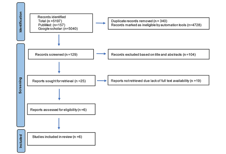
This systematic review followed the Preferred Reporting Items for Systematic Review and Meta-Analyses (PRISMA) guidelines and Population, Intervention, Comparison, Outcome (PICO) criteria. The PICO referred to veneers (P) fabricated with the CAD-CAM system and pressed ceramics (I) compared to the conventional feldspathic method present better marginal adaptation (O). PubMed and Google Scholar were explored for studies published between 1994 and 2020. The search strategy was a combination of medical subject heading terms “dental marginal adaptation.” “dental veneers,” “computer-aided design,” and “CAD-CAM” with the following text words “fit,” “gap,” “marginal,” “adaptation,” “accuracy,” “discrepancy,” “CAD,” “computer-aided,” “lithium silicate,” “feldspathic,” “leucite,” “milled,” and “composite.” All records identified were redeemed and imported into bibliographic software (Rayyan). Duplicates were removed. The entire search process is depicted in Figure 1.
Figure 1. Flow chart of the search strategy used in this systematic review.
Inclusion Criteria
In vitro and clinical prospective studies which investigated the marginal adaptation of CAD-CAM or heat-pressed veneers or conventional veneers were included in this review.
Exclusion Criteria
Case reports, case series, technique articles, abstracts, retrospective studies, review articles, studies only on composite veneering, and studies not published in the English language were excluded.
Data Extraction and Analysis
Two reviewers independently assessed all titles and abstracts according to the inclusion and exclusion criteria. The evaluation was performed jointly by both reviewers for the first 15 articles. Subsequently, the two reviewers continued with the evaluation independently. To avoid bias any studies identified by either reviewer during the initial screening were included. The full text of these selected studies was obtained for second-stage screening and submitted for data extraction. Information collected included journal name, year of publication, author names, type of preparation, type of porcelain, cementation, adaptation device, fabrication technique, follow-up period, type of study, and results (Table 1).
Table 1. Data extraction and analysis of the studies included in this systematic review.
CAD-CAM: computer-aided design-computer-aided machining; PLV: porcelain laminate veneer; SEM: scanning electron microscopy
| Journal, year | Author | Type of preparation | Type of porcelain | Cementation | Adaptation device | Fabrication technique | Follow up | Type of the study | Result |
| Journal of Dentistry, 2012 | Lin et al. [6] | Full veneer and traditional veneer | Leucite-reinforced ceramic veneer and conventional sintered feldspathic porcelain veneer | Clear self-cured acrylic resin | Keyence digital microscope at 200× | CAD-CAM and conventional feldspathic | - | In vitro | Traditional veneers designed with ProCAD porcelain demonstrated lesser horizontal gaps |
| Journal of Dentistry, 2012 | Aboushelib et al. [7] | Incisal lap preparation | Ceramic laminate veneers | Resin cement | SEM and stereomicroscope | CAD-CAM and pressed PLVs | 60 days | In vitro | Pressable ceramic laminate veneers exhibited superior marginal fidelity, uniform and thinner cement film thickness, and lesser microleakage in contrast to machinable ceramic veneers |
| Journal of Prosthodontics, 2013 | Jha et al. [8] | Window preparation | Refractory die technique, low-fusing feldspathic porcelain (IPS e. max Ceram), and lithium disilicate-reinforced glass ceramic (IPS e. max Press) | Dual-cure composite resin | SEM at 200× magnification | Conventional feldspathic and pressing technique | Seven days and three months | In vivo | Veneers of both groups showed a comparable marginal fidelity at the microscopic level at seven days and three months after cementation |
| Dental Research Journal, 2016 | Ghaffari et al. [9] | Incisal overlap preparation | Feldspathic laminate system (Du Ceram LFC) and in Ceram laminate veneer | Hueless glue | Stereomicroscope at 46× magnification | Refractory die technique and slip cast technique | - | In vitro | PLVs fabricated with Inceram had a marginal gap within an acceptable range |
| Journal of Prosthetic Dentistry, 2018 | Al-Dwairi et al. [10] | Full veneer preparation | Feldspathic glass ceramic, fine structure feldspar ceramic | Composite resin cement and variolink – N | SEM at 200× magnification | Pressed PLVs and CAD-CAM milling | - | In vitro | No statistically significant difference was found in gap measurements |
| Journal of Prosthodontics, 2017 | Yuce et al. [11] | Incisal overlap preparation | Nano-fluor apatite glass ceramic | Adhesive luting cement | Light optical microscopy at 40× magnification | CAD-CAM (cerec) and heat-pressed (E. Max press) | 6, 12, 18, and 24 months | In vivo | Marginal and internal adaptation of veneers were similar and within clinically acceptable ranges |
Results
The electronic search identified 5,197 articles that were transferred to the Rayyan software, and 4,728 articles were marked as ineligible. After removing duplicate articles, 129 articles were included. Of these, 104 articles were excluded based on title and abstracts. As the full text was not available 19 articles were excluded as they were not available to download, and, finally, six articles were included in this review. Details of the search strategy are presented in the PRISMA flow chart. Out of the six studies included, four were in vitro and two were in vivo. All six articles evaluated CAD-CAM veneers, three evaluated pressed veneers, and three evaluated conventional feldspathic veneers. Marginal adaptation was evaluated by scanning electron microscopy in three studies and stereomicroscope in another three studies.
Discussion
Anterior PLVs offer a viable treatment option for the management of esthetic conditions with minimal tooth preparation. The physical and mechanical properties of the veneer material, its adhesion to the tooth structure, and marginal integrity play a vital role in the clinical success of PLVs. Ample marginal adaptation is key to avoiding excessive gaps, which, in turn, can lead to leakage, dissolution of the luting agent, secondary caries, and failure of the restoration. According to this review, the marginal adaptation of pressed and milled PLVs was similar.
PLVs are traditionally fabricated using a layering technique that uses refractory dies to support the condensed layers of the ceramic slurry. This maneuver gives the ceramist complete command over the layers incorporated, resulting in a natural-appearing restoration. Conversely, it needs time and labor to produce precisely fitting restorations. The entire fabrication procedure is highly technique sensitive [12]. A new generation of ceramic materials was introduced to dentistry using different techniques such as CAD-CAM and pressing technology. Pressable ceramics are fabricated by burning out wax patterns using the conventional lost wax technique and melting and pressing ceramic ingots under controlled pressure, temperature, and vacuum using computer-programmed press ovens. These ovens are equipped with a pneumatic press that activates an alumina plunger used to compress molten ceramic ingots. Press-on ceramics allow accurate reproduction of the anatomical features carved in the wax pattern and controlled processing of the ceramic material resulting in an accurate restoration with minimal internal structural defects. Nowadays, CAD-CAM requires nothing more than a few keyboard clicks to design and fabricate accurate restorations. Nevertheless, the shade and color of machinable ceramic-produced ceramic veneers are limited by the color of the selected block used to mill these restorations.
External marginal adaptation of ceramic veneers, which is defined as the vertical distance between the finish line of the prepared tooth and the margins of the fabricated veneers, plays an important role in their success [13]. The differences between the mean marginal gap values of PLVs could be because of variations in the preparation design, fabrication technique, restoration thickness, design complexity, geometry, many PLV designs (window preparation or butt joint preparation or minimal to no preparation designs), adhesive luting agent, and marginal fit measurement method [14].
In this review, six articles were included, of which three used scanning electron microscopy for fit evaluation and two used stereomicroscopes. Microscopy permitted the two-dimensional evaluation of marginal gaps at tooth and veneer junctions.
Of the six articles, three compared the marginal adaptation of CAD-CAM veneers with conventional feldspathic veneers and reported different conclusions. Lin et al. found the conventional feldspathic method to have better results [6]. Jha et al. concluded that both techniques showed similar results [8]. Ghaffari et al. showed that CAD-CAM produced better results when compared to the conventional method [9]. In CAD-CAM, these marginal gaps may be from overgrinding and chipping thin porcelain margins due to the fragile nature of the material and the vibrations caused by milling.
Studies conducted by Aboushelib et al. and Al-Dwairi et al showed that pressed ceramics exhibited better marginal adaptation than CAD-CAM veneers [7,10]. However, in the in vivo study reported by Yuce et al., the fabrication method, whether CAD-CAM or heat-pressed, had no effect on the marginal and internal adaptation of PLVs; however the values were higher compared to the above in vitro studies [11].
It has been proven that preparation with butt joint design produces better marginal adaptation than the palatal chamfer with milled PLVs. The variability could be due to the complex geometry, higher curvature, and thinner incisal edge found with the palatal chamfer than with the butt joint design, negatively influencing the scanning and milling procedure and leading to larger marginal and internal gap discrepancies.
According to previous studies, marginal adaptation values of restorations should be between 100 and 120 µm to avoid cement wear. Other studies reported that an acceptable marginal adaptation value varied in clinical conditions, and up to 300 µm was accepted for ceramic restorations [11]. All studies included in this review, irrespective of the type of veneer, had marginal gaps within the acceptable range, though no further conclusions could be drawn.
Conclusions
The marginal fidelity of ceramic veneers issuing from the various fabrication techniques in this review, namely, conventional feldspathic veneers, CAD-CAM, and heat-pressed veneers, was found to be clinically acceptable. Feldspathic veneers exhibited better marginal adaptation compared to CAD-CAM veneers. However, between CAD-CAM and pressed veneers, varying results were observed. Because of limited literature, it was not feasible to establish a ranking of the different systems or conduct a proper comparison.
The content published in Cureus is the result of clinical experience and/or research by independent individuals or organizations. Cureus is not responsible for the scientific accuracy or reliability of data or conclusions published herein. All content published within Cureus is intended only for educational, research and reference purposes. Additionally, articles published within Cureus should not be deemed a suitable substitute for the advice of a qualified health care professional. Do not disregard or avoid professional medical advice due to content published within Cureus.
Footnotes
The authors have declared that no competing interests exist.
References
- 1.Etched porcelain veneers: the current state of the art. Calamia JR. https://pubmed.ncbi.nlm.nih.gov/3883393/ Quintessence Int. 1985;16:5–12. [PubMed] [Google Scholar]
- 2.The glossary of prosthodontic terms: ninth edition. J Prosthet Dent. 2017;117:0. doi: 10.1016/j.prosdent.2016.12.001. [DOI] [PubMed] [Google Scholar]
- 3.Marginal adaptation and fracture resistance of lithium disilicate laminate veneers on teeth with different preparation depths. Tuğcu E, Vanlıoğlu B, Özkan YK, Aslan YU. Int J Periodontics Restorative Dent. 2018;38:0. doi: 10.11607/prd.2995. [DOI] [PubMed] [Google Scholar]
- 4.Five-year clinical evaluation of 300 teeth restored with porcelain laminate veneers using total-etch and a modified self-etch adhesive system. Aykor A, Ozel E. Oper Dent. 2009;34:516–523. doi: 10.2341/08-038-C. [DOI] [PubMed] [Google Scholar]
- 5.Comparison of marginal fit between CAD-CAM and hot-press lithium disilicate crowns. Dolev E, Bitterman Y, Meirowitz A. J Prosthet Dent. 2019;121:124–128. doi: 10.1016/j.prosdent.2018.03.035. [DOI] [PubMed] [Google Scholar]
- 6.Fracture resistance and marginal discrepancy of porcelain laminate veneers influenced by preparation design and restorative material in vitro. Lin TM, Liu PR, Ramp LC, Essig ME, Givan DA, Pan YH. J Dent. 2012;40:202–209. doi: 10.1016/j.jdent.2011.12.008. [DOI] [PubMed] [Google Scholar]
- 7.Internal adaptation, marginal accuracy and microleakage of a pressable versus a machinable ceramic laminate veneers. Aboushelib MN, Elmahy WA, Ghazy MH. J Dent. 2012;40:670–677. doi: 10.1016/j.jdent.2012.04.019. [DOI] [PubMed] [Google Scholar]
- 8.Comparison of marginal fidelity and surface roughness of porcelain veneers fabricated by refractory die and pressing techniques. Jha R, Jain V, Das TK, Shah N, Pruthi G. J Prosthodont. 2013;22:439–444. doi: 10.1111/jopr.12032. [DOI] [PubMed] [Google Scholar]
- 9.Marginal adaptation of Spinell InCeram and feldspathic porcelain laminate veneers. Ghaffari T, Hamedi-Rad F, Fakhrzadeh V. Dent Res J (Isfahan) 2016;13:239–244. doi: 10.4103/1735-3327.182183. [DOI] [PMC free article] [PubMed] [Google Scholar]
- 10.A comparison of the marginal and internal fit of porcelain laminate veneers fabricated by pressing and CAD-CAM milling and cemented with 2 different resin cements. Al-Dwairi ZN, Alkhatatbeh RM, Baba NZ, Goodacre CJ. J Prosthet Dent. 2019;121:470–476. doi: 10.1016/j.prosdent.2018.04.008. [DOI] [PubMed] [Google Scholar]
- 11.Comparison of marginal and internal adaptation of heat-pressed and CAD/CAM porcelain laminate veneers and a 2-year follow-up. Yuce M, Ulusoy M, Turk AG. J Prosthodont. 2019;28:504–510. doi: 10.1111/jopr.12669. [DOI] [PubMed] [Google Scholar]
- 12.Comparison of marginal integrity of ceramic and composite veneer restorations luted with two different resin agents: an in vitro study. Celik C, Gemalmaz D. https://pubmed.ncbi.nlm.nih.gov/11887601/ Int J Prosthodont. 2002;15:59–64. [PubMed] [Google Scholar]
- 13.Marginal and internal fit of porcelain laminate veneers: a systematic review and meta-analysis [In Press] Baig MR, Qasim SS, Baskaradoss JK. J Prosthet Dent. 2022 doi: 10.1016/j.prosdent.2022.01.009. [DOI] [PubMed] [Google Scholar]
- 14.The success of dental veneers according to preparation design and material type. Alothman Y, Bamasoud MS. Open Access Maced J Med Sci. 2018;6:2402–2408. doi: 10.3889/oamjms.2018.353. [DOI] [PMC free article] [PubMed] [Google Scholar]


