Scleroderma is an autoimmune disease that causes fibrosis of the skin and other organs. A major obstacle to delivery of clinical care is the ability to objectively quantify fibrosis non-invasively over time. Vibrational optical coherence tomography (VOCT) is an imaging technique in which the skin is vibrated using audible sound waves and the maximum displacement of the skin can be determined from the OCT image. The resonant frequency is that in which maximum displacement occurs and is related to tissue stiffness1. We used VOCT to measure the resonant frequency of the skin longitudinally in a bleomycin mouse model of skin fibrosis and in a patient with diffuse scleroderma.
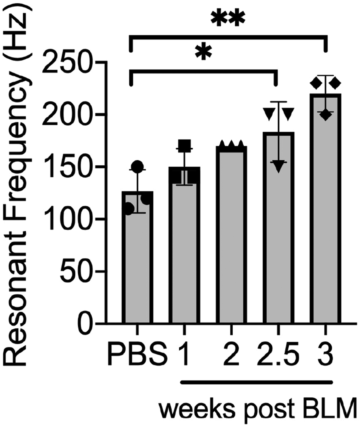
To model human scleroderma, we injected C57Bl/6 (B6) mice with bleomycin sulfate modified from Yamamoto et al2. A single subcutaneous dose of bleomycin 0.2 mg per 6–10 week old B6 female mouse induced histopathologic changes similar to human scleroderma at three weeks, including dermal thickening, loss of subcutaneous fat, and loss of CD34+ cells3. This is consistent with prior observations that B6 mice develop fibrosis by three weeks4 and a single dose of bleomycin can model pulmonary fibrosis5. We used VOCT to image B6 mice at different time points after subcutaneous injection with bleomycin on the posterior 1/3 of the back. Five age-matched cohorts each of three female mice were injected at staggered intervals prior to euthanasia and imaging with VOCT. Instrument sensitivity is accurate to approximately 10 Hz, so the mice were exposed to increasing sound frequencies from 30–300 Hz at 10 Hz intervals and OCT images recorded for calculation of transverse sample displacement. Mean resonant frequency of dermal collagen of mouse back skin injected with phosphate buffered saline vehicle was 127 Hz. In bleomycin-treated mice, resonant frequency was 150 Hz at 1 week, 170 Hz at 2 weeks, 183 Hz at 2.5 weeks, and 220 Hz by 3 weeks post injection. Means and standard deviations of resonant frequencies for each time point are plotted in Figure 1. VOCT was able to detect an increase in skin collagen resonant frequency as early as 1 week after bleomycin injection and steadily increased up to 3 weeks.
Figure 1. VOCT detects increasing resonant frequency at different times after BLM injection.

Resonant frequency of mouse skin at different time points after bleomycin injection. Data are means ± SD (*P < 0.05, **P < 0.01) analyzed with one-way analysis of variance (ANOVA) with Tukey multiple-comparisons test.
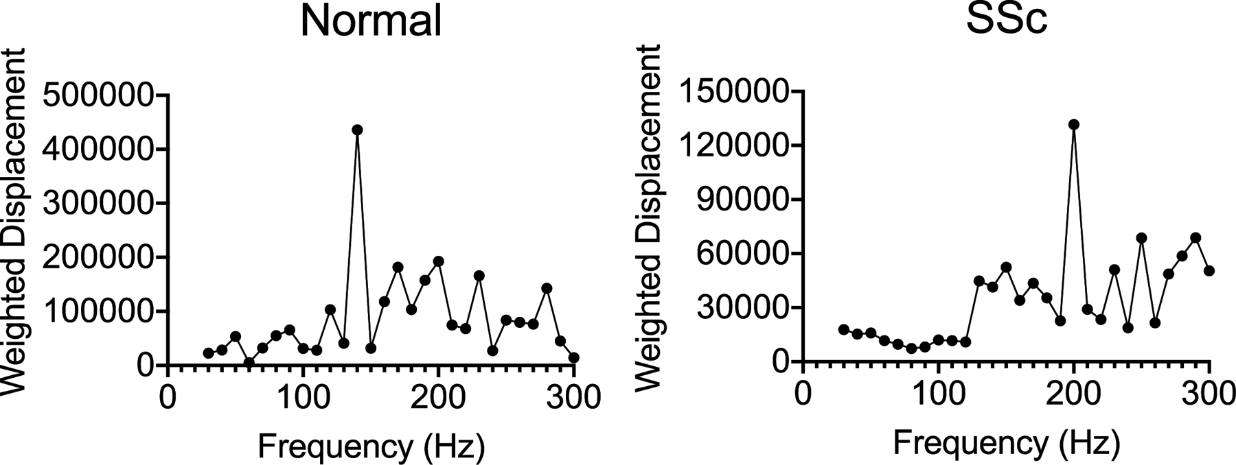
We tested VOCT in a patient with 3-year history of diffuse scleroderma with positive RNA Polymerase III antibody, kidney involvement, and mRSS of 7 treated with mycophenolate mofetil and extracorporeal photochemotherapy. We measured skin on his dorsal right second finger with VOCT, which showed a resonant frequency of 200 Hz compared to 140 Hz for the same location on normal skin from a healthy volunteer (Figure 2). VOCT imaging took less than 5 minutes to measure a single site. If present, hair trimming prior to imaging was needed to avoid artifacts. Similar to the increased resonant frequency of fibrotic mouse back skin, acral skin of human scleroderma showed increased resonant frequency of collagen by VOCT. This study is limited by measuring a single site of a patient with scleroderma. We plan future studies to validate VOCT as a non-invasive technique to quantify skin fibrosis in patients.
Figure 2. A patient with diffuse scleroderma has increased resonant skin frequency.

VOCT measurement of the dorsal finger skin of a patient with scleroderma (SSc) compared to normal control.
Acknowledgements
Dr. Odell is supported by career development awards from the Dermatology Foundation and CTSA Grant Number UL1 TR001863 from the National Center for Advancing Translational Science (NCATS), a component of the National Institutes of Health (NIH). The research contents are solely the responsibility of the authors and do not necessarily represent the official view of NIH. This work was supported by the Howard Hughes Medical Institute (R.A.F.).
Conflict of Interest
Drs. Odell and Flavell have no conflict of interest to declare. Dr. Silver is Chief Scientific Officer of OptoVibronex, LLC. and may in the future receive financial benefit from selling VOCT imaging devices.
Abbreviations:
- VOCT
Vibrational Optical Coherence Tomography
- ECM
extracellular matrix
- BLM
bleomycin
References
- 1.Akca BI, Chang EW, Kling S, Ramier A, Scarcelli G, Marcos S et al. Observation of sound-induced corneal vibrational modes by optical coherence tomography. Biomed Opt Express 2015;6:3313–9. [DOI] [PMC free article] [PubMed] [Google Scholar]
- 2.Yamamoto T, Takagawa S, Katayama I, Yamazaki K, Hamazaki Y, Shinkai H et al. Animal model of sclerotic skin. I: Local injections of bleomycin induce sclerotic skin mimicking scleroderma. J Invest Dermatol 1999;112:456–62. [DOI] [PubMed] [Google Scholar]
- 3.Aiba S, Tabata N, Ohtani H, Tagami H. CD34+ spindle-shaped cells selectively disappear from the skin lesion of scleroderma. Arch Dermatol 1994;130:593–7. [PubMed] [Google Scholar]
- 4.Lakos G, Takagawa S, Varga J. Animal models of scleroderma. Methods Mol Med 2004;102:377–93. [DOI] [PubMed] [Google Scholar]
- 5.Snider GL, Hayes JA, Korthy AL. Chronic interstitial pulmonary fibrosis produced in hamsters by endotracheal bleomycin: pathology and stereology. Am Rev Respir Dis 1978;117:1099–108. [DOI] [PubMed] [Google Scholar]


