Abstract
Multiple sclerosis (MS) is a chronic inflammatory disease of central nervous system leading to demyelination followed by neurological symptoms. Ferroptosis is a newly discovered pathogenic hallmark important for the progression of MS. However, the gene markers of ferroptosis in MS are still uncertain. In this study, mRNA expression profiles and clinical data of MS samples were retrieved from Gene Expression Omnibus database. Weighted gene co-expression network analysis and receiver operating characteristic curve analysis were utilized to identify ferroptosis-related gene (FRG) signatures of MS. Gene set enrichment analysis and gene set variation analysis were performed to explore the biological functions of single FRG signature. HMOX1, LPCAT3 and RPL8 were firstly identified as FRG signatures of MS with the predictive capacity confirmed. Gene set enrichment analysis and gene set variation analyses revealed that metabolism-related, immune and inflammation-related, microglia-related, oxidation-related, and mitochondria-related biological functions were enriched, providing implications of the mechanisms underlying ferroptosis in MS. This study presented a systematic analysis of FRG in MS and explored the potential ferroptosis targets for new interventional strategies in MS.
Keywords: ferroptosis, HMOX1, LPCAT3, multiple sclerosis, RPL8
1. Introduction
Multiple sclerosis (MS) is a chronic multifactorial inflammatory disease of the human central nervous system, which is characterized by perivascular inflammation, demyelination, oligodendrocyte death, and axonal and neuronal degeneration, eventually causing neurological symptoms with increased disability.[1] Approximately 85% of MS patients initially present with clinically isolated syndrome (CIS) or relapsing remitting MS (RRMS) course and the majority of them evolve to a secondary progressive MS (SPMS) course after 15 to 20 years. 10 to 15% of the patients experience a primary progressive MS (PPMS) course with slow and continuous deterioration without definable relapses.[2,3] The literature has confirmed several pathogenic mechanisms driving the progression of MS including continued compartmentalized inflammation by T-lymphocytes and B-lymphocytes and cells of innate immunity, mitochondrial damage, intense focal microglia activation, and oxidative stress, altogether leading to neurodegeneration with accumulation of disability.[4–6] While pathogenic mechanisms involved in progressive MS has helped to design more specific and precise therapeutic approaches such as the B-cell targeting monoclonal antibody ocrelizumab and sphingosine-1-receptor modulator siponimod, the treatment of progressive MS is relatively unsatisfactory.[7,8] One reason was the more intact blood-brain-barrier, more pronounced neurodegenerative aspects, and the more common representation of B-cell follicular structures underneath the meninges in progressive cases.[9,10] Moreover, the whole pathologic features of MS remain to be elucidated. Thus, novel therapeutic approaches need to target new divers of MS progression, combining anti-inflammatory strategies, remyelination promoting therapies, and neuroprotective medications simultaneously.
The literature has confirmed that another pathogenic hallmark important for the progression of MS might ferroptosis.[11] Ferroptosis is an iron-dependent form of programmed cell death driven by the lethal accumulation of lipid peroxidation, which is different from apoptosis, necroptosis, and autophagic cell death.[12,13] In MS, researchers have reported that ferroptosis amplifies inflammation, exacerbates mitochondrial dysfunction and oxidative stress, concomitant with immune cell infiltration and intense focal microglia activation, eventually leading to neurodegeneration in MS.[14] In recent years, researchers have identified that targeting ferroptosis might play an important role in MS treatment. For example, Clomipramine was reported to ameliorate clinical signs of acute and chronic phases with strong efficacy of reducing iron mediated neurotoxicity.[15] Numerous ferroptosis-related genes (FRGs) have been identified as modulators or markers of ferroptosis. As an inhibitor of ferroptosis, GPX4 was reported to be central to the prevention of ferroptotic damage in inflammatory demyelinating disorders such as experimental autoimmune encephalomyelitis.[16] However, there is a lack of systematic studies on the FRGs tightly linked with MS.[13,17–19]
In the present study, we determined FRG signatures associated with MS and investigated their enriched pathways and biological functions. We used 5 independent gene expression datasets from the gene expression omnibus database (GEO, www.ncbi.nlm.nih.gov/geo) and identified differentially expressed genes (DEGs). Gene Ontology (GO) enrichment and Kyoto encyclopedia of genes and genomes (KEGG) pathway analyses were further utilized to identify possible functions of the DEGs. These DEGs were then used to find FRGs associated with MS by weighted gene co-expression network analysis (WGCNA). Subsequently, the expression levels of these FRGs associated with MS were further assessed in GEO datasets for verification of FRG signatures in MS. Furthermore, gene set enrichment analysis (GSEA), and gene set variation analysis (GSVA) were utilized to explore potential biological functions of these FRG signatures. The protein–protein interaction (PPI) network based on the search tool for the retrieval of interacting genes (STRING) database and Cytoscape software were used to identify core genes interacted with FRG signatures. Overall, this work will provide further insight into FRGs associated with MS and should be helpful for further investigations of ferroptosis-related molecular mechanisms and therapy development of MS.
2. Materials and Methods
2.1. Microarray data
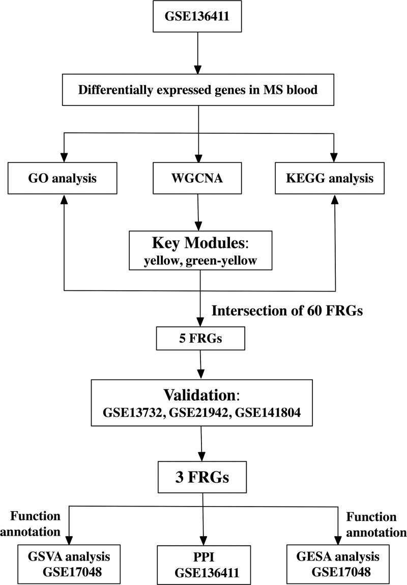
Figure 1 showed the overall workflow of this study. All microarray datasets were downloaded from GEO. We searched the GEO database for microarray datasets using the keyword “multiple sclerosis.” Datasets were included if they met the following criteria: Were from humans; Included blood expression data from MS and non-MS control samples; The number of rows in each platform was > 10,000; The number of MS samples was ≥ 10, the number of control samples was ≥ 10; and There were no repeated samples among datasets. After a careful review, 5 datasets (GSE136411,[20] GSE141804 [www.ncbi.nlm.nih.gov/geo/query/acc.cgi?acc=GSE141804], GSE13732,[21] GSE21942,[22] and GSE17048[23,24] were selected. Detailed information for these datasets was recorded and shown in Table 1.
Figure 1.
Study workflow. MS = multiple sclerosis, GSE = gene expression omnibus series, GO = gene ontology, KEGG = Kyoto encyclopedia of genes and genomes, WGCNA = weighted gene co-expression network analysis, FRG = ferroptosis-related gene, PPI = protein–protein interaction, GSEA = gene set enrichment analysis, GSVA = gene set variation analysis.
Table 1.
Characteristics of the included datasets.
| Dataset ID | Country | No. of samples in the dataset | GPL ID | Usage in the current study |
|---|---|---|---|---|
| GSE136411 | Italy | 60 CIS, 35 PPMS, 121 RRMS, 26 SPMS, 67 NC, and 27 OND | GPL10558 | Identification of DEGs, WGCNA, KEGG and GO analyses, and PPI construction |
| GSE141804 | Israel | 33 MS, and 10 NC | GPL571, GPL96 | Validation of FRG signatures for MS |
| GSE13732 | United States | 73 CIS, and 40 NC | GPL570 | Validation of FRG signatures for MS |
| GSE21942 | United Kingdom | 14 MS, and 15 NC | GPL570 | Validation of FRG signatures for MS |
| GSE17048 | Australia | 43 PPMS, 36 RRMS, 20 SPMS, and 45 NC | GPL6947 | GSEA, and GSVA |
GSE136411 was used for identification of differentially expressed genes (DEGs), weighted gene co-expression network analysis (WGCNA), Kyoto Encyclopedia of Genes and Genomes (KEGG) analysis, Gene Ontology (GO) analysis, and protein–protein interaction (PPI) network construction. GSE141804, GSE13732, and GSE21942 were used for validating the differential expression of FRG signatures for MS. GSE17048 was used for Gene Set Enrichment Analysis (GSEA) and Gene Set Variation Analysis (GSVA).
MS = multiple sclerosis, CIS = clinically isolated syndrome, FRG = ferroptosis-related gene, PPMS = primary progressive multiple sclerosis, RRMS = relapsing remitting multiple sclerosis, SPMS = secondary progressive multiple sclerosis; OND = other neurological disease, NC = normal control, GSE = Gene Expression Omnibus Series, GPL = Gene Expression Omnibus Platform, DEGs = differentially expressed genes, WGCNA = weighted gene co-expression network analysis, KEGG = Kyoto Encyclopedia of Genes and Genomes analysis, GO = Gene Ontology analysis, PPI = protein–protein interaction, GSEA = Gene Set Enrichment Analysis, GSVA = Gene Set Variation Analysis.
2.2. Identification of DEGs
Considering that dataset GSE136411 contained far more samples (60 CIS samples, 35 PPMS samples, 121 RRMS samples, 26 SPMS samples, 67 normal control [NC] samples, and 27 other neurological disease samples) than the other 4 datasets, GSE136411 was applied for identification of DEGs. The R package “limma” was utilized to conduct data normality by log2 transformation and perform the differentiation analysis of DEGs between the MS samples versus the control samples following the following criteria: (I) |log fold change (FC)| > 2 and (II) adjusted P value < 0.05 (adjusted by the false discovery rate [FDR] method).[25] The R package “OmicCircos” was used to visualize the expression patterns and chromosomal locations of the top 100 DEGs (top 50 up-regulated genes and top 50 down-regulated genes) from the differentiation analysis.[26]
2.3. Functional enrichment analysis of DEGs
To reveal potential biological functions of DEGs, we conducted Gene Ontology (GO) enrichment including the potential biological process, molecular function, and cellular component, and KEGG pathway analyses of the DEGs between 2 groups using the R package “clusterprofiler”.[27] GO terms or KEGG pathways with adjusted P < .05 were considered statistically significant and visualized by the R package “GOplot”.[28]
2.4. WGCNA
The co-expression module is a collection of genes with high topological overlap similarity. Genes in the same module often have a higher degree of co-expression. We extracted all DEGs with expression data retrieved from GSE136411 and used the R package “WGCNA” to construct a gene co-expression network for clinical trait–related modules and hub genes among the DEGs.[29] A matrix of similarity was constructed by calculating the correlations of all pairs of genes. Then, an appropriate soft-thresholding power β was selected by using the integrated function (pickSoftThreshold) in the WGCNA package. With this soft-thresholding power, the co-expression similarity was raised to achieve scale-free topology. In this study, we chose the soft-threshold β = 6 (scale-free R2 = 0.85). Subsequently, we transformed the adjacency matrix into a topological overlap matrix. The topological overlap matrix matrix is a method to quantitatively describe the similarity in nodes by comparing the weighted correlation between 2 nodes and other nodes. Next, we performed hierarchical clustering to identify modules, each containing at least 6 genes (minModuleSize = 6). The module eigengene, which was the first principal component of each module’s gene expression matrix, was obtained by the WGCNA to represent the expression profiles of module genes. Some highly similar modules with the height of module eigengene in the clustering lower than 0.25 were merged together. A clustering dendrogram was used to display the results of dynamic tree cut and merge. Gene significance (GS), as the mediator P value (GS = lg P) for each gene, represented the degree of linear correlation between gene expression of the module and clinical traits. Key modules highly correlated with CIS, RRMS, PPMS, or SPMS which were of higher GS values were extracted.
2.5. Identification of FRG signatures for ms
60 FRGs, which were reported and investigated to be involved in ferroptosis were retrieved from the previous literature and were provided in Supplemental Digital Content,[13,17–19] http://links.lww.com/MD/I124. Overlapping genes from key MS-related modules in WGCNA and 60 FRGs were identified as FRG signatures associated with MS in the current study and were selected for further analysis. To identify FRG signatures associated with MS, we applied Venn diagrams, which were constructed using http://bioinformatics.psb.ugent.be/webtools/Venn.
2.6. Validation of FRG signatures for MS in GEO datasets
Expression data of selected FRGs associated with MS were extracted from datasets GSE141804, GSE13732, and GSE21942, which were utilized to validate the differential expression of these FRGs in the blood of MS and NC samples. Significantly down-regulated or up-regulated FRGs in GSE141804, GSE13732, and GSE21942 were identified as FRG signatures for MS and were selected to measure their diagnostic values for MS. Plots in this section were all generated using the R package “ggpubr”.[30] Using the 3 datasets, receiver operating characteristic (ROC) curves were plotted and the areas under the curves (AUCs) were further calculated to assess the predictive accuracy of these ferroptosis specific markers for MS with the R package “rms” and “pROC” [31–33].
2.7. PPI
To systematically predict the protein association and PPI of FRG signatures for MS with other genes, the Spearman coefficients of expression of DEGs in GSE136411 dataset and each FRG signature were calculated, whilst the expression genes with P value < 0.05 were defined as FRG-signature-related genes. The top 100 related genes of each FRG signature according to P value were mapped into the online search tool STRING database (STRING, V11.0; https://string-db.org).[34] A combined score ≥ 0.7 of PPI pairs was considered significant. The results of STRING analysis were imported into Cytoscape software (http://www.cytoscape.org, version 3.7.1; Institute for Systems Biology, Seattle, WA) and the cluster analysis of FRG signatures was conducted using molecular complex detection plug-in, with all the parameters set as defaults.[35]
2.8. GSEA and GSVA
We utilized the R package “clusterprofiler” to perform GSEA of FRG signatures for MS on GSE17048 dataset. In addition, the “GSVA” R package was used to find the pathways most associated with FRG signatures for MS.[36] Based on the median expression of each FRG signature, 99 MS samples were divided into 2 groups (high expression vs low expression). P < .05 was regarded as statistically significant. The gene set “c2.cp.kegg.v7.1.symbols.gmt,” downloaded from the Molecular Signature Database (MSigDB, http://software.broadinstitute.org/gsea/msigdb/index.jsp), was selected as the reference gene set.
2.9. Statistical analysis
The Shapiro–Wilk statistic was used to test the normality of the distribution of data. Comparisons were analyzed with use of Student’s t tests or Wilcoxon’s rank-sum tests for continuous data. Multivariable stepwise logistic regression was used to identify prediction value of FRG signatures for MS. We computed the AUC with a 95% confidence interval (CI) by using 1000 bootstrap resampling.[37] All statistical tests were 2-tailed with a 5% level of statistical significance. Statistical analyses were conducted by using R software version 3.3.3 (Institute for Statistics and Mathematics, Vienna, Austria; https://www.r-project.org).
3. Results
3.1. Identification of DEGs
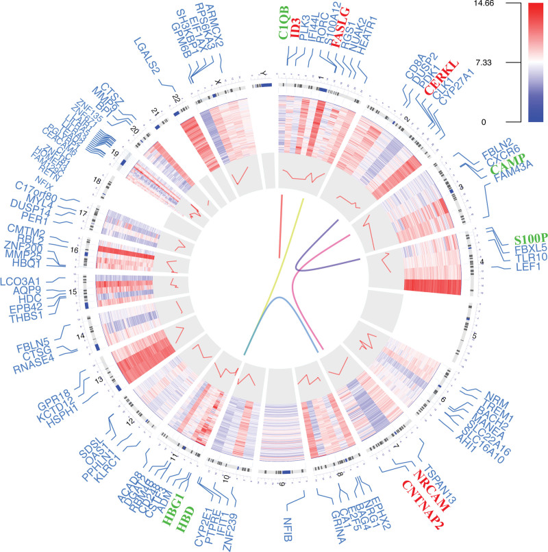
According to the sample information and data matrix of GSE136411, 3017 DEGs (1602 up-regulated and 1415 down-regulated) were identified (included in Supplemental Digital Content, http://links.lww.com/MD/I125). The polarity of genes described as “up-regulated” or “down- regulated” in this article is with respect to MS versus NC. The top 100 DEGs were chosen to visualize their chromosomal locations and expression patterns, as well as their logarithmic adjusted P values shown in the inner layer (Fig. 2). The top 5 up-regulated genes were NRCAM, CERKL, FASLG, ID3, and CNTNAP2, whereas the top 5 down-regulated genes were CAMP, S100P, HBD, C1QB, and HBG1 (Fig. 2).
Figure 2.
Circos plot of expression patterns and chromosomal positions of top 100 differentially expressed genes. The outer circle represented chromosomes, and lines coming from each gene point to their specific chromosomal locations. The multiple sclerosis microarray dataset from GSE136411 was represented in the inner circular heatmap. The red lines in the inner layer indicated -log10 (adjusted P value) of each gene. According to |log2 fold change|, the top five up-regulated genes (red) and the top five down-regulated genes (green) were connected with lines in the center of the Circos plot.
3.2. Functional enrichment analysis of DEGs
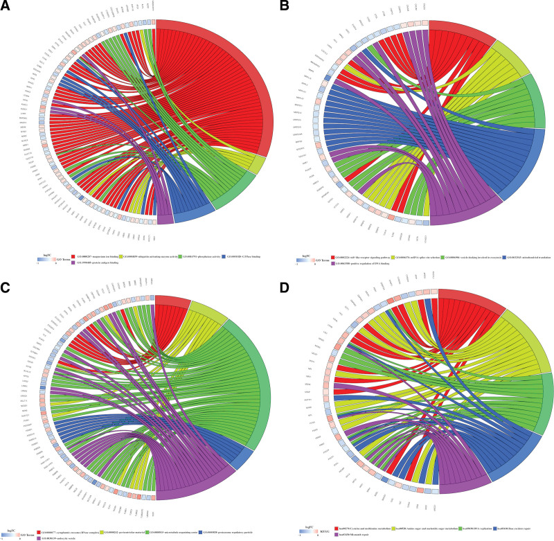
All DEGs were utilized to perform GO and KEGG analyses, and the top 5 of these terms based on their adjusted P values were shown in chord plots (Fig. 3). The top 5 molecular function terms for GO analysis were magnesium ion binding, GTPase binding, phosphatase activity, protein antigen binding, ubiquitin activating enzyme activity (Fig. 3A). The top 5 biological process terms for GO analysis were mitochondrial translation, positive regulation of DNA binding, mRNA splice site selection, vesicle docking involved in exocytosis, toll-like receptor signaling pathway (Fig. 3B). The top 5 cellular component terms for GO analysis were endocytic vesicle, cytoplasmic exosome (RNase complex), pericentriolar material, proteasome regulatory particle, microtubule organizing center (Fig. 3C). For KEGG pathway analysis, DEGs were mostly enriched in mismatch repair, DNA replication, base excision repair, cysteine and methionine metabolism, amino sugar and nucleotide sugar metabolism (Fig. 3D).
Figure 3.
Gene Ontology (GO) and Kyoto Encyclopedia of Genes and Genomes (KEGG) analyses of all differentially expressed genes. Chord plots indicate enrichment analysis of genes. (A) Molecular function of GO analysis. (B) Biological process of GO analysis. (C) Cellular component of GO analysis. (D) KEGG pathways. GO = gene ontology, KEGG = Kyoto encyclopedia of genes and genomes.
3.3. WGCNA and FRGs associated with ms
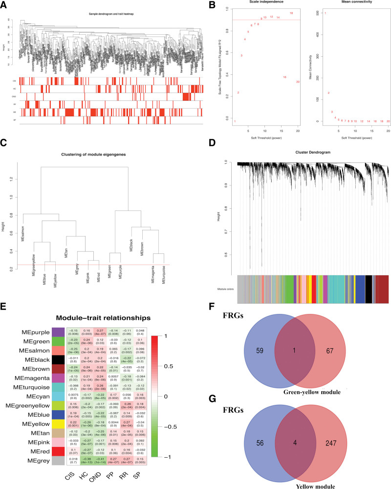
To find the key modules most associated with MS clinical traits, expression data of 3017 DEGs were extracted from GSE136411 was used to conduct WGCNA (Fig. 4). By setting soft-thresholding power as 6 (scale free R2 = 0.85) and cut height as 0.25, we eventually identified 15 modules (Fig. 4A–4D). From the heatmap of module–trait correlations, we found that the yellow and green-yellow modules were highly correlated with clinical traits, especially RR and CIS (yellow module for RR, correlation coefficient = 0.27, P < .001; yellow module for CIS, correlation coefficient = 0.22, P = .001; green-yellow module for RR, correlation coefficient = 0.26, P < .001, green-yellow module for CIS, correlation coefficient = 0.15, P = .006; Fig. 4E). The yellow module contained 4 FRGs: HMOX1, GOT1, LPCAT3, and SLC1A5, while the green-yellow module contained 1 IRG: RPL8 (Fig. 4F). Here, we found the following 5 FRGs associated with MS from the yellow and green-yellow modules: HMOX1, GOT1, LPCAT3, SLC1A5, and RPL8.
Figure 4.
Key module correlated with multiple sclerosis (MS) identified by weighted gene co-expression network analysis (WGCNA). (A) Clustering of samples to detect outliers. (B) Scale-free topology model (left) and mean connectivity (right) for finding the soft-thresholding power. The power selected is 6. (C) Clustering of all modules. The red line indicates the height cutoff (0.25). (D) Cluster dendrogram of genes. (E) Heatmap showed the relationships between different modules and clinical traits. (F) Venn diagram was used to pick up the intersection of green-yellow module genes and 60 FRGs. (G) Venn diagram was used to pick up the intersection of yellow module genes and 60 FRGs. FRG = ferroptosis-related gene, MS = multiple sclerosis, WGCNA = weighted gene co-expression network analysis.
3.4. FRG signatures for ms
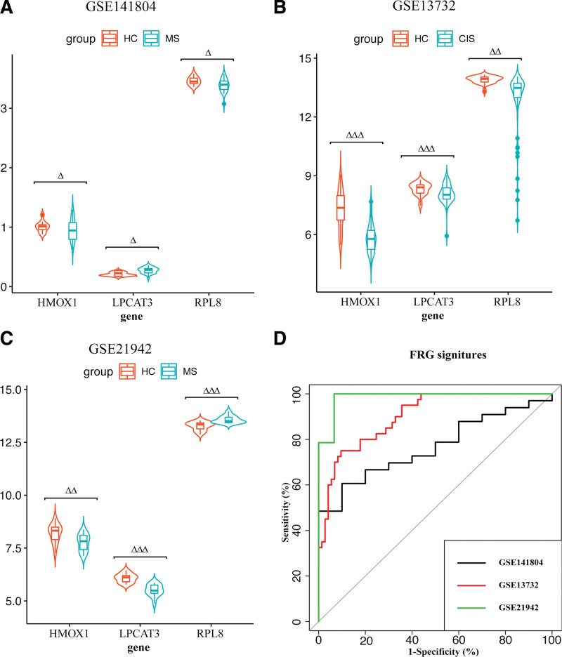
All 5 FRGs associated with MS underwent expression validation in GSE141804, GSE13732, and GSE21942 datasets. The expressions of HMOX1, LPCAT3, and RPL8 were significantly different from NC in MS samples in 3 datasets (Fig. 5A–5C), while no significant difference in the expressions of SLC1A5 and GOT1 between MS and NC was noted in in GSE13732 and GSE21942. HMOX1 was significantly down-regulated in MS from all 3 datasets. LPCAT3 was significantly up-regulated in MS from GSE141804, with significantly down-regulated in MS in GSE13732 and GSE21942. RPL8 was significantly down-regulated in MS from GSE141804 and GSE13732, with significantly up-regulated in MS in GSE21942. The ROC curves were plotted to illustrate the sensitivities and specificities of these 3 FRGs differentiating MS from NC. HMOX1, LPCAT3, and RPL8 exhibited striking diagnostic validity in the 3 datasets (Fig. 5D). In GSE141804, AUC for differentiating MS and NC samples is 0.76 (95% CI = 0.62–0.91). In GSE13732, AUC for differentiating MS and NC samples is 0.90 (95% CI = 0.85–0.96). In GSE21942, AUC for differentiating MS and NC samples is 0.99 (95% CI = 0.95–1.00). These results indicated that the following 3 FRGs: HMOX1, LPCAT3, and RPL8 were reliable ferroptosis specific markers in MS patients. The gene symbols, full names, and implications of these 3 FRG signatures for MS were shown in Table 2.[38–40]
Figure 5.
Validation of HMOX1, LPCAT3, and RPL8 in the Gene Expression Omnibus (GEO) datasets. (A) HMOX1, LPCAT3, and RPL8 gene expression differences between multiple sclerosis (MS) samples and normal control (NC) samples in GSE141804. (B) HMOX1, LPCAT3, and RPL8 gene expression differences between MS samples and NC samples in GSE13732. (C) HMOX1, LPCAT3, and RPL8 gene expression differences between MS samples and NC samples in GSE21942. The green box indicated MS samples, and the orange box indicated NC group. (D) Receiver operating characteristic (ROC) curves of HMOX1, LPCAT3, and RPL8 predicting MS. ROC curves showed the sensitivities and specificities of differentiating MS from NC with three ferroptosis-related gene (FRG) signatures in GSE141804, GSE13732, and GSE21942. The black line indicated GSE141804. The red line indicated GSE13732. The green line indicated GSE21942. ∆P < .05; ∆∆P < .01; ∆∆∆P < .001. FRG = ferroptosis-related gene, GEO = gene expression omnibus, GSE = gene expression omnibus series, CIS = clinically isolated syndrome, MS = multiple sclerosis, NC = normal control, ROC = receiver operating characteristic.
Table 2.
Implications of three FRG signatures for MS.
| Gene symbol | Full name | Implication | Location |
|---|---|---|---|
| HMOX1 | Heme oxygenase 1 | HMOX1 may play a significant role in the maintenance of immune homeostasis which is disrupted in autoimmune disorders, such as MS. | Chromosome 22 |
| LPCAT3 | Lysophosphatidylcholine acyltransferase 3 | Lysophospholipid acyltransferases (LPLATs) regulate the diversification of fatty acid composition in biological membranes. LPCATs are members of the LPLATs that play a role in inflammatory responses. | Chromosome 12 |
| RPL8 | Ribosomal protein L8 | Neuronal ferroptosis occurs during the acute phase of intracerebral hemorrhage in brain areas distant from the hematoma and that inhibition of ferroptosis by Fer-1 exerted a long-term cerebroprotective effect. | Chromosome 8 |
MS = multiple sclerosis, FRG = ferroptosis-related gene.
3.5. PPI network
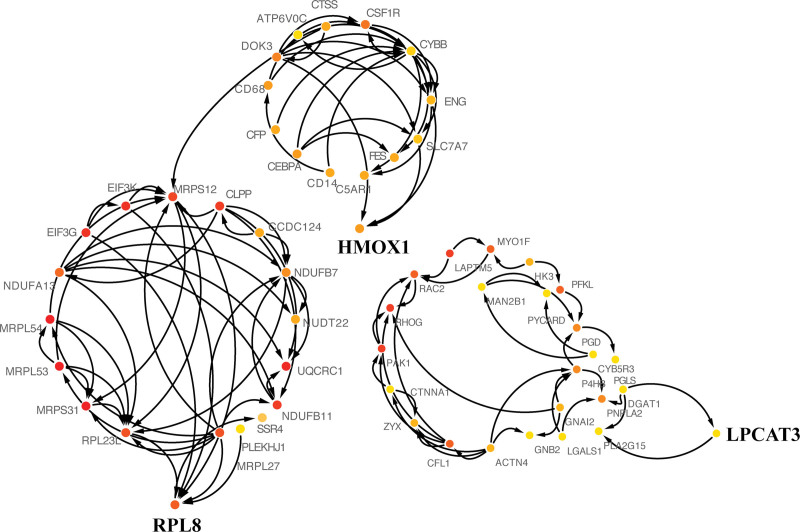
In order to screen out the core genes interacted with FRG signatures in MS samples, top 100 HMOX1-related genes, top 100 LPCAT3-related genes, and top 100 RPL8-related genes were uploaded to the STRING and PPI networks of FRG signatures and FRG-signature-related genes were constructed based on the STRING database. Clusters of HMOX1, LPCAT3, and RPL8 analyzed by molecular complex detection were visualized by Cytoscape, which were determined from the PPI networks (Fig. 6). A total of 53 genes were identified as core genes of HMOX1 (n = 13), LPCAT3 (n = 23), and RPL8 (n = 17), which might play significant key roles in biological function interaction with FRG signatures during the progress of MS.
Figure 6.
The Protein-Protein Interaction (PPI) networks downloaded from the STRING database indicated the interactions of single ferroptosis-related gene (FRG) signature among FRG-signature-related genes. FRG = ferroptosis-related gene, PPI = protein–protein interaction, search tool for the retrieval of interacting gene, STRING = search tool for the retrieval of interacting genes.
3.6. GSEA and GSVA results
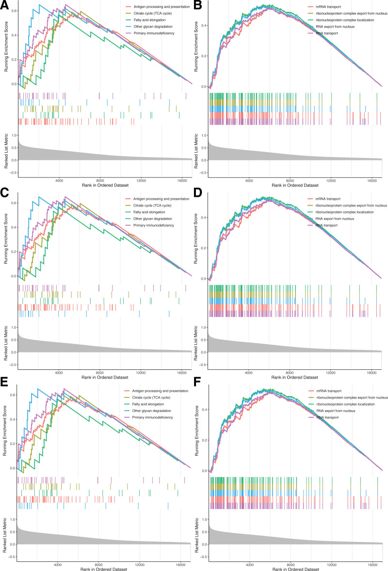
To further investigate the potential functions of HMOX1, LPCAT3, and RPL8 in MS, we performed GSEA and GSVA on the GSE17048 dataset. As shown in Figure 7, the top 5 enriched GSEA GO terms of high expression groups of HMOX1, LPCAT3, and RPL8 were “antigen processing and presentation,” “TCA cycle,” “fatty acid elongation,” “other glycan degradation,” and “primary immunodeficiency” (Fig. 7A, 7C, and 7E). Genes in high expression groups of HMOX1, LPCAT3, and RPL8 were all enriched in “mRNA transport,” “ribonucleoprotein complex export from nucleus,” “ribonucleoprotein complex localization,” “RNA export from nucleus,” and “RNA transport” KEGG pathways (Fig. 7B, 7D, and 7F).
Figure 7.
Gene set enrichment analysis (GSEA) of ferroptosis-related gene (FRG) signatures for multiple sclerosis (MS) in GSE17048 dataset. (A) Top five Gene Ontology (GO) terms (according to GSEA enrichment score) enriched in the high-expression group of HMOX1. (B) Top five Kyoto Encyclopedia of Genes and Genomes (KEGG) pathways (according to GSEA enrichment score) enriched in the high-expression group of HMOX1. (C) Top five GO terms enriched in the high-expression group of LPCAT3. (D) Top five KEGG pathways (according to GSEA enrichment score) enriched in the high-expression group of LPCAT3. (E) Top five GO terms enriched in the high-expression group of RPL8. (F) Top five KEGG pathways enriched in the high-expression group of RPL8. FRG = ferroptosis-related gene, GO = gene ontology, GSEA = gene set enrichment analysis, KEGG = Kyoto encyclopedia of genes and genomes, MS = multiple sclerosis.
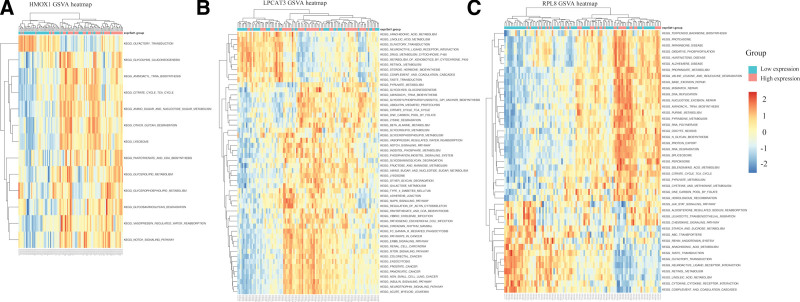
Also, many metabolism-related, immune and inflammation-related, microglia-related, oxidation-related, and mitochondria-related KEGG pathways such as amino acid and glutathione metabolism, lipid metabolism, pyruvate metabolism, JAK STAT signaling, oxidative phosphorylation, RNA degradation, and DNA replication pathways were enriched in the high-expression groups of these FRG signatures for MS in GSVA results, suggesting the contribution of ferroptosis in MS (Fig. 8).
Figure 8.
Gene Set Variation Analysis (GSVA)-derived clustering heatmaps of differentially expressed pathways for single ferroptosis-related gene (FRG) signatures. (A) HMOX1; (B) LPCAT3; (C) RPL8. FRG = ferroptosis-related gene, GSVA = gene set variation analysis.
4. Discussion
Increasing evidence has shown that ferroptosis, a recently discovered programmed cell death, plays a crucial role in progression of MS. However, profiling of it regulars across MS has yet to be clarified. In the current study, we systematically investigated FRGs associated with MS on the basis of WGCNA and 3 FRG signatures for MS were firstly constructed. The core genes interacted with each FRG signature were analyzed in PPI. Functional analyses revealed that biological functions related to metabolism, inflammation and immunity, microglia activation, oxidation, and mitochondria were enriched.
Previous studies demonstrated that ferroptotic cell death resulted from fatal lipid peroxidation.[22] In this regard, the accumulation of intracellular iron caused by the depletion of ferritin or iron transporters and subsequent peroxidation are fundamental mechanisms that lead to the accumulation of lipid peroxides and ferroptosis. Among the 3 identified FRG signatures, HMOX1 is a phase II enzyme which is widely recognized to metabolize heme into biliverdin/bilirubin, carbon monoxide, and ferrous iron, and it has been suggested to demonstrate cytoprotective effects or govern ferroptotic progression depends on the degree of ROS production and following oxidative damage in response to stimulatory cues.[38] LPCAT3, a trans-acylase, is one of important factors in ferroptosis. It was reported to participate in the maintenance of sufficient levels of oxidation substrates which is a significant required constituent of the ferroptotic program. On the other hand, LPCAT3 was also crucial in M1/M2-macrophage polarization by promoting M2 polarization.[39] In the previous rare studies, RPL8 encoded a component of the 60S ribosomal subunit presumably regulating mitochondrial fatty-acid metabolism and translation, which might be a specifically required gene for ferroptosis.[40] It was noteworthy that HMOX1 was significantly down-regulated in MS, and LPCAT3, RPL8 were expressed significantly differently from NC but similarly undetermined in MS in the present study. And there might be 3-fold main implications existing in the expressions of these 3 genes. First, the down-regulation of HMOX1 and uncertainty of LPCAT3 and RPL8 in MS approximately adhered to their bright and dark sides in ferroptosis. Second, the mentioned consistency additionally provided robust evidence that ferroptosis played a critical role in the progression of MS. Third, the current study further demonstrated that the ROC curve generated using these 3 FRGs accurately predicted MS with AUC of 0.76, 0.90, and 0.99. The coexistence of HMOX1, LPCAT3 and RPL8 might trigger ferroptosis, highlighting the pivotal roles of these FRGs in MS, and the potential of HMOX1-based, LPCAT3-based and RPL8-baesd MS therapy.
Although the mechanisms underlying MS progression to ferroptosis have been an intense area of research in the past few years, the whole and specific modulation between ferroptosis and MS remains elusive. GO and KEGG analyses of DEGs between MS and NC indicated that the DEGs corresponding to biological functions were closely related to inflammation and immunity such as protein antigen binding, mitochondrial dysfunction such as mitochondrial translation, microglia activation such as toll-like receptor signaling pathway, oxidation such as phosphatase activity, which was strongly consistent with aforementioned pathogenic mechanisms of MS. Based on 3 identified FRG signatures, we performed GSEA and GSVA analyses and interestingly discovered that the immunity and inflammation-related biological function such as primary immunodeficiency, microglia activation-related biological function such as JAK STAT signaling pathway, oxidation-related biological function such as oxidative phosphorylation pathway, mitochondria-related biological function such as DNA replication pathway were enriched in the high-expression groups of these FRG, which meant that ferroptosis was indeed closely related to MS and shared common pathways with MS in gene aspects. Remarkablely, this study demonstrated that many metabolism-related biological functions such as glycolysis gluconeogenesis, amino sugar and nucleotide sugar metabolism, pyruvate metabolism, and fatty acid elongation were significantly enriched in high expression of HMOX1, LPCAT3 and RPL8 groups in this study. As the understanding of complex biological processes of ferroptosis increases, it has been revealed that the initiation and execution of ferroptosis is not only closely connected with metal ions dysfunction, but also crosslinks with energy metabolism including amino acid, fatty acid, pyruvate, glutathione, phospholipids, and NADPH.[13] Moreover, the imbalance between energy production and consumption has been observed in demyelination during MS lesions including the activation of aerobic glycolysis, the increase of aerobic glycolysis and lactate production, and the decrease of pyruvate dehydrogenase activity.[41] Based on the enrichment and PPI results of 3 FRG signatures in the present study, 1 possible speculation is that the role of ferroptosis in myelin breakdown might not only be neurotoxicity concomitant with central nervous system inflammation, mitochondrial dysfunction, oxidative stress, and microglia activation, but also be a direct and important regulator of metabolism in demyelination in MS. And it is reasonable to assume that HMOX1, LPCAT3 and RPL8, as FRGs, are likely to be correlated with pathogenic mechanisms of metabolism, inflammation and immunity, mitochondrial dysfunction, oxidation, and microglia activation in MS. In addition, the core genes of HMOX1, LPCAT3, and RPL8 during the progress of MS were investigated in PPI, which further provided insights into the potential of FRG-based MS therapies.
There are several limitations of this study. First, 3 FRG signatures were all constructed and validated with retrospective data from GEO. We lack clinical data related to HMOX1, LPCAT3, and RPL8 in MS patients in the real world, which are warranted in future research. Future research with relatively large sample size will be necessary to detect a clinically meaningful effect of HMOX1, LPCAT3, and RPL8 in MS. Second, since current data only provide RNA-level quantifications for FRGs, whereas the ferroptosis process relies on proteins, there could be a variety of inaccuracies. Third, the detailed molecular mechanisms for ferroptosis are still unclear, and currently, the identified FRGs potential have various other functions; thus, the clinical utility of FRG signature might be limited and needs further evaluation.
5. Conclusion
In summary, this study was the first to investigate FRG signatures of MS, which proved to exhibit potential as a biomarker of MS patients in both the derivation and validation cohorts. HMOX1, LPCAT3 and RPL8 provided implications of the mechanisms underlying ferroptosis in MS, which might combine energy metabolism, inflammation and immunity, mitochondrial dysfunction, oxidative stress, as well as microglia activation. Future medications targeting HMOX1, LPCAT3 and RPL8 might eventually lead to personalized progressive MS therapy, a hopeful scenario for patients and treating neurologists.
Acknowledgements
We acknowledge all the authors for their helpful suggestions in this study during the COVID-19 outbreak.
Author contributions
Conceptualization: Si-Chun Gu, Can-Xing Yuan.
Data curation: Si-Chun Gu, Can-Xing Yuan, Chao Gu.
Formal analysis: Si-Chun Gu, Chao Gu.
Funding acquisition: Si-Chun Gu, Can-Xing Yuan.
Investigation: Si-Chun Gu, Can-Xing Yuan.
Methodology: Si-Chun Gu, Can-Xing Yuan.
Project administration: Si-Chun Gu.
Resources: Si-Chun Gu.
Software: Si-Chun Gu.
Supervision: Si-Chun Gu.
Validation: Si-Chun Gu.
Visualization: Si-Chun Gu.
Writing – original draft: Si-Chun Gu, Can-Xing Yuan, Chao Gu.
Writing – review & editing: Si-Chun Gu, Can-Xing Yuan, Chao Gu.
Supplementary Material
Abbreviations:
- AUC =
- areas under the curve
- CI =
- confidence interval
- CIS =
- clinically isolated syndrome
- DEGs =
- differentially expressed genes
- FRG =
- ferroptosis-related gene
- GEO =
- gene expression omnibus
- GO =
- gene ontology
- GS =
- gene significance
- GSEA =
- gene set enrichment analysis
- GSVA =
- gene set variation analysis
- KEGG =
- Kyoto encyclopedia of genes and genomes
- MS =
- multiple sclerosis
- NC =
- normal control
- PPI =
- protein–protein interaction
- PPMS =
- primary progressive multiple sclerosis
- ROC =
- receiver operating characteristic
- RRMS =
- relapsing remitting multiple sclerosis
- SPMS =
- secondary progressive multiple sclerosis
- STRING =
- search tool for the retrieval of interacting genes
- WGCNA =
- weighted gene co-expression network analysis
This work was supported by Science and Technology Department of Tibet [grant number XZ2019ZR-ZY47(Z)]; Shanghai Science and Technology Committee [grant number 19695840100]; the National Key R & D Program of China [grant number 2017YFC1310301]; New Frontier Technological Projects of Shanghai Shenkang Hospital Development Center [grant number SHDC12018131]; and Shanghai Sailing Program [grant number 2021YF1447800]. Funders had no role in study design, data collection, analysis, or decision to publish the manuscript.
The datasets generated during and/or analyzed during the current study are publicly available.
Since this was a retrospective medical record review study of public database, written informed consent was waived.
Supplemental Digital Content is available for this article.
The authors declare that the research was conducted in the absence of any commercial or financial relationships that could be construed as a potential conflict of interest.
How to cite this article: Gu S-C, Yuan C-X, Gu C. Identification of ferroptosis-related gene signatures associated with multiple sclerosis using weighted gene co-expression network analysis. Medicine 2022;XX:XX(e31802).
Contributor Information
Si-Chun Gu, Email: longhuaguchao@163.com.
Can-Xing Yuan, Email: cxyuan1964@163.com.
References
- [1].Ransohoff RM, Hafler DA, Lucchinetti CF. Multiple sclerosis-a quiet revolution. Nat Rev Neurol. 2015;11:134–42. [DOI] [PMC free article] [PubMed] [Google Scholar]
- [2].Weinshenker BG, Bass B, Rice GP, et al. The natural history of multiple sclerosis: a geographically based study. I. Clinical course and disability. Brain. 1989;112:133–46. [DOI] [PubMed] [Google Scholar]
- [3].Reich DS, Lucchinetti CF, Calabresi PA. Multiple Sclerosis. N Engl J Med. 2018;378:169–80. [DOI] [PMC free article] [PubMed] [Google Scholar]
- [4].Campbell GR, Ziabreva I, Reeve AK, et al. Mitochondrial DNA deletions and neurodegeneration in multiple sclerosis. Ann Neurol. 2011;69:481–92. [DOI] [PMC free article] [PubMed] [Google Scholar]
- [5].Fischer MT, Sharma R, Lim JL, et al. NADPH oxidase expression in active multiple sclerosis lesions in relation to oxidative tissue damage and mitochondrial injury. Brain. 2012;135:886–99. [DOI] [PMC free article] [PubMed] [Google Scholar]
- [6].Campbell GR, Worrall JT, Mahad DJ. The central role of mitochondria in axonal degeneration in multiple sclerosis. Mult Scler. 2014;20:1806–13. [DOI] [PubMed] [Google Scholar]
- [7].Kappos L, Bar-Or A, Cree BAC, et al. Siponimod versus placebo in secondary progressive multiple sclerosis (EXPAND): a double-blind, randomised, phase 3 study. Lancet. 2018;391:1263–73. [DOI] [PubMed] [Google Scholar]
- [8].Mayer L, Kappos L, Racke MK, et al. Ocrelizumab infusion experience in patients with relapsing and primary progressive multiple sclerosis: results from the phase 3 randomized OPERA I, OPERA II, and ORATORIO studies. Mult Scler Relat Disord. 2019;30:236–43. [DOI] [PubMed] [Google Scholar]
- [9].Serafini B, Rosicarelli B, Magliozzi R, et al. Detection of ectopic B-cell follicles with germinal centers in the meninges of patients with secondary progressive multiple sclerosis. Brain Pathol. 2004;14:164–74. [DOI] [PMC free article] [PubMed] [Google Scholar]
- [10].Fraussen J, de Bock L, Somers V. B cells and antibodies in progressive multiple sclerosis: contribution to neurodegeneration and progression. Autoimmun Rev. 2016;15:896–9. [DOI] [PubMed] [Google Scholar]
- [11].Mahad DH, Trapp BD, Lassmann H. Pathological mechanisms in progressive multiple sclerosis. Lancet Neurol. 2015;14:183–93. [DOI] [PubMed] [Google Scholar]
- [12].Angeli JPF, Shah R, Pratt DA, et al. Ferroptosis inhibition: mechanisms and opportunities. Trends Pharmacol Sci. 2017;38:489–98. [DOI] [PubMed] [Google Scholar]
- [13].Stockwell BR, Friedmann Angeli JP, Bayir H, et al. Ferroptosis: a regulated cell death nexus linking metabolism, redox biology, and disease. Cell. 2017;171:273–85. [DOI] [PMC free article] [PubMed] [Google Scholar]
- [14].Faissner S, Mishra M, Kaushik DK, et al. Systematic screening of generic drugs for progressive multiple sclerosis identifies clomipramine as a promising therapeutic. Nat Commun. 2017;8:1990. [DOI] [PMC free article] [PubMed] [Google Scholar]
- [15].Faissner S, Gold R. Progressive multiple sclerosis: latest therapeutic developments and future directions. Ther Adv Neurol Disord. 2019;12:1756286419878323. [DOI] [PMC free article] [PubMed] [Google Scholar]
- [16].Hu CL, Nydes M, Shanley KL, et al. Reduced expression of the ferroptosis inhibitor glutathione peroxidase-4 in multiple sclerosis and experimental autoimmune encephalomyelitis. J Neurochem. 2019;148:426–39. [DOI] [PMC free article] [PubMed] [Google Scholar]
- [17].Bersuker K, Hendricks JM, Li Z, et al. The CoQ oxidoreductase FSP1 acts parallel to GPX4 to inhibit ferroptosis. Nature. 2019;575:688–92. [DOI] [PMC free article] [PubMed] [Google Scholar]
- [18].Doll S, Freitas FP, Shah R, et al. FSP1 is a glutathione-independent ferroptosis suppressor. Nature. 2019;575:693–8. [DOI] [PubMed] [Google Scholar]
- [19].Hassannia B, Vandenabeele P, Vanden Berghe T. Targeting ferroptosis to iron out cancer. Cancer Cell. 2019;35:830–49. [DOI] [PubMed] [Google Scholar]
- [20].Acquaviva M, Menon R, Di Dario M, et al. Inferring multiple sclerosis stages from the blood transcriptome via machine learning. Cell Rep Med. 2020;1:100053. [DOI] [PMC free article] [PubMed] [Google Scholar]
- [21].Corvol JC, Pelletier D, Henry RG, et al. Abrogation of T cell quiescence characterizes patients at high risk for multiple sclerosis after the initial neurological event. Proc Natl Acad Sci USA. 2008;105:11839–44. [DOI] [PMC free article] [PubMed] [Google Scholar]
- [22].Kemppinen AK, Kaprio J, Palotie A, et al. Systematic review of genome-wide expression studies in multiple sclerosis. BMJ Open. 2011;1:e000053. [DOI] [PMC free article] [PubMed] [Google Scholar]
- [23].Gandhi KS, McKay FC, Cox M, et al. The multiple sclerosis whole blood mRNA transcriptome and genetic associations indicate dysregulation of specific T cell pathways in pathogenesis. Hum Mol Genet. 2010;19:2134–43. [DOI] [PubMed] [Google Scholar]
- [24].Riveros C, Mellor D, Gandhi KS, et al. A transcription factor map as revealed by a genome-wide gene expression analysis of whole-blood mRNA transcriptome in multiple sclerosis. PLoS One. 2010;5:e14176. [DOI] [PMC free article] [PubMed] [Google Scholar]
- [25].Ritchie ME, Phipson B, Wu D, et al. limma powers differential expression analyses for RNA-sequencing and microarray studies. Nucleic Acids Res. 2015;43:e47. [DOI] [PMC free article] [PubMed] [Google Scholar]
- [26].Hu Y, Yan C, Hsu CH, et al. OmicCircos: a simple-to-use R package for the circular visualization of multidimensional omics data. Cancer Inform. 2014;13:13–20. [DOI] [PMC free article] [PubMed] [Google Scholar]
- [27].Yu G, Wang LG, Han Y, et al. clusterProfiler: an R package for comparing biological themes among gene clusters. Omics. 2012;16:284–7. [DOI] [PMC free article] [PubMed] [Google Scholar]
- [28].Walter W, Sánchez-Cabo F, Ricote M. GOplot: an R package for visually combining expression data with functional analysis. Bioinformatics. 2015;31:2912–4. [DOI] [PubMed] [Google Scholar]
- [29].Langfelder P, Horvath S. WGCNA: an R package for weighted correlation network analysis. BMC Bioinf. 2008;9:559. [DOI] [PMC free article] [PubMed] [Google Scholar]
- [30].Zhang Y, Shen B, Zhuge L, et al. Identification of differentially expressed genes between the colon and ileum of patients with inflammatory bowel disease by gene co-expression analysis. J Int Med Res. 2020;48:300060519887268. [DOI] [PMC free article] [PubMed] [Google Scholar]
- [31].Mandrekar JN. Receiver operating characteristic curve in diagnostic test assessment. J Thorac Oncol. 2010;5:1315–6. [DOI] [PubMed] [Google Scholar]
- [32].Robin X, Turck N, Hainard A, et al. pROC: an open-source package for R and S+ to analyze and compare ROC curves. BMC Bioinf. 2011;12:77. [DOI] [PMC free article] [PubMed] [Google Scholar]
- [33].Zhang S, Tong YX, Zhang XH, et al. A novel and validated nomogram to predict overall survival for gastric neuroendocrine neoplasms. J Cancer. 2019;10:5944–54. [DOI] [PMC free article] [PubMed] [Google Scholar]
- [34].Szklarczyk D, Morris JH, Cook H, et al. The STRING database in 2017: quality-controlled protein-protein association networks, made broadly accessible. Nucleic Acids Res. 2017;45:D362–d368. [DOI] [PMC free article] [PubMed] [Google Scholar]
- [35].Bader GD, Hogue CW. An automated method for finding molecular complexes in large protein interaction networks. BMC Bioinf. 2003;4:2. [DOI] [PMC free article] [PubMed] [Google Scholar]
- [36].Hänzelmann S, Castelo R, Guinney J. GSVA: gene set variation analysis for microarray and RNA-seq data. BMC Bioinf. 2013;14:7. [DOI] [PMC free article] [PubMed] [Google Scholar]
- [37].Steyerberg EW, Bleeker SE, Moll HA, et al. Internal and external validation of predictive models: a simulation study of bias and precision in small samples. J Clin Epidemiol. 2003;56:441–7. [DOI] [PubMed] [Google Scholar]
- [38].Agúndez JA, García-Martín E, Martínez C, et al. Heme oxygenase-1 and 2 common genetic variants and risk for multiple sclerosis. Sci Rep. 2016;6:20830. [DOI] [PMC free article] [PubMed] [Google Scholar]
- [39].Taniguchi K, Hikiji H, Okinaga T, et al. Essential role of lysophosphatidylcholine acyltransferase 3 in the induction of macrophage polarization in PMA-treated U937 cells. J Cell Biochem. 2015;116:2840–8. [DOI] [PubMed] [Google Scholar]
- [40].Chen B, Chen Z, Liu M, et al. Inhibition of neuronal ferroptosis in the acute phase of intracerebral hemorrhage shows long-term cerebroprotective effects. Brain Res Bull. 2019;153:122–32. [DOI] [PubMed] [Google Scholar]
- [41].Vallée A, Lecarpentier Y, Guillevin R, et al. Demyelination in multiple sclerosis: reprogramming energy metabolism and potential PPARγ agonist treatment approaches. Int J Mol Sci. 2018;19. [DOI] [PMC free article] [PubMed] [Google Scholar]


