Abstract
Duct-dependent systemic circulation is accompanied by a right-to-left ductal shunt, at least during systole. Although observations of paradoxical continuous left-to-right shunts in duct-dependent systemic circulation have been reported, the mechanism remains unclear. We report a continuous left-to-right ductal shunt throughout the cardiac cycle during the initial recovery phase from circulatory collapse and right ventricular (RV) dysfunction due to ductal closure in an infant with hypoplastic left heart and severe aortic coarctation. Further recovery improved his RV function and changed the ductal flow from continuous left-to-right to bidirectional, which is usually seen in duct-dependent systemic circulation. Marked RV dysfunction may contribute to the continuous left-to-right ductal shunt. A continuous left-to-right ductal shunt should not be used to rule out duct-dependent systemic circulation.
Keywords: coarctation, ductus arteriosus, prostaglandin E 1, hypoplastic left heart
Due to marked advances in fetal screening echocardiography, many congenital heart disease (CHD) cases are diagnosed before birth. 1 Unfortunately, there are newborns not prenatally diagnosed who have duct-dependent systemic circulation. These cases develop circulatory collapse following postnatal ductus arteriosus (DA) constriction 2 ; hence, early diagnosis and treatment are crucial for their survival. In patients with duct-dependent systemic circulation, DA flow is predominantly from the pulmonary artery to the aorta (right-to-left shunt). With a postnatal drop in pulmonary vascular resistance, the diastolic shunt from the aorta to the pulmonary artery (left-to-right shunt) 3 gradually increases 4 ; however, to maintain systemic circulation, the amount of left-to-right shunt does not exceed that of the right-to-left shunt in the cardiac cycle.
Nevertheless, on rare occasions, paradoxical continuous left-to-right shunt in duct-dependent systemic circulation has been reported. 5 As the underlying mechanism remains unclear, we report an insightful case showing the rare circulatory hemodynamics of continuous left-to-right DA shunt in a patient with borderline hypoplastic left heart and aortic coarctation (CoA).
Case Report
Supplementary Video S1 Echocardiography on admission. Four-chamber, long-axis, and short-axis views show markedly impaired biventricular wall motion, small left atrium and left ventricle, and large atrial septal defect, right atrium, and right ventricle. Two-dimensional and simultaneous color Doppler shows hypoplastic aortic isthmus and coarctation of the aorta.
Supplementary Video S2 Color Doppler after prostaglandin use and before stabilization shows a paradoxical continuous left-to-right ductal shunt.
Supplementary Video S3 Color Doppler after stabilization shows typical bidirectional ductal shunt.
Supplementary Video S4 Four-chamber view shows improving biventricular wall motion after stabilization.
A male infant weighing 2,954 g was born at a gestational age of 38 6/7 weeks. Delivery occurred at a nearby obstetric clinic without any information on fetal screening ultrasound. Apgar scores were 9 at 1 minute and 10 at 5 minutes. He was first noticed to have uneasiness at 44 hours after delivery; 3 hours later, he became tachypneic with a respiratory rate of 70 breaths/min and was unable to feed anymore. Percutaneous arterial oxygen saturation (SpO 2 ) levels on the right arm gradually decreased to 88%. Oxygen administration failed to increase his SpO 2 level, and he was referred to our hospital 54 hours after delivery.
On admission, he cried weakly and had decreased activity and muscle tone. While the SpO 2 level on his right arm was 90%, his SpO 2 level on the lower extremities was unmeasurable. Bilateral air entry and respiratory sounds were clear with mild tachypnea (64 breaths/min). He had sinus tachycardia (176 bpm), and a gallop rhythm was audible. His abdomen was markedly distended, bilateral pulses of femoral and dorsalis pedis arteries were weak, and we could not measure the blood pressure of the lower extremities, while the blood pressure of the right arm was 73/52 (mean 57) mm Hg.
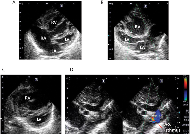
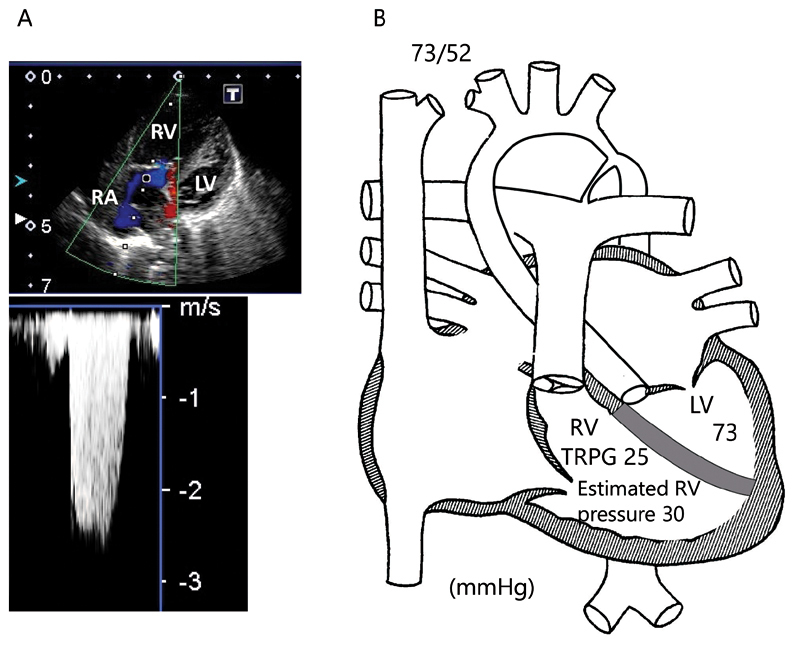
Venous blood gas from the lower extremities showed a pH of 6.966, partial pressure of carbon dioxide (pCO 2 ) of 54.1 mm Hg, bicarbonate (HCO 3 − ) of 11.7 mEq/L, base excess of −23.6 mEq/L, and lactate level of 13.9 mmol/L. He received sedation, intubation, and mechanical ventilation. Echocardiography revealed situs solitus and atrioventricular and ventriculoarterial concordance with a markedly dilated right ventricle (RV) and a small left ventricle (LV) ( Fig. 1 , Supplementary Video S1 ; available in the online version only). The size ( Z -value 6 ) of the valve annulus and the vessel diameters were as follows: 7.0 mm (−2.3) for the mitral valve, 15.1 mm (1.4) for the tricuspid valve, 4.1 mm (−4.4) for the aortic valve, 11.8 mm (2.5) for the pulmonary valve, 6.5 mm (−2.4) for the ascending aorta, 11.0 mm (2.2) for the main pulmonary artery, 4.0 mm (−4.0) for the transverse arch, and 1.7 mm (−6.8) for the isthmus. A large atrial septal defect (ASD) with massive left-to-right shunt, severe tricuspid regurgitation (TR) with a TR pressure gradient (TRPG) of 25 mm Hg ( Fig. 2 ), small mitral and aortic valves, narrowing of the entire aortic isthmus, and severe aortic CoA ( Fig. 1D , Supplementary Video S1 ; available in the online version only) were observed. The interventricular septum was intact. Biventricular wall motion was very poor ( Supplementary Video S1 ; available in the online version only). Despite the severe CoA ( Fig. 1D , Supplementary Video S1 ; available in the online version only), the DA was very narrow with left-to-right blood flow.
Fig. 1.

Echocardiography on admission. ( A ) Four-chamber view. ( B ) Long-axis view. ( C ) Short-axis view. These figures show the small LA and LV, and the large atrial septal defect, RA, and RV. ( D ) Two-dimensional and simultaneous color Doppler for aortic arch show hypoplastic aortic isthmus and aortic coarctation. Ao, aorta; LA, left atrium; LV, left ventricle; RA, right atrium; RV, right ventricle.
Fig. 2.

( A ) Continuous-wave Doppler signal for tricuspid regurgitation. The peak velocity is 2.5 m/s, and the TRPG is only 25 mm Hg. When the right atrial pressure was set to 5 mm Hg, 9 the estimated RV systolic pressure was 30 mm Hg. ( B ) Illustration of the cardiovascular structure and measured and estimated pressures (mm Hg). Estimated RV pressure is much lower than LV pressure. LV, left ventricle; RA, right atrium; RV, right ventricle; TRPG, tricuspid regurgitation pressure gradient.
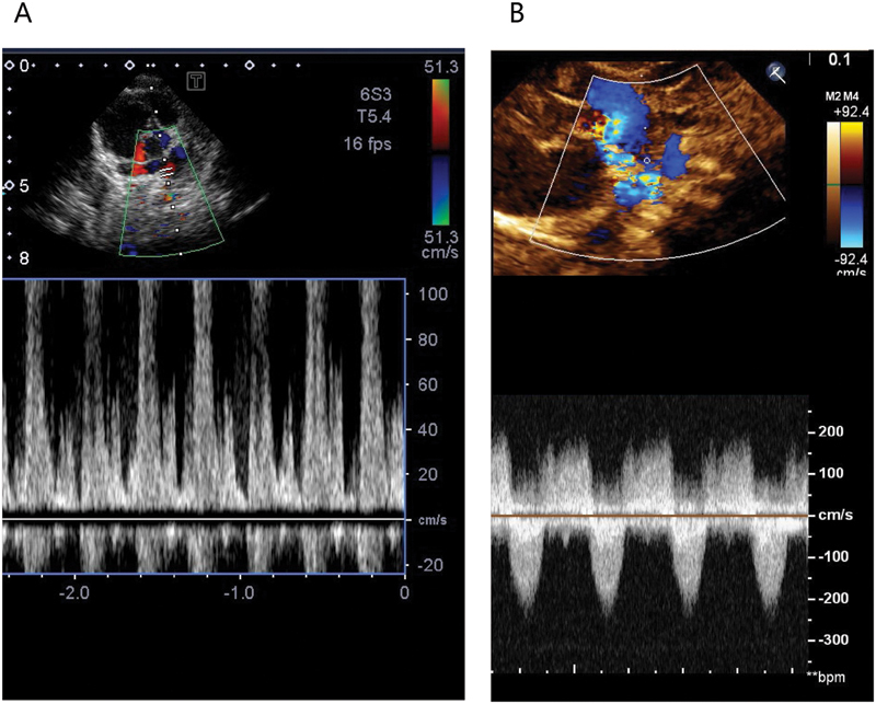
Immediately on diagnosis of a duct-dependent systemic circulation ( Fig. 2B ) and circulatory collapse due to ductal closure, the administration of prostaglandin E 1 (PGE 1 ) to maintain DA patency, and dopamine and dobutamine to improve cardiac function were initiated. Sodium bicarbonate was also used to correct metabolic acidosis. Subsequently, echocardiography revealed gradual improvement of ventricular wall motion, but a continuous left-to-right shunt ( Fig. 3A , Supplementary Video S2 ; available in the online version only) in the patent DA with a 4-mm width persisted despite duct-dependent systemic circulation. This paradoxical continuous left-to-right DA shunt possibly compromised systemic blood flow to the lower body. A left-to-right shunt across the ASD and DA synergistically caused high pulmonary blood flow along with low systemic blood flow. The RV fractional area change (FAC) was as low as 19%.
Fig. 3.

Doppler flow profile of the ductus arteriosus. ( A ) Pulse-wave Doppler image after prostaglandin use and before stabilization shows paradoxical continuous left-to-right ductal shunt. ( B ) Continuous-wave Doppler after stabilization shows typical bidirectional ductal shunt.
He was transferred to another hospital under manual bagging for further preoperative management and subsequent surgery. On arrival at the hospital, 59 hours after his delivery, his skin color had markedly improved. Arterial blood gas analysis showed pH of 7.183, pCO 2 of 36.0 mm Hg, HCO 3 − of 13.2 mEq/L, and BE of −14.1 mEq/L. The DA flow pattern also became bidirectional ( Fig. 3B , Supplementary Video S3 ; available in the online version only), as typically observed in a duct-dependent systemic circulation after a reduction in pulmonary vascular resistance. 4 RV wall motion significantly improved on echocardiography ( Supplementary Video S4 ; available in the online version only) with a RV FAC of 32%. This infant was further stabilized with N 2 inhalation therapy, 7 and bilateral pulmonary artery banding was performed at 5 days of age. He was extubated at 13 days of age, and postoperative recovery was achieved.
Discussion
An adequate right-to-left DA shunt is required to prevent circulatory collapse in a duct-dependent systemic circulation. However, there can be a paradoxical continuous left-to-right shunt in the duct-dependent systemic circulation ( Fig. 3A , Supplementary Video S2 ; available in the online version only), and the mechanism has not been clarified. To the best of our knowledge, this is the first case report indicating that marked RV dysfunction may contribute to the continuous ductal left-to-right shunt in patients with a duct-dependent systemic circulation.
A large diastolic left-to-right DA shunt usually develops after postnatal reduction in pulmonary vascular resistance. 4 Even on such occasions, systolic right-to-left DA flow is essential in CHD cases with duct-dependent systemic circulation. However, our patient showed a continuous left-to-right DA shunt throughout the cardiac cycle.
The mechanisms responsible for this paradoxical circulation are discussed. This infant had a hypoplastic LV, mitral and aortic valves, and aorta; CoA; as well as a large ASD, right atrium, and RV. In such cases, the RV becomes the main cardiac chamber that needs to adequately provide output, not only to the pulmonary circulation but also to the systemic circulation via a net right-to-left DA shunt during the cardiac cycle, which was eventually achieved after stabilization. However, this infant initially showed a paradoxical continuous left-to-right DA shunt throughout the entire cardiac cycle, despite duct-dependent systemic circulation. In the duct-dependent systemic circulation, ductal closing, as an initial trigger, reduces systemic blood flow and induces acidemia. The reduced coronary flow and acidemia synergistically impair ventricular function. Since RV coronary blood flow is supplied only by the right coronary artery, systemic RV has coronary artery supply mismatch. 8 Therefore, RV function should be more likely to be impaired. Echocardiography, in this case, revealed RV dysfunction and marked TR. RV FAC was as low as 19% despite the marked TR. When his blood pressure was 73/52 mm Hg in the right arm, the TRPG was only 25 mm Hg ( Fig. 2 ). When the right atrial pressure was set to 5 mm Hg, 9 the estimated RV systolic pressure was 30 mm Hg. Thus, the RV pressure was much lower than the pressure of hypoplastic LV during this phase. Consequently, the pulmonary arterial pressure appeared lower than the distal aortic pressure throughout the cardiac cycle, despite the CoA and hypoplastic LV ( Fig. 2 ). Lower pulmonary vascular resistance due to PGE 1 and prior oxygen use as well as higher systemic vascular resistance due to circulatory collapse could have contributed to this paradoxical shunt. The LV, which provides significant output to the aorta, could also be responsible for this paradoxical shunt because it should never occur in patients with aortic atresia.
The various treatment modalities modified the ductus opening status, systemic and pulmonary resistance, loading condition, and biventricular functions. After an improvement in RV function (RV FAC from 19 to 32%) and an increase in estimated RV pressure, this patient's paradoxical continuous left-to-right shunt disappeared. Thus, the paradoxical shunt is believed to be caused by the more severe and prolonged dysfunction of the RV 10 than that of the LV. A limitation of this case report is that detailed mechanisms, such as coronary perfusion status, remain to be elucidated.
Conclusion
In conclusion, a paradoxical continuous left-to-right ductal shunt throughout the cardiac cycle can occur under marked RV dysfunction, even in infants with CHD with duct-dependent systemic circulation. Optimization of circulatory and general status to improve RV function is essential to convert the shunt flow direction and to achieve further stabilization in such cases.
Acknowledgments
We thank all the doctors and medical staff involved in the treatment of this infant. We would like to thank Honyaku Center Inc. for English language editing.
Funding Statement
Funding None.
Footnotes
Conflict of Interest None declared.
References
- 1.American Heart Association Adults With Congenital Heart Disease Joint Committee of the Council on Cardiovascular Disease in the Young and Council on Clinical Cardiology, Council on Cardiovascular Surgery and Anesthesia, and Council on Cardiovascular and Stroke Nursing . Donofrio M T, Moon-Grady A J, Hornberger L K. Diagnosis and treatment of fetal cardiac disease: a scientific statement from the American Heart Association. Circulation. 2014;129(21):2183–2242. doi: 10.1161/01.cir.0000437597.44550.5d. [DOI] [PubMed] [Google Scholar]
- 2.Mellander M. Diagnosis and management of life-threatening cardiac malformations in the newborn. Semin Fetal Neonatal Med. 2013;18(05):302–310. doi: 10.1016/j.siny.2013.04.007. [DOI] [PubMed] [Google Scholar]
- 3.Lee M L, Wu M H, Wang J K, Chang C I, Lue H C. Flow characteristics in infants with hypoplastic left heart syndrome: an echocardiographic study. Zhonghua Min Guo Xiao Er Ke Yi Xue Hui Za Zhi. 1995;36(01):14–19. [PubMed] [Google Scholar]
- 4.Rychik J, Gullquist S D, Jacobs M L, Norwood W I. Doppler echocardiographic analysis of flow in the ductus arteriosus of infants with hypoplastic left heart syndrome: relationship of flow patterns to systemic oxygenation and size of interatrial communication. J Am Soc Echocardiogr. 1996;9(02):166–173. doi: 10.1016/s0894-7317(96)90024-3. [DOI] [PubMed] [Google Scholar]
- 5.Lu C W, Wang J K, Chang C I. Noninvasive diagnosis of aortic coarctation in neonates with patent ductus arteriosus. J Pediatr. 2006;148(02):217–221. doi: 10.1016/j.jpeds.2005.09.036. [DOI] [PubMed] [Google Scholar]
- 6.Ped(z) - Pediatric calculator Cardiac z-scoresAccessed July 31, 2022, at:https://www.pedz.de/de/pedz/mmode.html
- 7.Krushansky E, Burbano N, Morell V. Preoperative management in patients with single-ventricle physiology. Congenit Heart Dis. 2012;7(02):96–102. doi: 10.1111/j.1747-0803.2011.00584.x. [DOI] [PubMed] [Google Scholar]
- 8.Brida M, Diller G P, Gatzoulis M A. Systemic right ventricle in adults with congenital heart disease: anatomic and phenotypic spectrum and current approach to management. Circulation. 2018;137(05):508–518. doi: 10.1161/CIRCULATIONAHA.117.031544. [DOI] [PubMed] [Google Scholar]
- 9.European Special Interest Group ‘Neonatologist Performed Echocardiography’ (NPE) . de Boode W P, Singh Y, Molnar Z. Application of Neonatologist Performed Echocardiography in the assessment and management of persistent pulmonary hypertension of the newborn. Pediatr Res. 2018;84 01:68–77. doi: 10.1038/s41390-018-0082-0. [DOI] [PMC free article] [PubMed] [Google Scholar]
- 10.Oka S, Kondo U, Oshima A. Two extremely low birth weight infants who survived functional pulmonary atresia with normal intracardiac anatomy. AJP Rep. 2019;9(03):e310–e314. doi: 10.1055/s-0039-1697960. [DOI] [PMC free article] [PubMed] [Google Scholar]


