Abstract
Background
Ginsenoside compound K (GC-K) potentially alleviates ulcerative colitis involved in gut microbiota, which is significantly associated with the occurrence and development of colitis. However, the effect and mechanism of GC-K on anti-colitis in relation to gut microbiota are not clear. This study focused on the prevention and mechanism of GC-K on Dextran sulfate sodium (DSS)-induced colitis of mice pertinent to gut microbiota.
Methods
DSS was used to establish a chronic colitis mouse model. Body weight analysis, colon length measurement, HE staining, and inflammatory factors levels were processed in animal experiments. Flow cytometry was employed to analyze Th17/Treg cells in the mouse spleen and blood. 16S rRNA sequencing was utilized to analyze gut microbiota. Fecal microbiota transplantation (FMT) experiment was employed to verify the anti-colitis efficacy of GC-K by reshaping gut microbiota.
Results
GC-K significantly relieved colitis-related symptoms due to decreased disease activity index (DAI) scores, spleen weight, and increased colon length. Additionally, the tight junction proteins were increased, and the pro-inflammatory cytokines, such as TNF-α, IL-6, IL-1β and IL-17, were decreased after GC-K treatment. Furthermore, Bacteroides spp. significantly increased after modeling. Moreover, FMT experiments confirmed that GC-K-driven gut microbiota greatly relieved DSS-induced colitis.
Conclusion
GC-K alleviated colitis via the modulation of gut microbiota.
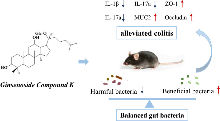
Graphical Abstract
Supplementary Information
The online version contains supplementary material available at 10.1186/s13020-022-00701-9.
Keywords: Ginsenoside compound K, Colitis, FMT, Gut microbiota
Highlights
Ginsenoside Compound K (GC-K) alleviated colitis in DSS-induced Mice.
GC-K modulated gut microbiota in DSS-induced mice.
GC-K relieved colitis via regulating gut microbiota.
GC-K-driven gut microbial effectively ameliorated colitis.
Supplementary Information
The online version contains supplementary material available at 10.1186/s13020-022-00701-9.
Introduction
Ulcerative colitis (UC), a chronic nonspecific intestinal inflammatory disease, clinically manifests as abdominal pain, bloody diarrhea, and even degrees of systemic symptoms, becoming a global disease in gastroenterology [1, 2]. Colitis triggers inflammatory cell infiltration followed by cytokine release syndrome and systemic inflammation. The pathological mechanism of colitis is not well-known, so colitis is symptomatically treated in the clinic along with adverse effects and easy recurrence. Therefore, it is essential to develop safe and effective drugs for colitis.
Although the underlying pathogenesis of UC is incompletely understood, numerous studies have demonstrated that gut microbiota plays a vital role in the pathogenesis of UC. Intestinal inflammation disappears in a spontaneous model of colitis in germ-free IL-10(−/−) mice [3, 4]. Meanwhile, the gut microbiota of UC patients maintains dysbiosis by comparing with healthy humans. The α-diversity of gut microbiota and some beneficial gut microbes, such as Faecalibacterium prausnitzii and Akkermansia muciniphila, were reduced in UC patients. F. prausnitzii can destroy the activation of the NF-κB pathway and block the production of IL-8 by producing butyrate, while A. muciniphila ameliorates colitis by regulating immune responses and gut barrier repair [5–7]. On the contrary, the abundance of pathogenic bacteria was increased, e.g., Bacteroides vulgatus, which exacerbates UC through secreting proteases [8]. Due to gut microbiota dysbiosis as a pathogenic factor that leads to an impaired intestinal barrier and promotes the onset of UC, targeting the gut microbiota could be a practical therapeutic approach for preventing and curing colitis.
Ginsenoside compound K (GC-K) is one of the primary metabolites of ginseng saponins bio-converted by gut microbiota in vivo [9–11], which possesses various pharmacological activities, such as anti-diabetic, anti-hepatic lipid accumulation, anti-inflammation, and antitumor effects. In addition, GC-K ameliorates colitis and inhibits inflammatory responses by suppressing NF-kB activation [12]. However, the absolute oral bioavailability of GC-K is only about 35% [13]. GC-K is catalyzed by β-glycosidase only secreted from the gut microbiota to generate its metabolite protopanaxadiol in vivo, which implies that GC-K interplays with gut microbiota after oral administration [14]. Furthermore, our previous study found that GC-K could suppress the tumor growth of AOM/DSS-induced colitis-associated colorectal cancer through the modulation of gut microbiota, partially by the up-regulation of A. muciniphila [9]. Accordingly, we hypothesized that GC-K could soothe colitis by regulating gut microbiota.
In this study, the effect of GC-K on gut microbiota was explored in DSS-induced colitis to verify whether gut microbiota mediated the progression. We observed that GC-K ameliorated experimental colitis and regulated gut microbial such as Bacteroides spp., which were enriched in the colitis group. In addition, co-incubation experiments further demonstrated the regulatory effect of GC-K on Bacteroides spp. Spearman correlation analysis showed that the progression of the UC phenotype was related to gut microbiota. To verify the beneficial effects of treatment-naïve gut microbiota on colitis, fecal microbiota transplantation (FMT) confirmed that gut microbiota from GC-K-dosed mice improved intestinal barrier function and allayed experimental colitis. Collectively, our study demonstrated the anti-inflammatory effects of GC-K in a DSS-induced colitis model mainly via regulating gut microbiota.
Materials and methods
Materials
Fluorescein isothiocyanate (FITC)-labeled dextran was obtained from Sigma-Aldrich (St. Louis, MO, USA). Antibodies of ZO-1 and Occludin were purchased from Proteintech Group, Inc. (Wuhan, Hubei, China). GC-K was provided by Chengdu Push Bio-technology Co., Ltd. (Sichuan, China).
Animals and experimental design
Dextran sulfate sodium (DSS; molecular weight, 36–50 kDa) and Sulfasalazine (SASP) were purchased from MP Biomedicals (San Jose, CA, USA) and Aladdin (Shanghai, China), respectively. Female C57BL/6 mice (6 weeks; 17–20 g) were obtained from Hunan SJA Laboratory Animal Co., Ltd. (Hunan, China). The Animal Ethics Committee of Central South University (No. 2020sydw1037) permitted the animal protocol. Mice after acclimation were divided into groups as control, DSS, DSS + LCK (20 mg/kg/day, low dose), DSS + HCK (60 mg/kg/day, high dose), and SASP (200 mg/kg/day, positive control) (Additional file 1: Fig. S1). Mice were fed with 2% DSS in drinking water for 5–7 days, replaced with autoclaved water for another 10–14 days for 3 cycles, and sacrificed on day 42.
Disease activity index (DAI) and histologic assessment
The DAI was scored every 2 days to evaluate the severity of DSS-induced colitis as described previously [15]. The distal colorectal segments were immersed in 4% paraformaldehyde and stained with hematoxylin and eosin (H&E) to perform a histopathology assay.
Quantitative real-time PCR analysis
According to previous studies [16], the total RNA of colon tissue was extracted by Trizol reagent. The concentration and purity of total RNA were determined by Nanodrop. SYBR Green Premix Ex TaqII was utilized for qPCR amplification. The relative regulation in target genes after normalization to β-action between two groups was calculated using the 2−ΔΔCt method. The qPCR primers of IL-17a, TNF-α, IL-β, IL-10, IL-6, Foxp3, ZO-1, Occludin, E-cadherin, and Mucin-2 were synthesized by Sanggon Biotech (Shanghai, China) (Additional file 2: Table S1).
Flow cytometry analysis
Spleens were collected, ground, and filtered using 70 μm cell strainers to obtain single-cell suspension. For Th17 cells measurement, cells were counted and stimulated by Leukocyte Activation Cocktail for 4 h. Then, Fixable Viability Dye eFluor ™ 780 (eBioscience) was utilized to identify the Viability of cells. After lysis of the red blood cells, cells were subsequently stained with antibodies as follows, (a) Anti-mouse CD45 Briliant Violet510, Anti-mouse CD3 FITC, Anti-mouse CD4 Briliant Violet421, (b) Anti-mouse IL-17A PE. For Treg cells measurement, cells were accordantly stained with antibodies as follows, (a) Anti-mouse CD45 Briliant Violet510, Anti-mouse CD3 FITC, Anti-mouse CD4 Briliant Violet421, Anti-mouse CD25 PE/Cy™, (b) Anti-mouse Foxp3 APC. All the antibodies were purchased from BD Biosciences (San Jose, CA, USA).
Western blotting
Proteins were extracted with radioimmunoprecipitation assay (RIPA) lysis buffer and then centrifuged at 12 000 rpm and 4 °C for 15 min. 200 μL of supernatant were subsequently added and mixed well with 40 μL of 5 × SDS-PAGE (100 °C, 10 min). Protein quantification was processed according to the BCA kit procedure. The polyvinylidene difluoride membranes were incubated with the specific primary antibodies, and the appropriate secondary antibodies for 1.5 h at room temperature. The blot was visually analyzed with enhanced chemiluminescence (ECL) substrate and a Bio-Rad imaging system.
Measurement of FITC-Dextran leakage
Mice were fasted overnight and gavaged with FITC-Dextan (Sigma) at 500 mg/kg. After administration of FITC-Dextran for 4 h, serum was collected to measure the fluorescence intensity.
16S ribosomal RNA gene sequencing and data analysis
After sequencing, data processing and quality control were operated using fastq-join (Version: 1.3.1) and pear (v0.9.11) to generate Raw Tags. Cutadapt (version 1.18) was used to get the Clean Tags from the original sequences. Usearch (Version11.0.667) software was used to operate OTU (Operational Taxonomic Units) clustering. Silva (Release132) database was used to pair OTUs sequences for taxonomic analysis. The α-diversity and β-diversity of the samples were analyzed by Mothur software and R language tools. LEfSe analysis was performed on LEfSe software to assess the adequate size (LED > 2 or 4) of each distinct taxa or OTU. Correlation analysis was conducted on Wekemo Bioincloud (https://www.bioincloud.tech/).
Bacterial strains and growth curve
All the bacterial strains were stored in 30% glycerol at − 80 °C until use. The bacteria were divided into the administration group (500 mg/mL or 20 μmol/mL of GC-K) and the control group (DMSO). Their growth dynamics were detected in an anaerobic chamber with mixed anaerobic gas (5% carbon dioxide, 5% hydrogen, 90% nitrogen).
Fecal microbiota transplantation
FMT was performed according to an established protocol with an appropriate modification [17]. Stools from mice treated with HCK or DSS were collected and stored at -80 °C. 100 mg of stools from donor mice were re-suspended in 1 mL of sterile saline. The solution was centrifuged at 600×g for 5 min to obtain the supernatant, which was then centrifuged at 12 000×g for 10 min. The pellet was re-suspended and used as transplant bacteria. Mice were fed with antibiotic water (0.5 g/L of ampicillin, 0.5 g/L of metronidazole, 0.5 g/L of neomycin, and 0.25 g/L of vancomycin) for 5 days. Transplantation was performed by oral gavage of 200 μL of bacterial fluid 3 times a week.
Statistical analysis
The data were expressed as the mean ± SEM and analyzed by GraphPad Prism 8.0 and SPSS software (Version 23). Significant differences between the two groups were evaluated by the two-tailed unpaired Student’s t-test, one-way ANOVA, or Kruskal–Wallis with the non-normal or non-parametric distribution. The statistical difference between the model and control group was denoted by "#", as well as the treatments and model groups by "*". The level of significance was set at p < 0.05 (* p < 0.05, ** p < 0.01, *** p < 0.001, and **** p < 0.0001, the same as "#").
Results
GC-K alleviated DSS-induced chronic colitis in mice
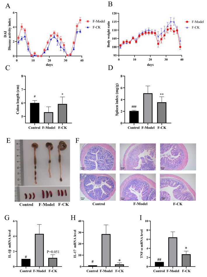
Chronic DSS treatment caused a reduction in body weight and an increase in DAI, which was reversed after being treated with GC-K (Fig. 1A). The body weight showed a slight increase with GC-K treatment (Fig. 1B). GC-K improved colon shortening, reduced spleen weight, and exhibited less immune cell infiltration and tissue damage by comparing with DSS Group (Fig. 1C–F).
Fig. 1.
GC-K alleviated DSS-induced chronic colitis. A Disease Activity Index during mouse model development; B Relative bodyweights during mouse model development; C Colon length in each group; D Spleen weight in each group (n = 10, 11). E Representative colon and spleen pictures from each group. F Representative H&E staining of colon tissue sections from each group. (** p < 0.01, *** p < 0.001 and **** p < 0.0001 vs. model group; ### p < 0.001 and #### p < 0.0001 vs. model group.)
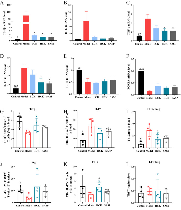
DSS-induced upregulations of pro-inflammatory cytokines such as IL-1β, IL-17a, TNF-α, and IL-6 were significantly downregulated by GC-K (Fig. 2A–D). In addition, the expression of anti-inflammatory cytokine IL-10 was reduced considerably by DSS (Fig. 2E), but neither GC-K nor SASP had any effect on the production of IL-10. GC-K down-regulated the expression of Foxp3 increased by DSS without significance (Fig. 2F). Th17 cells in the blood were significantly increased after modeling, while GC-K effectively reduced the quantities of Th17 cells and restored the Th17/Treg ratio (Fig. 2G–L). Meanwhile, the amounts of Treg cells in the blood and spleen were recovered with GC-K treatment.
Fig. 2.
GC-K decreased inflammatory cytokines production during DSS-induced colitis. Inflammatory factors IL-1β (A), IL-6 (B), TNF-α (C), IL-17a (D), IL-10 (E) and transcription factor Foxp3 (F) in mouse colon measured by RT-PCR (n = 4, 5). Flow cytometry analysis of Treg cells (CD3+CD4+CD25+Foxp3+) in blood (G) and spleen (J). Flow cytometry analysis of Th17 cells (CD3+CD4+IL-17a+) in blood (H) and spleen (K). Flow cytometry analysis of Th17/Treg cells in blood (I) and spleen (L). (* p < 0.1 vs. model group; # p < 0.1, ## p < 0.01, ### p < 0.001 and ### p < 0.0001 vs. model group.)
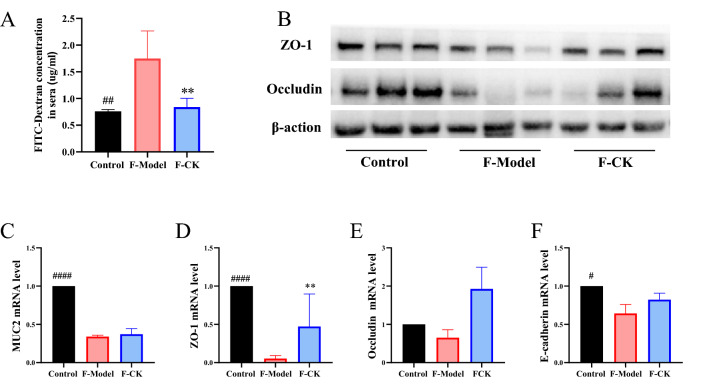
The concentration of FITC-dextran was increased in model group, while SASP and GC-K significantly reduced the concentration of dextrose in the plasma (Fig. 3A). Furthermore, GC-K reversed the expressions of Occludin, ZO-1, E-cadherin and MUC2, which implied that mucus layer thickness was restored (Fig. 3B–F).
Fig. 3.
GC-K improves intestinal barrier during DSS-induced colitis. Intestinal leakage measured by FITC-Dextran concentration in serum (A); Mucin MUC2 (B) and intestinal tight junction proteins E-cadherin (C), Occludin (D) and ZO-1 (E) in each group measured by RT-PCR (n = 4, 5). ZO-1 and Occludin in colon of different groups measured by Western blotting (F). (* p < 0.1, ** p < 0.01 and *** p < 0.001 vs. model group; # p < 0.1 and #### p < 0.0001 vs. model group.)
Alterations of the gut microbiome by GC-K treatment
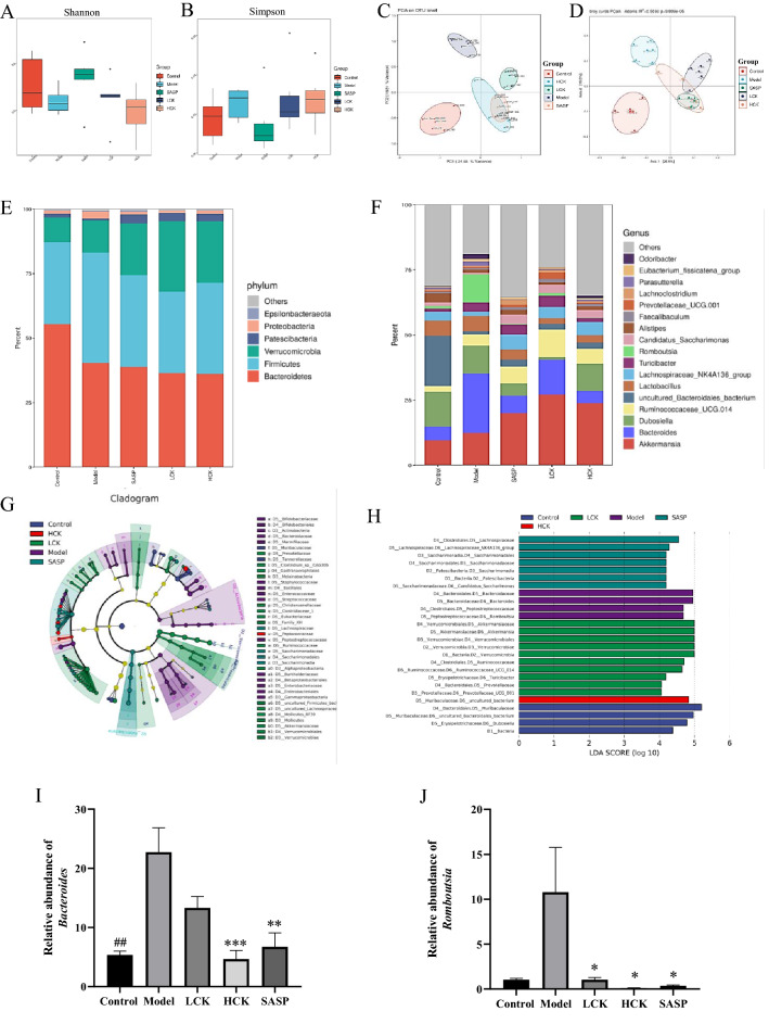
The α-diversity showed no significant difference among all groups (Fig. 4A, B). The β-diversity revealed distinct clustering of gut bacteria for each group (Fig. 4C, D). The results indicated that the profiles of gut microbiota were significantly different among each group.
Fig. 4.
16S rDNA sequencing revealed altered microbiota composition after GC-K treatment (n = 6). The α-diversity of each group measured by Shannon (A) and Simpson (B) index. PCA score plot analysis based on the relative abundance of OTUs (C) and bray curtis-based PCoA analysis (D) were used to evaluate the β-diversity of each group. Relative abundance of microbial taxa was determined at the phylum (E) and genus (F) level. Taxonomic cladogram obtained (G) and linear discriminant analysis score (H) from linear discriminant analysis effect size analysis of 16S rRNA sequences. Relative abundance of Bacteroides (I) and Rombsia (J) in each group. (* p < 0.1, ** p < 0.01 and *** p < 0.001 vs. model group; ## p < 0.1 vs. model group.)
Taxonomic histograms of gut bacteria on phylum and genus levels were shown in Fig. 4E, F. The phylum analysis revealed that GC-K increased the relative abundances of Verrucomicrobia and Patescibacteria and decreased Proteobacteria in DSS-induced colitis mice (Fig. 4F and Additional file 1: Fig. S3). In contrast, GC-K significantly increased the relative lots of Akkermansia, Candidatus_Saccharimonas, and Ruminococcaceae_UCG-014, and decreased Bacteroides and Rumboutsia in genus levels (Additional file 1: Fig. S4). The biomarkers for the Model group cluster were Bacteroides and Rumboutsia, while the biomarkers of the LCK group cluster were Akkermansia, Ruminococcaceae_UCG-014 and Turicibacter (Fig. 4G and H). The results showed that GC-K ameliorated gut microbiota dysbiosis in DSS-induced mice by significantly decreasing the abundance of Bacteroides.
To examine whether GC-K directly suppressed the growth of Bacteroides in vitro, the growth curves of 7 strains of Bacteroides co-incubated with GC-K were monitored (Additional file 1: Fig. S5). GC-K directly suppressed the growth of B. vulgatus in vitro, but had no significant effects on other strains of Bacteroides.
The transplant of GC-K-induced microbiota relieved colitis
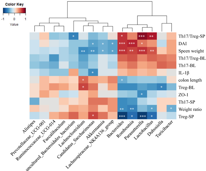
As shown in Fig. 5, a correlation map was constructed to discriminate the specific bacteria correlated with colitis disease indicators. Higher correlation scores indicated that the GC-K-driven gut bacteria potentially reversed colitis disease indicators.
Fig. 5.
Spearman correlation analysis between top 15 Taxonomic of gut microbiota and evaluation indexes of inflammatory bowel disease. (* p < 0.1, ** p < 0.01 and *** p < 0.001 vs. model group.)
FMT significantly alleviated DSS-induced colitis and decreased immune cell infiltration in colon tissue, and inflammatory factors expression of IL-1β, TNF-α and IL-17a (Fig. 6). Similar to the donor mice, the quantities of Treg and Th17 cells in the spleen were decreased significantly with DSS administration and increased after treated with GC-K (Additional file 1: Fig. S7). Furthermore, the FITC-dextran leakage experiment confirmed that FMT protected gut leakage. In addition, the expressions of Occludin, ZO-1, MUC2, and E-cadherin were increased, which indicated intestinal barrier function was restored (Fig. 7).
Fig. 6.
FMT alleviated DSS-induced chronic colitis. A Disease Activity Index during mouse model development. B Relative bodyweights during mouse model development. C Colon length in each group. D Spleen weight in each group (n = 6–9). E Representative colon and spleen pictures from each group. F Representative H&E staining of colon tissue sections from each group. Inflammatory factors IL-1β (G), TNF-α (H) and IL-17a (I) in mouse colon measured by RT-PCR, n=4. (* p < 0.1 and ** p < 0.01 vs. model group; # p < 0.1, ## p < 0.01 ### p < 0.001 vs. model group.)
Fig. 7.
FMT improves intestinal barrier during DSS-induced colitis. Intestinal leakage measured by FITC-Dextran concentration in serum (A) (n = 4). ZO-1 and Occludin in colon of different groups measured by Western blotting (B) Mucin MUC2 (C) and intestinal tight junction proteins ZO-1 (D), Occludin (F) and E-cadherin (G) in each group measured by RT-PCR (n = 3). (* p < 0.1, ** p < 0.01, *** p < 0.001 and **** p < 0.0001 vs. model group; ## p < 0.01 vs. model group.)
Discussion
Gut microbiota plays a vital role in maintaining body health [18]. Numerous studies have demonstrated that the gut microbiota is correlated to the occurrence and development of IBD [19], and the abundance and diversity of the gut microbiota of UC patients are decreased [20, 21]. Therefore, the regulation of gut microbiota has become a new therapeutic strategy of colitis.
GC-K is the primary metabolite of the protopanaxadiol ginsenosides bio-converted by gut microbiota [10, 11, 22], which plays a beneficial role in colitis [12]. Recently, natural products have been discovered to interplay with gut microbiota in vivo [23, 24]. Moreover, our previous work has confirmed GC-K suppressed the tumor growth of AOM/DSS-induced colitis-associated CRC via regulating gut microbiota [9]. Thus, the purpose of this work was to determine the role of gut microbiota in the treatment of UC with GC-K.
GC-K relieved the symptoms of weight loss and colon length shortening in DSS-induced mice. Compared with SASP, GC-K has advantages in weight and DAI scores. In colonic histopathology, GC-K was observed to reduce histologic inflammation. In addition, GC-K significantly decreased the mRNA expression of inflammatory cytokines (TNF-α, IL-6, IL-1β, and IL-17a) in colon tissue. The intestinal mucosal barrier could isolate the intestinal lumen from the environment to prevent the invasion of bacteria and toxic substances [25]. Still, an increase in proinflammatory cytokines further damages intestinal mucosal barrier function to make colitis worse [26, 27]. Occludin, ZO-1, and E-cadherin are crucial for connecting individual epithelial cells and maintaining the integrity of the epithelium [25]. The intestinal mucus layer is a protective gel-like substance covering the surface of the intestinal mucosa, which is the first barrier in the intestinal lumen [28]. Our experiment showed that DSS-induced colitis decreased the mRNA expressions of Occludin, ZO-1, E-cadherin, and MUC2 in colon tissue, which could be reversed by GC-K treatment.
According to 16S rRNA sequencing analysis, lower abundances of Lachnospiraceae_NK4A136_group and Candidatus_Saccharimonas and higher abundances of Bacteroides, Romboutsia, and Turicibacter were observed in the DSS group than in the control group, which were consistent with previous reports [29, 30]. GC-K reversed the intestinal dysbacteriosis caused by DSS. In addition, the abundance of Bacteroides and Romboutsia in GC-K significantly decreased. Spearman correlation analysis showed that Bacteroides mainly was correlated with the disease indicators of UC. In vitro, co-incubation experiments showed that GC-K significantly inhibited the growth of B. vulgatus. Studies have shown that the abundance of B. vulgatus in UC patients is increased substantially, and the proteases from B. vulgatus could be involved in UC pathogenesis [8]. The above results indicate that GC-K cotrolled the structure and abundance of gut bacteria and reduced the quantity of Bacteroides. However, the detailed mechanisms of GC-K regulated Bacteroides spp. remain unclear and need further investigation.
Finally, to further explore whether GC-K relieved colitis by modulating gut microbiota, we used FMT to verify the anti-colitis of GC-K in the DSS model. Before FMT, the pseudo-sterile mice were constructed with antibiotics, since gut microbiota may lead to colonization resistance. FMT relieved colonic inflammation and decreased intestinal permeability, which reproduced the anti-colitis effects of GC-K. Collectively, our data supported GC-K ameliorated colitis via regulating gut microbiota.
Conclusion
Our study evidenced that the anti-colitis effect of GC-K was related to gut microbiota, which was modulated by GC-K. Fecal microbiota transplantation experiments demonstrated that GC-K-driven gut microbiota significantly relieved DSS-induced colitis. In conclusion, GC-K showed anti-colitis effects via regulating gut microbiota, but the suitable mechanism needs further study.
Supplementary Information
Additional file 1: Fig. S1. The animal experimental protocol. DSS, dextran sulfate sodium; GC-K, ginsenoside compound K. Fig. S2. The frequency of Foxp3 + Treg cells among the CD4 + T cells (CD3 + CD4 + CD25 + cells) in spleen(A) and blood(B) of mice detected by flow cytometry; The frequency of IL-17a + Th17 cells among the CD4 + T cells (CD3 + CD4 + cells) in spleen(C) and blood(D) of mice detected by flow cytometry (n = 4, 5). Fig. S3. The relative abundance of the top 5 species at the phylum level in each group. Relative abundance of Bacteroidetes(a), Firmicutes(b), Verrucomicrobia(c), Patescibacteria(d) and Proteobacteria(e) in each group (n = 6). (* p < 0.1 and ** p < 0.01 vs. model group.) Fig. S4. Relative abundance of Akkermansia(a), Dubosiella(b), Lachnospiraceae_NK4A136(c), Ruminococcaceae_UCG-014(d), Turicibacter(e) and Candidatus_Saccharimonas(f) in each group (n = 6). (* p < 0.1 and ** p < 0.01 vs. model group.) Fig. S5 Effects of GC-K on the growth of representative strains in Bacteroides. (*** p < 0.001 vs. model group.) Fig. S6. Fecal microbial DNA concentration of mice before and after antibiotic administration. (** p < 0.01 vs. model group.) Fig. S7. Flow cytometry analysis of Treg cells (CD3 + CD4 + CD25 + Foxp3 +) and Th17 cells (CD3 + CD4 + IL-17a +) in spleen of mice in FMT group (n = 4). (* p < 0.1 and **** p < 0.0001 vs. model group; ### p < 0.001 vs. model group.)
Additional file 2: Table S1. The primer sequences used in real-time qPCR assays in colonic tissue.
Acknowledgements
Not applicable.
Abbreviations
- 16S rRNA
16S ribosomal RNA
- GC-K
Ginsenoside compound K
- LDA
Linear discriminative analysis
- LEfSe
The linear discriminative analysis effect size
- PCoA
Principal component analysis
- PC
Principal component
- RT-PCR
Real-time polymerase chain reaction
- QIIME
Quantitative Insights Into Microbial Ecology
Author contributions
WH designed the experiments; LW and LW participated in the experiments and analyzed the 16S rRNA sequencing data; MC, LW, LS, WZ, and FBT provided the technical support and advices for the study; LW and WH wrote the manuscript. All authors read and approved the final manuscript.
Funding
This research was supported by the National Natural Scientific Foundation of China (82074000, 81903784), the Hunan Provincial Natural Science Foundation of China (2020JJ4878), the Scientific Research Project of Department of Education of Hunan Province (20K136), Traditional Chinese medicine research project of Hunan Province (2021034).
Availability of data and materials
The research data generated from this study are included within the article and additional files.
Declarations
Ethics approval and consent to participate
Not applicable.
Consent for publication
Not applicable.
Competing interests
The authors declare that they have no competing interests.
Footnotes
Publisher's Note
Springer Nature remains neutral with regard to jurisdictional claims in published maps and institutional affiliations.
Contributor Information
Feng-Bo Tan, Email: fengbotan@csu.edu.cn.
Wei-Hua Huang, Email: endeavor34852@csu.edu.cn.
References
- 1.Singh V, Yeoh BS, Walker RE, Xiao X, Saha P, Golonka RM, Cai J, Bretin ACA, Cheng X, Liu Q, et al. Microbiota fermentation-NLRP3 axis shapes the impact of dietary fibres on intestinal inflammation. Gut. 2019;68(10):1801–1812. doi: 10.1136/gutjnl-2018-316250. [DOI] [PubMed] [Google Scholar]
- 2.Rangan P, Choi I, Wei M, Navarrete G, Guen E, Brandhorst S, Enyati N, Pasia G, Maesincee D, Ocon V, et al. Fasting-mimicking diet modulates microbiota and promotes intestinal regeneration to reduce inflammatory bowel disease pathology. Cell Rep. 2019;26(10):2704–2719. doi: 10.1016/j.celrep.2019.02.019. [DOI] [PMC free article] [PubMed] [Google Scholar]
- 3.Caruso R, Lo BC, Núñez G. Host–microbiota interactions in inflammatory bowel disease. Nat Rew Immunol. 2020;2020(7):411–426. doi: 10.1038/s41577-019-0268-7. [DOI] [PubMed] [Google Scholar]
- 4.Kühn R, Löhler J, Rennick D, Rajewsky K. Interleukin-10-deficient mice develop chronic enterocolitis. Cell. 1993;75(2):263–274. doi: 10.1016/0092-8674(93)80068-P. [DOI] [PubMed] [Google Scholar]
- 5.Zhai R, Xue X, Zhang L, Yang X, Zhao L, Zhang C. Strain-specific anti-inflammatory properties of two Akkermansia muciniphila strains on chronic colitis in mice. Front Cell Infect Microbiol. 2019;9:239. doi: 10.3389/fcimb.2019.00239. [DOI] [PMC free article] [PubMed] [Google Scholar]
- 6.Wang L, Tang L, Feng Y, Zhao S, Han M, Zhang C, Yuan G, Zhu J, Cao S, Wu Q, et al. A purified membrane protein from Akkermansia muciniphila or the pasteurised bacterium blunts colitis associated tumourigenesis by modulation of CD8(+) T cells in mice. Gut. 2020;69:1988–1997. doi: 10.1136/gutjnl-2019-320105. [DOI] [PMC free article] [PubMed] [Google Scholar]
- 7.Paredes-Sabja D, Kang C-S, Ban M, Choi E-J, Moon H-G, Jeon J-S, Kim D-K, Park S-K, Jeon SG, Roh T-Y, et al. Extracellular vesicles derived from gut microbiota, especially Akkermansia muciniphila, protect the progression of dextran sulfate sodium-induced colitis. PLoS ONE. 2013;8(10):e76520. doi: 10.1371/journal.pone.0076520. [DOI] [PMC free article] [PubMed] [Google Scholar]
- 8.Mills RH, Dulai PS, Vazquez-Baeza Y, Sauceda C, Daniel N, Gerner RR, Batachari LE, Malfavon M, Zhu Q, Weldon K, et al. Multi-omics analyses of the ulcerative colitis gut microbiome link Bacteroides vulgatus proteases with disease severity. Nat Microbiol. 2022;7(2):262–276. doi: 10.1038/s41564-021-01050-3. [DOI] [PMC free article] [PubMed] [Google Scholar]
- 9.Shao L, Guo YP, Wang L, Chen MY, Zhang W, Deng S, Huang WH. Effects of ginsenoside compound K on colitis-associated colorectal cancer and gut microbiota profiles in mice. Ann Transl Med. 2022;10(7):408–408. doi: 10.21037/atm-22-793. [DOI] [PMC free article] [PubMed] [Google Scholar]
- 10.Guo YP, Shao L, Chen MY, Qiao RF, Zhang W, Yuan JB, Huang WH. In vivo metabolic profiles of Panax notoginseng saponins mediated by gut microbiota in rats. J Agric Food Chem. 2020;68(25):6835–6844. doi: 10.1021/acs.jafc.0c01857. [DOI] [PubMed] [Google Scholar]
- 11.Chen MY, Shao L, Zhang W, Wang CZ, Zhou HH, Huang WH, Yuan CS. Metabolic analysis of Panax notoginseng saponins with gut microbiota-mediated biotransformation by HPLC-DAD-Q-TOF-MS/MS. J Pharm Biomed Anal. 2018;150:199–207. doi: 10.1016/j.jpba.2017.12.011. [DOI] [PubMed] [Google Scholar]
- 12.Li J, Zhong W, Wang W, Hu S, Yuan J, Zhang B, Hu T, Song G. Ginsenoside metabolite compound K promotes recovery of dextran sulfate sodium-induced colitis and inhibits inflammatory responses by suppressing NF-kappaB activation. PLoS ONE. 2014;9(2):e87810. doi: 10.1371/journal.pone.0087810. [DOI] [PMC free article] [PubMed] [Google Scholar]
- 13.Paek IB, Moon Y, Kim J, Ji HY, Kim SA, Sohn DH, Kim JB, Lee HS. Pharmacokinetics of a ginseng saponin metabolite compound K in rats. Biopharm Drug Dispos. 2006;27(1):39–45. doi: 10.1002/bdd.481. [DOI] [PubMed] [Google Scholar]
- 14.Guo YP, Shao L, Wang L, Chen MY, Zhang W, Huang WH. Bioconversion variation of ginsenoside CK mediated by human gut microbiota from healthy volunteers and colorectal cancer patients. Chin Med. 2021;16(1):28. doi: 10.1186/s13020-021-00436-z. [DOI] [PMC free article] [PubMed] [Google Scholar]
- 15.Fan L, Qi Y, Qu S, Chen X, Li A, Hendi M, Xu C, Wang L, Hou T, Si J, et al. B. adolescentis ameliorates chronic colitis by regulating Treg/Th2 response and gut microbiota remodeling. Gut Microbes. 2021;13(1):1–17. doi: 10.1080/19490976.2020.1826746. [DOI] [PMC free article] [PubMed] [Google Scholar]
- 16.Krych L, Kot W, Bendtsen KMS, Hansen AK, Vogensen FK, Nielsen DS. Have you tried spermine? A rapid and cost-effective method to eliminate dextran sodium sulfate inhibition of PCR and RT-PCR. J Microbiol Meth. 2018;144:1–7. doi: 10.1016/j.mimet.2017.10.015. [DOI] [PubMed] [Google Scholar]
- 17.Wu J, Wei Z, Cheng P, Qian C, Xu F, Yang Y, Wang A, Chen W, Sun Z, Lu Y. Rhein modulates host purine metabolism in intestine through gut microbiota and ameliorates experimental colitis. Theranostics. 2020;10(23):10665–10679. doi: 10.7150/thno.43528. [DOI] [PMC free article] [PubMed] [Google Scholar]
- 18.Abdollahi-Roodsaz S, Abramson SB, Scher JU. The metabolic role of the gut microbiota in health and rheumatic disease: mechanisms and interventions. Nat Rev Rheumatol. 2016;12(8):446–455. doi: 10.1038/nrrheum.2016.68. [DOI] [PubMed] [Google Scholar]
- 19.Sellon RK, Tonkonogy S, Schultz M, Dieleman LA, Grenther W, Balish E, Rennick DM, Sartor RB. Resident enteric bacteria are necessary for development of spontaneous colitis and immune system activation in interleukin-10-deficient mice. Infect Immun. 1998;66(11):5224–5231. doi: 10.1128/IAI.66.11.5224-5231.1998. [DOI] [PMC free article] [PubMed] [Google Scholar]
- 20.Pittayanon R, Lau JT, Leontiadis GI, Tse F, Yuan Y, Surette M, Moayyedi P. Differences in gut microbiota in patients with vs without inflammatory bowel diseases: a systematic review. Gastroenterology. 2020;158(4):930–946. doi: 10.1053/j.gastro.2019.11.294. [DOI] [PubMed] [Google Scholar]
- 21.Lavelle A, Sokol H. Gut microbiota-derived metabolites as key actors in inflammatory bowel disease. Nat Rev Gastroenterol Hepatol. 2020;2020(4):223–237. doi: 10.1038/s41575-019-0258-z. [DOI] [PubMed] [Google Scholar]
- 22.Zhu D, Zhou Q, Li H, Li S, Dong Z, Li D, Zhang W. Pharmacokinetic characteristics of steamed notoginseng by an efficient LC−MS/MS method for simultaneously quantifying twenty-three triterpenoids. J Agric Food Chem. 2018;66(30):8187–8198. doi: 10.1021/acs.jafc.8b03169. [DOI] [PubMed] [Google Scholar]
- 23.Chen L, Chen M-Y, Shao L, Zhang W, Rao T, Zhou H-H, Huang WH. Panax notoginseng saponins prevent colitis-associated colorectal cancer development: the role of gut microbiota. Chin J Nat Med. 2020;18(7):500–507. doi: 10.1016/S1875-5364(20)30060-1. [DOI] [PubMed] [Google Scholar]
- 24.Xu Y, Wang N, Tan HY, Li S, Zhang C, Zhang Z, Feng Y. Panax notoginseng saponins modulate the gut microbiota to promote thermogenesis and beige adipocyte reconstruction via leptin-mediated AMPKalpha/STAT3 signaling in diet-induced obesity. Theranostics. 2020;10(24):11302–11323. doi: 10.7150/thno.47746. [DOI] [PMC free article] [PubMed] [Google Scholar]
- 25.Luis AS, Jin C, Pereira GV, Glowacki RWP, Gugel SR, Singh S, Byrne DP, Pudlo NA, London JA, Basle A, et al. A single sulfatase is required to access colonic mucin by a gut bacterium. Nature. 2021;598(7880):332–337. doi: 10.1038/s41586-021-03967-5. [DOI] [PMC free article] [PubMed] [Google Scholar]
- 26.Bu F, Ding Y, Chen T, Wang Q, Wang R, Zhou JY, Jiang F, Zhang D, Xu M, Shi G, et al. Total flavone of Abelmoschus Manihot improves colitis by promoting the growth of Akkermansia in mice. Sci Rep. 2021;11(1):20787. doi: 10.1038/s41598-021-00070-7. [DOI] [PMC free article] [PubMed] [Google Scholar]
- 27.Machiels K, Joossens M, Sabino J, De Preter V, Arijs I, Eeckhaut V, Ballet V, Claes K, Van Immerseel F, Verbeke K, et al. A decrease of the butyrate-producing species Roseburia hominis and Faecalibacterium prausnitzii defines dysbiosis in patients with ulcerative colitis. Gut. 2014;63(8):1275–1283. doi: 10.1136/gutjnl-2013-304833. [DOI] [PubMed] [Google Scholar]
- 28.Kim YS, Ho SB. Intestinal goblet cells and mucins in health and disease: recent insights and progress. Curr Gastroenterol Rep. 2010;12(5):319–330. doi: 10.1007/s11894-010-0131-2. [DOI] [PMC free article] [PubMed] [Google Scholar]
- 29.Wexler HM. Bacteroides: the good, the bad, and the nitty-gritty. Clin Microbiol Rev. 2007;20(4):593–621. doi: 10.1128/CMR.00008-07. [DOI] [PMC free article] [PubMed] [Google Scholar]
- 30.Liu S, Zhao W, Lan P, Mou X. The microbiome in inflammatory bowel diseases: from pathogenesis to therapy. Protein Cell. 2020;12:331–345. doi: 10.1007/s13238-020-00745-3. [DOI] [PMC free article] [PubMed] [Google Scholar]
Associated Data
This section collects any data citations, data availability statements, or supplementary materials included in this article.
Supplementary Materials
Additional file 1: Fig. S1. The animal experimental protocol. DSS, dextran sulfate sodium; GC-K, ginsenoside compound K. Fig. S2. The frequency of Foxp3 + Treg cells among the CD4 + T cells (CD3 + CD4 + CD25 + cells) in spleen(A) and blood(B) of mice detected by flow cytometry; The frequency of IL-17a + Th17 cells among the CD4 + T cells (CD3 + CD4 + cells) in spleen(C) and blood(D) of mice detected by flow cytometry (n = 4, 5). Fig. S3. The relative abundance of the top 5 species at the phylum level in each group. Relative abundance of Bacteroidetes(a), Firmicutes(b), Verrucomicrobia(c), Patescibacteria(d) and Proteobacteria(e) in each group (n = 6). (* p < 0.1 and ** p < 0.01 vs. model group.) Fig. S4. Relative abundance of Akkermansia(a), Dubosiella(b), Lachnospiraceae_NK4A136(c), Ruminococcaceae_UCG-014(d), Turicibacter(e) and Candidatus_Saccharimonas(f) in each group (n = 6). (* p < 0.1 and ** p < 0.01 vs. model group.) Fig. S5 Effects of GC-K on the growth of representative strains in Bacteroides. (*** p < 0.001 vs. model group.) Fig. S6. Fecal microbial DNA concentration of mice before and after antibiotic administration. (** p < 0.01 vs. model group.) Fig. S7. Flow cytometry analysis of Treg cells (CD3 + CD4 + CD25 + Foxp3 +) and Th17 cells (CD3 + CD4 + IL-17a +) in spleen of mice in FMT group (n = 4). (* p < 0.1 and **** p < 0.0001 vs. model group; ### p < 0.001 vs. model group.)
Additional file 2: Table S1. The primer sequences used in real-time qPCR assays in colonic tissue.
Data Availability Statement
The research data generated from this study are included within the article and additional files.


