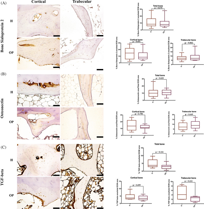
FIGURE 8.

Immunohistochemical staining of (A) bone sialoprotein 2, (B) Osteonectin and (C) TGF‐beta in healthy (H) and osteoporotic (OP) tissues (×20 magnification, 50 μm scale bar). (A) Immunohistochemical staining images and semi‐quantification of bone sialoprotein 2; (B) immunohistochemical staining images and semi‐quantification of Osteonectin; (C) immunohistochemical staining images and semi‐quantification of TGF‐beta
