



Abstract
Neurovascular 4D-Flow MRI has emerged as a powerful tool for comprehensive cerebrovascular hemodynamic characterization. Clinical studies in at risk populations such as aging adults indicate hemodynamic markers can be confounded by motion-induced bias. This study develops and characterizes a high fidelity 3D self-navigation approach for retrospective rigid motion correction of neurovascular 4D-Flow data. A 3D radial trajectory with pseudorandom ordering was combined with a multi-resolution low rank regularization approach to enable high spatiotemporal resolution self-navigators from extremely undersampled data. Phantom and volunteer experiments were performed at 3.0T to evaluate the ability to correct for different amounts of induced motions. In addition, the approach was applied to clinical-research exams from ongoing aging studies to characterize performance in the clinical setting. Simulations, phantom and volunteer experiments with motion correction produced images with increased vessel conspicuity, reduced image blurring, and decreased variability in quantitative measures. Clinical exams revealed significant changes in hemodynamic parameters including blood flow rates, flow pulsatility index, and lumen areas after motion correction in probed cerebral arteries (Flow: P<0.001 Lt ICA, P=0.002 Rt ICA, P=0.004 Lt MCA, P=0.004 Rt MCA; Area: P<0.001 Lt ICA, P<0.001 Rt ICA, P=0.004 Lt MCA, P=0.004 Rt MCA; flow pulsatility index: P=0.042 Rt ICA, P=0.002 Lt MCA). Motion induced bias can lead to significant overestimation of hemodynamic markers in cerebral arteries. The proposed method reduces measurement bias from rigid motion in neurovascular 4D-Flow MRI in challenging populations such as aging adults.
Keywords: 4D Flow MRI, head motion, bias, motion correction, quality control, angiography
1. Introduction
Technological advances in systems hardware and software have facilitated the translation of quantitative magnetic resonance imaging (qMRI) approaches from the research realm into clinical settings. The adoption of qMRI methods is driving novel characterization of disease; however, various studies indicate a high number of published neuroscientific and medicine literature may not be reproducible (Avinun et al., 2020; Keuken et al., 2017; Marek et al., 2022; Poldrack et al., 2017). For example, cortical thinning measures from high-resolution T1 imaging have been used to study distinct etiology driven patterns of neurodegeneration (Vidal-Pineiro et al., 2020). However, recent studies have shown morphometry measures can be significantly biased by subtle motion confounders that are unnoticeable by visual inspection (Reuter et al., 2015). Therefore, it is of utmost importance to develop robust qMRI methods with low measurement bias and variability even in the presence of confound effects such as patient motion.
Recently, intracranial four-dimensional (4D) Flow MRI has emerged as a powerful clinical tool for the characterization of cerebrovascular health (Aristova et al., 2022; Morgan et al., 2021; Turski et al., 2016; Wåhlin et al., 2022; Youn and Lee, 2022). With whole brain coverage, neurovascular 4D-Flow MRI can be used to probe vessels directly and quantify blood velocities in the brains feeding arteries and draining veins. 4D-Flow MRI has been utilized to understand vascular contributions to dementia (Rivera-Rivera et al., 2021b), to characterize blood flow patterns within aneurysms (Gottwald et al., 2020) and arteriovenous malformations (Aristova et al., 2019), and to quantify flow features related to atherosclerosis (Vali et al., 2019). Further, emerging paradigms are being developed to provide unique measures of low frequency flow oscillations (LFOs) (Rivera-Rivera et al., 2020), pulse wave velocity measures of arterial stiffness (Björnfot et al., 2021; Rivera-Rivera et al., 2021a; Vikner et al., 2021), and flow turbulence and disorder (Li et al., 2018; Schnell et al., 2017). These new applications and the need to characterize velocities in small vessels require 4D-Flow methods with high spatial and temporal resolution, which increases the required scan time and hence the sensitivity to patient motion. Therefore, it can be difficult to obtain accurate 4D-Flow metrics in vulnerable populations such as in geriatrics and pediatrics. Patient motion is of particular concern as motion itself may be correlated to age and disease, confounding effects of interest (Hausman et al., 2022; Pardoe et al., 2016).
A number of prospective and retrospective motion correction strategies have been devised to improve brain imaging quality (Godenschweger et al., 2016; Maknojia et al., 2019). Studies have identified motion correction to not only improve subjective image quality but also reduce motion-induced bias and variance in derived quantitative biomarkers (Kecskemeti et al., 2021, 2018; Tisdall et al., 2016). While prior work has demonstrated an approximately 30% error in derived flow due to respiratory motion in cardiothoracic 4D-Flow applications (Dyverfeldt and Ebbers, 2017), the effect and correction of bulk motion in neurovascular 4D-Flow MRI has not been previously studied.
One way to mitigate motion effects in neurovascular 4D-Flow MRI is to leverage k-space trajectories that are robust to motion, such as 3D radial sampling. Radial trajectories are inherently less sensitive to motion, producing image blurring and noise like artifact rather than coherent ghosting (Glover and Pauly, 1992). Radial trajectories also provide self-navigation for motion correction and have been used for motion corrected T1-weighted and quantitative T1 imaging (Anderson et al., 2013; Kecskemeti et al., 2018). In one such paradigm for motion correction, lower resolution images are reconstructed with high temporal binning and used to correct k-space data. While this strategy is compatible with 4D-Flow MRI when collected with 3D radial trajectories, it is hindered by the inherently longer TRs (~50%) and required time for velocity encodes. This results in an approximately 6x longer effective TR and hence lower frame rate for image navigators. However, recent developments in non-Cartesian constrained image reconstructions have demonstrated the feasibility of generating high temporal resolution images from very few measurements. To overcome the inherently reduced sampling efficiency of 4D-Flow MRI and achieve image based motion navigation with sufficient spatial and temporal resolution, this study leveraged pseudorandomly ordered 3D radials with a multi-scale low-rank (MSLR) reconstruction approach called “Extreme MRI” (Ong et al., 2020). Extreme MRI combines a compressed MSLR matrix factorization with a stochastic gradient descent approach for rapid, memory-efficient navigator image reconstruction. Extreme MRI has yet to be applied to image brain motion. We hypothesized that motion corrected neurovascular 4D-Flow data would portray improved vessel conspicuity and reduced measurement variability in cerebrovascular quantitative hemodynamic parameters. The approach was characterized and tested in simulations, flow phantom and controlled motion experiments in healthy volunteers. Finally, it was applied to artifact corrupted clinical exams from ongoing aging studies.
2. Materials and methods
The University of Wisconsin Institutional Review Board approved all study procedures and protocols for volunteer and clinical studies following the policies and guidance established by the campus Human Research Protection Program (HRPP). All study procedures were performed according to the Declaration of Helsinki, including obtaining written informed consent from each participant.
2.1. Imaging protocol
For all experiments and clinical studies 4D-Flow data were acquired on a 3.0T system (SIGNA Premier, GE Healthcare) using a radial acquisition with pseudorandom ordering where all velocity encodings are collected for a single line in k-space before moving on to the next (Johnson et al., 2008; Markl et al., 2012). Phantom experiments used a 30-channel surface coil (AIR, GE Healthcare), while human studies were conducted using a 48-channel head-coil (GE Healthcare). Scan parameters included: acquisition imaging volume = 22x22x16 cm3, TR/TE = 8.6/2.5 ms, number of projections ~11,000, scan time ~5.6 min, acquired spatial resolution = 0.7 mm isotropic, flip angle = 8°, bandwidth = 250 kHz, and Venc = 80 cm/s.
2.2. Motion correction
To estimate rigid head motion in neurovascular 4D-Flow MRI data, 3D radial sampling with pseudorandom ordering (interleaved) was leveraged with a multi-scale low rank (MSLR) reconstruction (Extreme MRI) to generate high fidelity 3D self-navigators (Figure 1). These navigators were used to estimate translation and rotation parameters throughout the scan, and subsequently used to correct k-space data. Navigator images were retrospectively reconstructed using GPU accelerated code in Python (SigPy (Ong et al., 2022)). Coil sensitivity maps were estimated with JSENSE (Ying and Sheng, 2007) from the composite data without any form of binning. Extreme MRI was performed to reconstruct a high temporal resolution navigator from one of the flow encodings (Ong et al., 2020). Extreme MRI incorporates stochastic gradient descent and a multi-scale low rank representation of the image, which is directly estimated from k-space. Multi-scale low rank reconstructions were performed with overlapping block sizes (128, 96, 64, 48) and an empirically tuned regularization parameter (λ = 1e-8). High-frame rate navigator images were reconstructed into 256 frames from extremely undersampled data (~43 projections per frame) for a temporal resolution of 1.3s and 1.4 mm isotropic spatial resolution in phantom, volunteer, and clinical exams. To compare quality and fidelity of navigators obtained from Extreme MRI reconstructions, navigators were also reconstructed using more traditional approaches including iterative SENSE (Pruessmann et al., 2001) and direct non-uniform fast Fourier transform (NUFFT) (Griswold et al., 2000) using data from a single volunteer.
Figure 1.

Framework for rigid motion corrected neurovascular 4D-Flow MRI. Randomized 3D radial data is utilized for a time-resolved (ungated) reconstruction with multi-scale low rank (MSLR) regularization support (Extreme MRI) to generate high fidelity 3D navigators from extremely undersampled data (~43 projections per frame). Navigators are registered to obtain motion estimates. Finally, motion estimates are used to correct k-space data and reconstruct motion corrected complex images for each of the different velocity encodings that are combined into motion corrected velocity images.
To estimate translation and rotation parameters from navigators, iterative rigid registration was performed using a grid search initialization to avoid local minima. Grid search initialization was empirically tuned to find x, y, and z translations between −5 to 5 mm (15 equidistant values) that minimized a loss function. A masked weighted mean square error loss function was utilized, where the mask was selected to remove the excitation slab edge. Following grid search initialization, iterative interpolation based registration was utilized with an optimized stochastic gradient descent (ADAM) optimization (e.g. learning rate 1e-4, loss threshold 1e-12, 3000 epochs). Registration was performed using the first navigator image as a reference frame, producing 3D rotation angles and translations for each frame.
Following motion estimation, motion correction was applied in k-space. First, the set of rotation angles were normalized to have a zero-mean rotation. This was performed to minimize the potential effects from velocity encoding rotations, which were not corrected for due to the non-linear and computationally expensive reconstructions that would be required to solve directly for velocities. Using estimated translations and zero-mean rotations, k-space data was rigid motion corrected through multiplication by a phase term describing translations in x, y, and z and a rotation of the coordinate system. A theoretical formulism of the registration and correction procedure can be found in Supplementary Material 1. The motion correction and reconstruction code can be found at: https://github.com/uwmri/flow_recon. The 3D registration code was written by the authors using auxiliary functions from PyTorch 1.12.
In this study, results depict translations with respect to the scan bore coordinates e.g. z-axis aligns with B0 and Euler rotational angles of: Ψ (psi) angle of rotation around the z-axis, Φ (phi) angle of rotation around the y-axis, and θ (theta) angle of rotation around the x-axis.
2.3. Image reconstruction and post processing
For human studies cardiac triggers were collected for each subject from an MRI compatible photoplethysmogram (GE Healthcare) worn on the subject’s finger during the MRI exam. 4D-Flow exams were reconstructed to 0.7 mm isotropic spatial resolution and 20 cardiac phases (temporal resolution ~50 ms). For flow pump experiments with constant flow (see below), the data was not cardiac-gated and thus only time-average reconstructed. Reconstructions were performed using standard NUFFT and a complex coil combine (Walsh et al., 2000). Velocity images were corrected for background phase errors and phase aliasing (Loecher et al., 2016). Phantoms and cerebral arteries, including the left and right internal carotid arteries (ICAs) and middle cerebral arteries (MCAs) from in vivo imaging, were segmented automatically in a MATLAB-based tool (Mathworks, Natick, MA) (Schrauben et al., 2015). Automatic segmentation contoured regions of interest (ROIs) using a k-means clustering algorithm under the assumption that any cross section will contain a low-signal background and a vessel region. This approach extracts the vascular tree using a centerline process with local cross-sectional cut-planes automatically placed in every centerline point perpendicular to the axial direction of the vessel. Flow rates were estimated from the product of cross-sectional areas and velocities from planes automatically segmented. Flow pulsatility index (PI), a surrogate marker of vascular compliance (de Riva et al., 2012), was estimated from cardiac-resolved velocity data and defined as (Qmax – Qmin) / Qmean, where Qmax, Qmin, and Qmean are the max, min, and mean blood flow values of the cardiac waveform. In summary, the following hemodynamic markers were extracted from 4D-Flow data: blood flow rates, PI, and cross-sectional areas.
2.4. Simulation experiments
Simulations were performed to characterize the motion correction approach. A high quality time-average 4D-Flow MRI dataset (spatial resolution 0.7 mm3 isotropic) with no apparent motion artifacts was used to create a dynamic series of complex images with rotations applied to simulate potential motion. For each motion case, 100 images were generated for each orthogonal velocity direction. These images were generated from copies of the example data. To simulate continuous motion, a cosine function was used to impart rotational motion around the z-axis with maximum rotations of 5, 10, 20, and 30 degrees and period of 1108s. All rotated images also rotated the velocity field directions (i.e. rotated relative to the simulation coordinates). First, experiments were performed to evaluate the effect of referencing and the impact of velocity encoding rotations. Images were generated by temporally averaging the data and comparing to the ground truth velocity (1) without motion correction, (2) with motion correction using a reference frame (i.e. the 1st frame), and (3) with motion correction using a zero-mean rotation reference. The reference frame also corresponded to the frame at maximum angular displacement. This was chosen deliberately to highlight the benefits of zero-mean rotation reference. Voxel-wise correlations of vessel velocities were obtained for all comparisons. Following this, the motion corrupted simulated images were transformed to k-space and sampled using the original MRI dataset k-space radial trajectories. Motion correction was first applied and compared using known rotations. The full motion correction was then applied including motion estimates from navigator images (256 frames, 2.2s per frame). This full motion correction simulation was also tested for stepwise motions to study the effect of distinct motion patterns on estimated parameters. A stepwise motion was simulated by a transition from 0 to a maximum rotation at the midpoint frame of the time series. Finally, the full motion correction pipeline was applied to a non-motion static simulation to study bias effects that could be induced by the approach.
2.5. Phantom experiments
Physical phantom data were acquired to characterize the neurovascular 4D-Flow rigid motion correction approach. The phantom consisted of a straight silicone tube embedded in a water box connected to a flow pump located outside the scan room (CompuFlow 1000 MR, Shelly). The pump supplied constant flow (~10 mL/s) of blood mimicking fluid along the phantom circuit. Blood mimicking fluid consisting of a mixture of 60% glycerol and 40% distilled water was used for a viscosity similar to that of blood (3.5cP) (Summers et al., 2005). The phantom was placed on a rigid rectangular cradle on the scanner table. One distal point of the cradle was rigidly fixed to the scanner table (e.g. pivot point) and motion was manually induced by small lateral rotations about the pivot point (phantom schematic, Figure 2). Six 4D-Flow MRI scans were acquired with different instances and magnitudes of motion including: static scan (no motion), one-time motion, motion every 60s, 30s, 15s and continuous motion.
Figure 2.

Schematic of the phantom setup.
2.6. Induced motion experiments
To evaluate performance of the rigid motion correction algorithm in vivo, four healthy participants (mean age = 36 ± 4y, 2F) were scanned twice with a neurovascular 4D-Flow sequence during the presence and absence of induced bulk motion. During the first 4D-Flow scan, volunteers were instructed to hold still. For the second scan, volunteers were trained to perform a maneuver that induced bulk head motion by switching their legs from being bent at the knee to flat on the table. Volunteers alternated between supine and knee bending positions every 30 seconds throughout the scan duration and were instructed to perform the maneuver at comfortable speeds. Volunteers received a visual signal from the scanner operator every 30s to indicate the start of a change in position.
2.7. Clinical Data
In addition to simulation, phantom and induced motion studies, the neurovascular 4D-Flow rigid motion correction approach was tested in clinical data from ongoing neuroimaging studies on aging and dementia. 4D-Flow data from 11 human subjects were selected for retrospective rigid motion correction based on low image quality of an unknown origin flagged by data intake quality assurance processes. Subject demographics are summarized in Table 1. These data were collected by the Wisconsin Alzheimer’s Disease Research Center and Wisconsin Registry for Alzheimer’s Prevention studies (Johnson et al., 2018).
Table 1.
Clinical subject demographics (N=11).
| Age (years) | 70 ± 6 |
| Female (n, %) | 9, (82%) |
| Education (years) | 16 ± 2 |
| SBP (mmHg) | 133 ± 14 |
| DBP (mmHg) | 76 ± 8 |
| HR (bpm) | 66 ± 9 |
| BMI (kg/m2) | 29.7 ± 6.6 |
| Clinical Diagnosis | 2 mild cognitively impaired, 9 cognitively normal |
2.8. Statistical analysis
Hemodynamic derived metrics from 4D-Flow MRI data were compared between scans before and after motion correction. Differences in blood flow rates, PI (in vivo studies only) and cross-sectional areas were assessed using Wilcoxon signed-rank test. Measurement variance differences were assessed with Levene’s test. Statistical analysis was performed in MATLAB and P < 0.05 was set as the threshold for statistical significance. For voxel-wise comparisons images were rigidly registered and velocities rotated. Velocities were extracted using a vessel mask generated from the union of binary masks derived from angiograms of cases to be compared. Voxel-wise correlations from masked vessel velocities were characterized using Pearson’s correlation coefficients and least squared regression lines.
3. Results
3.1. Simulation experiments
Results from simulations are summarized in Figures 3 and 4, and Supplementary Figures 1, 2 and 3. Higher correlation and agreement between corrected velocities and ground truth were measured when time series were rotated to a zero-mean average rotation compared to rotating images to a specific reference frame (frame 1) (Supplementary Figure 1). Uncorrected motions led to high degree of disagreements with ground truth velocities, which was worse for larger motions (Supplementary Figures 1 and 2). Velocity corrections in k-space using the known motions led to reconstruction of velocity images that demonstrated high levels of agreement with ground truth (Supplementary Figure 2). When testing the proposed motion correction approach using the full pipeline including motion estimation, voxel-wise velocity correlation coefficients of 0.99 were measured for both continuous and stepwise motion corrected velocities (Figure 3). For corrections of rotational continuous and stepwise motions on the order of 5°, ~2% velocity disagreements were observed with ground truth. For large rotations of 30°, there was an increase in the velocity error with an underestimation of 5-6%. In comparing the motion estimates, stepwise motions were recovered with high degree of fidelity (Figure 4, right panel); however, continuous motions were underestimated (Figure 4, left panel). Translational motions were correctly estimated to be near zero; however, some small deviations were observed for both continuous and stepwise motion involving large rotations of 20° and 30° (Supplementary Figure 3). Finally, the static simulation processed by the motion correction approach resulted in velocity correlation coefficients of 1.00 and linear model fit of y = 1.00x+0.50 cm/s with respect to ground truth.
Figure 3.

Voxel-wise velocity plots with overlaid least-square regression lines and Pearson correlation coefficients of the simulation experiments comparing the motion corrected velocities to ground truth for continuous (left column) and stepwise (right column) motions with different levels of maximum rotations (rows). Overall, for rotational motions on the order of 5°, differences of ~2% were found between both continuous and stepwise motion corrected data when compared to ground truth velocities. Larger rotations led to increased velocity differences between corrected and ground truth datasets. After motion correction, large rotations on the order of 30° led to 5-6% differences in velocities when compared to ground truth.
Figure 4.

Simulated input and recovered rotational motions from self-navigation using the proposed method for both continuous and stepwise motions. For the continuous case, recovered motions were underestimated indicating limitations of the multi-scale low rank Extreme MRI navigator reconstruction. Excellent agreement was observed between input and recovered stepwise motions. These results indicate continuous motions can be more challenging to resolve, while stepwise motions can be recovered with higher fidelity.
3.2. Phantom experiments
Phantom experiments results are summarized in Figures 5, 6, 7 and Table 2 (averaged along length of tube). Manually induced phantom motion led to variable translations and rotations throughout the different experiments (Figure 5). Induced phantom motion led to blurring of the straight tube angiogram and velocity images (Figure 6, right panel). The worst blurring was observed when the phantom was moved once and every 60s, corresponding to the scans that experienced the largest measured translations and rotations (Figure 7). However, after rigid motion correction, image blurring was reduced in all moving scans (Figure 6, left panel).
Figure 5.

Translations and rotations measured using 3D-navigators in phantom experiments for each of the six 4D-Flow scans (static, motion once, every 60s, 30s, 15s and continuously). The magnitude of motion varied with maximal values of 3 mm and 3° in the scans where the phantom was moved once and every 60s. The largest translational motion was measured along the direction of the induced motion (lateral movement, x-axis) and minimal displacement in z as the phantom was never lifted from the table. For all moving scans motion started after ~40s of imaging by design.
Figure 6.

Motion corrected and uncorrected complex difference (CD) angiograms and velocity images (z-component) derived from phantom experiments with varying levels of induced motion (static, motion once, every 60s, 30s, 15s and continuous). Regions of interest (ROIs) were extracted from a location proximal to where induced motion originated, therefore representing cross-sections that experienced greatest motions for each of the scans. Substantial distortion (blurring) of the velocity field was observed for all uncorrected scans except the static scan. After motion correction image blurring was notably reduced for all moving scans. Motion correction of the static scan did not induce any noticeable bias on the velocity fields.
Figure 7.

Quantitative summary of flow rates and cross-sectional areas is shown for phantom experiments along the length of the straight tube. Distance shown is relative to the distal end of the phantom, where 0 is closest to one end of the phantom and 100mm is closer to the pivot point. Overall, motion correction of the static scan did not add a noticeable bias to flow and area measures. Static vs moving scan flow-rate differences were reduced for most motion frequencies after motion correction. Flow-rate standard deviations in moving scans were reduced after motion correction. All cross-sectional area differences and standard deviations between the static and moving scans were reduced after motion correction of the moving scans.
Table 2.
Quantitative flow rates and cross-sectional area values averaged along the length of the straight tube from 4D-Flow data before and after motion correction.
| Type of motion | Flow (mL/cycle) | Area (mm2) | ||
|---|---|---|---|---|
| No MC | MC | No MC | MC | |
| static | 9.4 ± 0.2 | 9.4 ± 0.2 | 26.7 ± 0.6 | 26.7 ± 0.7 |
| once | 10.1 ± 1.0 | 9.3 ± 0.3 | 32.2 ± 6.8 | 26.4 ± 0.7 |
| 60s | 9.6 ± 0.6 | 9.2 ± 0.3 | 30.2 ± 4.2 | 28.0 ± 1.2 |
| 30s | 9.5 ± 0.5 | 9.4 ± 0.2 | 30.2 ± 4.6 | 27.1 ± 0.6 |
| 15s | 9.3 ± 0.3 | 9.2 ± 0.2 | 28.7 ± 2.5 | 27.0 ± 0.7 |
| continuous | 9.1 ± 0.2 | 9.1 ± 0.2 | 28.8 ± 1.1 | 27.7 ± 0.8 |
Quantitative flow rates and cross-sectional area values measured along the length of the straight tube from 4D-Flow data are summarized in Figure 7 and Table 2. Overall, similar flow and area profiles were measured in the static scan before and after motion correction, while flow and area differences and measurement variance between the static scan and moving scans were reduced after motion correction in most cases.
3.3. Controlled motion experiments in volunteers
Volunteer experiments are summarized in Figures 8, 9, 10 and 11. As shown in Figures 8 and 9, the moving task induced varying levels of motion on each volunteer. Volunteer 3 displayed the largest motion (max translation 5.8 mm, max rotation 1.4°) while volunteer 4 the smallest (max translation 0.9 mm, max rotation 2.1°). On Figure 8, angiograms generated from 4D-Flow data acquired during the motion inducing task without motion correction displayed degraded image quality including increased vessel blurring and noise. The worst vessel depiction was observed in volunteer 3, which was also the volunteer with greatest motion (Figure 9). Overall, after motion correction image quality for all participants improved. Further, motion correction improved the correlation and agreement of velocities between moving and static scans (Figure 10). For most volunteers, the corrected velocities disagreement with the static scan velocities was on the order of 2-3%; however, volunteer 3 only showed an improvement from ~18% disagreement before correction to 8% after correction. When comparing static scan motion corrected velocities with uncorrected static scan high correlations coefficients close to 1.00 and differences of 1-2% were observed (Figure 10, right column).
Figure 8:

Four healthy volunteers underwent two 4D-Flow scans with and without subject induced controlled motion. During the moving scan subjects alternated between supine and knee bending positions every 30 seconds throughout the scan duration. Subsequently 4D-Flow data were corrected and compared to the static scan. Overall, reduced blurring and increased vessel conspicuity was observed after motion correction on all the angiograms.
Figure 9:

Summary of motions in controlled volunteer experiments. Measured translations and rotations determined from the 3D navigator registration are shown for each of the four volunteers. Volunteer 3 displayed largest motion primarily on the z-direction, while volunteer 4 head motion was smallest. Rotations were variable across volunteers.
Figure 10:

Voxel-wise correlations of moving and static scans before and after motion correction for each of the four volunteers. The x-axis displays the velocities of the static scan without motion correction. Overall, moving scan data without motion correction resulted in disagreements on the order of 6-18% with static scans. Motion correction of moving scans increased correlation coefficients and agreement with static scan velocities in all cases. The level of agreement after motion correction was variable with better agreement in motion corrected data from volunteers that moved less (#1 and #4) compared to volunteers that moved more (#2 and #3). Correlations of motion corrected and uncorrected static scan data showed coefficients close to 1.00 (Volunteer 1 R2=0.999, Volunteer 2 R2=0.998, Volunteer 3 R2=0.999, and Volunteer 4 R2=0.999) for all volunteers and differences of 1-2%.
Figure 11:

Quantitative neurovascular 4D-Flow markers from healthy volunteers undergoing the induced motion experiments before and after motion correction of a moving (m,NoMC; m,MC) and static (s,NoMC; s,MC) scan. Quantitative measurements were extracted from vessel segments including bilateral cervical internal carotid arteries (ICAs) and middle cerebral arteries (MCAs), where quantitative markers including blood flow rates, flow pulsatility index (PI), and cross-sectional areas were derived from the average of 5 equidistant consecutively cross-sectional planes extracted automatically during segmentation. On moving scans (m,NoMC; m,MC), motion correction substantially reduced intra-subject variability while increased precision between moving and static scans for all quantitative markers. On the static scans (s,NoMC; s,MC), quantitative markers values were similar before and after MC correction.
Quantitative hemodynamics from 4D-Flow data for volunteer scans before and after motion correction are summarized in Figure 11 for large and small cerebral arteries (e.g., left and right ICAs and MCAs). After motion correction of moving scans, measurement differences between moving and static scans were reduced for all hemodynamic parameters including blood flow rates, PI, and cross-sectional area in all vessel segments. Standard deviation of hemodynamic parameters was also reduced. Moreover, no observable bias was induced in hemodynamic measures from static scans after motion correction.
In exemplary data from volunteer 3, higher navigator image quality, motion stability and fidelity were observed for Extreme MRI reconstructions (Figure 12). Navigators from iterative SENSE and NUFFT reconstructions had poor image quality and failed to resolve subject motion. NUFFT navigators performed worst at detecting motion compared to SENSE. Extreme MRI based motion corrected angiograms showed decreased image blurring and increased vessel sharpness compared to SENSE and NUFFT motion corrected images.
Figure 12:
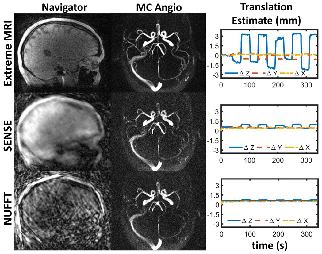
Navigators (left panel), generated from moving scan 4D-Flow data from volunteer 3, used to track motion (right panel) and enable motion corrected angiograms (middle panel) using three different navigator reconstructions schemes: Extreme MRI (multi-scale low rank (MSLR)), iterative SENSE, and direct NUFFT. Navigators were generated from ~43 projections per frame. Extreme MRI based navigators displayed greater image quality which led to high fidelity and stability motion tracking estimates and better angiogram motion correction. Undersampling artifact riddled SENSE and NUFFT navigators failed to resolved volunteer motion, leading to more blurred and noisy images.
3.4. Clinical study experiments
A summary of head motion translations and rotations for the 11 clinical subjects can be found in Supplementary Figure 4. Head motion was highly variable throughout the time course of the acquisition as shown in Figure 13 for exemplary cases with large motion. Exemplary dynamic navigator images for cases 4, 7, and 8 are presented in Supplementary Figure 5.
Figure 13.

Examples of observed motions variability in clinical cases during scanning. Translational and rotational head motion measures were derived from extremely undersampled (~40 projections per frame) 3D self-navigator registrations aided by Extreme MRI reconstruction. Subject demographics: Case 7: MCI subject, 71 yrs; Case 8: cognitively normal, 78 yrs; Case 4: cognitively normal, 82 yrs.
Uncorrected 4D-Flow magnitude and angiographic images displayed substantial blurring and reduced depiction of the vascular tree. After motion correction image blurring was reduced and vessel conspicuity increased in magnitude and angiographic images (Figure 14). In addition, after motion correction noise levels of the velocity field were also reduced leading to improvements in vessel segmentation (Figure 15).
Figure 14.

Example of magnitude (left panel) and angiographic (right panel) images derived from neurovascular 4D-Flow MRI data from clinical cases. After rigid head motion correction of k-space data, significant improvements in subjective magnitude and angiogram image quality were observed. Motion correction led to reduced blurring and increased vessel conspicuity (orange arrows). Subject demographics: Case 7: MCI subject, 71 yrs; Case 8: cognitively normal, 78 yrs; Case 4: cognitively normal, 82 yrs.
Figure 15.

Velocity streamlines along the cerebral arteries for clinical case 7 with and without motion correction (left and right panels). Subject motion led to substantial vessel blurring and noisy velocity fields. Subsequently these errors led to inaccuracies in vessel segmentation and quantification as observed on the right panel streamlines and MIP images (small overlay). After correcting k-space data for bulk motion, improved vessel segmentation and streamline quantification was achieved (left panel). In this example, both datasets (corrected and uncorrected) were segmented using a fixed threshold value.
Quantification of hemodynamic parameters from 4D-Flow data in the cerebral arteries of clinical exams are summarized in Figure 16. After motion correction all hemodynamic parameters including blood flow rates, cross-sectional area, and flow PI displayed a statistically significant reduction in all vessel segments including left and right ICAs and MCAs (Flow: P<0.001 Lt ICA, P=0.002 Rt ICA, P=0.004 Lt MCA, P=0.004 Rt MCA; Area: P<0.001 Lt ICA, P<0.001 Rt ICA, P=0.004 Lt MCA, P=0.004 Rt MCA; PI: P=0.042 Rt ICA, P=0.002 Lt MCA), except for the PI in the left ICA and right MCA which were reduced but not significantly (left ICA P=0.206, right MCA=0.053). Moreover, measurement variance in most hemodynamic markers were reduced after motion correction; however, significant variance differences were only found in the left ICA (P=0.014) and right MCA (P=0.021) cross-sectional areas.
Figure 16.

Summary of 4D-Flow MRI derived hemodynamic markers in various cerebral vessels before and after motion correction (MC) in 11 human subjects from ongoing aging research studies. Measurements were performed in the left and right internal carotid arteries (ICAs) and middle cerebral arteries (MCAs). All hemodynamic markers including blood flow rates, cross-sectional area, and flow pulsatility index (PI) were significantly reduced (P<0.05) in all vessel segments (except PI in the left ICA and right MCA) after motion correction. Most hemodynamic markers also displayed a reduction in measurement variability after motion correction.
4. Discussion
This study proposed and characterized a self-navigation approach for retrospective rigid motion correction of neurovascular 4D-Flow MRI. By leveraging the properties of pseudorandomly ordered radial k-space undersampling with a low rank reconstruction approach, high resolution 3D navigators from 4D-Flow data were used to estimate and correct rigid body motion. Motion tracking and correction feasibility was tested across a range of motions in simulation, phantom and volunteer experiments and applied to clinical exams from ongoing aging studies with artifacts of unknown nature. In all experiments motion correction led to substantial reductions in image blurring and increased vessel conspicuity. Further, hemodynamic parameters from motion corrected scans displayed reduced variability and increased agreement with static scans. Importantly, motion correction of static scans did not introduce noticeable bias in quantitative measures. Finally, motion correction improved image quality on clinical exams with original poor image quality from ongoing aging studies. Decreased blurring and noise in velocity data led to improved vessel segmentation. Hemodynamic markers of cerebrovascular health including blood flow rates, flow pulsatility index, and vessel lumen area were significantly different after motion correction in most vessel segments. Measurement variability was also reduced. These findings support the utilization of the proposed rigid motion correction method on ongoing clinical studies to correct measurement bias from motion confounders. Furthermore, the approach can be used to recover motion corrupted clinical exams with originally failed intake quality control.
Previous studies have demonstrated self-navigation schemes to estimate and correct for rigid body motion in 2D and 3D brain MRI (Anderson et al., 2013; Kecskemeti et al., 2018; Pipe, 1999). These methods benefited from different sampling strategies to generate low resolution navigators for motion tracking. In these approaches low resolution navigator images are reconstructed to determine inter-frame motion parameters estimated via co-registration analysis and retrospectively correct k-space data. While such approaches can successfully correct for rigid brain motion in structural imaging, self-navigation in neurovascular 4D-Flow MRI faces additional challenges. The inherently slower nature of 4D-Flow imaging due to longer TRs and different velocity encodings leads to increased levels of k-space undersampling. For example, (Kecskemeti et al., 2018) used self-navigator images with spatiotemporal resolutions of 2-10 mm and 2s reconstructed from ~400 radial projections per image for motion corrected quantitative T1 imaging, including submillimeter corrections. However, to enable high spatiotemporal navigators from neurovascular 4D-Flow MRI (e.g. 1.3s and 1.4mm) while maintaining clinically feasible scan times (e.g. 5.6 min), self-navigation images were reconstructed from ~43 projections per image, an extraordinary level of undersampling. From such few samples conventional image reconstruction approaches will generate artifact riddled navigator images with low motion fidelity as demonstrated in Figure 12. To overcome this limitation this study leveraged a MSLR reconstruction approach (Extreme MRI) (Ong et al., 2020) to enable high fidelity self-navigation for motion corrected neurovascular 4D-Flow. Extreme MRI can reconstruct large 3D dynamic image series with high levels of undersampling by considering all scales of correlations including local, global and sparse.
Image quality of clinical data from elderly adults was substantially improved after motion correction, revealing motion as a main source of artifacts. Motion correction reduced blurring and increased vessel depiction. Furthermore, characterization of velocity field streamlines revealed decreased velocity noise and improved vessel segmentation after motion correction. More importantly from a quantitative biomarker perspective, 4D-Flow derived hemodynamic markers of cerebrovascular health including blood flow rates, PI, and lumen areas significantly decreased after motion correction in all measured cerebral arteries except PI in the left ICA and right MCA (where PI was reduced but not significantly). PI was somewhat less sensitive to motion as peak flow measures can be more robust to image blurring. Standard deviations of all hemodynamic markers in most vessels were also reduced after correction. Together, these findings highlight the importance of incorporating motion correction approaches to neurovascular 4D-Flow MR imaging studies, especially in vulnerable populations where sub-millimeter motions can lead to a significant bias in hemodynamic markers of vascular health. Neurovascular 4D-Flow is particularly sensitive to such motions as vessels and the flow features are small. In this work, clinical data was collected from aging studies, however, pediatric and challenge studies (e.g. exercise) would likely benefit from the proposed rigid motion correction approach.
After motion correction volunteers moving scans demonstrated reduced vessel blurring, increased vessel conspicuity, and better agreement of quantitative parameters with static reference scans. While there were small differences of flow measures between motion corrected moving and reference scans, these can be expected due to natural variations in flow associated with neural and physiologic variability, especially given that the volunteers underwent a motor task with posture changes. Similar to clinical subjects, flow-rates, cross-sectional areas, and PI in volunteers moving scans showed a reduction in magnitude and standard deviations after motion correction. Velocity comparisons of static scans with and without motion correction revealed very high correlation coefficients for all volunteers; however, some small differences were observed. Since motion correction of static data in simulation experiments showed correlation coefficients and slopes of 1.00, we attribute in vivo static scan differences to the presence of small motions during the scan; however, the lack of a ground truth makes it impossible to disregard other potential sources of error. While the volunteer motion task may not completely represent incidental head motion during standard scans it provides some level of confidence in the correction. The motion task induced varying levels of head motion in each of the volunteers, with larger displacements than in phantoms or of those seen clinically. Volunteer 3 displayed the greatest amount of translational motion (5.7 mm), considerably more than the largest recorded clinical exam motion (2.7 mm). Interestingly, volunteer 3 showed an increase in right and left MCA flow-rates and cross-sectional areas after motion correction. Upon inspection of the angiograms, these results are explained by the high levels of MCAs blurring present on volunteer 3 moving scan. Massive MCAs blurring led to erroneous vessel segmentation roughly splitting each M1 segment into two distinct vessel segments explaining the observed lower flow-rates and areas pre-correction. Motion correction resolved the issue; however, corrected velocities agreement with the static scan were lowest for volunteer 3. Volunteer 4 motion estimates were relatively smaller compared to other volunteers, which can be attributed to the volunteer’s formal dance training leading to superior body control skills. Other volunteers lacked such experience.
Phantom experiments demonstrated high levels of motion fidelity and stability for most moving scans; however, no gold standard method was available to validate. We performed a hinged rotation to ensure some level of rotation but that precluded a simple linear motion stage experiment. Experiments where the phantom setup was moved once and every 60s displayed the greatest degree of image blurring and translational motion. This effectively creates a superposition of fewer motion states, whereas more continuous types of motion lead to an average out blurring artifact effect from the superposition of many motion states. On the phantom data, continuous motion had a somewhat limited effect on the flow measures, likely due to the averaging effect of 3D radial sampling and the lower displacement incurred. Notably, flow rates and area in that case were the closest to static scan values even before motion correction. Moreover, simulations indicate continuous motions might be more challenging to resolved and underestimated. After rigid motion correction image blurring was reduced in all moving cases. Again, there were some small differences between the measures which could be due to changes in flow from different positioning, changes in B0, or other motion effects aside from image translations and rotations. Hemodynamic parameter magnitude and variability were reduced after motion correction. Furthermore, after motion correction of moving scans, differences of flow rates and cross-sectional areas with static scan results were reduced.
Simulations of continuous and stepwise rotational motions showed rotation of k-space coordinates to a zero-mean average angle improves motion correction over rotation to a specific navigator reference frame. As expected, larger motions reduced velocities agreement, with ~2% differences between ground truth and corrected velocities for rotational motions on the order of 5° (which covers the maximum rotation observed in the clinical cohort) and differences of 5-6% for rotational motions of 30°. Simulation experiments also revealed the self-navigation approach underestimated continuous motions but recovered stepwise motions almost exactly. These findings indicate limitations from the self-navigator reconstruction (Extreme MRI) can obfuscate smaller continuous motions and lead to underestimation. This is likely a product of the inherently higher rank of the continuous motion, whereas the stepwise motion leads to a rank 2 representation of the temporal dynamics. However, corrected velocities agreement with ground truth were similar for both continuous and stepwise motions cases. Further work is required to compare navigators from Extreme MRI to optical or other gold standard measures.
In addition to retrospective rigid motion correction, prospective approaches could also be implemented to aid neurovascular 4D-Flow MRI. Prospective motion correction using optical tracking and self-navigation have been successfully implemented in 3D structural brain imaging (Godenschweger et al., 2016; Maknojia et al., 2019). These approaches can update scan parameters during the acquisition to image in head-relative coordinates despite subject motion or to re-acquired measurements. Using such approaches, researchers has shown significant improvements in morphometry measures (e.g. cortical thinning) even when motions were too small to produce noticeable image artifacts. However, prospective motion correction implementations can also be challenging and limited by additional scanner setup with optical tracking systems or longer acquisition times to acquire navigator data between TRs.
This study has several limitations. In this work, self-navigation for neurovascular 4D-Flow MRI motion correction was validated for radial trajectories, but while radial sampling is increasingly popular, some vendors utilize Cartesian sampling in 4D-Flow MRI products. More, the proposed approach corrects for rotational motions by rotating k-space coordinates to a zero-mean average rotation; however, non-trivial velocity encoded vector field rotations were not performed. Solving for velocity fields rotations would require the solution to a computationally intensive, non-linear problem that requires solving directly for velocity or phase. While some progress has been made for velocity estimation directly from k-space, the computational time is extremely limiting given the 100s transforms that would be required (Ong et al., 2018; Santelli et al., 2016). This is in contrast to diffusion imaging, where images are collected in a single shot and can be corrected in post-processing. The zero-mean average rotation approach was supported by simulation experiments. Still, in the presence of large rotational motions, velocity field rotations should be considered if computationally fast approaches can be devised. Simulation experiments also showed the motion correction approach recovers stepwise motions with higher fidelity than continuous motions, indicating temporal blurring limitations of the self-navigation. Furthermore, in this study, in vivo studies temporal resolution of navigators was 1.3s; however, higher frequencies of motion can occur. We targeted a temporal resolutions similar to those demonstrated in previous MSLR work leading to high levels of navigator undersampling (~43 projections per frame) (Ong et al., 2020). Higher temporal resolution would be desirable to reduce potential for intra-frame motion errors; however, for the present study datasets, fewer projections per frame led to convergence issues of the iterative reconstruction process. Reducing the spatial resolution of the navigator images could be used to improve the temporal resolution of the navigator images, although sensitivity to subtle motions could be lost (Kecskemeti et al., 2018). Non-flow motion-induced velocity encoding effects may include velocities of head movement; however, we assumed such head motions are small, lead to a zero-mean average, and are minimized after background phase correction. Finally, leveraging motion correction strategies that combine both retrospective with prospective motion tracking (e.g. optical tracking) might generate superior motion correction performance.
5. Conclusions
By combining radial sampling with pseudorandom projection ordering and MSLR reconstruction high fidelity navigators were generated from extremely undersampled data and were used to estimate and correct for rigid head motion in neurovascular 4D-Flow MRI. Studies in simulations, phantom and volunteer experiments demonstrated feasibility to correct for induced motions. More importantly, motion correction of neurovascular 4D-Flow MRI in exams from ongoing clinical studies on aging revealed statistically significant differences in hemodynamic parameter of cerebrovascular health after motion correction. These results indicate patient motion can significantly bias hemodynamic markers in cerebral arteries derived from quantitative MRI in ongoing longitudinal studies and vulnerable populations. The proposed method represents a promising approach to significantly reduce measurement variability from motion in neurovascular 4D-Flow MRI and can be used in vulnerable populations such as children, older adults, as well during motion challenges.
Supplementary Material
Acknowledgements
We gratefully acknowledge research support from GE Healthcare.
Funding
This work was supported by the Alzheimer’s Association [grant number AARFD-20-678095], and the National Institutes of Health [grant numbers R01AG075788, R21AG077337, R01AG021155, P30AG062715, UL1TR002373, and KL2TR002374].
Disclosure/Conflict of interest
S.C. Johnson serves on an advisory board for Roche Diagnostics for which he receives an honorarium and is principal investigator of an equipment grant from Roche. He receives research funding from Cerveau Technologies.
Footnotes
Data and code availability
Data from the core WRAP protocol study in partnership with the WADRC are accessible to qualified researchers via an online request form and data use agreement which can be linked from the Global Alzheimer’s Association Interactive Network web site (www.gaain.org). Prior to data access, users are required to agree to the data use agreement essential for data security, grant citation, and annual progress reports. Data from phantom and volunteers experiments are accessible via direct request to the authors. The motion correction and reconstruction code can be found at: https://github.com/uwmri/flow_recon.
CRediT authorship contribution statement
Leonardo A. Rivera-Rivera: Conceptualization, Funding acquisition, Investigation, Methodology, Software, Visualization, Writing original draft. Steve Kecskemeti: Methodology, Writing - review & editing. Mu-Lan Jen: Resources, Writing - review & editing. Zachary Miller: Resources, Writing - review & editing. Sterling C. Johnson: Funding acquisition, Resources, Writing - review & editing. Laura Eisenmenger: Resources, Writing - review & editing. Kevin M. Johnson: Conceptualization, Funding acquisition, Investigation, Methodology, Project supervision, Software, Visualization, Writing - review & editing.
References:
- Anderson AG, Velikina J, Block W, Wieben O, Samsonov A, 2013. Adaptive retrospective correction of motion artifacts in cranial MRI with multicoil three-dimensional radial acquisitions: Adaptive 3D Motion Correction. Magn. Reson. Med 69, 1094–1103. 10.1002/mrm.24348 [DOI] [PMC free article] [PubMed] [Google Scholar]
- Aristova M, Pang J, Ma Y, Ma L, Berhane H, Rayz V, Markl M, Schnell S, 2022. Accelerated dual-venc 4D flow MRI with variable high-venc spatial resolution for neurovascular applications. Magnetic Resonance in Medicine 88, 1643–1658. 10.1002/mrm.29306 [DOI] [PMC free article] [PubMed] [Google Scholar]
- Aristova M, Vali A, Ansari SA, Shaibani A, Alden TD, Hurley MC, Jahromi BS, Potts MB, Markl M, Schnell S, 2019. Standardized Evaluation of Cerebral Arteriovenous Malformations Using Flow Distribution Network Graphs and Dual-venc 4D Flow MRI. J Magn Reson Imaging 50, 1718–1730. 10.1002/jmri.26784 [DOI] [PMC free article] [PubMed] [Google Scholar]
- Avinun R, Israel S, Knodt AR, Hariri AR, 2020. Little evidence for associations between the Big Five personality traits and variability in brain gray or white matter. NeuroImage 220, 117092. 10.1016/j.neuroimage.2020.117092 [DOI] [PMC free article] [PubMed] [Google Scholar]
- Björnfot C, Garpebring A, Qvarlander S, Malm J, Eklund A, Wåhlin A, 2021. Assessing cerebral arterial pulse wave velocity using 4D flow MRI. J Cereb Blood Flow Metab 0271678X211008744. 10.1177/0271678X211008744 [DOI] [PMC free article] [PubMed] [Google Scholar]
- de Riva N, Budohoski KP, Smielewski P, Kasprowicz M, Zweifel C, Steiner LA, Reinhard M, Fábregas N, Pickard JD, Czosnyka M, 2012. Transcranial Doppler Pulsatility Index: What it is and What it Isn’t. Neurocritical Care 17, 58–66. 10.1007/s12028-012-9672-6 [DOI] [PubMed] [Google Scholar]
- Dyverfeldt P, Ebbers T, 2017. Comparison of respiratory motion suppression techniques for 4D flow MRI. Magn Reson Med 78, 1877–1882. 10.1002/mrm.26574 [DOI] [PMC free article] [PubMed] [Google Scholar]
- Glover GH, Pauly JM, 1992. Projection Reconstruction Techniques for Reduction of Motion Effects in MRI. Magn. Reson. Med 28, 275–289. 10.1002/mrm.1910280209 [DOI] [PubMed] [Google Scholar]
- Godenschweger F, Kägebein U, Stucht D, Yarach U, Sciarra A, Yakupov R, Lüsebrink F, Schulze P, Speck O, 2016. Motion correction in MRI of the brain. Physics in Medicine and Biology 61, R32–R56. 10.1088/0031-9155/61/5/r32 [DOI] [PMC free article] [PubMed] [Google Scholar]
- Gottwald LM, Töger J, Markenroth Bloch K, Peper ES, Coolen BF, Strijkers GJ, van Ooij P, Nederveen AJ, 2020. High Spatiotemporal Resolution 4D Flow MRI of Intracranial Aneurysms at 7T in 10 Minutes. AJNR Am J Neuroradiol 41, 1201–1208. 10.3174/ajnr.A6603 [DOI] [PMC free article] [PubMed] [Google Scholar]
- Griswold MA, Jakob PM, Nittka M, Goldfarb JW, Haase A, 2000. Partially parallel imaging with localized sensitivities (PILS). Magnetic Resonance in Medicine 44, 602–609. 10.1002/1522-2594(200010)44:4<602::AID-MRM14>3.0.CO;2-5 [DOI] [PubMed] [Google Scholar]
- Hausman HK, Hardcastle C, Kraft JN, Evangelista ND, Boutzoukas EM, O’Shea A, Albizu A, Langer K, Van Etten EJ, Bharadwaj PK, Song H, Smith SG, Porges E, Hishaw GA, Wu S, DeKosky S, Alexander GE, Marsiske M, Cohen R, Woods AJ, 2022. The association between head motion during functional magnetic resonance imaging and executive functioning in older adults. Neuroimage: Reports 2, 100085. 10.1016/j.ynirp.2022.100085 [DOI] [PMC free article] [PubMed] [Google Scholar]
- Johnson KM, Lum DP, Turski PA, Block WF, Mistretta CA, Wieben O, 2008. Improved 3D phase contrast MRI with off-resonance corrected dual echo VIPR. Magnetic Resonance in Medicine 60, 1329–1336. 10.1002/mrm.21763 [DOI] [PMC free article] [PubMed] [Google Scholar]
- Johnson SC, Koscik RL, Jonaitis EM, Clark LR, Mueller KD, Berman SE, Bendlin BB, Engelman CD, Okonkwo OC, Hogan KJ, Asthana S, Carlsson CM, Hermann BP, Sager MA, 2018. The Wisconsin Registry for Alzheimer’s Prevention: A review of findings and current directions. Alzheimer’s & Dementia: Diagnosis, Assessment & Disease Monitoring 10, 130–142. 10.1016/j.dadm.2017.11.007 [DOI] [PMC free article] [PubMed] [Google Scholar]
- Kecskemeti S, Freeman A, Travers BG, Alexander AL, 2021. FreeSurfer based cortical mapping and T1-relaxometry with MPnRAGE: Test-retest reliability with and without retrospective motion correction. NeuroImage 242, 118447. 10.1016/j.neuroimage.2021.118447 [DOI] [PMC free article] [PubMed] [Google Scholar]
- Kecskemeti S, Samsonov A, Velikina J, Field AS, Turski P, Rowley H, Lainhart JE, Alexander AL, 2018. Robust Motion Correction Strategy for Structural MRI in Unsedated Children Demonstrated with Three-dimensional Radial MPnRAGE. Radiology 289, 509–516. 10.1148/radiol.2018180180 [DOI] [PMC free article] [PubMed] [Google Scholar]
- Keuken MC, Ly A, Boekel W, Wagenmakers E-J, Belay L, Verhagen J, Brown SD, Forstmann BU, 2017. Corrigendum to “A purely confirmatory replication study of structural brain-behavior correlations” [Cortex 66 (2015) 115–133]. Cortex 93, 229–233. 10.1016/j.cortex.2017.03.007 [DOI] [PubMed] [Google Scholar]
- Li Y, Chen H, He L, Cao X, Wang X, Chen S, Li R, Yuan C, 2018. Hemodynamic assessments of venous pulsatile tinnitus using 4D-flow MRI. Neurology 91, e586. 10.1212/WNL.0000000000005948 [DOI] [PubMed] [Google Scholar]
- Loecher M, Schrauben E, Johnson KM, Wieben O, 2016. Phase unwrapping in 4D MR flow with a 4D single-step laplacian algorithm. J. Magn. Reson. Imaging 43, 833–842. 10.1002/jmri.25045 [DOI] [PubMed] [Google Scholar]
- Maknojia S, Churchill NW, Schweizer TA, Graham SJ, 2019. Resting State fMRI: Going Through the Motions. Frontiers in Neuroscience 13. [DOI] [PMC free article] [PubMed] [Google Scholar]
- Marek S, Tervo-Clemmens B, Calabro FJ, Montez DF, Kay BP, Hatoum AS, Donohue MR, Foran W, Miller RL, Hendrickson TJ, Malone SM, Kandala S, Feczko E, Miranda-Dominguez O, Graham AM, Earl EA, Perrone AJ, Cordova M, Doyle O, Moore LA, Conan GM, Uriarte J, Snider K, Lynch BJ, Wilgenbusch JC, Pengo T, Tam A, Chen J, Newbold DJ, Zheng A, Seider NA, Van AN, Metoki A, Chauvin RJ, Laumann TO, Greene DJ, Petersen SE, Garavan H, Thompson WK, Nichols TE, Yeo BTT, Barch DM, Luna B, Fair DA, Dosenbach NUF, 2022. Reproducible brain-wide association studies require thousands of individuals. Nature 603, 654–660. 10.1038/s41586-022-04492-9 [DOI] [PMC free article] [PubMed] [Google Scholar]
- Markl M, Frydrychowicz A, Kozerke S, Hope M, Wieben O, 2012. 4D Flow MRI. Journal of Magnetic Resonance Imaging 36, 1015–1036. [DOI] [PubMed] [Google Scholar]
- Morgan AG, Thrippleton MJ, Wardlaw JM, Marshall I, 2021. 4D flow MRI for non-invasive measurement of blood flow in the brain: A systematic review. J Cereb Blood Flow Metab 41, 206–218. 10.1177/0271678X20952014 [DOI] [PMC free article] [PubMed] [Google Scholar]
- Ong F, Cheng JY, Lustig M, 2018. General phase regularized reconstruction using phase cycling. Magnetic Resonance in Medicine 80, 112–125. 10.1002/mrm.27011 [DOI] [PMC free article] [PubMed] [Google Scholar]
- Ong F, Martin J, Grissom W, Srinivasan S, Johnson KM, Huynh C, Desai A, Li Z, Tamir J, Sandino C, Shimron E, Zeng D, Mickevicius N, 2022. mikgroup/sigpy: Minor release to trigger Zenodo for DOI. (v0.1.24). Zenodo. 10.5281/zenodo.5893788 [DOI] [Google Scholar]
- Ong F, Zhu X, Cheng JY, Johnson KM, Larson PEZ, Vasanawala SS, Lustig M, 2020. Extreme MRI: Large-scale volumetric dynamic imaging from continuous non-gated acquisitions. Magnetic Resonance in Medicine 84, 1763–1780. 10.1002/mrm.28235 [DOI] [PMC free article] [PubMed] [Google Scholar]
- Pardoe HR, Kucharsky Hiess R, Kuzniecky R, 2016. Motion and morphometry in clinical and nonclinical populations. NeuroImage 135, 177–185. 10.1016/j.neuroimage.2016.05.005 [DOI] [PubMed] [Google Scholar]
- Pipe JG, 1999. Motion correction with PROPELLER MRI: Application to head motion and free-breathing cardiac imaging. Magn. Reson. Med 42, 963–969. 10.1002/(SICI)1522-2594(199911)42:5<963::AID-MRM17>3.0.CO;2-L [DOI] [PubMed] [Google Scholar]
- Poldrack RA, Baker CI, Durnez J, Gorgolewski KJ, Matthews PM, Munafò MR, Nichols TE, Poline J-B, Vul E, Yarkoni T, 2017. Scanning the horizon: towards transparent and reproducible neuroimaging research. Nat Rev Neurosci 18, 115–126. 10.1038/nrn.2016.167 [DOI] [PMC free article] [PubMed] [Google Scholar]
- Pruessmann KP, Weiger M, Börnert P, Boesiger P, 2001. Advances in sensitivity encoding with arbitrary k-space trajectories. Magn. Reson. Med 46, 638–651. 10.1002/mrm.1241 [DOI] [PubMed] [Google Scholar]
- Reuter M, Tisdall MD, Qureshi A, Buckner RL, van der Kouwe AJW, Fischl B, 2015. Head motion during MRI acquisition reduces gray matter volume and thickness estimates. NeuroImage 107, 107–115. 10.1016/j.neuroimage.2014.12.006 [DOI] [PMC free article] [PubMed] [Google Scholar]
- Rivera-Rivera LA, Cody KA, Eisenmenger L, Cary P, Rowley HA, Carlsson CM, Johnson SC, Johnson KM, 2021a. Assessment of vascular stiffness in the internal carotid artery proximal to the carotid canal in Alzheimer’s disease using pulse wave velocity from low rank reconstructed 4D flow MRI. J Cereb Blood Flow Metab 41, 298–311. 10.1177/0271678X20910302 [DOI] [PMC free article] [PubMed] [Google Scholar]
- Rivera-Rivera LA, Cody KA, Rutkowski D, Cary P, Eisenmenger L, Rowley HA, Carlsson CM, Johnson SC, Johnson KM, 2020. Intracranial vascular flow oscillations in Alzheimer’s disease from 4D flow MRI. NeuroImage: Clinical 28, 102379. 10.1016/j.nicl.2020.102379 [DOI] [PMC free article] [PubMed] [Google Scholar]
- Rivera-Rivera LA, Eisenmenger L, Cody KA, Reher T, Betthauser T, Cadman RV, Rowley HA, Carlsson CM, Chin NA, Johnson SC, Johnson KM, 2021b. Cerebrovascular stiffness and flow dynamics in the presence of amyloid and tau biomarkers. Alzheimers Dement (Amst) 13, e12253–e12253. 10.1002/dad2.12253 [DOI] [PMC free article] [PubMed] [Google Scholar]
- Santelli C, Loecher M, Busch J, Wieben O, Schaeffter T, Kozerke S, 2016. Accelerating 4D flow MRI by exploiting vector field divergence regularization. Magnetic Resonance in Medicine 75, 115–125. 10.1002/mrm.25563 [DOI] [PubMed] [Google Scholar]
- Schnell S, Ansari SA, Wu C, Garcia J, Murphy IG, Rahman OA, Rahsepar AA, Aristova M, Collins JD, Carr JC, Markl M, 2017. Accelerated dual-venc 4D flow MRI for neurovascular applications. J Magn Reson Imaging 46, 102–114. 10.1002/jmri.25595 [DOI] [PMC free article] [PubMed] [Google Scholar]
- Schrauben E, Wåhlin A, Ambarki K, Spaak E, Malm J, Wieben O, Eklund A, 2015. Fast 4D flow MRI intracranial segmentation and quantification in tortuous arteries. Journal of magnetic resonance imaging : JMRI 42, 1458–1464. 10.1002/jmri.24900 [DOI] [PMC free article] [PubMed] [Google Scholar]
- Summers PE, Holdsworth DW, Nikolov HN, Rutt BK, Drangova M, 2005. Multisite trial of MR flow measurement: Phantom and protocol design. Journal of Magnetic Resonance Imaging 21, 620–631. 10.1002/jmri.20311 [DOI] [PubMed] [Google Scholar]
- Tisdall MD, Reuter M, Qureshi A, Buckner RL, Fischl B, van der Kouwe AJW, 2016. Prospective motion correction with volumetric navigators (vNavs) reduces the bias and variance in brain morphometry induced by subject motion. NeuroImage 127, 11–22. 10.1016/j.neuroimage.2015.11.054 [DOI] [PMC free article] [PubMed] [Google Scholar]
- Turski P, Scarano A, Hartman E, Clark Z, Schubert T, Rivera L, Wu Y, Wieben O, Johnson K, 2016. Neurovascular 4DFlow MRI (Phase Contrast MRA): emerging clinical applications. Neurovascular Imaging 2, 8. 10.1186/s40809-016-0019-0 [DOI] [Google Scholar]
- Vali A, Aristova M, Vakil P, Abdalla R, Prabhakaran S, Markl M, Ansari SA, Schnell S, 2019. Semi-automated analysis of 4D flow MRI to assess the hemodynamic impact of intracranial atherosclerotic disease. Magn Reson Med 82, 749–762. 10.1002/mrm.27747 [DOI] [PMC free article] [PubMed] [Google Scholar]
- Vidal-Pineiro D, Parker N, Shin J, French L, Grydeland H, Jackowski AP, Mowinckel AM, Patel Y, Pausova Z, Salum G, Sørensen Ø, Walhovd KB, Paus T, Fjell AM, the Alzheimer’s Disease Neuroimaging Initiative and the Australian Imaging Biomarkers and Lifestyle flagship study of ageing, 2020. Cellular correlates of cortical thinning throughout the lifespan. Scientific Reports 10, 21803. 10.1038/s41598-020-78471-3 [DOI] [PMC free article] [PubMed] [Google Scholar]
- Vikner T, Eklund A, Karalija N, Malm J, Riklund K, Lindenberger U, Bäckman L, Nyberg L, Wåhlin A, 2021. Cerebral arterial pulsatility is linked to hippocampal microvascular function and episodic memory in healthy older adults. J Cereb Blood Flow Metab 0271678X20980652. 10.1177/0271678X20980652 [DOI] [PMC free article] [PubMed] [Google Scholar]
- Wåhlin A, Eklund A, Malm J, 2022. 4D flow MRI hemodynamic biomarkers for cerebrovascular diseases. J Intern Med 291, 115–127. 10.1111/joim.13392 [DOI] [PubMed] [Google Scholar]
- Walsh DO, Gmitro AF, Marcellin MW, 2000. Adaptive reconstruction of phased array MR imagery. Magnetic Resonance in Medicine 43, 682–690. [DOI] [PubMed] [Google Scholar]
- Ying L, Sheng J, 2007. Joint image reconstruction and sensitivity estimation in SENSE (JSENSE). Magnetic Resonance in Medicine 57, 1196–1202. 10.1002/mrm.21245 [DOI] [PubMed] [Google Scholar]
- Youn SW, Lee J, 2022. From 2D to 4D Phase-Contrast MRI in the Neurovascular System: Will It Be a Quantum Jump or a Fancy Decoration? Journal of Magnetic Resonance Imaging 55, 347–372. 10.1002/jmri.27430 [DOI] [PubMed] [Google Scholar]
Associated Data
This section collects any data citations, data availability statements, or supplementary materials included in this article.