Abstract
Periodontal tissue is a highly dynamic and frequently stimulated area where homeostasis is easily destroyed, leading to proinflammatory periodontal diseases. Bacteria–bacteria and cell–bacteria interactions play pivotal roles in periodontal homeostasis and disease progression. Several reviews have comprehensively summarized the roles of bacteria and stem cells in periodontal homeostasis. However, they did not describe the roles of extracellular vesicles (EVs) from bacteria and cells. As communication mediators evolutionarily conserved from bacteria to eukaryotic cells, EVs secreted by bacteria or cells can mediate interactions between bacteria and their hosts, thereby offering great promise for the maintenance of periodontal homeostasis. This review offers an overview of EV biogenesis, the effects of EVs on periodontal homeostasis, and recent advances in EV-based periodontal regenerative strategies. Specifically, we document the pathogenic roles of bacteria-derived EVs (BEVs) in periodontal dyshomeostasis, focusing on plaque biofilm formation, immune evasion, inflammatory pathway activation and tissue destruction. Moreover, we summarize recent advancements in cell-derived EVs (CEVs) in periodontal homeostasis, emphasizing the multifunctional biological effects of CEVs on periodontal tissue regeneration. Finally, we discuss future challenges and practical perspectives for the clinical translation of EV-based therapies for periodontitis.
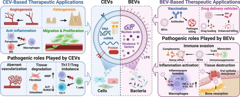
Graphical Abstract
Keywords: Cellular extracellular vesicles, Bacterial extracellular vesicles, Periodontal homeostasis Periodontitis, Periodontal regeneration
Introduction
An estimated 50% of the world's adult population is affected by periodontitis in its mildest form, and the incidence increases to more than 60% in people over 65 years old, which makes periodontitis a disease with the sixth highest prevalence [1, 2]. Initiated and propagated by dental plaque biofilms, periodontitis can lead to continuous and irreversible destruction of periodontal tissue [3]. Left untreated, periodontitis can cause persistent alveolar bone destruction that may lead to tooth loss in adults, significantly compromising mastication, speech, and self-esteem [4]. Moreover, a significant body of evidence has indicated that periodontitis affects the overall health of patients by increasing the risk for cardiovascular disease, Alzheimer’s disease, and diabetes [2, 5]. Several strategies involving conventional anti-infection therapy (e.g., scaling or root debridement) or guided tissue regeneration have been developed for periodontal bone repair. Unfortunately, while these clinical therapies for periodontitis succeed in preventing the progression of disease, they fail to restore lost periodontal tissues stably [6].
Tooth-supporting tissues [i.e., alveolar bone, periodontal ligaments (PDLs), cementum and, gingiva] are collectively known as the periodontium. To maintain the integrity of these tissues upon microbial stimulation, the periodontium undergoes continual remodeling [7]. In a healthy periodontal milieu, typical interactions between host cells and microorganisms trigger a protective immune response, contributing to the maintenance of periodontal homeostasis. For example, commensal bacteria can activate Toll-like receptors (TLRs) on host gingival epithelial cells, leading to enhanced production of defensins or cytokines that help to maintain oral health [8]. However, in periodontal disease, enhanced interactions of host cells with pathogens can cause persistent inflammation and ultimately lead to irreversible tissue destruction. When the epithelial barrier is breached, large volumes of invading bacteria can activate TLRs on periodontal immune and nonimmune cells, leading to the robust release of many proinflammatory cytokines (e.g., PGE2, interleukin (IL)-1β, IL-8, and IL-6) and other biological mediators [8, 9]. Furthermore, the influx of bacteria and uncontrolled proinflammatory cytokines can lead to high expression of Receptor Activator for Nuclear Factor-κB Ligand (RANKL) in PDL cells and osteoblasts, causing irreversible bone destruction [10]. These findings clearly demonstrate that cell–bacteria and cell‒cell interactions are major drivers of periodontal homeostasis and play key roles in disease progression.
As communication mediators evolutionarily conserved from bacteria to humans, extracellular vehicles (EVs) produced by eukaryotic host cells (CEVs) or bacteria (BEVs) play key roles in cell–bacteria, cell‒cell, and bacteria–bacteria interactions [11, 12]. EVs constitute a group of nanosized structures composed of a lipid membrane comprising diverse proteins, nucleic acids, and lipids [12]. Once internalized by neighboring or distant cells, EVs can exert positive or negative effects, depending on their cargoes, quantity, or targeting efficiency [13–15]. Several reviews presented excellent summaries of periodontitis pathogenesis years ago [16–18]. However, they did not describe the roles of EVs in the crosstalk between bacteria and host cells, which is critical to periodontal homeostasis. Therefore, a summary of the recent advances in uncovering the pathogenesis of periodontal disease from the perspective of EVs is urgently needed.
In addition to the roles they play in cellular communication, CEVs and BEVs can be modified and used as therapeutic agents or diagnostic tools for various diseases [19, 20]. Owing to their low immunogenicity, high safety, and multifunctional bioactivities, EVs can be applied in tissue engineering fields and have therefore attracted considerable attention. An increasing number of studies have demonstrated that stem cell-derived EVs participate in immunomodulation and show the potential to serve as alternatives to stem cell-based therapies [20–22]. Notably, the therapeutic potential of EVs derived from adipose-derived stem cells in periodontitis is being investigated (ClinicalTrials.gov identifier: NCT04270006). However, the translation of EVs from the laboratory to the clinic remains challenging; for example, techniques to isolate large amounts of EVs and treatments with delayed EV clearance in vivo are needed [23, 24]. Hence, increasing tissue curative effects by combining EVs with multiphasic biomaterials is a very active area of research.
Compared with previously published reviews [22], our manuscript focuses on the roles of EVs from bacteria and cells in periodontal homeostasis, and both the therapeutic and pathogenic roles of EVs in bacteria–bacteria and cell–bacteria interactions are summarized. Our review begins with a brief overview of the biology of cellular and bacterial EVs. Then, the roles of CEVs and BEVs in periodontal homeostasis will be discussed. In this section, the pathogenic roles of BEVs in bacteria–bacteria and cell–bacteria interactions will be presented, focusing on their effects on plaque biofilm formation, immune evasion, immune activation, and tissue destruction. Afterward, the multifunctional biological effects of CEVs in periodontal homeostasis and periodontal tissue regeneration will be summarized, emphasizing the multifunctional biological effects of CEVs on periodontal tissue regeneration. Finally, the remaining challenges in EV-based therapeutic applications and some insightful perspectives on the modification and application of EVs to reconstruct the periodontium will be briefly introduced, which will benefit the development of EV-based therapies for periodontal regeneration in the future.
Biogenesis of CEVs and BEVs
CEV biogenesis
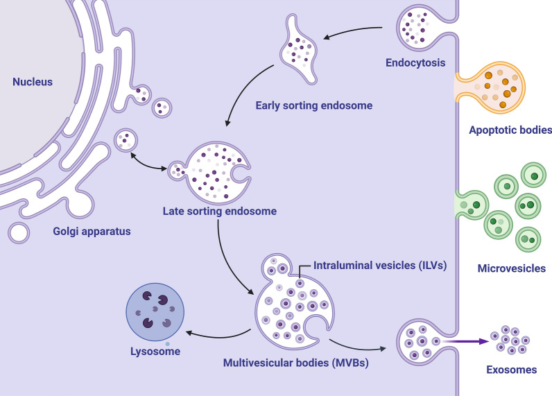
Although CEVs share similar physical characteristics, they are highly heterogeneous membrane-bound vesicles that vary in biogenesis, size, and biological responses [25]. Following the nomenclature of the International Society for Extracellular Vesicles, we use the term CEVs for all membrane-bound vesicles generated by cells. According to the most up-to-date biogenesis studies on CEVs, the three main types are exosomes (40–160 nm), microvesicles (50–1,000 nm), and apoptotic bodies (50–5,000 nm). Exosomes are sequentially formed from early sorting endosomes, late sorting endosomes, and multivesicular endosomes (MVEs) containing intraluminal vesicles (ILVs). In the final step, MVEs fuse with the plasma membrane and release ILVs as exosomes [26]. Microvesicles are produced by direct outward budding and fission of the plasma membrane surface [27]. Apoptotic bodies are produced during cellular apoptosis (Fig. 1).
Fig. 1.
Biogenesis of extracellular vesicles (EVs) in eukaryotic host cells. Three types of extracellular vesicles have been characterized to date and are shown: apoptotic bodies (50–5000 nm) produced by apoptotic cells, microvesicles (50–1000 nm) formed through outward budding and fission of the plasma membrane, and exosomes (40–160 nm) released from eukaryotic cells via the fusion of multivesicular bodies with the plasma membrane. ILVs intraluminal vesicles, MVBs multivesicular bodies. Created with BioRender.com
BEV biogenesis
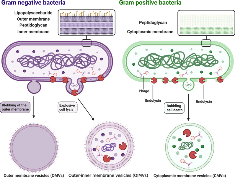
Similar to eukaryotic cells, lipid membrane vesicles (20–400 nm) can also be secreted by both Gram-positive and Gram-negative bacteria. Vesicles generated by bacteria can be classically categorized into three types on the basis of their structure and composition. (1) Outer membrane vesicles (OMVs) are generated by blebbing of the outer membrane in Gram-negative bacteria. OMVs mainly comprise an asymmetrical lipid bilayer with an outer leaflet carrying many lipopolysaccharides (LPSs). Due to barriers created by peptidoglycans and the inner membrane, OMVs cannot access cytoplasmic contents and are therefore enriched with only outer membrane proteins and do not carry nucleic acids or cytosolic proteins [27, 28]. (2) Outer-inner membrane vesicles (OIMVs) are formed when autolysins or endolysins degrade the peptidoglycan layer of Gram-negative bacteria, allowing the inner membrane to protrude into the periplasmic space, which enables cytoplasmic contents to enter forming vesicles. Unlike OMVs, OIMVs contain two bilayers of membranes: one derived from the inner membrane and the other derived from the outer membrane. Among BEVs, they are the sole carriers of DNA from Gram-negative bacteria [29]. (3) Cytoplasmic membrane vesicles (CMVs) are generated by Gram-positive bacteria, which lack an outer membrane. Hence, CMVs contain cytoplasmic contents (Fig. 2) [29, 30]. In this article, the term BEVs refers to any type of vesicle released by Gram-positive or Gram-negative bacteria.
Fig. 2.
Formation of distinct bacterial membrane vesicles. Three major types of vesicles are generated by bacteria: OMVs, OIMVs, and CMVs. OMVs are produced by the blebbing of the outer membranes by living gram-negative bacteria. They do not access cytoplasmic components. OIMVs are generated by membrane fragments of dying and bursting bacteria and contain cytoplasmic components due to endolysin-induced peptidoglycan degradation. CMVs are generated by gram-positive bacteria, which lack an outer membrane. OMVs outer membrane vesicles, OIMVs outer-inner membrane vesicles, CMVs cytoplasmic membrane vesicles. Created with BioRender.com
Pathogenic roles of EVs in periodontal homeostasis
Pathogenic roles of CEVs in periodontal homeostasis
The pathogenic roles of CEVs have been documented in a number of diseases, including Parkinson’s disease and Alzheimer’s disease [31, 32]. However, the detrimental effects of CEVs on periodontal homeostasis have not been well recognized, and most studies published thus far have concentrated on their therapeutic applications in periodontitis [33]. The maintenance of periodontal homeostasis relies on the host response to the invaded periodontopathogens [34], wherein CEVs play key roles in the development of the host response. Periodontitis shares common characteristics, including an inflammatory response, aberrant angiogenesis, and tissue destruction. Recently, emerging evidence has indicated that CEVs released by host cells can play negative roles in the host immune response and cause aberrant angiogenesis and tissue destruction. For example, CEVs released from inflamed periodontal ligament stem cells (PDLSCs) have been reported to enhance Th17-cell activation while inhibiting regulatory T-cell (Treg) activation by targeting Sirtuin-1, leading to a Th17-cell/Treg imbalance and a proinflammatory response [35–37]. Moreover, CEVs from LPS-pretreated periodontal ligament fibroblasts (PDLFs) have been found to upregulate the expression of tumor necrosis factor (TNF)-α and IL-6 in osteoblasts [38]. In addition to the host immune response, aberrant angiogenesis is a feature of periodontitis, which is closely associated with progressive inflammation. By activating miR-17-5p/VEGFA signaling pathways, CEVs from inflamed PDLSCs significantly promoted tube formation in a cell model of human umbilical vein endothelial cells (HUVECs) [39], indicating that they may contribute to anomalous vascularization in periodontitis. With the progression of inflammation, tissue destruction is inevitable, and there have been reports of EVs being involved in tissue degradation. For example, EVs released from PDLFs pretreated with LPS have been found to inhibit osteogenesis [38]. Furthermore, EVs released from biofilm-stimulated gingival epithelial cells significantly upregulated MMP-1 and MMP-3 expression in human gingival fibroblasts (HGFs). Upregulated MMP-1 and MMP-3 in HGFs promoted a tissue-destructive phenotype and reduced extracellular matrix (ECM) production, leading to increased tissue degradation [40].
In summary, CEVs play essential roles in modulating the host immune response and related tissue destruction. Despite these findings, the harmful effects of host cell-derived EVs on periodontitis-induced bone resorption are still unknown. Therefore, the pathogenic role of CEVs in periodontitis is largely unclear, and more research is required to investigate the roles of CEVs in periodontitis pathogenesis.
Pathogenic roles of BEVs in periodontal homeostasis
Compared with CEVs, BEVs have been widely recognized as carriers of the bacterial virulence repertoire, which plays a role in the pathogenesis of various diseases [41–43]. Indeed, an increasing body of evidence has indicated that BEVs generated by periodontopathogens initiate and accelerate the progression of periodontal disease by delivering a large number of virulence factors, including LPS, lipids, and outer membrane proteins, to host cells [27]. By interacting with other bacteria and host cells, BEVs not only promote the survival and evasion of periodontopathogens, but also exacerbate inflammation and cause periodontal destruction [44].
Enrichment of virulence factors
Since BEVs are mainly derived from the membrane of bacteria, their structures are composed of phospholipid-rich membranes that can carry large amounts of bacteria-derived virulence factors. These virulence factors include membrane-associated molecules (e.g., LPS and gingipain), cytosolic proteins (e.g., peptidyl arginine deiminase), and nucleic acids (e.g., RNA and DNA) [45, 46]. BEVs enhanced the pathogenic effects of their parent bacterial cells by carrying highly concentrated virulence factors [47, 48]. Compared with the effect of parent Porphyromonas gingivalis cells, BEVs released from P. gingivalis showed much higher invasive activity [49] and resulted in higher expression of cytokines, including IFN-β, IL-12p70, IL-6, IL-10, and TNF-α, in macrophages. Moreover, BEVs activated inflammasome formation through caspase-11 and led to macrophage pyroptosis, but their parent bacteria could not exert similar effects [50]. The enhanced pathogenic effects of P. gingivalis BEVs are not entirely due to the asymmetric bilayer with LPS exposed on the outer leaflet, since they were also effective in increasing IL-1β secretion and activating the formation of inflammasomes in monocytes, which cannot be attributed to LPS alone [51]. In addition to P. gingivalis BEVs, compared to their parent bacterial cells, BEVs from Tannerella forsythia induced higher expression levels of monocyte chemoattractant protein-1 (MCP-1), IL-8 and IL-6 in PDLSCs and led to higher TNF-α and IL-8 expression in macrophages [52].
In summary, periodontopathogens can exert more devastating effects during periodontitis progression when virulence factors are enriched in BEVs. However, many important but unanswered questions about the increased pathogenic effects of BEVs remain. For example, why do BEVs generated by periodontopathogens present enriched virulence factors, and what are the mechanisms that lead to this enrichment? Further investigation is urgently needed to discover the underlying mechanisms of BEV-mediated enrichment of virulence factors. The answers may contribute to our understanding of bacteria-driven diseases, including but not limited to periodontitis.
Plaque biofilm formation
Although the etiology of periodontitis is still unknown, the formation of plaque biofilms is considered to play central roles in initiating and accelerating the development of periodontitis [53]. The plaque biofilm-encapsulated ECM protects bacteria from various external stimuli and countering mechanisms in host cells that disperse and eradicate bacteria. Increasing evidence has indicated that BEVs participate in plaque biofilm formation by promoting bacteria–bacteria interactions, aiding in bacterial adhesion to host epithelial cells, and promoting the transport of nonmotile bacteria [54]. Due to the BEV-packed gingipains or adhesin enrichment, P. gingivalis BEVs significantly increased the coaggregation of other bacteria (e.g., Staphylococcus aureus and other Streptococcus spp.) on the tooth surface and in gingival crevices [55]. In addition, P. gingivalis BEVs facilitated the attachment and invasion of other bacteria (e.g., T. forsythia) on host epithelial cells [56]. Furthermore, P. gingivalis BEVs enabled the coaggregation of nonmotile bacteria with motile bacteria (e.g., Treponema denticola and Lachnoanaerobaculum saburreum), which led to the movement of nonmotile bacteria with motile bacteria to form polymicrobial biofilms [57]. The aforementioned studies support the findings that BEVs from periodontopathogens contribute to bacterial aggregation and plaque biofilm formation.
Immune evasion
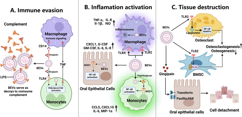
BEVs facilitate bacterial survival by invading the host, triggering the host response during periodontitis in numerous ways. First, BEVs can serve as decoys, protecting invading pathogens from bactericidal factors such as complement system compounds. For example, BEVs from either A. actinomycetemcomitans or P. gingivalis suppressed the activity of bactericidal factors in human serum, and BEVs released from A. actinomycetemcomitans, a complement immune system target, consumed complement components in an LPS-dependent manner, thereby shielding the orgnism from the bactericidal activity of complement factors [58, 59]. Second, BEVs can inhibit the functions of immune cells, thereby facilitating bacterial evasion of host immune defense. For example, BEVs released from P. gingivalis degraded the LPS-detecting CD14 receptor on human macrophage-like cells, reducing proinflammatory signaling in a gingipain-dependent manner [60]. Moreover, gingipains in P. gingivalis BEVs recognized Toll-like Receptor 4 and activated the PI3K/AKT/mTOR pathway in monocytes to induce selective TNF deficiency, hampering immune cell recognition of microbes (Fig. 3A) [61]. Third, in addition to evading innate immune cells (e.g., macrophages and monocytes), BEVs can facilitate periodontopathogen evasion of adaptive immune cells, such as T cells. Notably, small RNAs (similar in size to miRNAs) carried by BEVs released from periodontopathogens (e.g., A. actinomycetemcomitans or P. gingivalis) suppressed the production of certain cytokines, including IL-13 and IL-5, by Jurkat T cells, suggesting that signaling molecules carried by BEVs mediate immune evasion via bacteria-to-human interactions [62]. In summary, these findings suggest that BEVs aid in periodontopathogen evasion of the host immune system by serving as decoys to reduce bactericidal factor effectiveness and inhibiting the function of immune cells, thus favoring the survival of periodontopathogens.
Fig. 3.
Role of extracellular vesicles (EVs) and bacterial extracellular vesicles (BEVs) in the pathogenesis of periodontitis. A The effects of BEVs on immune invasion. BEVs can serve as decoys that disrupt the complement system, thereby protecting periodontopathogens from complement bactericidal effects. Moreover, BEVs can suppress microbial recognition by macrophages/monocytes by interacting with host cell membrane receptors. B BEVs released from periodontopathogens can bind to the pathogen recognition receptors (PRRs) of host cells, including TLR2, TLR4, and NOD1, triggering the activation of downstream proinflammatory signalling pathways. C BEVs released from periodontopathogens cause periodontal tissue destruction. BEVs can deliver virulence factors, including gingipain and cytolethal distending toxin (CDT), to oral epithelial cells, causing cell dysfunction and detachment. Additionally, BEVs can inhibit the osteogenesis of bone marrow stromal cells (BMSCs) and induce the osteoclastogenesis of osteoclast precursors, leading to bone tissue destruction. BEVs bacterial extracellular vesicles, PRR pathogen recognition receptor, TLR2 Toll-like receptor 2, TLR4 Toll-like receptor 4, NOD1 nucleotide-binding oligomerization domain-containing protein 1, CDT cytolethal distending toxin, BMSCs bone marrow stromal cells. Created with BioRender.com
Activation of inflammatory responses
Initiated by plaque biofilm bacteria, periodontitis is characterized by persistent tissue-destroying inflammation [63–65]. Since they can be enriched with virulence factors, BEVs can activate and amplify the host immune response in a manner similar to their parent bacteria. As the first line of defense against infection, the oral mucosal epithelium can be activated by BEVs to trigger innate immune signalling in multiple ways (Fig. 3B) [66]. The surface-exposed LPS or porins/lipoproteins on the outer leaflets of OMVs bind to epithelial cell pathogen recognition receptors (PRRs), including TLRs, and activate downstream proinflammatory signaling pathways [67]. For example, BEVs released from Filifactor alocis, a newly discovered periodontopathogen, activated TLR2 in human oral epithelial cells and enabled at least a 1.5-fold increase in the expression of proinflammatory cytokines, including IL-6, IL-8, G-CSF, GM-CSF, and chemokine (C-X-C motif) ligand-1 (CXCL-1) [68, 69]. When BEVs enter epithelial cells, virulence factors (e.g., peptidoglycan) carried by BEVs can activate an intracellular PRR, nucleotide-binding oligomerization domain-containing protein 1 (NOD1), leading to innate immune responses in epithelial cells [70]. For example, peptidoglycan delivered into host cells by BEVs activated NF-κB responses via a NOD-dependent mechanism and resulted in increased induction of innate immune signaling [71].
When BEVs pass the oral epithelial barrier (i.e., the gingival epithelium) and enter the underlying submucosal tissues, they interact directly with host immune cells, such as dendritic cells, monocytes and macrophages [72], leading to the activation of innate or adaptive immune responses [73, 74]. Similar to those on epithelial cells, the PRRs on host immune cells, including TLRs and NOD-like receptors, recognize BEV-carried danger-associated molecular patterns (e.g., LPS, peptidoglycans, proteins and nucleic acids) to trigger a cascade of inflammatory responses [71, 75, 76]. For example, BEVs produced from the periodontopathogenic “red complex”, consisting of P. gingivalis, T. forsythia and T. denticola, bound to PRRs on macrophages and monocytes, thereby leading to excessive activation of NF-κB and increased TNF-α, IL-1β, and IL-8 production [77]. Moreover, the BEVs produced by different parent bacterial cells in “red complexes” stimulated PRRs to different degrees. Compared with those released from either T. denticola or T. forsythia, BEVs released from P. gingivalis induced stronger TLR2- and TLR4-specific responses and more moderate responses in cells expressing NOD2, NOD1, TLR8, TLR7, and TLR4, confirming the dominant roles of P. gingivalis and related BEVs in the pathogenesis of periodontitis. In contrast, BEVs released from T. denticola or T. forsythia induced only weak responses [77]. When entering immune cells, the BEVs released from A. actinomycetemcomitans promoted the induction of cytoplasmic peptidoglycan sensor activity and led to NOD1-dependent NF-κB activation in monocytes, which significantly increased the production of chemokine (C–C motif) ligand (CCL)-5, CXCL-10, IL-6 and macrophage inflammatory protein (MIP)-1α [78]. These findings strongly suggest that BEVs are important pathogenic factors of bacteria, and the roles of BEVs in the development of periodontitis may differ from each other. Moreover, the ways that BEVs activate PRPs in immune cells determine downstream immune response signaling. However, most of these findings were based on studies performed in vitro, and further investigations under in vivo conditions are still needed.
Destruction of periodontal tissues
In addition to their activating effects on inflammation, periodontopathogens can produce various virulence factors that are involved in periodontal tissue destruction. As mentioned in the previous section, BEVs released from periodontopathogens can serve as vehicles for high concentrations of toxins, including gingipains and cytolethal distending toxin (CDT), causing dysfunction and detachment of host oral cells [79]. For example, P. gingivalis BEV-associated gingipains could compromise the function of epithelial cells by degrading integrin-related molecules and transferrin receptors [80, 81]. Moreover, the BEV-encapsulated gingipains could disrupt the fibronectin-integrin interactions in HGFs, leading to the formation of periodontal pockets [82]. CDT is associated with cell cycle arrest and apoptosis. The BEV-mediated release of CDT killed HGFs, possibly damaging the sulcular/junctional epithelium [83]. Apart from negatively affecting soft tissue, BEVs can transfer toxic components to osteoblasts and osteoclasts, leading to periodontal bone dyshomeostasis. By activating the TLR2 downstream signaling of the ERK and JNK pathways, BEVs released from F. alocis inhibited osteogenic differentiation and increased the RANKL/OPG ratio in bone marrow stromal cells (BMSCs) to promote osteoclast differentiation, resulting in increased bone resorption [84, 85] (Fig. 3C). Furthermore, increasing evidence has indicated that BEVs lead to periodontal bone loss indirectly by inducing hyperactivation of the immune response. BEVs released by F. nucleatum could induce M0-like macrophage switching to M1 macrophages, significantly increasing the production of proinflammatory cytokines. Moreover, in vivo studies have indicated that F. nucleatum BEVs increased osteoclast numbers and aggravated alveolar bone loss in a mouse model of periodontitis [86]. Overall, BEVs can transport virulence factors to host cells, impairing oral epithelial barrier structure and accelerating periodontal bone loss, thereby resulting in periodontal tissue destruction.
Development of systemic disease
Due to protection conferred by vesicle membrane structures, BEVs can diffuse into the bloodstream, enter host cells, and transfer toxic components to distant organs. Therefore, BEVs secreted by periodontopathogens not only contribute to the development of periodontitis but also play relevant roles in the progression of a variety of systemic diseases, including Alzheimer’s disease, diabetes, and cardiovascular disease, which may explain to some extent the involvement of periodontitis in other systemic diseases [87, 88].
Mounting evidence has indicated that P. gingivalis BEVs are risk factors for cardiovascular diseases such as atherosclerosis, wherein endothelial dysfunction, endothelial permeability, and calcium deposits play key roles during atherosclerosis development. Compared with parent bacteria, nanosized OMVs could easily migrate in the blood vessels where the parent bacteria cannot reach, causing vascular damage and contributing to the atherosclerosis process [89]. P. gingivalis BEVs have been reported to aggravate endothelial dysfunction by activating Rho kinase-induced ERK1/2 and p38 MAPK pathways [90]. Researchers have found that P. gingivalis BEVs decreased the expression of the endothelial adhesion molecule PECAM-1 (CD31), leading to enhanced endothelial permeability in vitro and in vivo [91, 92]. Furthermore, BEVs secreted from P. gingivalis activated the ERK1/2-RUNX2 pathway to induce calcification of vascular smooth muscle cells, thereby accelerating the development of atherosclerosis [93].
Alzheimer's disease, an inflammatory neurodegenerative condition, is characterized by myeloid plaques and neurofibrillary tangles. Accumulative evidence has shown that periodontitis is closely linked to Alzheimer's disease, suggesting that periodontitis may play a role in the progression of Alzheimer's disease via the transfer of periodontopathogenic BEVs [94]. For the first time, Han et al. demonstrated that BEVs from A. actinomycetemcomitans crossed the blood‒brain barrier and reached the brains of mice. By delivering extracellular RNA cargoes, BEVs significantly increased the expression of TNF-α [95]. The BEV-mediated integrity loss and permeability of the human blood‒brain barrier have also been reported by other researchers [96]. Furthermore, the Han et al. group found that BEVs released from A. actinomycetemcomitans could be taken up by brain immune cells, such as macrophages and microglial cells. In vitro experiments have shown that the extracellular RNA cargoes carried by BEVs enhanced the expression of IL-6 in microglial cells by activating NF-κB pathways [95, 97].
The metabolic disorder diabetes mellitus is characterized by elevated blood glucose. BEVs released from P. gingivalis can be delivered to the liver, where they accumulate, significantly reducing the synthesis of hepatic glycogen, attenuating insulin sensitivity, and resulting in high blood glucose levels. Mechanistically, gingipains in P. gingivalis OMVs could negatively regulate Akt/GSK-3β pathway activation, thereby attenuating insulin sensitivity and glycogen synthesis in liver cells [98].
In summary, these studies reveal a novel mechanism through which systemic diseases and periodontitis are closely related, implying that BEVs play vital roles in the association between systemic disease and periodontitis. However, clinical evidence and more detailed mechanistic studies are still required to test and validate these findings.
CEV-based therapeutic applications in periodontal regeneration
Periodontitis is characterized by continual and irreversible destruction of tissues supporting the tooth. Regenerating the lost periodontium, including the PDLs, alveolar bone, and cementum is the ultimate goal of periodontal treatment [3, 99]. Due to their regenerative capability and immunomodulatory effects, stem cell-based strategies are among the most promising regenerative therapies for periodontal regeneration [3, 100, 101]. However, stem cell-based therapies are limited due to great challenges, including the high cost of cell expansion, uncontrolled cell proliferation, the potential for tumorigenesis, and immunogenicity [102, 103]. Moreover, maintaining cell viability and controlling cell differentiation following cell transplantation in situ have yet to be achieved due to the complex periodontal microenvironment [104]. Mounting evidence has indicated that the therapeutic effects of stem cells are mainly derived from the release of CEVs [105, 106], and that CEVs can recapitulate the therapeutic effects of their parent cells [107, 108]. Therefore, stem cell-derived EV therapies are potential substitutes for stem cell-based therapies [109, 110]. Compared with those of their parent stem cells or other biomolecules, CEV-based regenerative therapies offer the following key advantages: (1) CEV-based strategies pose fewer risks, including unwanted transformation and immunogenicity [111]; (2) CEVs exert multifunctional effects to target diverse therapeutic mechanisms due to their cargoes, including abundant proteins, nucleic acids, and lipids (Table 1). Herein, we summarize the multifunctional biological effects of dental stem cell-derived CEVs on periodontal regeneration.
Table 1.
CEV-based therapeutic applications in periodontal regeneration
| CEV origin | CEV cargos | Involved pathway | Administration methods | Functional effects | Refs |
|---|---|---|---|---|---|
| PDLSC | miR-590-3p | miR-590-3p/TLR4 | Local injection | Inhibit macrophage pyroptosis | [113] |
| DPSC | miR-1246 | NF-κB p65, p38 MAPK | Chitosan hydrogel | Promote anti-inflammatory macrophage phenotype | [114] |
| TNF-α treated GMSC | CD73, miR-1260b | miR-1260b/Wnt5a/RANKL, JNK | Local injection | Promote macrophages toward M2 polarization, inhibit osteoclastogenesis | [116] |
| MSC | CD73 | AKT, ERK | Collagen sponge | Promote PDL cell migration and proliferation | [118] |
| SCAP | Cdc42 | – | Local injection | Promote angiogenesis | [119] |
| SHED | Wnt3a, BMP2 | AMPK, Wnt/β-catenin, BMP/Smad | β-TCP, Local injection | Promote osteogenesis and angiogenesis, repress inflammatory cytokines expression |
[110] [120] [121] |
| PDLSC | Wnt | – | β-TCP, Matrigel | Promote osteogenesis | [123] |
| LPS-Preconditioned DFC | Wnt3a, BMP2 | RANKL/ OPG | Gelatin/laponite hydrogel | Promote PDLC proliferation, migration, and osteogenic differentiation, inhibit osteoclast formation | [127] |
Anti-inflammatory effects
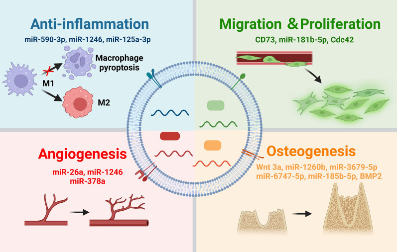
The resolution of inflammation is a prerequisite for periodontal tissue regeneration [112]. CEVs released from various types of oral stem cells have shown anti-inflammatory abilities, contributing to effective periodontitis treatment (Fig. 4). For example, CEVs released by stem cells from human exfoliated deciduous teeth (SHED-EVs) repressed IL-6 and TNF-α gene expression in bone marrow mesenchymal stem cells (BMMSCs) and prevented alveolar bone loss in mouse models of periodontitis [110]. In a similar study based on a murine model of periodontitis, CEVs released from PDLSCs decreased the production of IL-18, TNF-α, and IL-1β to alleviate bone loss in periodontitis. Mechanistically, PDLSC-derived CEVs expressed a stable level of miR-590-3p, which can suppress TLR4 downstream signaling pathways, thereby inhibiting macrophage pyroptosis [113]. In addition, CEVs released from dental pulp stem cells (DPSC-EVs) induced macrophage polarization to an anti-inflammatory phenotype. The immunomodulatory effects of DPSC-EVs were mainly the result of miR-125-3p and miR-1246 carried by DPSC-EVs. By incorporating chitosan, these DPSC-EVs accelerated the healing of the damaged periodontal epithelium and alveolar bone [114, 115].
Fig. 4.
The bioactive effects of CEVs and their cargoes on anti-inflammatory responses, angiogenesis, cell migration and proliferation, and osteogenesis. Functional nucleic acid and protein cargoes carried by CEVs are classified into four panels on the basis of their effects. Created with BioRender.com
Since CEV-encapsulated bioactive molecules, including nucleotides, proteins, or lipids manifest the biological effects of CEVs, strategies to change the content compositions of CEVs may significantly enhance the anti-inflammatory functions of CEVs. To this end, Nakao et al. reported that TNF-α pretreatment increased not only the amount of CEVs secreted from gingival tissue-derived MSCs (GMSCs) but also the immunomodulatory effects of GMSC-EVs by inducing a higher anti-inflammatory M2 macrophage polarization rate. Mechanistically, highly expressed exosomal CD73 in TNF-α-pretreated GMSC-EVs is responsible for enhanced M2 polarization [116]. In another report, cyclic stretch force enhanced the inhibitory effects of PDL-derived CEVs on IL-1β production in LPS-stimulated macrophages by suppressing the nuclear translocation of NF-κB and the binding of NF-κB-p65 to DNA [117]. These findings suggest that preconditioning parent cells with periodontitis-related stimuli, including mechanical stress and inflammatory factors, is an effective way to enhance the anti-inflammatory effects of CEVs, providing an easy-to-use and effective strategy for CEV modification.
Regulation of cell proliferation, migration, osteogenesis, and angiogenesis
The proliferation, migration, and differentiation of endogenous stem/progenitor cells at an injury site, as well as angiogenesis, are indispensable steps in periodontal regeneration (Fig. 4). Chew et al. reported that MSC-derived CEVs promoted periodontal tissue regeneration by increasing cellular proliferation and migration. In vitro studies indicated that MSC-EVs activated prosurvival AKT and ERK signaling in PDL cells by presenting exosomal CD73 proteins [118]. In addition to MSC-EVs, CEVs derived from stem cells in apical papilla (SCAP-EVs) promoted the migration of HUVECs by transferring cell division cycle 42 (Cdc42) to these cells. As a result, SCAP-EVs accelerated palatal gingiva regeneration by promoting angiogenesis [119]. SHED-EVs have also been reported to promote the proliferation and migration of HUVECs and BMSCs and to enhance the angiogenesis-related differentiation of HUVECs and osteogenesis-related differentiation of BMSCs [120]. Additional mechanistic investigations suggested that the bioactivities of SHED-EVs were mainly due to the encapsulated Wnt3a and BMP2. By increasing nuclear β-catenin protein abundance and upregulating Smad1/5/8 phosphorylation, SHED-EVs activated Wnt/β-catenin and BMP/Smad signaling in stem cells respectively, and significantly promoted the osteogenic differentiation of BMSCs [120, 121]. In addition to engagement with osteogenic differentiation, miR-26a in SHED-EVs could promote angiogenesis via the TGF-β/SMAD2/3 pathway [122]. The combination of SHED-EVs with a β-TCP scaffold induced both neovascularization and new bone formation, thus promoting alveolar bone regeneration in a rat model of periodontal defects [120]. The biological effects of PDLSC-EVs (PDLSC-EVs) have been extensively investigated. In a rat model of periodontitis, PDLSC-EVs restored the osteogenic differentiation ability of PDLSCs in inflamed periodontal tissue by activating the Wnt signaling pathway, thereby accelerating bone healing [123]. By carrying miR-1246, CEVs induced angiogenesis by activating Smad 1/5/8 signaling [124].
The pro-osteogenic or pro-angiogenic effects of MSC-EVs can be enhanced via modification of their parent cells before CEV isolation. These modifications mainly include gene modifications, mechanical stimulation, and inflammation preconditioning. For example, Xu et al. recently showed that CEVs secreted by P2X7 receptor-encoding gene-modified PDLSCs rescued inflammation-impaired osteogenesis of PDLSCs. Gene modification could increase the abundance of miR-6747-5p, miR-6515-5p, and miR-3679-5p in CEVs, which could bind to the GREB-1 protein in different ways [125]. Lv et al. found that CEVs derived from osteocytes exposed to mechanical strain induced significantly increased proliferation of PDLSCs through the activation of miR-185b-5p/PTEN/AKT pathways and promoted osteogenic differentiation mediated via BMP/Runx2 in an inflammatory environment [126]. CEVs released from LPS-pretreated dental folic cells (DFCs) facilitated the migration, proliferation, and osteogenic differentiation of inflamed PDLSCs, accelerating periodontal regeneration in an inflammatory microenvironment [127]. Our group indicated that CEVs secreted by dental pulp stem cells compromised by periodontitis significantly enhanced angiogenesis via miR-378a they carried. Angiogenesis-related microRNAs (miRNAs) carried by CEVs could an silence Sufu expression in endothelial cells and activate Hedgehog/Gli1 signaling to stimulate endothelial cell proliferation, migration, and tube formation [128, 129].
Collectively, these studies highlight the multiplicativity of CEVs in regulating the behaviors of stem cells. Moreover, the bioactive effects of CEVs, such as pro-osteogenic and pro-angiogenetic effects of CEVs, can be modified and enhanced by preconditioning parental cells. Given that tissue regeneration is a complex and cascading process requiring coordinated control of multiple factors (e.g., cell migration, differentiation, and angiogenesis), CEVs represent promising agents for regenerative medicine, including but not limited to periodontal regeneration.
The prospects for BEV-based therapeutic applications in periodontal regeneration
Although the pathogenic roles of BEVs in periodontitis have been widely studied and recognized, the potential applications of periodontopathogen-derived BEVs are still in their infancy. Compared with stem cell-derived EVs, BEVs show unique advantages such as cost-effective production. Large-scale isolation and purification of CEVs require extensive labor and time costs, which has hindered their clinical translation. In contrast, BEVs can be easily produced in cost-effective ways via large-scale cultivation of bacteria [130]. Specifically, bioreactors and hypervesiculating mutant strains have been successfully developed to increase the production of BEVs [131, 132]. To achieve widespread clinical use in periodontitis, a nonlife-threatening disease with a high prevalence, the treatment must be both cost-effective and scientifically sound [6]. From this point of view, the low cost and large-scale production of BEVs make BEV-based therapies more promising for periodontitis treatment.
As a bacteria-driven disease, periodontitis can be treated by abrogating the survival of periodontopathogens and preventing the formation of plaque biofilms. Due to the impermeability of the host cell membrane to antibiotics, working concentrations of antibiotics fail to kill intracellular bacteria [133, 134]. Notably, the periodontopathogens P. gingivalis and A. actinomycetemcomitans were grown intracellularly in human buccal epithelial cells [135], and antibiotics alone did not eliminate them [136]. Due to their high permeability with respect to host cells, BEVs can kill intracellular pathogens more effectively than antibiotics [137]. Compared with soluble antibiotics, such as gentamicin, BEVs have more killing power by delivering autolysin and peptidoglycan hydrolases [138, 139]. In addition, BEVs can be employed for antibiotic drug delivery to enhance the uptake of antibiotics, leading to superior antibacterial efficiency of conventional antibiotics [137]. Therefore, we suggest that BEVs be employed for antibiotic drug delivery or as potential antibacterial substances for effectively killing intracellular periodontopathogens. Moreover, BEVs can prevent the formation of plaque biofilms by inhibiting bacterial adhesion to host cells, further alleviating antibiotic resistance development [140]. Specifically, adhesins in BEVs inherited from their parent bacteria can inhibit bacterial adhesion to host cells. For instance, BEV-coated nanoparticles derived from Helicobacter pylori effectively inhibited H. pylori adhesion to gastric epithelial cell tissues in a dose-dependent fashion [141].
With bioengineering or detoxification, OMVs can also be used as biological carriers for vaccines. It has been reported that P. gingivalis OMVs retain the immunodominant determinant of P. gingivalis, as demonstrated by the increased production of salivary IgA, serum IgG and IgA in mice following intranasal administration [142]. The mucosal immune response elicited by P. gingivalis OMVs could further enhance the clearance of P. gingivalis in an oral infection model [143]. The strong immunogenicity of P. gingivalis OMVs is mainly derived from LPS and A-LPS-modified proteins in OMVs, and absorption of serum with LPS results in a dramatic reduction in immmunoreactivity [144]. Despite these progresses, the application of OMVs as vaccination is still in its infancy and extensive work is required to decrease their side effects.
Challenges and future directions
The lack of standard and cost-effective methods for separating and purifying EVs represents a major barrier to EV-based therapies. Methods currently used to separate and purify EVs include ultracentrifugation, precipitation, ultrafiltration, chromatography, and immunoaffinity capture. Among these methods, separating EVs by ultracentrifugation is the gold standard method. However, from a clinical-scale manufacturing perspective, ultracentrifugation is not ideal because of its low productivity and purity outputs [24]. Tangential-flow filtration (TFF), a method for concentrating EVs from a medium based on size, is a promising method. Compared with ultracentrifugation, TFF concentrates up to 100-fold more EVs while enhancing the removal of unwanted albumin [145, 146]. Among the reported methods for EV purification, immunoaffinity capture might be the most promising method since it is based on antibody-coupled magnetic beads and can yield highly purified EVs [147]. Due to the heterogeneity of EVs, the surface protein composition of EVs with various origins can be quite different [148–150]. Among the reported surface markers, transmembrane proteins such as CD9, CD63, and CD81 have been widely utilized for immunoaffinity-based EV extraction [151]. Future EV manufacturing methods require both high yield and high purity, and a combined separation/purification approach may be better than single-step procedures. Therefore, among these methods, combinations of TFF and immunoaffinity capture or other steps for high purification represent the best methods to produce clinical-grade EVs.
The rapid clearance of EVs following systemic administration is also a challenge for EV-based regenerative strategies and cannot be ignored. After systemic administration, EVs can be cleared rapidly in the body, and most EVs cannot reach target sites to exert their bioactive effects [152]. Prolonging the retention of EVs in a desired area by immobilizing EVs within biomaterials may enhance EV therapeutic efficacy. Specifically, anchoring EVs in biomaterials can extend their bioactivity following administration, control their release, and potentially improve therapeutic efficacy [153–155]. More importantly, given the complexity of periodontal regeneration, which includes the oriented insertion of newly formed periodontal ligaments in bone and cementum, EVs alone cannot coordinate the signaling regulation needed during the aligned formation of the bone–ligament–cementum complex. However, well-designed multiphasic scaffolds with precise compartments for tissue regeneration and integration are promising for mediating spatiotemporal events during periodontal regeneration [156, 157]. Therefore, we believe that advanced multiphasic biomaterial scaffolds combined with spatiotemporally released bioactive EVs represent a potential application for periodontal regeneration.
In most studies, the pathogenic roles of EVs in periodontitis are recognized based on in vitro cell culture systems. Therefore, in vivo experiments are urgently required to provide strong evidence of their effects. Moreover, most studies use bulk separation of heterogeneous EVs obtained from culture medium or clinical samples to analyze the biological effects of EVs; however, effects due to the heterogeneity of the biology, structure, and function of single vesicles are generally not considered in these studies. Recent technological advances with in vivo imaging and single-vesicle analysis have enabled researchers to overcome these two main limitations and provide us with novel tools to study the roles of EVs in periodontal homeostasis at the single-vesicle level [148, 158]. Advancing our knowledge regarding the role of EVs in periodontal homeostasis based on these advanced approaches can shed new light on treatment options for periodontal disease.
Conclusion and perspective
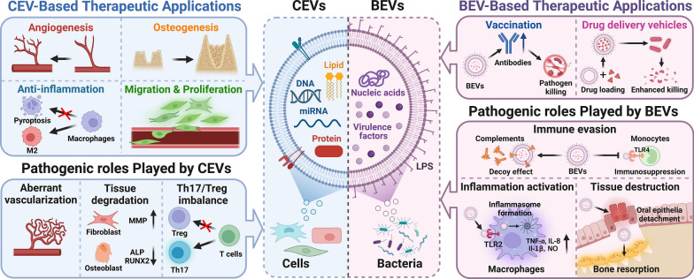
Overall, since EVs secreted by bacteria or cells play key roles in the interaction between the bacteria and host, the role of EVs in maintaining periodontal homeostasis is unquestionable. In this article, we thoroughly reviewed the pathogenic and therapeutic roles of BEVs and CEVs in periodontal homeostasis. By delivering different virulence factors, BEVs released from periodontal pathogens are envisaged as important missing pieces needed to reveal the imbalance in periodontal homeostasis and factors in periodontal disease progression. The multifunctional bioactivities of CEVs provide novel avenues by which to regain the loss of periodontal homeostasis and promote periodontal regeneration (Fig. 5). Despite great progress, the clinical translation of EVs is still in its infancy due to longstanding challenges, such as a lack of cost-effective production and rapid clearance in vivo. With the development of advanced biomaterials, the combination of EVs and multiphasic scaffolds holds great promise for periodontal regeneration.
Fig. 5.
The roles of cell-derived extracellular vesicles (CEVs) and bacteria-derived extracellular vesicles (BEVs) in periodontal homeostasis. BEVs released from periodontopathogens contain virulence factors resulting in immune evasion, inflammation activation and tissue destruction. BEVs can also be used as biological carrier for vaccines and antibiotic drugs. CEVs released from cells contain bioactive cargos which can exert multiple biological effects, such as anti-inflammation, pro-migration & proliferation, pro-angiogenesis, and pro-osteogenesis. However, CEVs could also exert pathogenic effects under certain conditions. Created with BioRender.com
Acknowledgements
Not applicable.
Abbreviations
- EVs
Extracellular vesicles
- BEVs
Bacteria-derived EVs
- CEVs
Cell-derived EVs
- PDLs
Periodontal ligaments
- TLRs
Toll-like receptors
- IL
Interleukin
- RANKL
Receptor Activator for Nuclear Factor-κB Ligand
- MVEs
Multivesicular endosomes
- ILVs
Intraluminal vesicles
- OMVs
Outer membrane vesicles
- LPSs
Lipopolysaccharides
- OIMVs
Outer-inner membrane vesicles
- CMVs
Cytoplasmic membrane vesicles
- PDLSCs
Periodontal ligament stem cells
- Treg
Regulatory T-cell
- PDLFs
Periodontal ligament fibroblasts
- TNF-α
Tumor necrosis factor-α
- HUVECs
Human umbilical vein endothelial cells
- HGFs
Human gingival fibroblasts
- ECM
Extracellular matrix
- MCP-1
Monocyte chemoattractant protein-1
- PRPs
Pathogen recognition receptors
- CXCL
Chemokine (C-X-C motif) ligand
- NOD1
Nucleotide-binding oligomerization domain-containing protein 1
- CCL
Chemokine (C–C motif) ligand
- MIP-1α
Macrophage inflammatory protein-1α
- CDT
Cytolethal distending toxin
- BMSCs
Bone marrow stromal cells
- SHED
Stem cells from human exfoliated deciduous teeth
- DPSC
Dental pulp stem cells
- GMSCs
Gingival tissue-derived MSCs
- SCAP
Stem cells in apical papilla
- Cdc42
Cell division cycle 42
- DFCs
Dental folic cells
- TFF
Tangential-flow filtration
Author contributions
XTH, FMC and XL contributed to the conceptualization, writing-review & editing, funding acquisition, validation and supervision of the manuscript. DKD, XTH, JJZ and DG contributed to writing—original draft, methodology and data curation. DKD, XL, DG and JJZ helped design the figures. RXW, YT and YY provided guidance and revised the manuscript. All authors read and approved the final manuscript.
Funding
This work was supported by the National Natural Science Foundation of China (Grant Nos. 82130026, 82001102, and 81970947), the Young Elite Scientist Support Program administered by CSA (2021PYRC001), the Young Elite Scientist Sponsorship Program by CAST (2022QNRC001) and the Natural Science Basic Research Program of Shaanxi (2022JQ-959).
Availability of data and materials
Not applicable.
Declarations
Ethics approval and consent to participate
Not applicable.
Consent for publication
All authors gave their consent for publication
Competing interests
The authors declare that they have no competing interests.
Footnotes
Publisher's Note
Springer Nature remains neutral with regard to jurisdictional claims in published maps and institutional affiliations.
Dao-Kun Deng, Jiu-Jiu Zhang and Dian Gan contributed equally to this work
Contributor Information
Xuan Li, Email: li_xuan15@163.com.
Fa-Ming Chen, Email: cfmsunhh@fmmu.edu.cn.
Xiao-Tao He, Email: hexiao__tao@163.com.
References
- 1.Tonetti MS, Jepsen S, Jin L, Otomo-Corgel J. Impact of the global burden of periodontal diseases on health, nutrition and wellbeing of mankind: a call for global action. J Clin Periodontol. 2017;44:456–462. doi: 10.1111/jcpe.12732. [DOI] [PubMed] [Google Scholar]
- 2.Genco RJ, Sanz M. Clinical and public health implications of periodontal and systemic diseases: an overview. Periodontol. 2000;2020(83):7–13. doi: 10.1111/prd.12344. [DOI] [PubMed] [Google Scholar]
- 3.Xu XY, Li X, Wang J, He XT, Sun HH, Chen FM. Concise review: periodontal tissue regeneration using stem cells: strategies and translational considerations. Stem Cells Transl Med. 2019;8:392–403. doi: 10.1002/sctm.18-0181. [DOI] [PMC free article] [PubMed] [Google Scholar]
- 4.Ravidà A, Qazi M, Rodriguez MV, Galli M, Saleh MHA, Troiano G, Wang HL. The influence of the interaction between staging, grading and extent on tooth loss due to periodontitis. J Clin Periodontol. 2021;48:648–658. doi: 10.1111/jcpe.13430. [DOI] [PubMed] [Google Scholar]
- 5.Hajishengallis G. Periodontitis: from microbial immune subversion to systemic inflammation. Nat Rev Immunol. 2015;15:30–44. doi: 10.1038/nri3785. [DOI] [PMC free article] [PubMed] [Google Scholar]
- 6.Chen FM, Wu LA, Zhang M, Zhang R, Sun HH. Homing of endogenous stem/progenitor cells for in situ tissue regeneration: promises, strategies, and translational perspectives. Biomaterials. 2011;32:3189–3209. doi: 10.1016/j.biomaterials.2010.12.032. [DOI] [PubMed] [Google Scholar]
- 7.Luan X, Zhou X, Trombetta-eSilva J, Francis M, Gaharwar AK, Atsawasuwan P, Diekwisch TGH. MicroRNAs and periodontal homeostasis. J Dent Res. 2017;96:491–500. doi: 10.1177/0022034516685711. [DOI] [PMC free article] [PubMed] [Google Scholar]
- 8.Hans M, Hans VM. Toll-like receptors and their dual role in periodontitis: a review. J Oral Sci. 2011;53:263–271. doi: 10.2334/josnusd.53.263. [DOI] [PubMed] [Google Scholar]
- 9.Wang IC, Sugai JV, Majzoub J, Johnston J, Giannobile WV, Wang HL. Pro-inflammatory profiles in cardiovascular disease patients with peri-implantitis. J Periodontol. 2022;93:824–836. doi: 10.1002/JPER.21-0419. [DOI] [PubMed] [Google Scholar]
- 10.Tsukasaki M, Komatsu N, Nagashima K, Nitta T, Pluemsakunthai W, Shukunami C, Iwakura Y, Nakashima T, Okamoto K, Takayanagi H. Host defense against oral microbiota by bone-damaging T cells. Nat Commun. 2018;9:701. doi: 10.1038/s41467-018-03147-6. [DOI] [PMC free article] [PubMed] [Google Scholar]
- 11.Deatherage BL, Cookson BT. Membrane vesicle release in bacteria, eukaryotes, and archaea: a conserved yet underappreciated aspect of microbial life. Infect Immun. 2012;80:1948–1957. doi: 10.1128/IAI.06014-11. [DOI] [PMC free article] [PubMed] [Google Scholar]
- 12.Woith E, Fuhrmann G, Melzig MF. Extracellular vesicles-connecting kingdoms. Int J Mol Sci. 2019;20:5695. doi: 10.3390/ijms20225695. [DOI] [PMC free article] [PubMed] [Google Scholar]
- 13.Coumans FAW, Brisson AR, Buzas EI, Dignat-George F, Drees EEE, El-Andaloussi S, Emanueli C, Gasecka A, Hendrix A, Hill AF, Lacroix R, Lee Y, van Leeuwen TG, Mackman N, Mäger I, Nolan JP, van der Pol E, Pegtel DM, Sahoo S, Siljander PRM, Sturk G, de Wever O, Nieuwland R. Methodological guidelines to study extracellular vesicles. Circ Res. 2017;120:1632–1648. doi: 10.1161/CIRCRESAHA.117.309417. [DOI] [PubMed] [Google Scholar]
- 14.van Niel G, D'Angelo G, Raposo G. Shedding light on the cell biology of extracellular vesicles. Nat Rev Mol Cell Biol. 2018;19:213–228. doi: 10.1038/nrm.2017.125. [DOI] [PubMed] [Google Scholar]
- 15.Marar C, Starich B, Wirtz D. Extracellular vesicles in immunomodulation and tumor progression. Nat Immunol. 2021;22:560–570. doi: 10.1038/s41590-021-00899-0. [DOI] [PMC free article] [PubMed] [Google Scholar]
- 16.Kornman KS. Mapping the pathogenesis of periodontitis: a new look. J Periodontol. 2008;79(Suppl 8):1560–1568. doi: 10.1902/jop.2008.080213. [DOI] [PubMed] [Google Scholar]
- 17.Yucel-Lindberg T, Båge T. Inflammatory mediators in the pathogenesis of periodontitis. Expert Rev Mol Med. 2013;15:e7. doi: 10.1017/erm.2013.8. [DOI] [PubMed] [Google Scholar]
- 18.Meyle J, Chapple I. Molecular aspects of the pathogenesis of periodontitis. Periodontol. 2000;2015(69):7–17. doi: 10.1111/prd.12104. [DOI] [PubMed] [Google Scholar]
- 19.Stremersch S, De Smedt SC, Raemdonck K. Therapeutic and diagnostic applications of extracellular vesicles. J Control Release. 2016;244:167–183. doi: 10.1016/j.jconrel.2016.07.054. [DOI] [PubMed] [Google Scholar]
- 20.Wiklander OPB, Brennan MÁ, Lötvall J, Breakefield XO, El Andaloussi S. Advances in therapeutic applications of extracellular vesicles. Sci Transl Med. 2019;11:eaav8521. doi: 10.1126/scitranslmed.aav8521. [DOI] [PMC free article] [PubMed] [Google Scholar]
- 21.Riazifar M, Pone EJ, Lötvall J, Zhao W. Stem cell extracellular vesicles: extended messages of regeneration. Annu Rev Pharmacol Toxicol. 2017;57:125–154. doi: 10.1146/annurev-pharmtox-061616-030146. [DOI] [PMC free article] [PubMed] [Google Scholar]
- 22.Hua S, Bartold PM, Gulati K, Moran CS, Ivanovski S, Han P. Periodontal and dental pulp cell-derived small extracellular vesicles: a review of the current status. Nanomaterials (Basel) 2021;11:1858. doi: 10.3390/nano11071858. [DOI] [PMC free article] [PubMed] [Google Scholar]
- 23.Han C, Zhou J, Liang C, Liu B, Pan X, Zhang Y, Wang Y, Yan B, Xie W, Liu F, et al. Human umbilical cord mesenchymal stem cell derived exosomes encapsulated in functional peptide hydrogels promote cardiac repair. Biomater Sci. 2019;7:2920–2933. doi: 10.1039/c9bm00101h. [DOI] [PubMed] [Google Scholar]
- 24.Ahn SH, Ryu SW, Choi H, You S, Park J, Choi C. Manufacturing therapeutic exosomes: from bench to industry. Mol Cells. 2022;45:284–290. doi: 10.14348/molcells.2022.2033. [DOI] [PMC free article] [PubMed] [Google Scholar]
- 25.Kalluri R, LeBleu VS. The biology, function, and biomedical applications of exosomes. Science. 2020;367:eaau6977. doi: 10.1126/science.aau6977. [DOI] [PMC free article] [PubMed] [Google Scholar]
- 26.Wen C, Seeger RC, Fabbri M, Wang L, Wayne AS, Jong AY. Biological roles and potential applications of immune cell-derived extracellular vesicles. J Extracell Vesicles. 2017;6:1400370. doi: 10.1080/20013078.2017.1400370. [DOI] [PMC free article] [PubMed] [Google Scholar]
- 27.Tiku V, Tan MW. Host immunity and cellular responses to bacterial outer membrane vesicles. Trends Immunol. 2021;42:1024–1036. doi: 10.1016/j.it.2021.09.006. [DOI] [PubMed] [Google Scholar]
- 28.Xie J, Li Q, Haesebrouck F, Van Hoecke L, Vandenbroucke RE. The tremendous biomedical potential of bacterial extracellular vesicles. Trends Biotechnol. 2022;40:1173–1194. doi: 10.1016/j.tibtech.2022.03.005. [DOI] [PubMed] [Google Scholar]
- 29.Toyofuku M, Nomura N, Eberl L. Types and origins of bacterial membrane vesicles. Nat Rev Microbiol. 2019;17:13–24. doi: 10.1038/s41579-018-0112-2. [DOI] [PubMed] [Google Scholar]
- 30.Toyofuku M, Cárcamo-Oyarce G, Yamamoto T, Eisenstein F, Hsiao CC, Kurosawa M, Gademann K, Pilhofer M, Nomura N, Eberl L. Prophage-triggered membrane vesicle formation through peptidoglycan damage in Bacillus subtilis. Nat Commun. 2017;8:481. doi: 10.1038/s41467-017-00492-w. [DOI] [PMC free article] [PubMed] [Google Scholar]
- 31.Hafiane A, Daskalopoulou SS. Extracellular vesicles characteristics and emerging roles in atherosclerotic cardiovascular disease. Metabolism. 2018;85:213–222. doi: 10.1016/j.metabol.2018.04.008. [DOI] [PubMed] [Google Scholar]
- 32.Takeuchi T. Pathogenic and protective roles of extracellular vesicles in neurodegenerative diseases. J Biochem. 2021;169:181–186. doi: 10.1093/jb/mvaa131. [DOI] [PubMed] [Google Scholar]
- 33.Novello S, Pellen-Mussi P, Jeanne S. Mesenchymal stem cell-derived small extracellular vesicles as cell-free therapy: perspectives in periodontal regeneration. J Periodontal Res. 2021;56:433–442. doi: 10.1111/jre.12866. [DOI] [PubMed] [Google Scholar]
- 34.Bartold PM, Van Dyke TE. Periodontitis: a host-mediated disruption of microbial homeostasis. Unlearning learned concepts. Periodontol. 2000;2013(62):203–217. doi: 10.1111/j.1600-0757.2012.00450.x. [DOI] [PMC free article] [PubMed] [Google Scholar]
- 35.Wang L, Wang J, Jin Y, Gao H, Lin X. Oral administration of all-trans retinoic acid suppresses experimental periodontitis by modulating the Th17/Treg imbalance. J Periodontol. 2014;85:740–750. doi: 10.1902/jop.2013.130132. [DOI] [PubMed] [Google Scholar]
- 36.Wang L, Guan N, Jin Y, Lin X, Gao H. Subcutaneous vaccination with Porphyromonas gingivalis ameliorates periodontitis by modulating Th17/Treg imbalance in a murine model. Int Immunopharmacol. 2015;25:65–73. doi: 10.1016/j.intimp.2015.01.007. [DOI] [PubMed] [Google Scholar]
- 37.Zheng Y, Dong C, Yang J, Jin Y, Zheng W, Zhou Q, Liang Y, Bao L, Feng G, Ji J, Feng X, Gu Z. Exosomal microRNA-155-5p from PDLSCs regulated Th17/Treg balance by targeting sirtuin-1 in chronic periodontitis. J Cell Physiol. 2019;234:20662–20674. doi: 10.1002/jcp.28671. [DOI] [PubMed] [Google Scholar]
- 38.Zhao M, Dai W, Wang H, Xue C, Feng J, He Y, Wang P, Li S, Bai D, Shu R. Periodontal ligament fibroblasts regulate osteoblasts by exosome secretion induced by inflammatory stimuli. Arch Oral Biol. 2019;105:27–34. doi: 10.1016/j.archoralbio.2019.06.002. [DOI] [PubMed] [Google Scholar]
- 39.Zhang Z, Shuai Y, Zhou F, Yin J, Hu J, Guo S, Wang Y, Liu W. PDLSCs regulate angiogenesis of periodontal ligaments via VEGF transferred by exosomes in periodontitis. Int J Med Sci. 2020;17:558–567. doi: 10.7150/ijms.40918. [DOI] [PMC free article] [PubMed] [Google Scholar]
- 40.Bi J, Koivisto L, Owen G, Huang P, Wang Z, Shen Y, Bi L, Rokka A, Haapasalo M, Heino J, Häkkinen L, Larjava HS. Epithelial microvesicles promote an inflammatory phenotype in fibroblasts. J Dent Res. 2016;95:680–688. doi: 10.1177/0022034516633172. [DOI] [PubMed] [Google Scholar]
- 41.Nowotny A, Behling UH, Hammond B, Lai CH, Listgarten M, Pham PH, Sanavi F. Release of toxic microvesicles by Actinobacillus actinomycetemcomitans. Infect Immun. 1982;37:151–154. doi: 10.1128/iai.37.1.151-154.1982. [DOI] [PMC free article] [PubMed] [Google Scholar]
- 42.Grenier D, Mayrand D. Functional characterization of extracellular vesicles produced by Bacteroides gingivalis. Infect Immun. 1987;55:111–117. doi: 10.1128/iai.55.1.111-117.1987. [DOI] [PMC free article] [PubMed] [Google Scholar]
- 43.Ma L, Cao Z. Membrane vesicles from periodontal pathogens and their potential roles in periodontal disease and systemic illnesses. J Periodontal Res. 2021;56:646–655. doi: 10.1111/jre.12884. [DOI] [PubMed] [Google Scholar]
- 44.Pathirana RD, Kaparakis-Liaskos M. Bacterial membrane vesicles: biogenesis, immune regulation and pathogenesis. Cell Microbiol. 2016;18:1518–1524. doi: 10.1111/cmi.12658. [DOI] [PubMed] [Google Scholar]
- 45.Kato S, Kowashi Y, Demuth DR. Outer membrane-like vesicles secreted by Actinobacillus actinomycetemcomitans are enriched in leukotoxin. Microb Pathog. 2002;32:1–13. doi: 10.1006/mpat.2001.0474. [DOI] [PubMed] [Google Scholar]
- 46.Veith PD, Chen YY, Gorasia DG, Chen D, Glew MD, O'Brien-Simpson NM, Cecil JD, Holden JA, Reynolds EC. Porphyromonas gingivalis outer membrane vesicles exclusively contain outer membrane and periplasmic proteins and carry a cargo enriched with virulence factors. J Proteome Res. 2014;13:2420–2432. doi: 10.1021/pr401227e. [DOI] [PubMed] [Google Scholar]
- 47.Jin JS, Kwon SO, Moon DC, Gurung M, Lee JH, Kim SI, Lee JC. Acinetobacter baumannii secretes cytotoxic outer membrane protein A via outer membrane vesicles. PLoS ONE. 2011;6:e17027. doi: 10.1371/journal.pone.0017027. [DOI] [PMC free article] [PubMed] [Google Scholar]
- 48.Unal CM, Schaar V, Riesbeck K. Bacterial outer membrane vesicles in disease and preventive medicine. Semin Immunopathol. 2011;33:395–408. doi: 10.1007/s00281-010-0231-y. [DOI] [PubMed] [Google Scholar]
- 49.Ho MH, Chen CH, Goodwin JS, Wang BY, Xie H. Functional advantages of Porphyromonas gingivalis vesicles. PLoS ONE. 2015;10:e0123448. doi: 10.1371/journal.pone.0123448. [DOI] [PMC free article] [PubMed] [Google Scholar]
- 50.Fleetwood AJ, Lee MKS, Singleton W, Achuthan A, Lee MC, O'Brien-Simpson NM, Cook AD, Murphy AJ, Dashper SG, Reynolds EC, Hamilton JA. Metabolic remodeling, inflammasome activation, and pyroptosis in macrophages stimulated by Porphyromonas gingivalis and its outer membrane vesicles. Front Cell Infect Microbiol. 2017;7:351. doi: 10.3389/fcimb.2017.00351. [DOI] [PMC free article] [PubMed] [Google Scholar]
- 51.Cecil JD, O'Brien-Simpson NM, Lenzo JC, Holden JA, Singleton W, Perez-Gonzalez A, Mansell A, Reynolds EC. Outer membrane vesicles prime and activate macrophage inflammasomes and cytokine secretion in vitro and in vivo. Front Immunol. 2017;8:1017. doi: 10.3389/fimmu.2017.01017. [DOI] [PMC free article] [PubMed] [Google Scholar]
- 52.Friedrich V, Gruber C, Nimeth I, Pabinger S, Sekot G, Posch G, Altmann F, Messner P, Andrukhov O, Schäffer C. Outer membrane vesicles of Tannerella forsythia: biogenesis, composition, and virulence. Mol Oral Microbiol. 2015;30:451–473. doi: 10.1111/omi.12104. [DOI] [PMC free article] [PubMed] [Google Scholar]
- 53.Hajishengallis G, Darveau RP, Curtis MA. The keystone-pathogen hypothesis. Nat Rev Microbiol. 2012;10:717–725. doi: 10.1038/nrmicro2873. [DOI] [PMC free article] [PubMed] [Google Scholar]
- 54.Gui MJ, Dashper SG, Slakeski N, Chen YY, Reynolds EC. Spheres of influence: Porphyromonas gingivalis outer membrane vesicles. Mol Oral Microbiol. 2016;31:365–378. doi: 10.1111/omi.12134. [DOI] [PubMed] [Google Scholar]
- 55.Kamaguchi A, Nakayama K, Ichiyama S, Nakamura R, Watanabe T, Ohta M, Baba H, Ohyama T. Effect of Porphyromonas gingivalis vesicles on coaggregation of Staphylococcus aureus to oral microorganisms. Curr Microbiol. 2003;47:485–491. doi: 10.1007/s00284-003-4069-6. [DOI] [PubMed] [Google Scholar]
- 56.Inagaki S, Onishi S, Kuramitsu HK, Sharma A. Porphyromonas gingivalis vesicles enhance attachment, and the leucine-rich repeat BspA protein is required for invasion of epithelial cells by "Tannerella forsythia". Infect Immun. 2006;74:5023–5028. doi: 10.1128/IAI.00062-06. [DOI] [PMC free article] [PubMed] [Google Scholar]
- 57.Grenier D. Porphyromonas gingivalis outer membrane vesicles mediate coaggregation and piggybacking of Treponema denticola and Lachnoanaerobaculum saburreum. Int J Dent. 2013;2013:305476. doi: 10.1155/2013/305476. [DOI] [PMC free article] [PubMed] [Google Scholar]
- 58.Lindholm M, Min Aung K, Nyunt Wai S, Oscarsson J. Role of OmpA1 and OmpA2 in Aggregatibacter actinomycetemcomitans and Aggregatibacter aphrophilus serum resistance. J Oral Microbiol. 2018;11:1536192. doi: 10.1080/20002297.2018.1536192. [DOI] [PMC free article] [PubMed] [Google Scholar]
- 59.Lindholm M, Metsäniitty M, Granström E, Oscarsson J. Outer membrane vesicle-mediated serum protection in Aggregatibacter actinomycetemcomitans. J Oral Microbiol. 2020;12:1747857. doi: 10.1080/20002297.2020.1747857. [DOI] [PMC free article] [PubMed] [Google Scholar]
- 60.Duncan L, Yoshioka M, Chandad F, Grenier D. Loss of lipopolysaccharide receptor CD14 from the surface of human macrophage-like cells mediated by Porphyromonas gingivalis outer membrane vesicles. Microb Pathog. 2004;36:319–325. doi: 10.1016/j.micpath.2004.02.004. [DOI] [PubMed] [Google Scholar]
- 61.Waller T, Kesper L, Hirschfeld J, Dommisch H, Kölpin J, Oldenburg J, Uebele J, Hoerauf A, Deschner J, Jepsen S, et al. Porphyromonas gingivalis outer membrane vesicles induce selective tumor necrosis factor tolerance in a toll-like receptor 4- and mTOR-dependent manner. Infect Immun. 2016;84:1194–1204. doi: 10.1128/IAI.01390-15. [DOI] [PMC free article] [PubMed] [Google Scholar]
- 62.Choi JW, Kim SC, Hong SH, Lee HJ. Secretable small RNAs via outer membrane vesicles in periodontal pathogens. J Dent Res. 2017;96:458–466. doi: 10.1177/0022034516685071. [DOI] [PubMed] [Google Scholar]
- 63.Graves DT, Cochran D. The contribution of interleukin-1 and tumor necrosis factor to periodontal tissue destruction. J Periodontol. 2003;74:391–401. doi: 10.1902/jop.2003.74.3.391. [DOI] [PubMed] [Google Scholar]
- 64.Darveau RP. Periodontitis: a polymicrobial disruption of host homeostasis. Nat Rev Microbiol. 2010;8:481–490. doi: 10.1038/nrmicro2337. [DOI] [PubMed] [Google Scholar]
- 65.Ebersole JL, Graves CL, Gonzalez OA, Dawson D, 3rd, Morford LA, Huja PE, Hartsfield JK, Jr, Huja SS, Pandruvada S, Wallet SM. Aging, inflammation, immunity and periodontal disease. Periodontol. 2000;2016(72):54–75. doi: 10.1111/prd.12135. [DOI] [PubMed] [Google Scholar]
- 66.Ismail S, Hampton MB, Keenan JI. Helicobacter pylori outer membrane vesicles modulate proliferation and interleukin-8 production by gastric epithelial cells. Infect Immun. 2003;71:5670–5675. doi: 10.1128/IAI.71.10.5670-5675.2003. [DOI] [PMC free article] [PubMed] [Google Scholar]
- 67.Marion CR, Lee J, Sharma L, Park KS, Lee C, Liu W, Liu P, Feng J, Gho YS, Dela Cruz CS. Toll-like receptors 2 and 4 modulate pulmonary inflammation and host factors mediated by outer membrane vesicles derived from Acinetobacter baumannii. Infect Immun. 2019;87:e00243–e319. doi: 10.1128/IAI.00243-19. [DOI] [PMC free article] [PubMed] [Google Scholar]
- 68.Aruni AW, Mishra A, Dou Y, Chioma O, Hamilton BN, Fletcher HM. Filifactor alocis—a new emerging periodontal pathogen. Microbes Infect. 2015;17:517–530. doi: 10.1016/j.micinf.2015.03.011. [DOI] [PMC free article] [PubMed] [Google Scholar]
- 69.Kim HY, Lim Y, An SJ, Choi BK. Characterization and immunostimulatory activity of extracellular vesicles from Filifactor alocis. Mol Oral Microbiol. 2020;35:1–9. doi: 10.1111/omi.12272. [DOI] [PubMed] [Google Scholar]
- 70.Fritz JH, Ferrero RL, Philpott DJ, Girardin SE. Nod-like proteins in immunity, inflammation and disease. Nat Immunol. 2006;7:1250–1257. doi: 10.1038/ni1412. [DOI] [PubMed] [Google Scholar]
- 71.Kaparakis M, Turnbull L, Carneiro L, Firth S, Coleman HA, Parkington HC, Le Bourhis L, Karrar A, Viala J, Mak J, Hutton ML, Davies JK, Crack PJ, Hertzog PJ, Philpott DJ, Girardin SE, Whitchurch CB, Ferrero RL. Bacterial membrane vesicles deliver peptidoglycan to NOD1 in epithelial cells. Cell Microbiol. 2010;12:372–385. doi: 10.1111/j.1462-5822.2009.01404.x. [DOI] [PubMed] [Google Scholar]
- 72.Kaparakis-Liaskos M, Ferrero RL. Immune modulation by bacterial outer membrane vesicles. Nat Rev Immunol. 2015;15:375–387. doi: 10.1038/nri3837. [DOI] [PubMed] [Google Scholar]
- 73.Almubarak A, Tanagala KKK, Papapanou PN, Lalla E, Momen-Heravi F. Disruption of monocyte and macrophage homeostasis in periodontitis. Front Immunol. 2020;11:330. doi: 10.3389/fimmu.2020.00330. [DOI] [PMC free article] [PubMed] [Google Scholar]
- 74.Hajishengallis G. New developments in neutrophil biology and periodontitis. Periodontol. 2000;2020(82):78–92. doi: 10.1111/prd.12313. [DOI] [PubMed] [Google Scholar]
- 75.Kuehn MJ, Kesty NC. Bacterial outer membrane vesicles and the host-pathogen interaction. Genes Dev. 2005;19:2645–2655. doi: 10.1101/gad.1299905. [DOI] [PubMed] [Google Scholar]
- 76.Zingl FG, Leitner DR, Thapa HB, Schild S. Outer membrane vesicles as versatile tools for therapeutic approaches. microLife. 2021;2:uqab006. doi: 10.1093/femsml/uqab006. [DOI] [PMC free article] [PubMed] [Google Scholar]
- 77.Cecil JD, O’Brien-Simpson NM, Lenzo JC, Holden JA, Chen YY, Singleton W, Gause KT, Yan Y, Caruso F, Reynolds EC. Differential responses of pattern recognition receptors to outer membrane vesicles of three periodontal pathogens. PLoS ONE. 2016;11:e0151967. doi: 10.1371/journal.pone.0151967. [DOI] [PMC free article] [PubMed] [Google Scholar]
- 78.Thay B, Damm A, Kufer TA, Wai SN, Oscarsson J. Aggregatibacter actinomycetemcomitans outer membrane vesicles are internalized in human host cells and trigger NOD1- and NOD2-dependent NF-κB activation. Infect Immun. 2014;82:4034–4046. doi: 10.1128/IAI.01980-14. [DOI] [PMC free article] [PubMed] [Google Scholar]
- 79.Nakao R, Takashiba S, Kosono S, Yoshida M, Watanabe H, Ohnishi M, Senpuku H. Effect of Porphyromonas gingivalis outer membrane vesicles on gingipain-mediated detachment of cultured oral epithelial cells and immune responses. Microbes Infect. 2014;16:6–16. doi: 10.1016/j.micinf.2013.10.005. [DOI] [PubMed] [Google Scholar]
- 80.Bartruff JB, Yukna RA, Layman DL. Outer membrane vesicles from Porphyromonas gingivalis affect the growth and function of cultured human gingival fibroblasts and umbilical vein endothelial cells. J Periodontol. 2005;76:972–979. doi: 10.1902/jop.2005.76.6.972. [DOI] [PubMed] [Google Scholar]
- 81.Furuta N, Takeuchi H, Amano A. Entry of Porphyromonas gingivalis outer membrane vesicles into epithelial cells causes cellular functional impairment. Infect Immun. 2009;77:4761–4770. doi: 10.1128/IAI.00841-09. [DOI] [PMC free article] [PubMed] [Google Scholar]
- 82.Baba A, Abe N, Kadowaki T, Nakanishi H, Ohishi M, Asao T, Yamamoto K. Arg-gingipain is responsible for the degradation of cell adhesion molecules of human gingival fibroblasts and their death induced by Porphyromonas gingivalis. Biol Chem. 2001;382:817–824. doi: 10.1515/BC.2001.099. [DOI] [PubMed] [Google Scholar]
- 83.Rompikuntal PK, Thay B, Khan MK, Alanko J, Penttinen AM, Asikainen S, Wai SN, Oscarsson J. Perinuclear localization of internalized outer membrane vesicles carrying active cytolethal distending toxin from Aggregatibacter actinomycetemcomitans. Infect Immun. 2012;80:31–42. doi: 10.1128/IAI.06069-11. [DOI] [PMC free article] [PubMed] [Google Scholar]
- 84.Song MK, Kim HY, Choi BK, Kim HH. Filifactor alocis-derived extracellular vesicles inhibit osteogenesis through TLR2 signaling. Mol Oral Microbiol. 2020;35:202–210. doi: 10.1111/omi.12307. [DOI] [PubMed] [Google Scholar]
- 85.Kim HY, Song MK, Gho YS, Kim HH, Choi BK. Extracellular vesicles derived from the periodontal pathogen Filifactor alocis induce systemic bone loss through Toll-like receptor 2. J Extracell Vesicles. 2021;10:e12157. doi: 10.1002/jev2.12157. [DOI] [PMC free article] [PubMed] [Google Scholar]
- 86.Chen G, Sun Q, Cai Q, Zhou H. Outer membrane vesicles from Fusobacterium nucleatum switch M0-like macrophages toward the M1 phenotype to destroy periodontal tissues in mice. Front Microbiol. 2022;13:815638. doi: 10.3389/fmicb.2022.815638. [DOI] [PMC free article] [PubMed] [Google Scholar]
- 87.Cullinan MP, Seymour GJ. Periodontal disease and systemic illness: will the evidence ever be enough? Periodontol. 2000;2013(62):271–286. doi: 10.1111/prd.12007. [DOI] [PubMed] [Google Scholar]
- 88.Gaudilliere DK, Culos A, Djebali K, Tsai AS, Ganio EA, Choi WM, Han X, Maghaireh A, Choisy B, Baca Q, et al. Systemic immunologic consequences of chronic periodontitis. J Dent Res. 2019;98:985–993. doi: 10.1177/0022034519857714. [DOI] [PMC free article] [PubMed] [Google Scholar]
- 89.Zhang Z, Liu D, Liu S, Zhang S, Pan Y. The Role of Porphyromonas gingivalis outer membrane vesicles in periodontal disease and related systemic diseases. Front Cell Infect Microbiol. 2021;10:585917. doi: 10.3389/fcimb.2020.585917. [DOI] [PMC free article] [PubMed] [Google Scholar]
- 90.Jia Y, Guo B, Yang W, Zhao Q, Jia W, Wu Y. Rho kinase mediates Porphyromonas gingivalis outer membrane vesicle-induced suppression of endothelial nitric oxide synthase through ERK1/2 and p38 MAPK. Arch Oral Biol. 2015;60:488–495. doi: 10.1016/j.archoralbio.2014.12.009. [DOI] [PubMed] [Google Scholar]
- 91.Chistiakov DA, Orekhov AN, Bobryshev YV. Endothelial barrier and its abnormalities in cardiovascular disease. Front Physiol. 2015;6:365. doi: 10.3389/fphys.2015.0036. [DOI] [PMC free article] [PubMed] [Google Scholar]
- 92.Farrugia C, Stafford GP, Murdoch C. Porphyromonas gingivalis outer membrane vesicles increase vascular permeability. J Dent Res. 2020;99:1494–1501. doi: 10.1177/0022034520943187. [DOI] [PMC free article] [PubMed] [Google Scholar]
- 93.Yang WW, Guo B, Jia WY, Jia Y. Porphyromonas gingivalis-derived outer membrane vesicles promote calcification of vascular smooth muscle cells through ERK1/2-RUNX2. FEBS Open Bio. 2016;6:1310–1319. doi: 10.1002/2211-5463.12151. [DOI] [PMC free article] [PubMed] [Google Scholar]
- 94.Singhrao SK, Olsen I. Are Porphyromonas gingivalis outer membrane vesicles microbullets for sporadic Alzheimer's disease manifestation? J Alzheimers Dis Rep. 2018;2:219–228. doi: 10.3233/ADR-180080. [DOI] [PMC free article] [PubMed] [Google Scholar]
- 95.Han EC, Choi SY, Lee Y, Park JW, Hong SH, Lee HJ. Extracellular RNAs in periodontopathogenic outer membrane vesicles promote TNF-α production in human macrophages and cross the blood-brain barrier in mice. FASEB J. 2019;33:13412–13422. doi: 10.1096/fj.201901575R. [DOI] [PMC free article] [PubMed] [Google Scholar]
- 96.Pritchard AB, Fabian Z, Lawrence CL, Morton G, Crean S, Alder JE. An investigation into the effects of outer membrane vesicles and lipopolysaccharide of Porphyromonas gingivalis on blood–brain barrier integrity, permeability, and disruption of scaffolding proteins in a human in vitro model. J Alzheimers Dis. 2022;86:343–364. doi: 10.3233/JAD-215054. [DOI] [PubMed] [Google Scholar]
- 97.Ha JY, Choi SY, Lee JH, Hong SH, Lee HJ. Delivery of periodontopathogenic extracellular vesicles to brain monocytes and microglial IL-6 promotion by RNA cargo. Front Mol Biosci. 2020;7:596366. doi: 10.3389/fmolb.2020.596366. [DOI] [PMC free article] [PubMed] [Google Scholar]
- 98.Seyama M, Yoshida K, Yoshida K, Fujiwara N, Ono K, Eguchi T, Kawai H, Guo J, Weng Y, Haoze Y, et al. Outer membrane vesicles of Porphyromonas gingivalis attenuate insulin sensitivity by delivering gingipains to the liver. Biochim Biophys Acta Mol Basis Dis. 2020;1866:165731. doi: 10.1016/j.bbadis.2020.165731. [DOI] [PubMed] [Google Scholar]
- 99.Seo BM, Miura M, Gronthos S, Bartold PM, Batouli S, Brahim J, Young M, Robey PG, Wang CY, Shi S. Investigation of multipotent postnatal stem cells from human periodontal ligament. Lancet. 2004;364:149–155. doi: 10.1016/S0140-6736(04)16627-0. [DOI] [PubMed] [Google Scholar]
- 100.Tassi SA, Sergio NZ, Misawa MYO, Villar CC. Efficacy of stem cells on periodontal regeneration: systematic review of pre-clinical studies. J Periodontal Res. 2017;52:793–812. doi: 10.1111/jre.12455. [DOI] [PubMed] [Google Scholar]
- 101.Song N, Scholtemeijer M, Shah K. Mesenchymal stem cell immunomodulation: mechanisms and therapeutic potential. Trends Pharmacol Sci. 2020;41:653–664. doi: 10.1016/j.tips.2020.06.009. [DOI] [PMC free article] [PubMed] [Google Scholar]
- 102.Volarevic V, Markovic BS, Gazdic M, Volarevic A, Jovicic N, Arsenijevic N, Armstrong L, Djonov V, Lako M, Stojkovic M. Ethical and safety issues of stem cell-based therapy. Int J Med Sci. 2018;15:36–45. doi: 10.7150/ijms.21666. [DOI] [PMC free article] [PubMed] [Google Scholar]
- 103.Yamanaka S. Pluripotent stem cell-based cell therapy-promise and challenges. Cell Stem Cell. 2020;27:523–531. doi: 10.1016/j.stem.2020.09.014. [DOI] [PubMed] [Google Scholar]
- 104.Zarubova J, Hasani-Sadrabadi MM, Dashtimoghadam E, Zhang X, Ansari S, Li S, Moshaverinia A. Engineered delivery of dental stem-cell-derived extracellular vesicles for periodontal tissue regeneration. Adv Healthc Mater. 2022;11:e2102593. doi: 10.1002/adhm.202102593. [DOI] [PMC free article] [PubMed] [Google Scholar]
- 105.Rani S, Ryan AE, Griffin MD, Ritter T. Mesenchymal stem cell-derived extracellular vesicles: toward cell-free therapeutic applications. Mol Ther. 2015;23:812–823. doi: 10.1038/mt.2015.44. [DOI] [PMC free article] [PubMed] [Google Scholar]
- 106.Li CJ, Fang QH, Liu ML, Lin JN. Current understanding of the role of adipose-derived extracellular vesicles in metabolic homeostasis and diseases: communication from the distance between cells/tissues. Theranostics. 2020;10:7422–7435. doi: 10.7150/thno.42167. [DOI] [PMC free article] [PubMed] [Google Scholar]
- 107.Veerman RE, GüçlülerAkpinar G, Eldh M, Gabrielsson S. Immune cell-derived extracellular vesicles—functions and therapeutic applications. Trends Mol Med. 2019;25:382–394. doi: 10.1016/j.molmed.2019.02.003. [DOI] [PubMed] [Google Scholar]
- 108.Maumus M, Rozier P, Boulestreau J, Jorgensen C, Noël D. Mesenchymal stem cell-derived extracellular vesicles: opportunities and challenges for clinical translation. Front Bioeng Biotechnol. 2020;8:997. doi: 10.3389/fbioe.2020.00997. [DOI] [PMC free article] [PubMed] [Google Scholar]
- 109.Mohammed E, Khalil E, Sabry D. Effect of adipose-derived stem cells and their exo as adjunctive therapy to nonsurgical periodontal treatment: a histologic and histomorphometric study in rats. Biomolecules. 2018;8:167. doi: 10.3390/biom8040167. [DOI] [PMC free article] [PubMed] [Google Scholar]
- 110.Wei J, Song Y, Du Z, Yu F, Zhang Y, Jiang N, Ge X. Exosomes derived from human exfoliated deciduous teeth ameliorate adult bone loss in mice through promoting osteogenesis. J Mol Histol. 2020;51:455–466. doi: 10.1007/s10735-020-09896-3. [DOI] [PubMed] [Google Scholar]
- 111.Vonk LA, van Dooremalen SFJ, Liv N, Klumperman J, Coffer PJ, Saris DBF, Lorenowicz MJ. Mesenchymal stromal/stem cell-derived extracellular vesicles promote human cartilage regeneration in vitro. Theranostics. 2018;8:906–920. doi: 10.7150/thno.20746. [DOI] [PMC free article] [PubMed] [Google Scholar]
- 112.Nuñez J, Vignoletti F, Caffesse RG, Sanz M. Cellular therapy in periodontal regeneration. Periodontol. 2000;2019(79):107–116. doi: 10.1111/prd.12250. [DOI] [PubMed] [Google Scholar]
- 113.Han XD, Chen HM, Li C. Effect of human periodontal ligament stem cell-derived extracellular vesicles on macrophage pyroptosis and periodontal inflammatory injury in periodontitis. Cells Tissues Organs. 2022;211:57–72. doi: 10.1159/000519569. [DOI] [PubMed] [Google Scholar]
- 114.Shen Z, Kuang S, Zhang Y, Yang M, Qin W, Shi X, Lin Z. Chitosan hydrogel incorporated with dental pulp stem cell-derived exosomes alleviates periodontitis in mice via a macrophage-dependent mechanism. Bioact Mater. 2020;5:1113–1126. doi: 10.1016/j.bioactmat.2020.07.002. [DOI] [PMC free article] [PubMed] [Google Scholar]
- 115.Zheng J, Kong Y, Hu X, Li Z, Li Y, Zhong Y, Wei X, Ling J. MicroRNA-enriched small extracellular vesicles possess odonto-immunomodulatory properties for modulating the immune response of macrophages and promoting odontogenesis. Stem Cell Res Ther. 2020;11:517. doi: 10.1186/s13287-020-02039-1. [DOI] [PMC free article] [PubMed] [Google Scholar]
- 116.Nakao Y, Fukuda T, Zhang Q, Sanui T, Shinjo T, Kou X, Chen C, Liu D, Watanabe Y, Hayashi C, et al. Exosomes from TNF-α-treated human gingiva-derived MSCs enhance M2 macrophage polarization and inhibit periodontal bone loss. Acta Biomater. 2021;122:306–324. doi: 10.1016/j.actbio.2020.12.046. [DOI] [PMC free article] [PubMed] [Google Scholar]
- 117.Wang Z, Maruyama K, Sakisaka Y, Suzuki S, Tada H, Suto M, Saito M, Yamada S, Nemoto E. Cyclic stretch force induces periodontal ligament cells to secrete exosomes that suppress IL-1β production through the inhibition of the NF-κB signaling pathway in macrophages. Front Immunol. 2019;10:1310. doi: 10.3389/fimmu.2019.01310. [DOI] [PMC free article] [PubMed] [Google Scholar]
- 118.Chew JRJ, Chuah SJ, Teo KYW, Zhang S, Lai RC, Fu JH, Lim LP, Lim SK, Toh WS. Mesenchymal stem cell exosomes enhance periodontal ligament cell functions and promote periodontal regeneration. Acta Biomater. 2019;89:252–264. doi: 10.1016/j.actbio.2019.03.021. [DOI] [PubMed] [Google Scholar]
- 119.Liu Y, Zhuang X, Yu S, Yang N, Zeng J, Liu X, Chen X. Exosomes derived from stem cells from apical papilla promote craniofacial soft tissue regeneration by enhancing Cdc42-mediated vascularization. Stem Cell Res Ther. 2021;12:76. doi: 10.1186/s13287-021-02151-w. [DOI] [PMC free article] [PubMed] [Google Scholar]
- 120.Wu J, Chen L, Wang R, Song Z, Shen Z, Zhao Y, Huang S, Lin Z. Exosomes secreted by stem cells from human exfoliated deciduous teeth promote alveolar bone defect repair through the regulation of angiogenesis and osteogenesis. ACS Biomater Sci Eng. 2019;5:3561–3571. doi: 10.1021/acsbiomaterials.9b00607. [DOI] [PubMed] [Google Scholar]
- 121.Wang M, Li J, Ye Y, He S, Song J. SHED-derived conditioned exosomes enhance the osteogenic differentiation of PDLSCs via Wnt and BMP signaling in vitro. Differentiation. 2020;111:1–11. doi: 10.1016/j.diff.2019.10.003. [DOI] [PubMed] [Google Scholar]
- 122.Wu M, Liu X, Li Z, Huang X, Guo H, Guo X, Yang X, Li B, Xuan K, Jin Y. SHED aggregate exosomes shuttled miR-26a promote angiogenesis in pulp regeneration via TGF-β/SMAD2/3 signalling. Cell Prolif. 2021;54:e13074. doi: 10.1111/cpr.13074. [DOI] [PMC free article] [PubMed] [Google Scholar]
- 123.Lei F, Li M, Lin T, Zhou H, Wang F, Su X. Treatment of inflammatory bone loss in periodontitis by stem cell-derived exosomes. Acta Biomater. 2022;141:333–343. doi: 10.1016/j.actbio.2021.12.035. [DOI] [PubMed] [Google Scholar]
- 124.Yamada N, Tsujimura N, Kumazaki M, Shinohara H, Taniguchi K, Nakagawa Y, Naoe T, Akao Y. Colorectal cancer cell-derived microvesicles containing microRNA-1246 promote angiogenesis by activating Smad 1/5/8 signaling elicited by PML down-regulation in endothelial cells. Biochim Biophys Acta. 2014;1839:1256–1272. doi: 10.1016/j.bbagrm.2014.09.002. [DOI] [PubMed] [Google Scholar]
- 125.Xu XY, Tian BM, Xia Y, Xia YL, Li X, Zhou H, Tan YZ, Chen FM. Exosomes derived from P2X7 receptor gene-modified cells rescue inflammation-compromised periodontal ligament stem cells from dysfunction. Stem Cells Transl Med. 2020;9:1414–1430. doi: 10.1002/sctm.19-0418. [DOI] [PMC free article] [PubMed] [Google Scholar]
- 126.Lv PY, Gao PF, Tian GJ, Yang YY, Mo FF, Wang ZH, Sun L, Kuang MJ, Wang YL. Osteocyte-derived exosomes induced by mechanical strain promote human periodontal ligament stem cell proliferation and osteogenic differentiation via the miR-181b-5p/PTEN/AKT signaling pathway. Stem Cell Res Ther. 2020;11:295. doi: 10.1186/s13287-020-01815-3. [DOI] [PMC free article] [PubMed] [Google Scholar] [Retracted]
- 127.Shi W, Guo S, Liu L, Liu Q, Huo F, Ding Y, Tian W. Small extracellular vesicles from lipopolysaccharide-preconditioned dental follicle cells promote periodontal regeneration in an inflammatory microenvironment. ACS Biomater Sci Eng. 2020;6:5797–5810. doi: 10.1021/acsbiomaterials.0c00882. [DOI] [PubMed] [Google Scholar]
- 128.Zhou H, Li X, Wu RX, He XT, An Y, Xu XY, Sun HH, Wu LA, Chen FM. Periodontitis-compromised dental pulp stem cells secrete extracellular vesicles carrying miRNA-378a promote local angiogenesis by targeting Sufu to activate the Hedgehog/Gli1 signalling. Cell Prolif. 2021;54:e13026. doi: 10.1111/cpr.13026. [DOI] [PMC free article] [PubMed] [Google Scholar]
- 129.Zhou H, Zhang L, Chen Y, Zhu CH, Chen FM, Li A. Research progress on the hedgehog signalling pathway in regulating bone formation and homeostasis. Cell Prolif. 2022;55:e13162. doi: 10.1111/cpr.13162. [DOI] [PMC free article] [PubMed] [Google Scholar]
- 130.Bitto NJ, Kaparakis-Liaskos M. The therapeutic benefit of bacterial membrane vesicles. Int J Mol Sci. 2017;18:1287. doi: 10.3390/ijms18061287. [DOI] [PMC free article] [PubMed] [Google Scholar]
- 131.Baart GJ, de Jong G, Philippi M, van’t Riet K, van der Pol LA, Beuvery EC, Tramper J, Martens DE. Scale-up for bulk production of vaccine against meningococcal disease. Vaccine. 2007;25:6399–6408. doi: 10.1016/j.vaccine.2007.06.008. [DOI] [PubMed] [Google Scholar]
- 132.Turner L, Praszkier J, Hutton ML, Steer D, Ramm G, Kaparakis-Liaskos M, Ferrero RL. Increased outer membrane vesicle formation in a Helicobacter pylori tolB mutant. Helicobacter. 2015;20:269–283. doi: 10.1111/hel.12196. [DOI] [PubMed] [Google Scholar]
- 133.Lehar SM, Pillow T, Xu M, Staben L, Kajihara KK, Vandlen R, DePalatis L, Raab H, Hazenbos WL, Morisaki JH, et al. Novel antibody-antibiotic conjugate eliminates intracellular S. aureus. Nature. 2015;527:323–328. doi: 10.1038/nature16057. [DOI] [PubMed] [Google Scholar]
- 134.Kamaruzzaman NF, Kendall S, Good L. Targeting the hard to reach: challenges and novel strategies in the treatment of intracellular bacterial infections. Br J Pharmacol. 2017;174:2225–2236. doi: 10.1111/bph.13664. [DOI] [PMC free article] [PubMed] [Google Scholar]
- 135.Ardila CM, Granada MI, Guzmán IC. Antibiotic resistance of subgingival species in chronic periodontitis patients. J Periodontal Res. 2010;45:557–563. doi: 10.1111/j.1600-0765.2010.01274.x. [DOI] [PubMed] [Google Scholar]
- 136.Eick S, Pfister W. Efficacy of antibiotics against periodontopathogenic bacteria within epithelial cells: an in vitro study. J Periodontol. 2004;75:1327–1334. doi: 10.1902/jop.2004.75.10.1327. [DOI] [PubMed] [Google Scholar]
- 137.Wu S, Huang Y, Yan J, Li Y, Wang J, Yang YY, Yuan P, Ding X. Bacterial outer membrane-coated mesoporous silica nanoparticles for targeted delivery of antibiotic rifampicin against gram-negative bacterial infection in vivo. Adv Funct Mater. 2021;31:2103442. doi: 10.1002/adfm.202103442. [DOI] [Google Scholar]
- 138.Li Z, Clarke AJ, Beveridge TJ. Gram-negative bacteria produce membrane vesicles which are capable of killing other bacteria. J Bacteriol. 1998;180:5478–5483. doi: 10.1128/JB.180.20.5478-5483.1998. [DOI] [PMC free article] [PubMed] [Google Scholar]
- 139.MacDonald KL, Beveridge TJ. Bactericidal effect of gentamicin-induced membrane vesicles derived from Pseudomonas aeruginosa PAO1 on gram-positive bacteria. Can J Microbiol. 2002;48:810–820. doi: 10.1139/w02-077. [DOI] [PubMed] [Google Scholar]
- 140.Ofek I, Hasty DL, Sharon N. Anti-adhesion therapy of bacterial diseases: prospects and problems. FEMS Immunol Med Microbiol. 2003;38:181–191. doi: 10.1016/S0928-8244(03)00228-1. [DOI] [PubMed] [Google Scholar]
- 141.Zhang Y, Chen Y, Lo C, Zhuang J, Angsantikul P, Zhang Q, Wei X, Zhou Z, Obonyo M, Fang RH, et al. Inhibition of pathogen adhesion by bacterial outer membrane-coated nanoparticles. Angew Chem Int Ed Engl. 2019;58:11404–11408. doi: 10.1002/anie.201906280. [DOI] [PubMed] [Google Scholar]
- 142.Nakao R, Hasegawa H, Dongying B, Ohnishi M, Senpuku H. Assessment of outer membrane vesicles of periodontopathic bacterium Porphyromonas gingivalis as possible mucosal immunogen. Vaccine. 2016;34:4626–4634. doi: 10.1016/j.vaccine.2016.06.016. [DOI] [PubMed] [Google Scholar]
- 143.Nakao R, Hasegawa H, Ochiai K, Takashiba S, Ainai A, Ohnishi M, Watanabe H, Senpuku H. Outer membrane vesicles of Porphyromonas gingivalis elicit a mucosal immune response. PLoS ONE. 2011;6:e26163. doi: 10.1371/journal.pone.0026163. [DOI] [PMC free article] [PubMed] [Google Scholar]
- 144.Bai D, Nakao R, Ito A, Uematsu H, Senpuku H. Immunoreactive antigens recognized in serum samples from mice intranasally immunized with Porphyromonas gingivalis outer membrane vesicles. Pathog Dis. 2015;73:ftu006. doi: 10.1093/femspd/ftu006. [DOI] [PubMed] [Google Scholar]
- 145.Busatto S, Vilanilam G, Ticer T, Lin WL, Dickson DW, Shapiro S, Bergese P, Wolfram J. Tangential flow filtration for highly efficient concentration of extracellular vesicles from large volumes of fluid. Cells. 2018;7:273. doi: 10.3390/cells7120273. [DOI] [PMC free article] [PubMed] [Google Scholar]
- 146.He F, Li L, Fan R, Wang X, Chen X, Xu Y. Extracellular vesicles: an emerging regenerative treatment for oral disease. Front Cell Dev Biol. 2021;9:669011. doi: 10.3389/fcell.2021.669011. [DOI] [PMC free article] [PubMed] [Google Scholar]
- 147.Maria Y, Konoshenko EA, Lekchnov AV, Laktionov PP. Isolation of extracellular vesicles: general methodologies and latest trends. BioMed Res Int. 2018;2018:1–27. doi: 10.1155/2018/8545347. [DOI] [PMC free article] [PubMed] [Google Scholar]
- 148.Bordanaba-Florit G, Royo F, Kruglik SG, Falcón-Pérez JM. Using single-vesicle technologies to unravel the heterogeneity of extracellular vesicles. Nat Protoc. 2021;16:3163–3185. doi: 10.1038/s41596-021-00551-z. [DOI] [PubMed] [Google Scholar]
- 149.Théry C, Witwer KW, Aikawa E, Alcaraz MJ, Anderson JD, Andriantsitohaina R, Antoniou A, Arab T, Archer F, Atkin-Smith GK, et al. Minimal information for studies of extracellular vesicles 2018 (MISEV2018): a position statement of the International Society for Extracellular Vesicles and update of the MISEV2014 guidelines. J Extracell Vesicles. 2018;7:1535750. doi: 10.1080/20013078.2018.1535750. [DOI] [PMC free article] [PubMed] [Google Scholar]
- 150.Rai A, Fang H, Claridge B, Simpson RJ, Greening DW. Proteomic dissection of large extracellular vesicle surfaceome unravels interactive surface platform. J Extracell Vesicles. 2021;10:e12164. doi: 10.1002/jev2.12164. [DOI] [PMC free article] [PubMed] [Google Scholar]
- 151.Karimi N, Dalirfardouei R, Dias T, Lötvall J, Lässer C. Tetraspanins distinguish separate extracellular vesicle subpopulations in human serum and plasma—contributions of platelet extracellular vesicles in plasma samples. J Extracell Vesicles. 2022;11:e12213. doi: 10.1002/jev2.12213. [DOI] [PMC free article] [PubMed] [Google Scholar]
- 152.Xing Y, Yerneni SS, Wang W, Taylor RE, Campbell PG, Ren X. Engineering pro-angiogenic biomaterials via chemoselective extracellular vesicle immobilization. Biomaterials. 2022;281:121357. doi: 10.1016/j.biomaterials.2021.121357. [DOI] [PubMed] [Google Scholar]
- 153.Chen Y, Huang J, Chen R, Yang L, Wang J, Liu B, Du L, Yi Y, Jia J, Xu Y, et al. Sustained release of dermal papilla-derived extracellular vesicles from injectable microgel promotes hair growth. Theranostics. 2020;10:1454–1478. doi: 10.7150/thno.39566. [DOI] [PMC free article] [PubMed] [Google Scholar]
- 154.Swanson WB, Zhang Z, Xiu K, Gong T, Eberle M, Wang Z, Ma PX. Scaffolds with controlled release of pro-mineralization exosomes to promote craniofacial bone healing without cell transplantation. Acta Biomater. 2020;118:215–232. doi: 10.1016/j.actbio.2020.09.052. [DOI] [PMC free article] [PubMed] [Google Scholar]
- 155.Liu A, Lin D, Zhao H, Chen L, Cai B, Lin K, Shen SG. Optimized BMSC-derived osteoinductive exosomes immobilized in hierarchical scaffold via lyophilization for bone repair through Bmpr2/Acvr2b competitive receptor-activated Smad pathway. Biomaterials. 2021;272:120718. doi: 10.1016/j.biomaterials.2021.120718. [DOI] [PubMed] [Google Scholar]
- 156.Ivanovski S, Vaquette C, Gronthos S, Hutmacher DW, Bartold PM. Multiphasic scaffolds for periodontal tissue engineering. J Dent Res. 2014;93:1212–1221. doi: 10.1177/0022034514544301. [DOI] [PMC free article] [PubMed] [Google Scholar]
- 157.Vaquette C, Saifzadeh S, Farag A, Hutmacher DW, Ivanovski S. Periodontal tissue engineering with a multiphasic construct and cell sheets. J Dent Res. 2019;98:673–681. doi: 10.1177/0022034519837967. [DOI] [PubMed] [Google Scholar]
- 158.Verweij FJ, Balaj L, Boulanger CM, Carter DRF, Compeer EB, D'Angelo G, El Andaloussi S, Goetz JG, Gross JC, Hyenne V, et al. The power of imaging to understand extracellular vesicle biology in vivo. Nat Methods. 2021;18:1013–1026. doi: 10.1038/s41592-021-01206-3. [DOI] [PMC free article] [PubMed] [Google Scholar]
Associated Data
This section collects any data citations, data availability statements, or supplementary materials included in this article.
Data Availability Statement
Not applicable.


