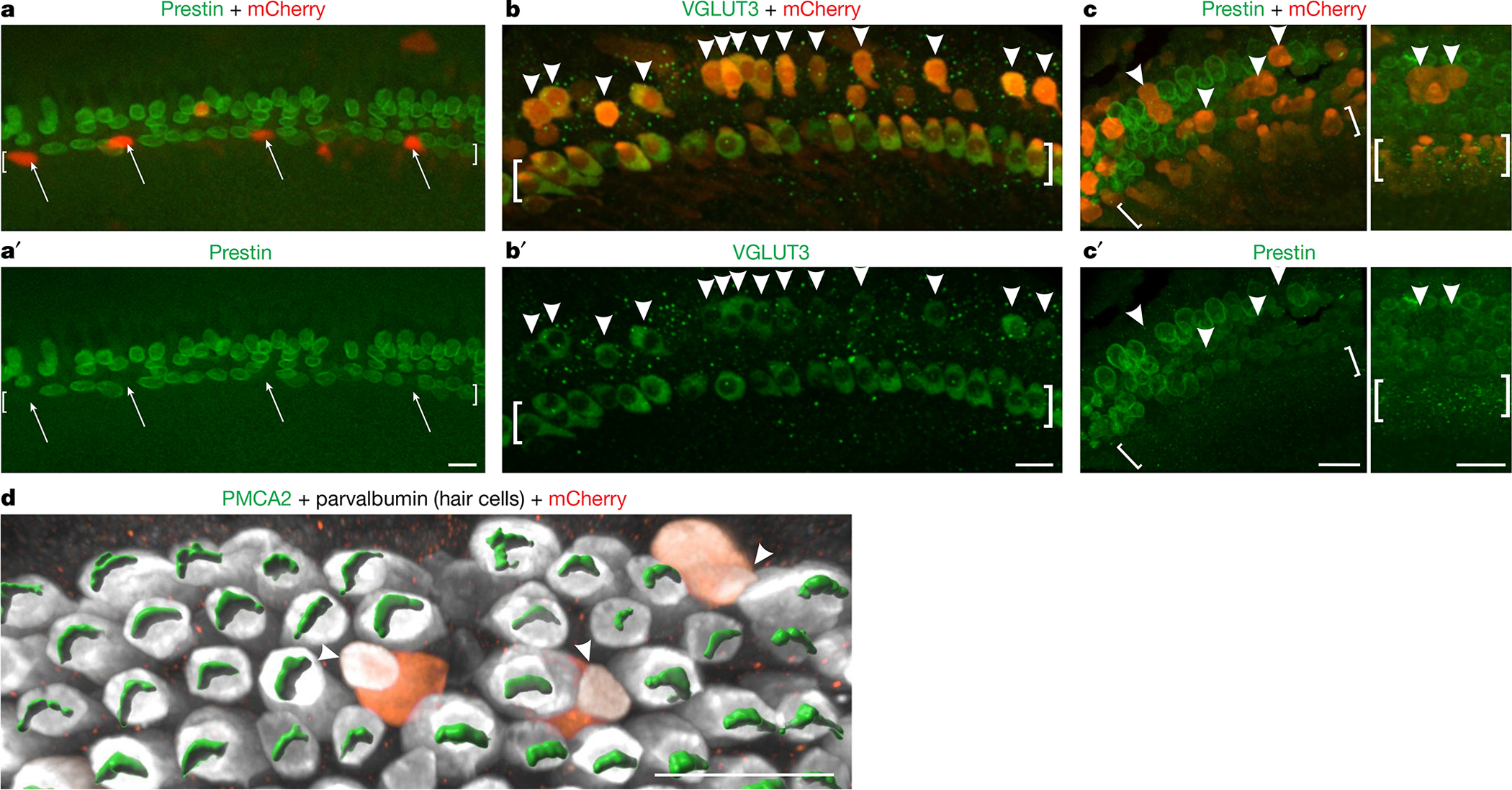
Fig. 4 |. Ectopic expression of TBX2 in OHCs results in their conversion into IHCs.

a, a′, Organ of Corti explants from neonatal Fgf8creER/+; Tbx2F/F mice were established at P0, exposed to Anc80 AAVs expressing TBX2-IRES-mCherry and 4-hydroxytamoxifen (to ablate Tbx2) from 1 DIV to 3 DIV, fixed at 7 DIV and immunostained to detect the OHC marker prestin. Untransfected IHCs expressed prestin, as expected, owing to their loss of TBX2. However, transfected IHCs (mCherry+, arrows) showed no prestin expression, demonstrating that ectopic TBX2 compensated for the loss of endogenous TBX2. b–d, Organ of Corti explants from wild-type mice were established at E17.5, exposed to Anc80 AAVs expressing TBX2-IRES-mCherry from 1 DIV to 3 DIV and fixed at 6 DIV (b–c′) or 7 DIV (d). Cells in the position of OHCs that were transfected (mCherry+, arrowheads) expressed the IHC marker VGLUT3 (b, b′) but not the OHC markers prestin (c, c′) or PMCA2 (d), as visualized with Imaris software. The image in d shows only the outer compartment. Square brackets surround the IHCs. The images shown are examples of results obtained with at least three separate cultures (n = 3 biologically independent samples). Scale bars, 20 μm.
