Abstract
Objective:
Radiotherapy continues to play an important role in the management of breast cancer. This study compared the dosimetric differences between the techniques of intensity modulated radiotherapy (IMRT) and volumetric modulated arc therapy (VMAT) in breast cancer patients who had radiotherapy after mastectomy.
Materials and Methods:
Forty post-mastectomy patients (19 right-sided breast and 21 left-sided breast) treated with the IMRT technique using 7-9 fields who were re-planned with VMAT using 2 coplanar arc on the Varian Vital beam linear accelerator between January, 2020 and August, 2021 were included in this study. The patients received 42 Gy in 15 fractions to the chest wall, lymph nodes and supraclavicular nodes. The dosimetric parameter for planning target volume (PTV), organs at risk (OAR) and the integral dose to the body were analysed. Student’s t-test for two independent means was used to analyse the dosimetric differences between the plans.
Results:
Clinical goals were achieved for both techniques. In terms of PTV coverage at 95% (IMRT: 712.17±233) vs (VMAT: 694.9±214) and the homogeneity index (IMRT: 0.075±0.04) vs (VMAT: 0.104±0.03), IMRT resulted in better dose coverage and homogeneity than VMAT. However, with the conformity index, no significant difference was seen. As regards the OARs, the mean doses, V5, V10, V20, V30, and V40 for the Ipsilateral-lung were lower in IMRT plans than in VMAT plans with a non-significant variation (p-values = 0.141, 0.416, 0.954, 0.443, and 1 respectively). Regarding the mean dose to the heart, low-dose volumes V5, V10, and high-dose volume V30 were significantly reduced in IMRT compared to VMAT. When comparing the dose to the contralateral breast, IMRT achieved a significantly lower mean dose than VMAT (2.9 vs 3.62, p = 0.0148). For MU, VMAT showed lower MU compared to IMRT with a non-significant difference.
Conclusion:
With IMRT, better PTV coverage, homogeneity and OAR sparing were observed. Additionally, VMAT resulted in a lower delivery time than IMRT. Overall, both techniques offered dosimetric qualities that were clinically acceptable.
Keywords: Cancer, conformity, homogeneity, mastectomy, radiotherapy
Key Points
• The dosimetric properties of intensity modulated radiotherapy (IMRT) and volumetric modulated arc therapy (VMAT) for post-mastectomy patients were evaluated on 40 patients.
• Dosimetric paramaters of planning target volume and organs at risks were obtained and evaluated from the DVH.
• Quality of plan was analyzed including the integral dose to normal healthy tissue.
• Both techniques achieved clinical goals, VMAT reduce monitor unit than IMRT.
Introduction
Three-dimensional conformal radiotherapy (3D-CRT) had been the standard technique for several years until the advent of more sophisticated machines which have resulted in advanced treatment techniques. These advancements in recent decades have improved radiotherapy treatments for breast cancer. The 3D-CRT poses some dosimetric challenges in delivering a uniform dose to the target due to the overlaying concave shape of the target, which can result in more dose to the adjacent structure, especially when treating the left-side chest wall (1). Further improvements in technology have enabled the intensity modulation of beams, permitting fluence across the radiotherapy fields, a technique known as intensity modulated radiotherapy (IMRT). Through beam modulation, regular and irregular shaped dose distribution can be attained, leading to an improvement in cosmetic results and minimizing toxicity to normal tissues (2). It also increases the therapeutic goals via improved target dose homogeneity and conformity for breast cancer treatment with the added sparing of the surrounding normal tissues (3).
An innovative modification of IMRT which allows optimum three-dimensional dose distribution to be delivered to the target in a single or multiple gantry rotation was introduced in 2007 (4). This novel technique, termed volumetric modulated arc therapy (VMAT), is an arc-based technique which leads to highly conformal dose distributions by employing beam fluence modulation, variable dose rate, and gantry speed. While VMAT results in similar or better planned target volume (PTV) coverage and better sparing of organs at risk (OARs) in comparison to IMRT, its major advantages are fewer deliveries of monitor units (MUs) and reduced total treatment time. Hence, it aids the fast delivery of treatment. Chest wall irradiation is complicated when compared to whole breast treatment due to its shape post-mastectomy. Hence, in this study, we aimed to dosimetrically evaluate the impacts of IMRT and VMAT on post-mastectomy patients.
Materials and Methods
Patient Enrolment
The computed tomography (CT) simulation cross-section data of 40 post-mastectomy patients (19 right-sided breast and 21 left-sided breast) referred for radiotherapy with invasive ductal carcinoma (T1–T3 N0–N2) to the ipsilateral chest wall, axillary nodes, and supraclave and who had been treated with the IMRT technique using the Varian Vital beam linear accelerator between January, 2020 and August, 2021 were used in this study. The ages of the patients were within the 25–64 years range. All of the patients were prescribed a total dose of 42 Gy in 15 fractions to the chest wall. A re-plan of the same set of patients treated with IMRT was carried out with the VMAT technique for the purpose of this research.
At the time of the CT simulation, the patients were positioned supine on an angled breast board with the sternum parallel to the couch and both arms raised above their heads. The simulation was carried out using a GE CT (Optima 580; GE Healthcare, Waukesha, WI, USA) of 16 slices and 2.5 mm thickness. The Eclipse treatment planning system (version 15.6.05) was used for contouring and treatment planning, while the anisotropic analytic Algorithm was used for dose calculation.
Target Delineation
The Clinical Target Volume (CTV) which included the chest wall (CW), axiliary nodes (AN), intermammary node (IM) and supraclave (SC), were delineated manually from the axial-CT images and outlined by a radiation oncologist following the radiation therapy oncology group (RTOG) recommendation. The PTV of the CW, AN, and SC was linked to the reference frame of the machine and was delineated by expanding from the CTV with a uniform 0.5 cm margin to account for physiological and daily set-up variations/uncertainty. The total PTV (PTVtot) consisted of the PTVCW, PTVAN, and PTVSC, all of which were limited to the skin surface. The heart, ipsilateral lungs, contralateral lungs, contralateral breast, spinal cord, and thyroid were contoured as critical organs and non-tumour tissue. Figure 1 describes the target and OAR delineation.
Figure 1.
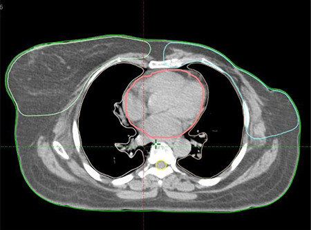
Target and OAR delineation
OAR: organs at risk
Planning
For each patient, one IMRT and one VMAT plan were created and optimization was achieved using the Photon Optimizer algorithm (version 15.6.05) with objectives specified accordingly to the planning goal. A typical IMRT plan consists of 7–9 photon fields spaced according to the planner’s discretion at a single isocenter using 6 MV energy. The gantry angles were individually selected for each patient’s CT dataset to achieve optimal dose target coverage and minimize entry and existing dose to the OARs. During the intensity optimization, dose constraints and priority were set for the PTV, NS ring control, and OARs following the quantitative analysis of normal tissue effects in the clinic (QUANTEC) analysis and RTOG report 62 guidelines as shown in Table 1 below. A 0.5 cm tissue equivalent bolus was placed over the PTV-CW to ensure sufficient target coverage near the CW surface.
Table 1. Planning goal for OAR.
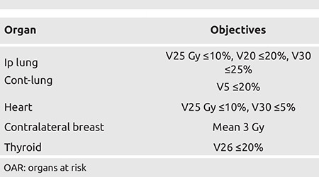
Each plan was optimized so that 95% of the PTV would receive 95% of the prescribed dose (i.e., V95=95%). The doses were calculated using the anisotropic analyses algorithm (version 15.6.05) and efforts were taken to maintain the 3D dose max below 107%.
Additionally, VMAT plans were generated using the same isocenter and energy level as their corresponding IMRT plans, employing two partial coplanar arcs and 30-degree collimation with a starting angle of 179° and an ending angle of 181°. These plans were optimized according to institutional practice, following the same dose objectives as used for the IMRT planning technique. The IMRT field arrangement and VMAT arc arrangement are shown in Figure 2 below. The planning goals for the plans are described in Table 1.
Figure 2.
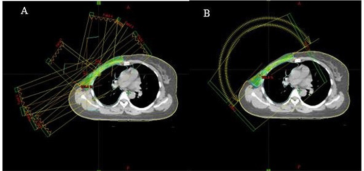
Field arrangement for (A) IMRT, (B) VMAT plans
VMAT: volumetric modulated arc therapy; IMRT: intensity modulated radiotherapy
Plan Quality
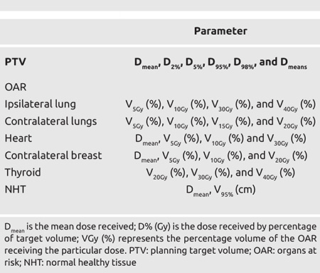
For plan quality comparison, the dose homogeneity index (HI), conformity index (CI), integral dose (ID), MU and dose to OAR using the parameters obtained from the dose volume histogram are shown in Table 2. The CI was computed according to the definition proposed by RTOG (5) and estimated as:
Table 2. Dosimetric parameters.

Where V95% is the volume of the target receiving 95% of the prescribed dose and TV is the total volume of the target. The closer the value of CI is to 1, the more conformity there is to the plan. This study utilized two distinct HI formulas (HI1 and HI2). HI1 was obtained based on the definition proposed by ICRU-83 (6) and is presented below.

Where D2% and D98% represent the minimum dose received by 2% and 98% of the target volume, indicating the maximal and minimal doses to the target, respectively, and D95% represents the dose received by 95% of the target. The closer the value of HI is to 0, the more homogenous the plan. HI2 is calculated as given below (7). In this mode, the closer the value is to 1, the better the homogeneity.

ID is calculated as Dmean (Gy)*V(L), where Dmean (Gy) is the mean dose and V is the volume of the organ. Normal healthy tissue (NHT) was delineated by subtracting the target volumes from the body volume.
NHT= BODY - PTV. The percentage volume of the NHT receiving 5 Gy was obtained from the DVH.
Statistical Analysis
Student’s t-test for two independent means was used to analyse the dosimetric differences between the plans. It was carried out on the social sciences window software version 18 (IBM Corp. Armonk, NY, USA), and p-values <0.05 were considered statistically significant.
Ethics Approval
This study protocol was reviewed and approved by the Lagos University Teaching Hospital Health Research Ethics Committee (LUTHHREC) ADM/DSCST/HREC/APP/4664.
Informed Consent
All patients were informed and their consent was taken.
Results
The IMRT plan for each patient was reviewed and approved by the radiation oncologist before delivery of treatment. In total, 40 IMRT plans (21 left-sided and 19 right-sided) and 40 VMAT plans were created for this study and the dosimetric results of the two techniques are presented in the tables below.
PTV Dose Analysis
Both treatment techniques achieved 95% PTV coverage with a non-statistical difference. Table 3 summarizes the PTV result in terms of mean dose, min dose, max dose, D5, D95, CI, HI, and GI. The VMAT and IMRT techniques had no significant difference in CI (0.962 vs 0.981, p = 0.084). A similar result was obtained with the mean dose (42.226 Gy vs 42.39 Gy, p = 0.211). However, when compared to the VMAT plan, the IMRT technique showed more dose homogeneity as the difference between them is statistically significant (H1: 0.075 vs 0.104, p = 0.0003; H2: 1.056 vs 1.082, p = 0.0005). Statistically significant comparisons were also seen for the max dose D2 (43.52 Gy vs 43.88 Gy, p = 0.0037) and min dose D98 Gy (39.77 Gy vs 39.52 Gy, p = 0.0007). The comparisons of dose volumes between the two techniques are shown in Figure 3.
Table 3. Comparison of dose coverage for PTVtotal for both planning techniques.
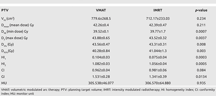
Figure 3.
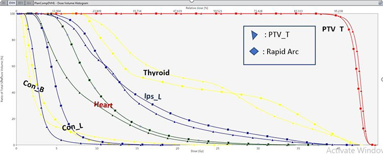
DVH comparison of IMRT and VMAT plan
VMAT: volumetric modulated arc therapy; IMRT: intensity modulated radiotherapy; PTV: planning target volume
Dose Analysis of OARs
In Table 4, the dosimetric parameters of the OAR observed in IMRT and VMAT plans are summarised. The mean doses, V5, V10, V20, V30, and V40 for the Ip-lung were lower in the IMRT plans than in the VMAT plans with an insignificant variation (p-values = 0.141, 0.416, 0.954, 0.443, and 1 respectively). However, in both techniques, the doses were within clinically acceptable limits. For the contralateral lungs, both techniques yielded similar results for V10, V15, and V20. However, the mean dose and V5 of the cont-lungs were significantly spared in the IMRT plan in comparison to the VMAT plan (mean dose: 4.6 Gy vs 5.6 Gy, p = 0.001; V5: 38.78% vs 52.32%, p = 0.001).
Table 4. OAR dose comparison between both planning techniques.
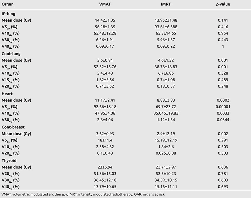
The mean dose to the heart, low-dose volume V5, V10, and high-dose volume V30 were significantly reduced in IMRT in comparison to VMAT. In comparing the dose to the contralateral breast, IMRT achieved a significantly lower mean dose than VMAT (2.9 vs 3.62, p = 0.0148). However, there was no significant difference in terms of V5, V10, and V20. VMAT, on the other hand, indicated a low mean dose and volume dose for thyroid, although there was no significant difference between the two plans.
The average MU for each fixed angle beam in the IMRT plan was 303.34, while that for each partial arc trajectory was 307.54 in the VMAT plan. There was no significant difference in MU for both plans.
The planning volumes for each patient were well inside the planning CT scans, so the irradiated normal tissues were included in the CT volumes. Table 5 shows the ID to the non-tumour tissue (IDNTT) and no significant difference in normal tissue ID was observed (p = 0.493) in either technique.
Table 5. Comparison of dose coverage for body and non-tumour tissue for both planning techniques.

Discussion and Conclusion
Conformal techniques have proven to be of great benefit in radiotherapy for mastectomy breast cancer. It is essential to evaluate the dosimetric properties of these techniques. In recent times, such studies (8) have evaluated the dosimetric properties of 3D-CRT and IMRT in post-mastectomy irradiated patients. Additionally, several trials have made comparisons of the VMAT technique, which uses an arc trajectory, against the fixed angle beam IMRT technique. However, this has led to a debate on which technique should be employed in radiotherapy. The current study compares the above-mentioned radiotherapy techniques often utilized in the treatment of post-mastectomy breast cancer and evaluates these plans using the dosimetric parameters obtained from the DVH. A plan with good target coverage has the benefit of maximizing the efficacy and improving the local control to ensure homogenous dose coverage by avoiding cold spots (PTV receiving less than 90% of the prescribed dose) and hot spots (outside PTV receiving a dose greater than PTV) as well as minimizing normal long-term tissue toxicity. The findings from our work showed that both plans met the target coverage with a non-significant difference in the conformity index, which indicates successful avoidance of hot spots (i.e., areas of relative overdose). However, VMAT showed significantly lower dose homogeneity in PTVtotal than IMRT, indicating that the IMRT plan reduces the cold spot issue to a greater extent, which might decrease local reoccurrence.
Radiation doses to the ipsilateral lungs can result in induced pneumonitis, a deterministic effect of breast cancer treatment (9), hence, the need for proper optimization of the lung during planning. Conventionally, the dosimetry parameters influencing radiation-induced pneumonitis include V5 Gy, V10 Gy, and V20 Gy. However, the main predictors among these parameters remain debatable. Yorke et al. (10) researched dosimetric factors of radiation-induced pneumonitis and reported that the V5 Gy and V10 Gy of the lung may be effective predictors, whereas Caudell et al. (11) concluded that V20 Gy and radiation-induced pneumonitis are related. In this study, the volume dose of both plans was within the QUANTEC recommendations, and there was no significant difference between the VMAT and IMRT plans for all the dose parameters of the ipsilateral lung (mean dose, V5 Gy, V10 Gy, V30 Gy, and V40 Gy), indicating that both techniques reduced the radiation dose while ensuring sufficient radiation to the target area, which may reduce the incidence of radiation-induced injury.
To prevent cardiac morbidity, it is essential to limit the heart dose as much as is reasonably achievable in patients, particularly those with left-sided breast cancer. However, the required level of sparing is unclear. In this study, the IMRT significantly outperformed the VMAT in sparing the heart in cases of the left-sided CW (based on mean dose, V5 Gy and V10 Gy; p<0.00001), both offered similar heart sparing in cases of the right-sided CW.
The minimization of the irradiation of the contralateral breast needs to be highly prioritized. This is required to reduce the possibility of radiation-induced carcinogenesis (12). Although there are risk models which quantify the relationship between low-medium dose levels and the induction of secondary cancer (13, 14), clinical validation is inadequate. As a result, optimization is required (i.e., applying the ALARA principle). As shown in the results presented above, the mean dose to the contralateral breast differed significantly where the IMRT plan complied with the QUANTEC restriction of less than 3 Gy, although the dose-volume (V5, V10, and V20) was similar.
The delivery of low-dose irradiation to healthy tissue has been estimated to double the risk of subsequent malignancy, and this risk increases with increasing dosages (15). According to the findings of this study, it was observed that VMAT resulted in a significant reduction in the mean dose to the healthy tissue compared to IMRT (p = 0.00001). Based on the report by D’Souza and Rosen (16), the non-tumour integral dosage is mostly determined by beam margin size and energy, with the fractionation scheme playing a minor role. Smaller margin size and higher energy result in a constant reduction in non-tumour tissue ID, regardless of the number of beams. This study observed a similar non-tumour ID (p = 0.493) as the same energy and fractionation scheme were utilized.
The results of this study are in line with the findings of Dumane et al. (17), who compared the plan quality of three techniques (IMRT, VMAT, and 3D-CRT) on the right CW. According to their study, HI and PTV coverage were found to be best with IMRT, while IMRT and VMAT improved conformity similar to the 3D-CRT plan (improved by as much as 25%). OAR are spared more with VMAT in comparison to IMRT (by as much as 17.1% decrease for the ipsilateral lung and 16.22% for the contralateral lung). The study by Ma et al. (18) on dosimetric comparison of three radiotherapy techniques (3D-CRT, IMRT, and VMAT) agrees with our results as their IMRT plan achieved better homogeneity than the other plans (IMRT: 0.114 vs VMAT: 0.143, p = 0.002; IMRT vs 3D, p = 0.001) while both IMRT and VMAT achieve similar CI (p = 0.425). Also, the mean dose to the contralateral breast was higher in VMAT (5.79 Gy) than in IMRT (2.81 Gy), with p = 0.016.
In contrast to our findings, Johansen et al. (19) reported that better dose homogeneity and PTV conformity were observed in VMAT (HI and CI; p<0.05). In addition, the mean dose to the contralateral breast was lower in VMAT than in the other techniques (IMRT and conventional plan), however, these differences were not significant. Past studies have reported lower MUs in VMAT than in IMRT. The higher the MU, the longer the beam-on time and vice versa. Our findings agree with the report published by Zhang et al. (2). From their findings, VMAT reduced the number of monitored units by 24% and treatment time by 53%. They also reported that VMAT achieved better normal tissue sparing than IMRT, although both techniques (VMAT and IMRT) showed similar PTV dose homogeneity (p = 0.048). The average MU for each fixed angle beam in the IMRT plan was 306.57±64.88, while that for each partial arc trajectory in the VMAT plan was 305.538±46.077. This implies that the overall delivery time for the VMAT plan is lower than that of IMRT, although the MU result showed no significant difference.
The deep inspiration breath-hold technique to reduce the heart dose in breast cancer management has been studied (20), however this was not the focus of our study. This study’s design was not intended to evaluate the advantage of one modality over another in terms of toxicity. A lengthy follow-up would be necessary to address the effects of inverse planning techniques on survival.
From a dosimetric perspective, it is concluded that both plans investigated in this study offer quality patient treatments. However, the IMRT plans achieved a better dosimetric advantage for the CW owing to enhanced PTV coverage, better dose homogeneity, and enhanced sparing of the OAR, such as the contralateral breast, heart, and lungs compared with VMAT. On the other hand, VMAT, while maintaining a good degree of conformity similar to IMRT, had the advantage of a lower MU than IMRT, thereby decreasing the overall treatment plan times.
Acknowledgments
We would like to sincerely appreciate Mr. Abdallah Kotkat and Mr. Ibrahim El-Hamamsi for their guidance in the course of this research.
Footnotes
Ethics Committee Approval: This study protocol was reviewed and approved by the Lagos University Teaching Hospital Health Research Ethics Committee (LUTHHREC) ADM/DSCST/HREC/APP/4664.
Informed Consent: All patients were informed and their consent was taken.
Peer-review: Internally and externally peer-reviewed.
Authorship Contributions
Surgical and Medical Practices: M.H., O.S.; Concept: S.A., M.A.; Design: S.A., M.A., M.H.; Data Collection or Processing: N.A., R.J., E.A., R.L.; Analysis or Interpretation: N.A., A.A.; Literature Search: A.J., I.A.; Writing: N.A., R.J.
Conflict of Interest: No conflict of interest was declared by the authors.
Financial Disclosure: The authors declared that this study received no financial support.
References
- 1.Hjelstuen MH, Mjaaland I, Vikström J, Dybvik KI. Radiation during deep inspiration allows loco-regional treatment of left breast and axillary-, supraclavicular- and internal mammary lymph nodes without compromising target coverage or dose restrictions to organs at risk. Acta Oncol. 2012;51:333–344. doi: 10.3109/0284186X.2011.618510. [DOI] [PubMed] [Google Scholar]
- 2.Zhang Q, Yu XL, Hu WG, Chen JY, Wang JZ, Ye JS, et al. Dosimetric comparison for volumetric modulated arc therapy and intensitymodulated radiotherapy on the left-sided chest wall and internal mammary nodes irradiation in treating post-mastectomy breast cancer. Radiol Oncol. 2015;49:91–98. doi: 10.2478/raon-2014-0033. [DOI] [PMC free article] [PubMed] [Google Scholar]
- 3.Dogan N, Cuttino L, Lloyd R, Bump EA, Arthur DW. Optimized dose coverage of regional lymph nodes in breast cancer: the role of intensity-modulated radiotherapy. Int J Radiat Oncol Biol Phys. 2007;68:1238–1250. doi: 10.1016/j.ijrobp.2007.03.059. [DOI] [PubMed] [Google Scholar]
- 4.Pasler M, Georg D, Bartelt S, Lutterbach J. Node-positive left-sided breast cancer: does VMAT improve treatment plan quality with respect to IMRT? Strahlenther Onkol. 2013;189:380–386. doi: 10.1007/s00066-012-0281-2. [DOI] [PubMed] [Google Scholar]
- 5.Shaw E, Kline R, Gillin M, Souhami L, Hirschfeld A, Dinapoli R, et al. Radiation Therapy Oncology Group: radiosurgery quality assurance guidelines. Int J Radiat Oncol Biol Phys. 1993;27:1231–1239. doi: 10.1016/0360-3016(93)90548-a. [DOI] [PubMed] [Google Scholar]
- 6.Hodapp N. Der ICRU-Report 83: Verordnung, Dokumentation und Kommunikation der fluenzmodulierten Photonenstrahlentherapie (IMRT) [The ICRU Report 83: prescribing, recording and reporting photon-beam intensity-modulated radiation therapy (IMRT)] Strahlenther Onkol. 2012;188:97–99. doi: 10.1007/s00066-011-0015-x. [DOI] [PubMed] [Google Scholar]
- 7.Lai Y, Chen Y, Wu S, Shi L, Fu L, Ha H, et al. Modified Volumetric Modulated Arc Therapy in Left Sided Breast Cancer After Radical Mastectomy With Flattening Filter Free Versus Flattened Beams. Medicine (Baltimore) 2016;95:e3295. doi: 10.1097/MD.0000000000003295. [DOI] [PMC free article] [PubMed] [Google Scholar]
- 8.Adeneye S, Akpochafor M, Adegboyega B, Alabi A, Adedewe N, Joseph A, et al. Evaluation of Three-Dimensional Conformal Radiotherapy and Intensity Modulated Radiotherapy Techniques for Left Breast Post-Mastectomy Patients: Our Experience in Nigerian Sovereign Investment Authority-Lagos University Teaching Hospital Cancer Center, South-West Nigeria. Eur J Breast Health. 2021;17:247–252. doi: 10.4274/ejbh.galenos.2021.6357. [DOI] [PMC free article] [PubMed] [Google Scholar]
- 9.Tajvidi M, Sirous M, Sirous R, Hajian P. Partial frequency of radiation pneumonitis and its association with the energy and treatment technique in patients with breast cancer, Isfahan, Iran. J Res Med Sci. 2013;18:413–416. [PMC free article] [PubMed] [Google Scholar]
- 10.Yorke ED, Jackson A, Rosenzweig KE, Braban L, Leibel SA, Ling CC. Correlation of dosimetric factors and radiation pneumonitis for nonsmall- cell lung cancer patients in a recently completed dose escalation study. Int J Radiat Oncol Biol Phys. 2005;63:672–682. doi: 10.1016/j.ijrobp.2005.03.026. [DOI] [PubMed] [Google Scholar]
- 11.Caudell JJ, De Los Santos JF, Keene KS, Fiveash JB, Wang W, Carlisle JD, et al. A dosimetric comparison of electronic compensation, conventional intensity modulated radiotherapy, and tomotherapy in patients with early-stage carcinoma of the left breast. Int J Radiat Oncol Biol Phys. 2007;68:1505–1511. doi: 10.1016/j.ijrobp.2007.04.026. [DOI] [PubMed] [Google Scholar]
- 12.Zhao H, He M, Cheng G, Han D, Wu N, Shi D, et al. A comparative dosimetric study of left sided breast cancer after breast-conserving surgery treated with VMAT and IMRT. Radiat Oncol. 2015;10:231. doi: 10.1186/s13014-015-0531-4. [DOI] [PMC free article] [PubMed] [Google Scholar]
- 13.Schneider U, Sumila M, Robotka J, Gruber G, Mack A, Besserer J. Dose-response relationship for breast cancer induction at radiotherapy dose. Radiat Oncol. 2011;6:67. doi: 10.1186/1748-717X-6-67. [DOI] [PMC free article] [PubMed] [Google Scholar]
- 14.Berrington de Gonzalez A, Gilbert E, Curtis R, Inskip P, Kleinerman R, Morton L, et al. Second solid cancers after radiation therapy: a systematic review of the epidemiologic studies of the radiation dose-response relationship. Int J Radiat Oncol Biol Phys. 2013;86:224–233. doi: 10.1016/j.ijrobp.2012.09.001. [DOI] [PMC free article] [PubMed] [Google Scholar]
- 15.Hall EJ. Intensity-modulated radiation therapy, protons, and the risk of second cancers. Int J Radiat Oncol Biol Phys. 2006;65:1–7. doi: 10.1016/j.ijrobp.2006.01.027. [DOI] [PubMed] [Google Scholar]
- 16.D’Souza WD, Rosen II. Nontumor integral dose variation in conventional radiotherapy treatment planning. Med Phys. 2003;30:2065–2071. doi: 10.1118/1.1591991. [DOI] [PubMed] [Google Scholar]
- 17.Dumane VA, Hunt MA, Green S, Lo YC, Bakst RL. Dosimetric Comparison of Volumetric Modulated Arc Therapy, Static Field Intensity Modulated Radiation Therapy, and 3D Conformal Planning for the Treatment of a Right-Sided Reconstructed Chest Wall and Regional Nodal Case. Journal of Radiotherapy. 2014;9:1–13. [Google Scholar]
- 18.Ma C, Zhang W, Lu J, Wu L, Wu F, Huang B, et al. Dosimetric Comparison and Evaluation of Three Radiotherapy Techniques for Use after Modified Radical Mastectomy for Locally Advanced Left-sided Breast Cancer. Sci Rep. 2015;5:12274. doi: 10.1038/srep12274. [DOI] [PMC free article] [PubMed] [Google Scholar]
- 19.Johansen S, Cozzi L, Olsen DR. A planning comparison of dose patterns in organs at risk and predicted risk for radiation induced malignancy in the contralateral breast following radiation therapy of primary breast using conventional, IMRT and volumetric modulated arc treatment techniques. Acta Oncol. 2009;48:495–503. doi: 10.1080/02841860802657227. [DOI] [PubMed] [Google Scholar]
- 20.Dumane VA, Saksornchai K, Zhou Y, Hong L, Powell S, Ho AY. Reduction in low-dose to normal tissue with the addition of deep inspiration breath hold (DIBH) to volumetric modulated arc therapy (VMAT) in breast cancer patients with implant reconstruction receiving regional nodal irradiation. Radiat Oncol. 2018;13:187. doi: 10.1186/s13014-018-1132-9. [DOI] [PMC free article] [PubMed] [Google Scholar]


