Abstract
Congenital epulis (CE) is an extremely rare benign tumor of the gingiva that is found on the alveolar ridge of newborns, and the main treatment option is simple excision. Postoperative prognosis is very good, and spontaneous regression may occur despite incomplete excision. This report presented a rare case of CE and its healing process after surgery under local anesthesia. The treatment plan was decided upon through consultation between a medical team and the patient’s family, with surgical excision for the main lesion, which benefitted from surgery, and follow-up for a very small-sized lesion, which was thought to be appropriate for a newborn. No recurrence was found after its removal, and favorable healing was observed.
Keywords: Congenital epulis, Newborn
I. Introduction
Congenital epulis (CE) is an extremely rare benign tumor of the gingiva that is found on the alveolar ridge of newborns, and its etiology or pathogenesis remains unclear1. CE occurs mainly on the maxillary ridge with a female predilection, and its incidence rate is as low as 6/1,000,0002. Depending on its size, the tumor can induce disturbances in feeding and respiration. CE is generally treated by surgical excision, and recurrence is uncommon3.
II. Case Report
A 2-day-old female infant was referred from the Department of Pediatrics to the Department of Oral and Maxillofacial Surgery for evaluation and treatment of an intraoral mass. The patient was born by spontaneous vaginal delivery 31 hours after premature rupture of the membranes at 39 weeks’ gestation. The infant weighed 3,160 g and was hospitalized in the newborn intensive care unit (NICU), where she received phototherapy for hyperbilirubinemia.
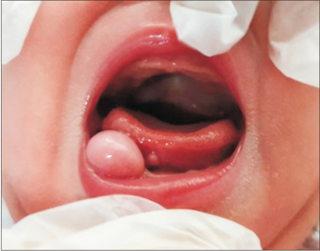
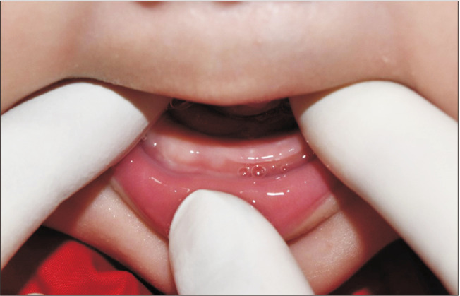
Intraoral examination revealed a 1-cm×1-cm-sized, smooth-surfaced pedunculated mass on the right mandibular alveolar ridge. Postero-lingually, about 2-mm-sized small mass was also identified in the sublingual region.(Fig. 1) The patient’s mother complained of discomfort during breastfeeding, and the location of the mass was found to be unfavorable to breastfeeding. Since it was thought to be advantageous to remove the lesion during hospitalization in the NICU, a treatment plan for surgical excision under local anesthesia was decided upon, after consultation between a medical team and the patient’s family.
Fig. 1.
Initial clinical photography. A 1-cm×1-cm-sized, smooth-surfaced pedunculated mass on the right mandibular alveolar ridge and postero-lingually; about 2-mm-sized small mass also in the sublingual region.
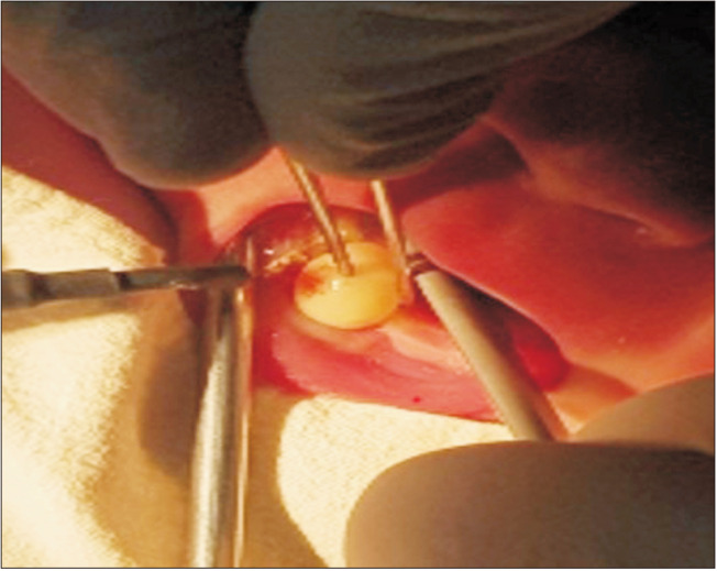
The surgery was performed in the NICU at a bedside by excising the main lesion using an electrocautery device under local anesthesia.(Fig. 2) It was decided to follow up on the sublingual mass, considering that its small size reduced the benefit of surgical intervention. Postoperatively, the wound showed good healing with no significant complications or signs of infection.
Fig. 2.
The lesion being excised with an electric cautery device during surgery.
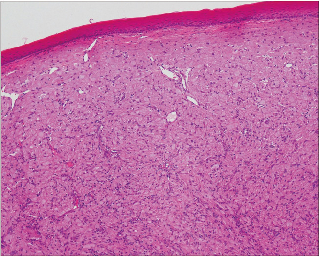
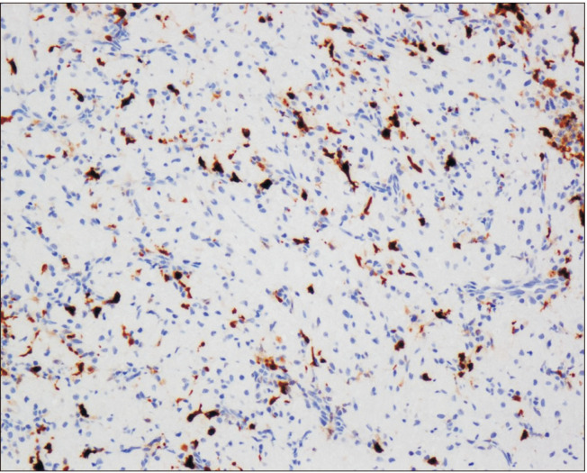
Histopathological examination revealed granular tumor cells with abundant cytoplasm upon H&E staining.(Fig. 3) Tumor cells were negative for S-100 protein; sustentacular cells that support tumor structure were observed in the basal layer between the tumor cell nests.(Fig. 4) Finally, a diagnosis of CE was rendered. Five months after surgery, the patient’s mandibular ridge showed good healing without a defect, and its width and height were normal.(Fig. 5) Complete regression occurred in the sublingual lesion upon follow-up.
Fig. 3.
The microscopic features of histopathological examination revealed granular tumor cells with abundant cytoplasm upon H&E staining (×100).
Fig. 4.
Immunohistochemical properties of lesion cells were negative for S-100 protein. Sustentacular cells that support tumor structure were observed in the basal layer between the tumor cell nests (×100).
Fig. 5.
Clinical photography taken five months after surgery. Mandibular ridge showed good healing without a defect, and its width and height were normal.
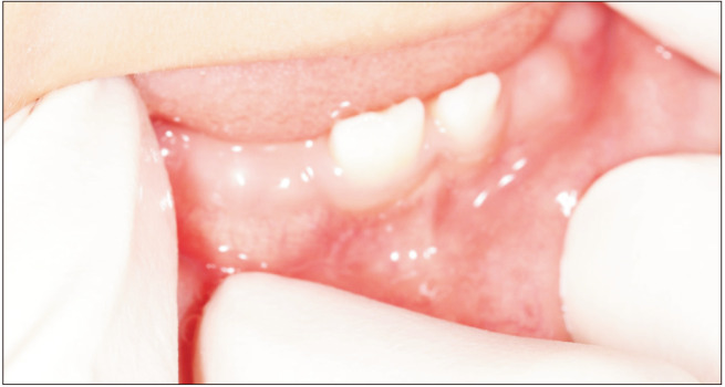
Good healing of the surgery site was also observed one year after surgery.(Fig. 6)
Fig. 6.
Clinical photography taken one year after surgery. Mandibular ridge showed good healing.
III. Discussion
Yuwanati et al.4 reported that CE occurs twice as commonly in the maxillary ridge as in the mandibular ridge, and the most common site is the canine region. Although a female preponderance has been reported, no direct association with specific hormones has been identified, since endogenous hormone receptors have not been found in the lesions4. Vered et al.5 stated that CE is regarded as a reactive lesion to local metabolism or irritation, showing no neoplastic features. The word “epulis” of CE signifies the hyperplastic gingival tissue.
CE mainly presents as a single mass; only 10% of the cases are observed as multiple lesions6. CE may be mainly smooth, pedunculated, or sometimes lobulated, and has a fleshy pink color, and its size varies from several mm to 9 cm6. CE can be detected using magnetic resonance imaging and ultrasound as prenatal images before childbirth, but prenatal imaging study can be used as an aid to diagnosis and is not critical for differential diagnosis with other intraoral masses7. The pathogenesis of CE has not been established. The possibility of a neurogenic origin has been suggested8. In an electron microscopy study, similarities to stromal cells such as fibroblasts and histiocytes suggest the possibility that CE has a reactive origin9.
CE is known to occur alone with no associations with specific congenital anomalies10. CE should be distinguished from granular cell tumor (GCT), which has histological similarity. GCT occurs mainly in adults, and the common intraoral site is the tongue. The main features of CE that distinguish it from GCT are tumor location, patient age, the absence of cytoplasmic hyaline granules, solid growth pattern, and negativity for S-100 staining4. GCT also shows an indolent aspect, but the distinction between CE and GCT is significant as it is reported as malignant in about 0.5% to 2% of cases11.
Regarding treatment, several CE cases showing spontaneous regression have been reported12. However, the possibility of difficulties in breathing and feeding should be considered, which may warrant surgical excision, depending on tumor size, or close follow-up. There has been a report of a case of dysphagia caused by blockage of the oral cavity of a child by CE13.
The main treatment option for CE is simple excision under local or general anesthesia. Postoperative prognosis is very good, and spontaneous regression may occur despite incomplete excision14.
This report presented a rare case of CE and its healing process after surgery. No malignant transformation and recurrence were found after its removal, and favorable healing was observed without intraoral structural and functional disorders. The treatment plan was decided upon through the consultation between a medical team and the patient’s family, with surgical excision for a main lesion, which benefitted from surgery, and follow-up for a very small-sized lesion, which was thought to be appropriate for a newborn.
Considering the present case and previous literature, surgical excision may be conducted to prevent feeding and breathing disturbances depending on tumor size, and small-sized lesions can be followed-up without surgical intervention, with the anticipation of spontaneous regression.
Footnotes
Authors’ Contributions
M.J.K. and S.H.K. obtained data and wrote the manuscript. M.J.K. and S.H.K. drafted the manuscript. M.J.K. and S.H.K. participated in article design and coordination and carefully reviewed and revised the manuscript. All the authors read and approved the final manuscript.
Ethics Approval and Consent to Participate
The study was approved by the Institutional Review Board (IRB) of National Health Insurance Service Ilsan Hospital (No. NHIMC 2022-05-001), and the informed consent was waived by the IRB.
Conflict of Interest
No potential conflict of interest relevant to this article was reported.
References
- 1.Childers EL, Fanburg-Smith JC. Congenital epulis of the newborn: 10 new cases of a rare oral tumor. Ann Diagn Pathol. 2011;15:157–61. doi: 10.1016/j.anndiagpath.2010.10.003. https://doi.org/10.1016/j.anndiagpath.2010.10.003. [DOI] [PubMed] [Google Scholar]
- 2.Bosanquet D, Roblin G. Congenital epulis: a case report and estimation of incidence. Int J Otolaryngol. 2009;2009:508780. doi: 10.1155/2009/508780. https://doi.org/10.1155/2009/508780. [DOI] [PMC free article] [PubMed] [Google Scholar]
- 3.Klingler PJ, Seelig MH, DeVault KR, Wetscher GJ, Floch NR, Branton SA, et al. Ingested foreign bodies within the appendix: a 100-year review of the literature. Dig Dis. 1998;16:308–14. doi: 10.1159/000016880. https://doi.org/10.1159/000016880. [DOI] [PubMed] [Google Scholar]
- 4.Yuwanati M, Mhaske S, Mhaske A. Congenital granular cell tumor - a rare entity. J Neonatal Surg. 2015;4:17. doi: 10.47338/jns.v4.170. [DOI] [PMC free article] [PubMed] [Google Scholar]
- 5.Vered M, Dobriyan A, Buchner A. Congenital granular cell epulis presents an immunohistochemical profile that distinguishes it from the granular cell tumor of the adult. Virchows Arch. 2009;454:303–10. doi: 10.1007/s00428-009-0733-y. https://doi.org/10.1007/s00428-009-0733-y. [DOI] [PubMed] [Google Scholar]
- 6.Wittebole A, Bayet B, Veyckemans F, Gosseye S, Vanwijck R. Congenital epulis of the newborn. Acta Chir Belg. 2003;103:235–7. doi: 10.1080/00015458.2003.11679415. https://doi.org/10.1080/00015458.2003.11679415. [DOI] [PubMed] [Google Scholar]
- 7.Roy S, Sinsky A, Williams B, Desilets V, Patenaude YG. Congenital epulis: prenatal imaging with MRI and ultrasound. Pediatr Radiol. 2003;33:800–3. doi: 10.1007/s00247-003-1024-4. https://doi.org/10.1007/s00247-003-1024-4. [DOI] [PubMed] [Google Scholar]
- 8.Stewart CM, Watson RE, Eversole LR, Fischlschweiger W, Leider AS. Oral granular cell tumors: a clinicopathologic and immunocytochemical study. Oral Surg Oral Med Oral Pathol. 1988;65:427–35. doi: 10.1016/0030-4220(88)90357-X. https://doi.org/10.1016/0030-4220(88)90357-x. [DOI] [PubMed] [Google Scholar]
- 9.Lack EE, Perez-Atayde AR, McGill TJ, Vawter GF. Gingival granular cell tumor of the newborn (congenital "epulis"): ultrastructural observations relating to histogenesis. Hum Pathol. 1982;13:686–9. doi: 10.1016/S0046-8177(82)80018-X. https://doi.org/10.1016/s0046-8177(82)80018-x. [DOI] [PubMed] [Google Scholar]
- 10.Bertozzi M, Nardi N, Falcone F, Prestipino M, Appignani A. An unusual case of asymptomatic appendicular cutting foreign body. Pediatr Med Chir. 2010;32:223–5. [PubMed] [Google Scholar]
- 11.Elkousy H, Harrelson J, Dodd L, Martinez S, Scully S. Granular cell tumors of the extremities. Clin Orthop Relat Res. 2000;380:191–8. doi: 10.1097/00003086-200011000-00026. https://doi.org/10.1097/00003086-200011000-00026. [DOI] [PubMed] [Google Scholar]
- 12.Sakai VT, Oliveira TM, Silva TC, Moretti AB, Santos CF, Machado MA. Complete spontaneous regression of congenital epulis in a baby by 8 months of age. Int J Paediatr Dent. 2007;17:309–12. doi: 10.1111/j.1365-263X.2006.00795.x. https://doi.org/10.1111/j.1365-263X.2006.00795.x. [DOI] [PubMed] [Google Scholar]
- 13.López de Lacalle JM, Aguirre I, Irizabal JC, Nogues A. Congenital epulis: prenatal diagnosis by ultrasound. Pediatr Radiol. 2001;31:453–4. doi: 10.1007/s002470000416. https://doi.org/10.1007/s002470000416. [DOI] [PubMed] [Google Scholar]
- 14.Fister P, Volavsek M, Novosel Sever M, Jazbec J. A newborn baby with a tumor protruding from the mouth. Diagnosis: congenital gingival granular cell tumor. Acta Dermatovenerol Alp Pannonica Adriat. 2007;16:128–30. [PubMed] [Google Scholar]


