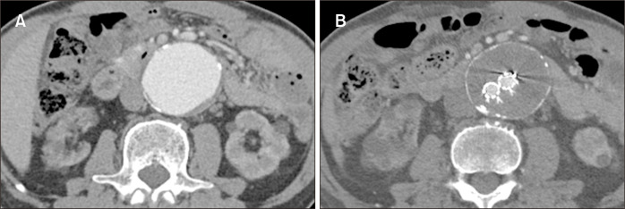
A 71-year-old male presented with abdominal discomfort for 7 days. His medical history included hypertension, coronary artery disease, and hemodialysis for 5 years. Three years earlier, he underwent endovascular aneurysm repair (EVAR) for an abdominal aortic aneurysm (AAA) with a low-profile INCRAFT stent graft (30 mm main body; Cordis Corp., Santa Clara, CA, USA). The AAA diameter was 56 mm at the time of EVAR, and the sac remained stable throughout the follow-up (Fig. 1).
Fig. 1.
(A) Computed tomography showed an abdominal aortic aneurysm with the anteroposterior diameter measuring 56 mm. (B) After endovascular aneurysm repair, the aneurysm sac was stable without endoleak for 3 years.
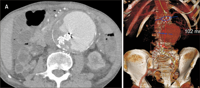
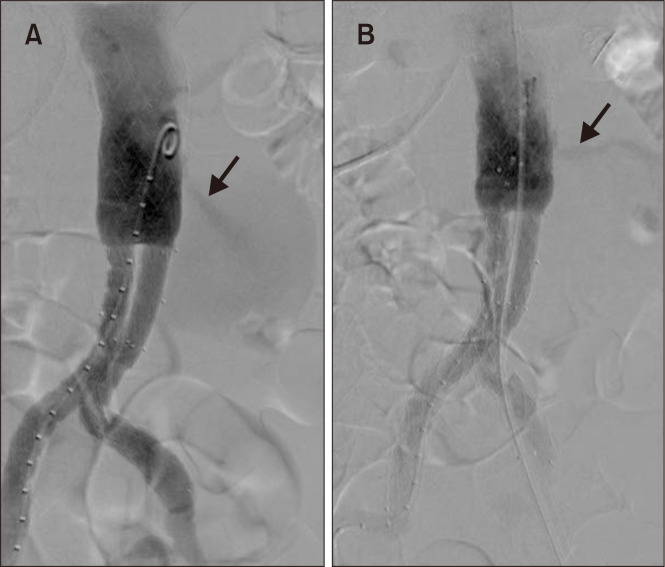
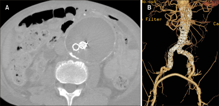
However, the computed tomography at this presentation revealed an AAA sac enlargement up to 83 mm with a large amount of perigraft endoleak (Fig. 2). Because separation of the main body and limbs was not detected, we suspected type I or IIIb endoleak and decided to perform an urgent endovascular repair. The angiography revealed an endoleak jetting from the left side of the main body (Fig. 3A, Supplementary Video 1), but no evidence of Ia endoleak. We initially suspected an endoleak from the mid portion of the main body; therefore, a 32-mm aortic cuff (Endurant II; Medtronic, Minneapolis, MN, USA) was deployed from 1 cm proximal to the previous main body to distally. However, the endoleak persisted (Fig. 3B, Supplementary Video 2), and its origin was suspected to be from the lower main body or flow divider of bifurcated endograft. Therefore, aorto-uni-iliac (AUI) endograft (Endurant II) with a proximal diameter of 32 mm and a length of 102 mm was deployed to cover the entire stent graft within the sac, followed by embolization of the right limb and femoro-femoral bypass. The final angiography revealed no endoleak, and the follow-up images revealed sac shrinkage without endoleak (Fig. 4).
Fig. 2.
(A) Computed tomography prior to reintervention revealed a sac enlargement measuring 83 mm with large amount of perigraft endoleak on axial image. (B) Three-dimensional reconstruction image showed the endoleak in the middle of the main body.
Fig. 3.
(A) Digital subtraction angiography demonstrated type IIIb endoleak (arrow) jetting from the left side of the main body of the bifurcated stent graft at initial angiography. (B) After placement of the extension cuff, the endoleak persisted unexpectedly.
Fig. 4.
(A) Axial computed tomography image 1 month after reintervention demonstrated disappearance of the endoleak with sac shrinkage to 78 mm and occluded right limb due to embolization with vascular plugs (Amplatzer; Abbott, Plymouth, MN, USA). (B) Three-dimensional reconstruction image showed the endograft and new femoro-femoral bypass graft.
With the advancement of EVAR devices, type IIIb endoleaks have been decreasing [1]. However, although rare, type IIIb endoleaks are serious because they may repressurize the sac and lead to secondary rupture [2]. Flow divider should be considered as a possible location for type IIIb endoleak [3], and AUI may be an option in select patients.
SUPPLEMENTARY MATERIALS
Supplementary Videos can be found via https://doi.org/10.5758/vsi.220055.
REFERENCES
- 1.Maleux G, Poorteman L, Laenen A, Saint-Lèbes B, Houthoofd S, Fourneau I, et al. Incidence, etiology, and management of type III endoleak after endovascular aortic repair. J Vasc Surg. 2017;66:1056–1064. doi: 10.1016/j.jvs.2017.01.056. [DOI] [PubMed] [Google Scholar]
- 2.Leopardi M, Salerno A, Scarpelli P, Ventura M. Type III B endoleak leading to aortic rupture after endovascular repair: analysis of errors in follow up and treatment. CVIR Endovasc. 2018;1:9. doi: 10.1186/s42155-018-0020-6. [DOI] [PMC free article] [PubMed] [Google Scholar]
- 3.Park JH, Rha SW. Early-onset persistent type IIIb endoleak following incraft stent graft implantation in an abdominal aortic aneurysm patient. Catheter Cardiovasc Interv. 2022;99:1358–1362. doi: 10.1002/ccd.30060. [DOI] [PubMed] [Google Scholar]
Associated Data
This section collects any data citations, data availability statements, or supplementary materials included in this article.


