PLOS ONE logoLink to PLOS ONE
. 2023 Jan 3;18(1):e0279578. doi: 10.1371/journal.pone.0279578

SARS-CoV-2 replicates and displays oncolytic properties in clear cell and papillary renal cell carcinoma

Oi Kuan Choong 1,2, Rasmus Jakobsson 1,3, Anna Grenabo Bergdahl 4,5, Sofia Brunet 2,6, Ambjörn Kärmander 2,6, Jesper Waldenström 2,6, Yvonne Arvidsson 1,7, Gülay Altiparmak 1,7, Jonas A Nilsson 2,8,9, Joakim Karlsson 2,8,9, Kristina Nyström 2,6,*, Martin E Johansson 1,2,7,*
Editor: Birke Bartosch10
PMCID: PMC9810192  PMID: 36595529

Abstract

The SARS-CoV-2 virus is currently causing a global pandemic. Infection may result in a systemic disease called COVID-19, affecting primarily the respiratory tract. Often the gastrointestinal tract and kidneys also become involved. Angiotensin converting enzyme 2 (ACE2) serves as the receptor for SARS-CoV-2. The membrane proteins, Transmembrane serine protease 2 (TMPRSS2) and Neuropilin 1 (NRP1) are accessory proteins facilitating the virus entry. In this study we show that the human proximal kidney tubules, express these factors. We hypothesized that cancers derived from proximal tubules as clear cell (CCRCC) and papillary renal cell carcinoma (PRCC), retain the expression of the SARS-CoV-2 entry factors making these cancers susceptible to SARS-CoV-2 infection. We used bioinformatics, western blotting, and assessment of tissue micro arrays (TMA) including 263 cases of CCRCC, 139 cases of PRCC and 18 cases of chromophobe RCC to demonstrate that the majority of CCRCC and PRCC cases retained the RNA and protein expression of the entry factors for SARS-CoV-2. We furthermore show that SARS-CoV-2 virus propagated robustly in primary cultures of CCRCC and PRCC cells with a visible virus cytopathogenic effect correlating with viral RNA expression levels. We also noted that the delta-variant of SARS-CoV-2 causes cancer cells to form syncytia in-vitro. This phenomenon was also identified histologically in CCRCC tissue from a patient that had been hospitalized for COVID-19, twelve months prior to nephrectomy. Our data provide insights into SARS-CoV-2 infectivity in renal cell carcinoma and that the virus causes a distinct cytopathogenic effect.

Introduction

In 2019 a novel coronavirus was identified in China. It was subsequently named Severe Acute Respiratory Syndrome Coronavirus 2 (SARS-CoV-2) and has the capacity to cause a severe respiratory and systemic disease, called Corona Virus Disease 2019 (COVID-19). SARS-CoV-2 is a positive-sense single-stranded RNA virus. It has been shown that the virus uses the plasma membrane protein Angiotensin Converting Enzyme 2 (ACE2) as receptor to enter the host cell [1, 2]. Virus uptake has also been shown to depend on proteolytic cleavage and priming of the viral spike proteins by enzymes such as FURIN, Transmembrane protease serine 2 (TMPRSS2) or Cathepsin L (CATL). Recently it has also been reported that Neuropilin 1 (NRP1) constitutes an important final viral entry factor by enhancement of the ACE2 binding by the primed spike protein [3]. Co-expression of ACE2 and TMPRSS2 mRNA has been reported in cornea, nasopharynx, lung and gastrointestinal tissues [2]. COVID-19 is indeed a systemic disease however and SARS-CoV-2 has also been shown to cause renal impairment, with several potential mechanisms of injury, including direct kidney tropism [4]. It has previously been shown that FURIN is expressed by the tubular cells of the human kidney [5] and ACE2, NRP1 and TMPRSS2 mRNA and protein have been shown to be expressed by human kidney tissue [68]. A previous bioinformatical study has shown that esophageal squamous cell carcinoma, cervical cancer and papillary renal cell carcinoma express ACE2 and TMPRSS2 mRNA [9]. The two major forms of renal cell carcinoma (RCC), clear cell renal cell carcinoma (CCRCC) and papillary renal cell carcinoma (PRCC) represent about 90% of all RCC. Both originate from the proximal tubules, whereas the third most common RCC, chromophobe RCC (CHRCC) stems from the cortical collecting ducts [10]. Recently introduced targeted therapies offer ways to slow the disease progression, but surgery remains the only curative treatment. In this study we investigated the hypothesis that the expression of the key viral entry factors may be retained by CCRCC and PRCC thereby rendering the RCC cells susceptible to SARS-CoV-2 infection.

Materials and methods

Tissue procurement and primary cell culture

Tissue for primary cell culture and protein analysis was obtained from nephrectomies performed due to kidney malignancy and in compliance with ethical regulations and following informed patient consent. The patients were treatment naïve and had not received any treatment prior to surgery performed on curative intent. Ethical approval was granted by the Swedish Ethical Review Authority (LU680‐08, LU289‐07, 2019–00905). Clinical pathological data of the included cases are shown in the S1 Table.

For normal renal tubular cell culture, macroscopically normal tissue was sampled farthest from the tumor site. Cancer tissue was chosen from areas where the tumor seemed clearly viable and without signs of necrosis. Both normal kidney and tumor tissue was confirmed histologically by an experienced urological pathologist. Tissues were collected in serum-free DMEM medium (GE Healthcare, Little Chalfont, UK) supplemented with 1% penicillin-streptomycin (GE Healthcare) and transferred on ice to the cell culture facility. Tissue samples were minced into smaller pieces with scissors and digested overnight in full DMEM medium containing Collagenase Type I (300 U/ml, Thermo Fischer Scientific, Waltham, MA, USA) and DNase I, type II (200U/ml, Sigma Aldrich, St Louis, MO, USA). Following incubation, the digested tissues were treated with 0.125% Trypsin (GE Healthcare) and triturated through a sterile 5 ml pipette. Thereafter cells were sequentially passed through 40 and 20 μm strainers to ensure a single cell solution. Cells were cultured in DMEM supplemented with 1% penicillin-streptomycin and 10% fetal calf serum, at 37°C, 5% CO2 in a humidified incubator. Samples for preparation of lysates for Western blotting were chosen from the same tissue areas as those for primary culture.

Bioinformatics and gene expression analysis

RNA-seq read counts for 9724 primary tumors from 32 TCGA cohorts, calculated with HTSeq based on alignments to the hg38 human reference genome assembly, were downloaded using TCGAbiolinks (v. 2.17.1) [11] in R (v. 3.6.1) and normalized to RPKM values relative to the longest reference transcript of each gene using the rpkm function in edgeR (v. 3.28.1) [12] with the parameters log = F, prior.count = 0. Transcript lengths were obtained using biomaRt (v. 2.42.1) [13]. In box plots, edges of boxes represent the first and third quartiles, whiskers represent the smallest/largest data points at most 1.5 times inter-quartile range from the lower and upper bound, respectively.

Tissue micro arrays and histological analysis

Tissue micro arrays (TMA) of clear cell, papillary and chromophobe renal cell carcinoma were used to assess the histological expression of ACE2, TMPRSS2 and NRP1. The CCRCC TMA was constructed from 263 cases of CCRCC. From 30 of these cases, material from metachronous metastases was also included. The papillary renal cell carcinoma TMA was assembled from 139 cases of PRCC and the TMA from chromophobe RCC was constructed from 18 cases of chromophobe renal cell carcinoma. For all three TMAs, cases were re-evaluated by an experienced urological pathologist (M.J) and representative areas were marked for inclusion. Two 1 mm punches per case were sampled and added to the recipient block. The TMA blocks were sectioned at 3 μm. Histological material for Hematoxylin/Eosin staining and immunohistochemistry was sectioned at 3 μm and stained according to standard procedure. For immunohistochemistry, sections were pretreated using a Dako PT-Link with EnVision FLEX Target Retrieval Solution (high pH). Immunohistochemistry was performed using a Dako Autostainer Link with EnVision FLEX reagents according to the manufacturer’s instructions (DakoCytomation, Glostrup, Denmark). The RCC4 cell line was paraffin embedded using the Cellient automated cell block system according to the manufacturer´s instructions (Hologic, Mississauga, Canada). Antibodies used for immunohistochemistry were rabbit anti-ACE2 (1:500, HPA000288, Atlas antibodies, Stockholm, Sweden), mouse anti-TMPRSS2 (1:500, sc-515727, Santa Cruz Biotechnology, Texas, United States) and rabbit anti-NRP1 (1:4000, ab81321, Abcam, Cambridge, United Kingdom).

Western blotting

Approximately 50mg of tissue samples were lysed in RIPA lysis buffer (Millipore, Massachusetts, United States) supplemented with protease inhibitor (Sigma Aldrich, St Louis, MO, United States) by TissueLyser II (Qiagen, Hilden, Germany). Total protein was collected and quantified using Pierce BCA protein assay kit (23225, Thermo Fisher Scientific, MA, United States). Protein samples were separated in NuPAGE Bis-Tris gel electrophoresis system (ThermoFisher Scientific, Massachusetts, United States) and transferred to PVDF membrane. The membranes were incubated with mouse anti-TMPRSS2 (1:1000, MA5-35756, Thermo Fisher Scientific, MA, United States), rabbit anti-ACE2 (1:1000, HPA000288, Atlas antibodies, Stockholm, Sweden), rabbit anti-NRP1 (1:1000, ab81321, abcam, Cambridge, United Kingdom), mouse anti-β actin (1:5000, ab8226, abcam, Cambridge, United Kingdom), respectively. The membranes were detected and imaged using Amersham ImageQuant 800 system (Cytiva, Uppsala, Sweden).

Primary cell exposure to SARS-CoV-2 virus

The SARS-CoV-2 DE-Gbg20 strain was used (MW092768). Virus was cultured on Vero cells (ATCC CCL-81). The Vero cells were cultured in DMEM supplemented with 2% heat-inactivated FCS, and 100 U of penicillin and 60 μg/ml of streptomycin during virus infection. The virus was titrated on Vero cells and 100 TCID50 was used for infection. Virus was added to each well of 24 well plates of three individual normal kidney, three CCRCC and three PRCC cell cultures in triplicate and the infection was monitored for cytopathic effect (CPE). 100ul supernatant was collected from wells at 0, 24, 48 and 72 hours.

The DE-Gbg20 strain and the delta variant, as confirmed by NGS sequencing, cultured in Vero cells, were used to infect normal kidney cells and CCRCC from three individuals. 100 and 1000 TCID50 were used for infection in triplicate as well as mock-infected cells. CPE was monitored every 24 hours and photographed using a CytoSMART Lux2 microscope (CytoSMART Technologies, Eindhoven, The Netherlands).

RNA extraction and real time PCR

Supernatant was lysed in RLT lysis buffer (Qiagen, Hilden, Germany) according to the manufacturer’s instructions and the samples were further heat-inactivated at 56°C for one hour. RNA was isolated using RNeasy mini protocol (Qiagen, Hilden, Germany). The real-time PCR was performed on a 7300 Real-Time PCR machine (Applied Biosystems) and all samples were tested in triplicate. The reaction was performed in a 20 μl reaction mixture containing Superscript III Platinum One-Step qRT-PCR Kit (Invitrogen) and 0.3 μM of each primer, forward primer GTCATGTGTGGCGGTTCACT and reverse primer CAACACTATTAGCATAAGCAGTTGT and 0.2 μM of probe [FAM] CAGGTGGAACCTCATCAGGAGATGC [BHQ1], all located in the RdRp of SARS-CoV-2. The qPCR was initiated with reverse transcription at 46°C for 30 min followed by one cycle of 95°C for 10 min and 45 cycles of 95°C for 15 sec and 56°C 1 min. A plasmid containing the target sequence was used as a control in four 10-fold serial dilutions and from this the viral genome copies per ml were calculated.

Histological analysis of CCRCC from a patient previously hospitalized for COVID-19

The Department of Urology at Sahlgrenska University Hospital performed a search for patients that had undergone nephrectomy due to renal cell carcinoma with a previously confirmed SARS-CoV-2 infection. One case was identified. The patient was male, 52 years of age and had been hospitalized for COVID-19, 10 months prior to surgery. Infection had been confirmed by PCR and subsequent positive serology. Analysis of the histological material was performed by an experienced urological pathologist (M.J).

Results

Expression of ACE2, TMPRSS2 and NRP1 is distinct in clear cell and papillary renal cell carcinoma and ACE2 and NRP1 display the highest mRNA expression across 32 distinct cancer forms

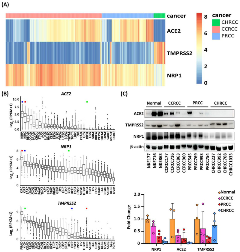
To test the hypothesis that the SARS-CoV-2 virus displays tropism not only to kidney cells, but also to renal cell carcinoma, we utilized the publicly available transcriptomic data sets deposited by The Cancer Genome Atlas project (TCGA) to assess the transcriptional levels of ACE2, TMPRSS2 and NRP1 mRNA in individual CCRCC, PRCC and CHRCC cases. The results were grouped according to RCC type (Fig 1A). The mRNA levels for ACE2 and NRP1 proved to be high in both CCRCC and PRCC, whereas levels for TMPRSS2 were lower, but significant. The reverse relation was seen for CHRCC where ACE2 and NRP1 levels were low but TMPRSS2 highly expressed. We furthermore analyzed the transcriptional levels of ACE2, TMPRSS2 and NRP1 mRNA across 32 different types of malignancies deposited at the TCGA. Of all cancer forms analyzed, CCRCC and PRCC displayed the highest expression levels for ACE2 and NRP1 mRNA, whereas again TMPRSS2 expression was lower but not negligible (Fig 1B). To elucidate the expression of the actual proteins, we performed western blot analysis of protein lysates from normal kidney cortex (n = 3), CCRCC tissue (n = 4), PRCC tissue (n = 4) and CHRCC tissue (n = 4). We discovered that ACE2, TMPRSS2 and NRP1 protein was detected in normal cortical tissue, CCRCC and PRCC (Fig 1C). The ACE2 protein was clearly detectable by three out of four CCRCC and PRCC cases and was undetectable in CHRCC lysates. NRP1 expression was strongly expressed by three of four CCRCC and PRCC cases, and at lower intensity by chromophobe RCC. Samples from CCRCC, PRCC and CHRCC expressed TMPRSS2 protein.

Fig 1. Expression of ACE2, TMPRSS2 and NRP1 in clear cell, papillary and chromophobe renal cell carcinoma and across 32 distinct cancer forms.

Fig 1

(A) Bioinformatic analysis of the TCGA data sets for renal cell carcinoma showing mRNA expression levels for the ACE2, TMPRSS2 and NRP1 genes in individual RCC cases for chromophobe renal cell carcinoma (CHRCC), clear cell renal cell carcinoma (CCRCC) and papillary renal cell carcinoma (PRCC). (B) Comparison of the mRNA expression levels for ACE2, TMPRSS2 and NRP1 across 32 different types of malignancies deposited at the TCGA atlas. KIRC denotes CCRCC (red square), KIRP denotes PRCC (blue square) and KICH denotes CHRCC (green square). (C) Immunoblotting results for ACE2, TMPRSS2 and NRP1 proteins in lysates from normal tissue, CCRCC, PRCC and CHRCC. Results from densitometry of the protein bands is shown below the blots.

ACE2, TMPRSS2 and NRP1 expression by proximal kidney tubules is retained by clear cell and papillary renal cell carcinoma tissue

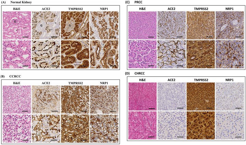
To allow for high-throughput analysis of ACE2, TMPRSS2 and NRP1 protein expression in RCC, we stained tissue micro arrays (TMA) with 263 cases of CCRCC, 139 cases of PRCC, 18 cases of CHRCC and human kidney tissue as control with antibodies against ACE2, TMPRSS2 and NRP1. We found that all three proteins were localized to the apical plasma membranes of the proximal tubules of normal human kidney (Fig 2A). ACE2 and NRP1 could not be detected in other tubular segments, whereas a faint TMPRSS2 signal was detected in the collecting ducts. Clear cell renal cell carcinoma and papillary renal cell carcinoma both originate from the proximal tubules, whereas chromophobe renal cell carcinoma originates from the intercalated cortical collecting duct cells. Of the CCRCC cases, 76%, 81% and 85% were positive for ACE2, TMPRSS2 and NRP1 respectively. PRCC displayed 93%, 56% and 66% positivity for ACE2, TMPRSS2 and NRP1 (Fig 2B and 2C, Table 1). CHRCC was negative for ACE2 and NRP1 but displayed a faint TMPRSS2 positivity in 50% of the cases, (Fig 2D, Table 1). Cells from the CCRCC derived cell line RCC4 were also fixed by formalin and paraffin embedded. No ACE2, TMPRSS2 and NRP1 staining was detected in the plasma membranes of the RCC4 cells. A distinct nuclear signal for ACE2 was however noted, (S1 Fig).

Fig 2. Expression of ACE2, TMPRSS2 and NRP1 in normal kidney and renal cell carcinoma tissue.

Fig 2

Haematoxylin & eosin staining of tissues and immunohistochemistry staining for ACE2, TMPRSS2 and NRP1. (A) Normal kidney displays staining of the apical plasma membranes of the proximal tubules for all three proteins. G = glomerulus, arrows = examples of proximal tubules and asterisk = collecting duct (negative). (B) Clear cell renal cell carcinoma tissue shows a diffuse staining of the plasma membranes for ACE2, TMPRSS2 and NRP1. (C) Papillary renal cell carcinoma displays an apical staining pattern for all three proteins. The lower rows of the panels show higher magnification of the images above. (D) Chromophobe renal cell carcinoma displayed negative staining for ACE2 and NRP1 whereas TMPRSS2 was strongly positive in the cancer cells. Upper panel scale bar = 100μm, lower panel scale bar = 40μm. (CCRCC = clear cell renal cell carcinoma, PRCC = papillary renal cell carcinoma, CHRCC = chromophobe renal cell carcinoma).

Table 1. Assessment of protein expression of ACE2, TMPRSS2 and NRP1 in clear cell, papillary and chromophobe renal cell carcinoma.

PROTEIN CCRCC (N = 263) TMA PRCC (n = 139) CHRCC (N = 18)
ACE2 76% (201/263) 93% (125/134) 0% (0/18)
TMPRSS2 81% (207/255) 56% (74/131) 50% (9/18)
NRP1 85% (220/258) 66% (91/137) 0% (0/18)

TMA, tissue microarray; CCRCC, clear cell renal cell carcinoma; PRCC, papillary renal cell carcinoma; CHRCC, chromophobe renal cell carcinoma. Due to random drop out of TMA-cores the number of assessed cores may vary slightly.

SARS-CoV-2 virus replicates in cultured renal cell carcinoma cells and causes a distinct virus cytopathogenic effect

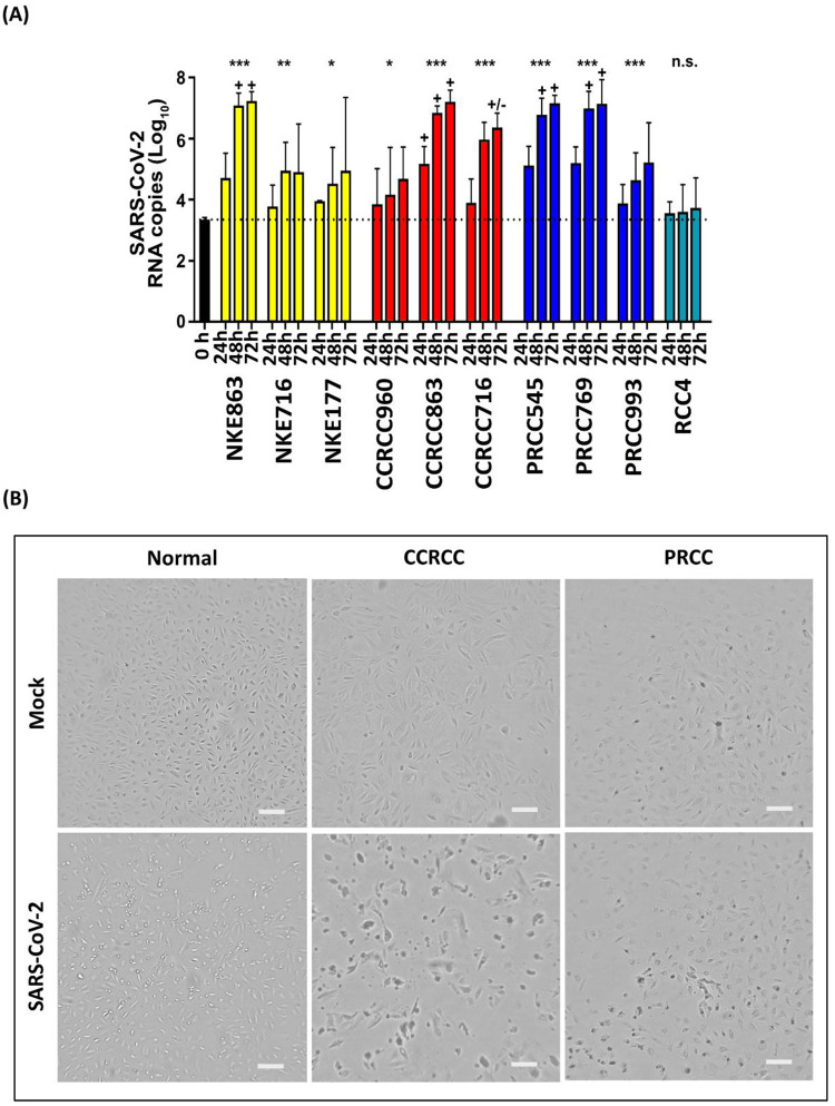
Having established that the key receptor and cofactors for SARS-CoV-2 binding were expressed by normal kidney, CCRCC tissue and PRCC tissue, we proceeded to investigate if primary RCC cancer cells, primary normal tubular cells and the CCRCC cell line RCC4 would internalize and propagate SARS-CoV-2 virus. The RCC4 cell line was included in order to evaluate if cell lines retain entry factor expression. We used primary RCC cells from the same cases that had been previously assessed by western blotting for expression of the entry factors. Primary cultures from 3 individual normal kidneys, 3 CCRCC cases and 3 PRCC cases were established. The cells were subsequently exposed to SARS-CoV-2 virus and propagation was monitored by virus negative strand PCR and visual inspection of the cultures at 24, 48 and 72 hours. Results showed that SARS-CoV-2 infected and propagated in primary normal kidney cells, CCRCC cells and PRCC cells as determined by real-time PCR and visual inspection. The RCC4 cell line failed to demonstrate infection detectable by PCR (Fig 3A). One normal kidney culture (NKE863), one CCRCC (CCRCC863) and two PRCC cell cultures (PRCC545 & PRCC769) also displayed distinct visible signs of virus cytopathogenic effect (CPE) at 48 hours (Fig 3B) and 72 hours post infection. This also correlated with the highest expression of viral RNA (Fig 3A). Additionally, one of the SARS-CoV-2 infected CCRCC cell cultures (CCRCC716) resulted in a moderate CPE. This culture also displayed slightly lower RNA levels, than in cultures where a clear CPE was visible (Fig 3A). Two normal kidney cultures (NKE716 & NKE177), one CCRCC (CCRCC960) and one PRCC (PRCC993) showed a limited increase in viral RNA and no CPE was visible. However, one of these normal kidney cultures demonstrated high expression of ACE (Fig 1C).

Fig 3. SARS-CoV-2 virus replicates in cultured renal cell carcinoma cells and causes a distinct virus cytopathogenic effect.

Fig 3

(A) Negative strand qPCR analysis of SARS-CoV-2 viral RNA copy number at 24h, 48h and 72h post-infection in primary normal tubular epithelial cells, CCRCC cells, PRCC cells and the renal cell carcinoma cell line RCC4. (B) Representative images of virus cytopathogenic effect (CPE) for normal (NKE863), clear cell renal cell carcinoma (CCRCC) (CCRCC863) and papillary renal cell carcinoma (PRCC) (PRCC545) at 48 hours after infection. The CPE causes rounding up of cells, followed by detachment from the growth surface. Mock is control. Scale bar = 200μm.

Unusual histological features in clear cell renal cell carcinoma tissue from a patient with previous COVID-19

Patients with COVID-19 are not candidates for nephrectomy. We however identified one CCRCC case from our records where the patient had undergone a serologically confirmed and hospital treated COVID-19 episode 12 months prior to nephrectomy. Histologically, the case was a clear cell carcinoma with very unusual features (S2A Fig). About 80% of the tumor was necrotic (S2B Fig). The remaining part displayed odd features of cancer cell fusion where the CCRCC cells had fused into syncytia (S2C and S2D Fig). The cancer cells in these areas also were discohesive and rounded with signs of degeneration. PCR for SARS-CoV-2 proved negative in the cancer tissue.

Visible formation of syncytia in primary CCRCC cells exposed to the delta virus variant

Following up on the finding of potential syncytial changes in SARS-CoV-2 exposed CCRCC tissue we investigated the in vitro effects of two SARS-CoV-2 variants, D614G & Delta. These were inoculated with normal renal tubular cell and CCRCC primary cell cultures, respectively (S3 Fig). Formation of CCRCC cell syncytia was clearly observable after 48 hours of incubation in clear cell renal cell carcinoma cells infected by the Delta variant in vitro (S3B Fig).

Discussion

COVID-19 is the systemic syndrome caused by severe infection by the SARS-CoV-2 virus. The virus enters the host via the respiratory tract, where many cell types express the receptors and proteins necessary for virus uptake. The most important of these proteins is the actual receptor for the virus, the ACE2 protein. Cofactors such as NRP1 and TMPRSS2 have been shown to be crucial as well however. ACE2 is relatively broadly expressed and the gastrointestinal tract expresses ACE2 mRNA in orders of magnitude above the respiratory tract levels [14]. Co-expression of receptor and co-factors is seen in a relatively small number of tissues and organs however. The gastrointestinal tract has been shown to be a site for active viral uptake. Furthermore, kidney injury is often diagnosed during COVID-19 disease. This was initially regarded as secondary to major systemic illness, but since then reports of direct renal infection have been published. We show that the necessary proteins for viral uptake, indeed are expressed by the proximal tubules of the human kidney. The proximal tubular cells are cells of origin for CCRCC and PRCC and thus for the major types of RCC. Importantly, we found that the expression is retained in a majority of CCRCC and PRCC cases, as assessed by immunohistochemistry combined with bioinformatical analysis of tumor mRNA levels for these genes deposited in the TCGA data sets. We found that CCRCC and PRCC clearly stand out in the TCGA data sets. The expressional levels for ACE2 and NRP1 mRNA occupy first and second position across all cancers analysed for CCRCC and PRCC, whereas TMPRSS2 was more modestly expressed. We could furthermore demonstrate that the virus replicated and displayed a clear virus cytopathogenic effect in a majority of the primary cancer cells. Uptake and effect were not always clearly correlated with ACE2 expression levels as determined by western blot. This may indicate additional factors playing a role in causing cytopathogenic effects in cell culture. The effect on the primary RCC cells was similar in magnitude to that of the kidney derived cell lines used to propagate SARS-CoV-2 virus in the clinical setting at the department of virology. Other cell lines used, such as calu3 require higher viral load for CPE and high replication. Virus replication was not shown to occur in the RCC4 cell line that proved to be negative for plasma membrane bound ACE2 and cofactors. This could be an argument that primary RCC cells are required to study this phenomenon. We are aware of the fact that the present paper describes in-vitro data, albeit based on primary cancer cells. We sought to amend this by identifying all patients with confirmed COVID-19 that had undergone a subsequent nephrectomy due to malignancy. It turned out that this patient category is quite small, since only one case was identified, where the COVID-19 diagnosis was made on clinical grounds and confirmed by serology. Histological assessment showed that the cancer tissue displayed very unusual features. The viable cancer cells displayed a rounded-up morphology and discohesive growth pattern with extensive necrotic zones. In areas cancer cells had fused into peculiar multinuclear syncytial cells, again with discohesive growth pattern. The RCC tissue was negative regarding SARS-CoV-2 RNA, but this is as expected since the COVID-19 episode predated the surgery with 12 months. It however led us to investigate in vitro effects of SARS-CoV-2 variants on CCRCC cells. We could observe a distinct syncytialization effect on the CCRCC cells following exposure to the delta variant. We are aware of the anecdotal nature of this RCC case and can therefore not draw any conclusions regarding potential effects in human RCC. We suggest however that this patient category may be important to follow in order to establish a potential causal connection where virus infection of the cancer cells may cause tumor lysis in the patient. In agreement with this reasoning, a recent study has shown that three patients with colorectal cancer did observe tumor regression while suffering from Covid-19 [15]. Recently it has been found that not only the actual oncolytic cellular effect is of value. Of similar or greater importance is the possibility of an enhanced immune response elicited by the cancer cells undergoing viral infection [16]. The virally affected cells release antigens that can be recognized by the immune system. This approach has become interesting as an adjunct to immune-oncological treatment protocols. At present, there is only one registered viral oncolytic treatment: talimogene laherparepvec (Imlygic). It is based on an attenuated herpes virus that has been modified to selectively replicate in melanoma cells and to stimulate a T-cell mediated immune response. It is administered by direct injection into the tumor [17]. Whether SARS-CoV-2 uptake and propagation in RCC cells could elicit a similar response is uncertain. The isolated case described above does not prove causality for SARS-CoV-2 induced effects on CCRCC in patients. Furthermore, usage of this uptake mechanism is questionable due to the pathogenicitity of the virus and lack of selectivity due to expression in normal organs. We conclude however that SARS-CoV-2 infects and displays virus cytopathogenic effects in clear cell and papillary renal cell carcinoma in-vitro. The cytopathogenic effect causes RCC cell lysis and the delta-variant of the virus causes syncytialization of the cancer cells.

Supporting information

S1 Fig. Expression of ACE2, TMPRSS2 and NRP1 in the RCC4 cell line.

None of the proteins could be detected at the expected location in the CCRCC cell line RCC4. ACE2 displayed nuclear positivity, NRP1 a faint cytoplasmic staining, whereas TMPRSS2 was negative. Upper panel scale bar = 100μm, lower panel scale bar = 40μm.

(TIF)

S2 Fig. Unusual histology in clear cell renal cell carcinoma from a COVID-19 patient.

(A) The basic histopathological pattern of the cancer case was clear cell renal cell carcinoma. (B) In about 60% of the tumor tissue extensive areas of necrosis could be seen. (C and D) Areas displaying very unusual features where the cancer cells had coalesced into syncytial, multinuclear and discohesive cancer cells with fusion of the lipid laded cytoplasms. All images were stained by Hematoxylin/Eosin, Scale bars in A-C: 100 μm, in D: 200 μm.

(TIF)

S3 Fig. In-vitro formation of syncytia in primary CCRCC cells exposed to virus variants.

Representative images of (A) normal tubular epithelial cells and (B) clear cell renal cell carcinoma cells after exposure to the SARS CoV-2 variants D614G or delta. Uninfected is mock control. After 48 hours of exposure to the delta variant a distinct formation of syncytia is seen in the CCRCC cultures. Hatched lines mark syncytial cancer cells. Scale bar = 200μm.

(TIF)

S1 Raw images. Uncropped western blot.

(ZIP)

S1 Table. Clinical characteristics of the patients included in the study.

(DOCX)

Data Availability

All relevant data are within the manuscript and its Supporting information files.

Funding Statement

MJ: Grant number 21 1767 Pj 01 H from The Swedish Cancer Society (Cancerfonden) MJ: The National Association against Kidney Diseases. MJ: Grant number 71390 Swedish Government Funding of Clinical Research within the National Health Service (ALF), MJ: The strategic research programme BioCARE, RJ: Grant number 273289 The Research Fund (R&D) at Skaraborg Hospital, Skövde, Sweden and the Healthcare Committee, Region Västra Götaland, Sweden. The funders had no role in study design, data collection and analysis, decision to publish, or preparation of the manuscript.

References

  • 1.Hoffmann M, Kleine-Weber H, Schroeder S, Krüger N, Herrler T, Erichsen S, et al. SARS-CoV-2 cell entry depends on ACE2 and TMPRSS2 and is blocked by a clinically proven protease inhibitor. Cell. 2020;181(2):271–80.e8. Epub 2020/03/07. doi: 10.1016/j.cell.2020.02.052 . [DOI] [PMC free article] [PubMed] [Google Scholar]
  • 2.Sungnak W, Huang N, Becavin C, Berg M, Queen R, Litvinukova M, et al. SARS-CoV-2 entry factors are highly expressed in nasal epithelial cells together with innate immune genes. Nat Med. 2020;26(5):681–7. Epub 2020/04/25. doi: 10.1038/s41591-020-0868-6 . [DOI] [PMC free article] [PubMed] [Google Scholar]
  • 3.Daly JL, Simonetti B, Klein K, Chen KE, Williamson MK, Anton-Plagaro C, et al. Neuropilin-1 is a host factor for SARS-CoV-2 infection. Science. 2020;370(6518):861–5. Epub 2020/10/22. doi: 10.1126/science.abd3072 . [DOI] [PMC free article] [PubMed] [Google Scholar]
  • 4.Gupta A, Madhavan MV, Sehgal K, Nair N, Mahajan S, Sehrawat TS, et al. Extrapulmonary manifestations of COVID-19. Nat Med. 2020;26(7):1017–32. Epub 2020/07/12. doi: 10.1038/s41591-020-0968-3 . [DOI] [PubMed] [Google Scholar]
  • 5.Mayer G, Boileau G, Bendayan M. Sorting of furin in polarized epithelial and endothelial cells: expression beyond the Golgi apparatus. J Histochem Cytochem. 2004;52(5):567–79. Epub 2004/04/22. doi: 10.1177/002215540405200502 . [DOI] [PubMed] [Google Scholar]
  • 6.Puelles VG, Lutgehetmann M, Lindenmeyer MT, Sperhake JP, Wong MN, Allweiss L, et al. Multiorgan and Renal Tropism of SARS-CoV-2. N Engl J Med. 2020;383(6):590–2. Epub 2020/05/14. doi: 10.1056/NEJMc2011400 . [DOI] [PMC free article] [PubMed] [Google Scholar]
  • 7.Harper SJ, Xing CY, Whittle C, Parry R, Gillatt D, Peat D, et al. Expression of neuropilin-1 by human glomerular epithelial cells in vitro and in vivo. Clin Sci (Lond). 2001;101(4):439–46. Epub 2001/09/22. . [PubMed] [Google Scholar]
  • 8.Chen QL, Li JQ, Xiang ZD, Lang Y, Guo GJ, Liu ZH. Localization of Cell Receptor-Related Genes of SARS-CoV-2 in the Kidney through Single-Cell Transcriptome Analysis. Kidney Dis (Basel). 2020;6(4):258–70. Epub 2020/09/10. doi: 10.1159/000508162 . [DOI] [PMC free article] [PubMed] [Google Scholar]
  • 9.Li Y, Xu Q, Ma L, Wu D, Gao J, Chen G, et al. Systematic profiling of ACE2 expression in diverse physiological and pathological conditions for COVID-19/SARS-CoV-2. J Cell Mol Med. 2020;24(16):9478–82. Epub 2020/07/09. doi: 10.1111/jcmm.15607 . [DOI] [PMC free article] [PubMed] [Google Scholar]
  • 10.Lindgren D, Eriksson P, Krawczyk K, Nilsson H, Hansson J, Veerla S, et al. Cell-Type-Specific Gene Programs of the Normal Human Nephron Define Kidney Cancer Subtypes. Cell Rep. 2017;20(6):1476–89. Epub 2017/08/10. doi: 10.1016/j.celrep.2017.07.043 . [DOI] [PubMed] [Google Scholar]
  • 11.Colaprico A, Silva TC, Olsen C, Garofano L, Cava C, Garolini D, et al. TCGAbiolinks: an R/Bioconductor package for integrative analysis of TCGA data. Nucleic Acids Res. 2016;44(8):e71. Epub 2015/12/26. doi: 10.1093/nar/gkv1507 . [DOI] [PMC free article] [PubMed] [Google Scholar]
  • 12.Robinson MD, McCarthy DJ, Smyth GK. edgeR: a Bioconductor package for differential expression analysis of digital gene expression data. Bioinformatics. 2010;26(1):139–40. Epub 2009/11/17. doi: 10.1093/bioinformatics/btp616 . [DOI] [PMC free article] [PubMed] [Google Scholar]
  • 13.Durinck S, Moreau Y, Kasprzyk A, Davis S, De Moor B, Brazma A, et al. BioMart and Bioconductor: a powerful link between biological databases and microarray data analysis. Bioinformatics. 2005;21(16):3439–40. Epub 2005/08/06. doi: 10.1093/bioinformatics/bti525 . [DOI] [PubMed] [Google Scholar]
  • 14.Zhang H, Kang Z, Gong H, Xu D, Wang J, Li Z, et al. Digestive system is a potential route of COVID-19: an analysis of single-cell coexpression pattern of key proteins in viral entry process. Gut. 2020;69(6):1010–8. doi: 10.1136/gutjnl-2020-320953 [DOI] [Google Scholar]
  • 15.Ottaiano A, Scala S, D’Alterio C, Trotta A, Bello A, Rea G, et al. Unexpected tumor reduction in metastatic colorectal cancer patients during SARS-Cov-2 infection. Therapeutic Advances in Medical Oncology. 2021;13:17588359211011455. doi: 10.1177/17588359211011455 . [DOI] [PMC free article] [PubMed] [Google Scholar]
  • 16.Brown MC, Holl EK, Boczkowski D, Dobrikova E, Mosaheb M, Chandramohan V, et al. Cancer immunotherapy with recombinant poliovirus induces IFN-dominant activation of dendritic cells and tumor antigen–specific CTLs. Science Translational Medicine. 2017;9(408):eaan4220. doi: 10.1126/scitranslmed.aan4220 [DOI] [PMC free article] [PubMed] [Google Scholar]
  • 17.Andtbacka RHI, Collichio F, Harrington KJ, Middleton MR, Downey G, Ӧhrling K, et al. Final analyses of OPTiM: a randomized phase III trial of talimogene laherparepvec versus granulocyte-macrophage colony-stimulating factor in unresectable stage III–IV melanoma. J Immunother Cancer. 2019;7(1):145. doi: 10.1186/s40425-019-0623-z [DOI] [PMC free article] [PubMed] [Google Scholar]

Decision Letter 0

Birke Bartosch

Transfer Alert

This paper was transferred from another journal. As a result, its full editorial history (including decision letters, peer reviews and author responses) may not be present.

4 Jul 2022

PONE-D-22-06970SARS-CoV-2 replicates and displays oncolytic properties in clear cell and papillary renal cell carcinomaPLOS ONE

Dear Dr. Johansson,

Thank you for submitting your manuscript to PLOS ONE. After careful consideration, we feel that it has merit but does not fully meet PLOS ONE’s publication criteria as it currently stands. Therefore, we invite you to submit a revised version of the manuscript that addresses all comments raised by the two reviewers and in particular their comments on syncytia formation and the mechanisms underlying the described cytopathic effects.

Please submit your revised manuscript by Aug 18 2022 11:59PM. If you will need more time than this to complete your revisions, please reply to this message or contact the journal office at plosone@plos.org. When you're ready to submit your revision, log on to https://www.editorialmanager.com/pone/ and select the 'Submissions Needing Revision' folder to locate your manuscript file.

Please include the following items when submitting your revised manuscript:

  • A rebuttal letter that responds to each point raised by the academic editor and reviewer(s). You should upload this letter as a separate file labeled 'Response to Reviewers'.

  • A marked-up copy of your manuscript that highlights changes made to the original version. You should upload this as a separate file labeled 'Revised Manuscript with Track Changes'.

  • An unmarked version of your revised paper without tracked changes. You should upload this as a separate file labeled 'Manuscript'.

If you would like to make changes to your financial disclosure, please include your updated statement in your cover letter. Guidelines for resubmitting your figure files are available below the reviewer comments at the end of this letter.

If applicable, we recommend that you deposit your laboratory protocols in protocols.io to enhance the reproducibility of your results. Protocols.io assigns your protocol its own identifier (DOI) so that it can be cited independently in the future. For instructions see: https://journals.plos.org/plosone/s/submission-guidelines#loc-laboratory-protocols. Additionally, PLOS ONE offers an option for publishing peer-reviewed Lab Protocol articles, which describe protocols hosted on protocols.io. Read more information on sharing protocols at https://plos.org/protocols?utm_medium=editorial-email&utm_source=authorletters&utm_campaign=protocols.

We look forward to receiving your revised manuscript.

Kind regards,

Birke Bartosch

Academic Editor

PLOS ONE

Journal requirements:

When submitting your revision, we need you to address these additional requirements.

1. Please ensure that your manuscript meets PLOS ONE's style requirements, including those for file naming. The PLOS ONE style templates can be found at

https://journals.plos.org/plosone/s/file?id=wjVg/PLOSOne_formatting_sample_main_body.pdf and

https://journals.plos.org/plosone/s/file?id=ba62/PLOSOne_formatting_sample_title_authors_affiliations.pdf".

2. We note that the grant information you provided in the ‘Funding Information’ and ‘Financial Disclosure’ sections do not match. When you resubmit, please ensure that you provide the correct grant numbers for the awards you received for your study in the ‘Funding Information’ section.

3. Thank you for stating the following in the Acknowledgments Section of your manuscript:

“This study was supported by grants from the Swedish Cancer Society (Cancerfonden), the National Association against Kidney Diseases, Swedish Government Funding of Clinical Research within the National Health Service (ALF), the strategic research program BioCARE, the Research Fund (R&D) at Skaraborg Hospital, Skövde, Sweden and the Healthcare Committee, Region Västra Götaland, Sweden.”

We note that you have provided funding information that is not currently declared in your Funding Statement. However, funding information should not appear in the Acknowledgments section or other areas of your manuscript. We will only publish funding information present in the Funding Statement section of the online submission form.

Please remove any funding-related text from the manuscript and let us know how you would like to update your Funding Statement. Currently, your Funding Statement reads as follows:

“RJ: the Healthcare Committee, Region Västra Götaland, Sweden - 273289.

The funders had no role in study design, data collection and analysis, decision to publish, or preparation of the manuscript.”

Please include your amended statements within your cover letter; we will change the online submission form on your behalf.

4. We note that you have stated that you will provide repository information for your data at acceptance. Should your manuscript be accepted for publication, we will hold it until you provide the relevant accession numbers or DOIs necessary to access your data. If you wish to make changes to your Data Availability statement, please describe these changes in your cover letter and we will update your Data Availability statement to reflect the information you provide.

5. PLOS ONE now requires that authors provide the original uncropped and unadjusted images underlying all blot or gel results reported in a submission’s figures or Supporting Information files. This policy and the journal’s other requirements for blot/gel reporting and figure preparation are described in detail at https://journals.plos.org/plosone/s/figures#loc-blot-and-gel-reporting-requirements and https://journals.plos.org/plosone/s/figures#loc-preparing-figures-from-image-files. When you submit your revised manuscript, please ensure that your figures adhere fully to these guidelines and provide the original underlying images for all blot or gel data reported in your submission. See the following link for instructions on providing the original image data: https://journals.plos.org/plosone/s/figures#loc-original-images-for-blots-and-gels.

In your cover letter, please note whether your blot/gel image data are in Supporting Information or posted at a public data repository, provide the repository URL if relevant, and provide specific details as to which raw blot/gel images, if any, are not available. Email us at plosone@plos.org if you have any questions.

[Note: HTML markup is below. Please do not edit.]

Reviewers' comments:

Reviewer's Responses to Questions

Comments to the Author

1. Is the manuscript technically sound, and do the data support the conclusions?

The manuscript must describe a technically sound piece of scientific research with data that supports the conclusions. Experiments must have been conducted rigorously, with appropriate controls, replication, and sample sizes. The conclusions must be drawn appropriately based on the data presented.

Reviewer #1: No

Reviewer #2: Yes

**********

2. Has the statistical analysis been performed appropriately and rigorously?

Reviewer #1: N/A

Reviewer #2: Yes

**********

3. Have the authors made all data underlying the findings in their manuscript fully available?

The PLOS Data policy requires authors to make all data underlying the findings described in their manuscript fully available without restriction, with rare exception (please refer to the Data Availability Statement in the manuscript PDF file). The data should be provided as part of the manuscript or its supporting information, or deposited to a public repository. For example, in addition to summary statistics, the data points behind means, medians and variance measures should be available. If there are restrictions on publicly sharing data—e.g. participant privacy or use of data from a third party—those must be specified.

Reviewer #1: Yes

Reviewer #2: Yes

**********

4. Is the manuscript presented in an intelligible fashion and written in standard English?

PLOS ONE does not copyedit accepted manuscripts, so the language in submitted articles must be clear, correct, and unambiguous. Any typographical or grammatical errors should be corrected at revision, so please note any specific errors here.

Reviewer #1: Yes

Reviewer #2: Yes

**********

5. Review Comments to the Author

Please use the space provided to explain your answers to the questions above. You may also include additional comments for the author, including concerns about dual publication, research ethics, or publication ethics. (Please upload your review as an attachment if it exceeds 20,000 characters)

Reviewer #1: The study presented by Choong et al. implies that SARS-CoV-2 may replicate in renal cell carcinoma cells (CCRCC and PRCC) because these they express virus entry factors. Infection with SARS-Cov-2 resulted in cytopathic effect – syncytia formation. The manuscript is well written but I am afraid that some of the data presented suffer flaws and at some points there is overinterpretation of the results.

Here are my comments concerning the study:

1. Line 71: the authors conclude that their data “provide insights into SARS-CoV-2 infectivity in renal cell carcinoma and define a potential viral entry mechanism that could be used to target renal carcinoma”. However, the latter is not shown experimentally nor sufficiently discussion in the Discussion section. Moreover, in the Discussion section, the authors widely stress on oncolytic viral therapy implicating SARS-CoV-2. Such notion is not supported experimentally. Do cancer cells strongly overexpress viral entry factors compared to normal cells? How will be the specific targeting of cancer cells achieved? Indeed, measles virus (the vaccine strain!) has been used in such therapies, but this is justified by the strong expression of MCP (CD46) in cancer cells and the non-pathogenicity of the virus which is not the case with SARS-CoV-2.

2. Concerning the Discussion, the authors are not the first to show that SARS-CoV-2 entry factors are found in renal cells. This are just some examples of other studies: PMID:32903321 (TMPRSS2), PMID:19736548, PMID:11566082 (NRP1), and for ACE-2 there are multiple papers.

3. Figure 1C: I am afraid I do not see the expression of TMPRSS2 in normal tissues on the blot as stated by the authors: line 226. The authors also stated that “NRP1 expression was robust across all cases” (Line 228). This is not the case for CHRCC and some of PRCC samples. A better blot must be provided, and also a quantification of the blot by densitometry.

4. Figure 2A: Use arrows to indicate different histological structures.

5. Line 246: “We also formalin” The verb is missing…

6. Figure 3B: The data of syncytia formation in PRCC is not convincing (the microscopic image). Moreover, there is no correlation between the cell culture data (slightly visible syncytia) and the PCR data (strong expression) for PRCC.

7. I do not understand the including of the case study of 1 patient within this report. There is absolutely no proof that syncytia formation is due to SARS-CoV-2 (that the patient had 1 year before). Research for viral antigens was not performed. Moreover, syncytia formation is directly linked to cell lysis. This is even seen in the in vitro data on FigS3 B (almost no cells left).

8. In the discussion section (Lines 312-316) the authors say that “uptake and effect were not always clearly correlated to ACE-2 expression and that additional factors play role in causing cytopathogenic effect (CPE) in cell culture”. CPE is usually due to expression of viral receptors on uninfected cells and the expression of viral glycoproteins in infected cells. What is the expression of spike in these cells?

Reviewer #2: The manuscript by Choong et al describes a very promising phenomenon: oncolytic activity of SARS-CoV-2 against renal carcinoma variants. The authors used bioinformatic analysis of existing RNAseq data and showed that both normal renal cells and cells of two renal carcinoma types expressed both ACE2 receptor and NRP1 co-factor required for SARS-CoV-2 entry. Then they verified these results by western blot analysis using normal and tumor renal cells. A correct (using a negative strand-specific RNA amplification) RT-qPCR analysis revealed that the same cell lines were permissive for virus, and in cases of high infection titers cytophathic effect is observed. The authors demonstrate formation of syncytia in renal tissue from a patient with previous COVID-19 infection, despite signs of the infection at a moment of analysis. In the discussion the authors speculate that SARS-CoV-2 can act as oncolutic viruses and may trigger tumor lysis in cancer patients with COVID-19, as observed for colon cancer patients.

The manuscript is carefully and clearly written. All conclusions are based on presented experimental data.

The topic definitely merits publication. Other groups have shown that SARS-CoV-2 does not always display cytophatic effect (i.e. in case of hepatocellular carcinoma Huh7 cell line).

I would suggest publishing the study after answering two short questions:

1. Was cytophatic effect quantified Ii.e. by MTT assay, staining with annexin V or propidium iodide)?

2. Can the authors speculate about the mechanisms that control cytopathic effect of the virus? or in authors opinion the effect is merely due to high replication rate? In our personal experience some cells lines survive during the infection, despite high replication rates.

**********

6. PLOS authors have the option to publish the peer review history of their article (what does this mean?). If published, this will include your full peer review and any attached files.

If you choose “no”, your identity will remain anonymous but your review may still be made public.

Do you want your identity to be public for this peer review? For information about this choice, including consent withdrawal, please see our Privacy Policy.

Reviewer #1: No

Reviewer #2: No

**********

[NOTE: If reviewer comments were submitted as an attachment file, they will be attached to this email and accessible via the submission site. Please log into your account, locate the manuscript record, and check for the action link "View Attachments". If this link does not appear, there are no attachment files.]

While revising your submission, please upload your figure files to the Preflight Analysis and Conversion Engine (PACE) digital diagnostic tool, https://pacev2.apexcovantage.com/. PACE helps ensure that figures meet PLOS requirements. To use PACE, you must first register as a user. Registration is free. Then, login and navigate to the UPLOAD tab, where you will find detailed instructions on how to use the tool. If you encounter any issues or have any questions when using PACE, please email PLOS at figures@plos.org. Please note that Supporting Information files do not need this step.

PLoS One. 2023 Jan 3;18(1):e0279578. doi: 10.1371/journal.pone.0279578.r002

Author response to Decision Letter 0


29 Sep 2022

The work and valuable constructive comments from the reviewers is highly appreciated and the text that follows is the same as in the response to reviewers file.

Response to the reviewer comments to the author

Reviewer #1: The study presented by Choong et al. implies that SARS-CoV-2 may replicate in renal cell carcinoma cells (CCRCC and PRCC) because these they express virus entry factors. Infection with SARS-Cov-2 resulted in cytopathic effect – syncytia formation. The manuscript is well written but I am afraid that some of the data presented suffer flaws and at some points there is overinterpretation of the results.

Here are my comments concerning the study:

1. Line 71: the authors conclude that their data “provide insights into SARS-CoV-2 infectivity in renal cell carcinoma and define a potential viral entry mechanism that could be used to target renal carcinoma”. However, the latter is not shown experimentally nor sufficiently discussion in the Discussion section. Moreover, in the Discussion section, the authors widely stress on oncolytic viral therapy implicating SARS-CoV-2. Such notion is not supported experimentally. Do cancer cells strongly overexpress viral entry factors compared to normal cells? How will be the specific targeting of cancer cells achieved? Indeed, measles virus (the vaccine strain!) has been used in such therapies, but this is justified by the strong expression of MCP (CD46) in cancer cells and the non-pathogenicity of the virus which is not the case with SARS-CoV-2.

Response: We thank the reviewer for this comment. We agree that we have overstated the interpretation of our results and that a more measured approach serves the manuscript better. To this end we have rewritten line 70-71 (Our data provide insights into SARS-CoV-2 infectivity in renal cell carcinoma and define a potential viral entry mechanism that could be used to target renal cell carcinoma.).

The new wording in line 70-71 is: Our data provide insights into SARS-CoV-2 infectivity in renal cell carcinoma and that the virus causes a distinct cytopathogenic effect.

We have also rewritten relevant parts of the discussion regarding oncolytic therapy in order to temper the conclusions. Specifically, we deleted the sentence beginning on line 346: (We suggest that the mechanism presented in this paper may be used in a similar fashion) and line 347-48: (Another possibility may be to use the receptor apparatus of a modified attenuated virus to deliver therapeutic nucleotides or a similar payload to the cancer cells).

The new text is: “Whether SARS-CoV-2 uptake and propagation in RCC cells could elicit a similar response is uncertain. Usage of this uptake mechanism is questionable due to the the pathogenicitity of the virus and lack of selectivity due to expression in normal organs.” Line 356-359.

Based on bioinformatic analysis of the TCGA data set we do believe that RCC is unusual in its expression of the entry factors for SARS-CoV-2. This we can corroborate using tissue micro arrays of a large number of RCC cases. This in turn reflects the native expression of these factors in the proximal tubular epithelium of the kidney, a colocalization not shared by many organs. There are however other tissues expressing these factors, so it is not entirely specific and selective for delivery to RCC cells. The overly speculative concluding sentence of the discussion section has therefore also been deleted, line 350-351: (We suggest that the uptake mechanism for SARS-CoV-2 virus may be exploited to mediate uptake of therapeutic compounds or nucleic acids into renal cell carcinoma) and changed to: “The cytopathogenic effect causes RCC cell lysis and the delta-variant of the virus causes syncytialization of the cancer cells.”, now line 363-364.

2. Concerning the Discussion, the authors are not the first to show that SARS-CoV-2 entry factors are found in renal cells. This are just some examples of other studies: PMID:32903321 (TMPRSS2), PMID:19736548, PMID:11566082 (NRP1), and for ACE-2 there are multiple papers.

Response: We thank the reviewer for this point, we are aware of this, but regret that we have failed to communicate this in the manuscript. We do mention in the introduction, that the factors are expressed at mRNA level in kidney tissue, but regarding protein expression we were less clear. We have added the references PMID 11566082 and PMID 32903321 to the reference list in order to more clearly acknowledge previous histological work. The sentence describing this (line 90-92) has been changed to account for this. New sentence in line 92-93.

3. Figure 1C: I am afraid I do not see the expression of TMPRSS2 in normal tissues on the blot as stated by the authors: line 226. The authors also stated that “NRP1 expression was robust across all cases” (Line 228). This is not the case for CHRCC and some of PRCC samples. A better blot must be provided, and also a quantification of the blot by densitometry.

Response: We agree that the western blot for TMPRSS2 is definitely not the easiest to interpret. To amend for this, we tested alternative antibodies for TMPRSS2 on the protein lysates. We chose anti-TMPRSS2 from Thermo Fisher (1:1000, MA5-35756). This resulted in a much better blot with improved signal and we have replaced the inferior blot with the new one in a new Figure 1C. Information regarding this has been added to the materials and methods section in the manuscript (line 167-168).

As suggested by the reviewer, we also performed densitometry of the bands in order to quantify ACE2, NRP1 and TMPRSS2, the results from this have also been added to figure 1C and to the figure legend (line 478-479). Finally, we have changed line 226-228 in the results section to reflect the improvements suggested by the reviewer. It now reads:

The ACE2 protein was clearly detectable by three out of four CCRCC and PRCC cases and was undetectable in CHRCC lysates. NRP1 expression was strongly expressed by three of four CCRCC and PRCC cases, and at lower intensity by chromophobe RCC. Samples from CCRCC, PRCC and CHRCC expressed TMPRSS2 protein. (now line 229-232)

4. Figure 2A: Use arrows to indicate different histological structures.

Response: This suggestion improves evaluation of the images. For clarity figure 2A has been added the letter G to indicate glomeruli, arrows for examples of proximal tubules and asterisks for collecting ducts. This has also been added to the figure legend at line 485-486.

5. Line 246: “We also formalin” The verb is missing…

Response: we have changed the sentence to the more clear-cut sentence: “Cells from the CCRCC derived cell line RCC4 were also fixed by formalin and paraffin embedded.” This is now the text at line 251-253.

6. Figure 3B: The data of syncytia formation in PRCC is not convincing (the microscopic image). Moreover, there is no correlation between the cell culture data (slightly visible syncytia) and the PCR data (strong expression) for PRCC.

Response: We are grateful for the opportunity to clarify the figures. Supplemental Figure 3B does not show syncytia in cultured PRCC cells, it shows the formation of syncytia in cultured primary CCRCC cells. We agree that the image could be improved and we have therefore hatched the perimeter of two distinct syncytialised CCRCC cells in supplemental figure 3B and description added to figure legend (line 525). Judging from figure 1C and figure 3A, the levels of attachment proteins and SARS-Cov-2 replication seem roughly similar between normal tubular epithelial cells and primary CCRCC cells. The protein expression is slightly lower in the CCRCC cells, but not significantly so.

7. I do not understand the including of the case study of 1 patient within this report. There is absolutely no proof that syncytia formation is due to SARS-CoV-2 (that the patient had 1 year before). Research for viral antigens was not performed. Moreover, syncytia formation is directly linked to cell lysis. This is even seen in the in vitro data on FigS3 B (almost no cells left).

Response: We agree with the concerns of the reviewer. We included the case, knowing that the number is too low to form a conclusion, especially since the active infection was a year previously. Having discovered that SARS-CoV-2 virus infects RCC cells in vitro, ideally, we would have liked to test the uptake in another system. The gold standard would be to infect a PdX mouse model with SARS-CoV-2. Since this is out of reach at this point, we reached out to the department of Urology asking if they had performed surgery on a patient with a proven SARS-CoV infection. They could only identifty one cases from their records. The histology being very unusual, we decided to add it to the paper in order to notify the pathology community about the possible morphology of a passing SARS-CoV-2 infection in RCC. The syncytia of the RCC-cases were definitely associated with cell lysis and outright necrosis of the RCC-tissue. This led us to design the experiment where we assessed if this could be repeated in vitro by a SARS-CoV-2.

As pointed out, we definitely understand the concerns of the reviewer and have added an additional sentence underscoring the speculative nature of the presented case at line 340-342 with the following wording:

We are aware of the anecdotal nature of this RCC case and can therefore not draw any conclusions regarding potential effects in human RCC.

We have also deleted the following sentence (line 331): We suggest that this finding strengthens our assumption that SARS-CoV-2 might infect RCC also in patients

8. In the discussion section (Lines 312-316) the authors say that “uptake and effect were not always clearly correlated to ACE-2 expression and that additional factors play role in causing cytopathogenic effect (CPE) in cell culture”. CPE is usually due to expression of viral receptors on uninfected cells and the expression of viral glycoproteins in infected cells. What is the expression of spike in these cells?

Response: This is a very interesting comment and suggestion. We would like to assess the expression of spike protein and accessory glycoproteins in the infected RCC and normal epithelial cells. However, we have not secured histological material or protein lysates from the infection assay, which precludes adding this data to the present study.

_____________________________________________________________________

Reviewer #2: The manuscript by Choong et al describes a very promising phenomenon: oncolytic activity of SARS-CoV-2 against renal carcinoma variants. The authors used bioinformatic analysis of existing RNAseq data and showed that both normal renal cells and cells of two renal carcinoma types expressed both ACE2 receptor and NRP1 co-factor required for SARS-CoV-2 entry. Then they verified these results by western blot analysis using normal and tumor renal cells. A correct (using a negative strand-specific RNA amplification) RT-qPCR analysis revealed that the same cell lines were permissive for virus, and in cases of high infection titers cytophathic effect is observed. The authors demonstrate formation of syncytia in renal tissue from a patient with previous COVID-19 infection, despite signs of the infection at a moment of analysis. In the discussion the authors speculate that SARS-CoV-2 can act as oncolutic viruses and may trigger tumor lysis in cancer patients with COVID-19, as observed for colon cancer patients. The manuscript is carefully and clearly written. All conclusions are based on presented experimental data. The topic definitely merits publication. Other groups have shown that SARS-CoV-2 does not always display cytophatic effect (i.e., in case of hepatocellular carcinoma Huh7 cell line).

I would suggest publishing the study after answering two short questions:

1. Was cytophatic effect quantified Ii.e. by MTT assay, staining with annexin V or propidium iodide)?

Response: This is a very good point. We used the method preferred by the virologists at the virology department of the university hospital. Therefore, we assessed the cells by inspection and photography. The cytopathic effect was pronounced and easy to see and compared to infection controls and uninfected cells. Virus replication in cell culture as determined by visual cytopathic effect was further confirmed by RT-PCR. We did not run the suggested assays, partially due to infectivity issues.

2. Can the authors speculate about the mechanisms that control cytopathic effect of the virus? or in authors opinion the effect is merely due to high replication rate? In our personal experience some cells lines survive during the infection, despite high replication rates.

Response: For SARS-CoV-2 exposed kidney derived cell lines we observe a clear correlation between a high replication rate and CPE. For other cell lines such as calu3 more virus is necessary to induce CPE and a high viral replication rate.

A sentence regarding this has been added to the discussion section starting at line 330 with the following wording:

The effect on the primary RCC cells was similar in magnitude to that of the kidney derived cell lines used to propagate SARS-CoV-2 virus in the clinical setting at the department of virology. Other cell lines used, such as calu3 require higher viral load for CPE and high replication (322-325).

In conclusion we hope that the changes and additions described above have met the expectations of the editor and the reviewers. We wish to thank all involved for their work in the constructive review of this manuscript.

Attachment

Submitted filename: Response to Reviewers PONE D 22 06970 version TC.docx

Decision Letter 1

Birke Bartosch

28 Oct 2022

PONE-D-22-06970R1SARS-CoV-2 replicates and displays oncolytic properties in clear cell and papillary renal cell carcinomaPLOS ONE

Dear Dr. Johansson,

Thank you for submitting your manuscript to PLOS ONE. After careful consideration, we feel that it has merit but would like to propose some further (very minor) changes, as outlined by reviewer 1 and in the editor comments further below, as we feel that these changes would further improve the manuscript.

Please submit your revised manuscript by Dec 12 2022 11:59PM. If you will need more time than this to complete your revisions, please reply to this message or contact the journal office at plosone@plos.org. When you're ready to submit your revision, log on to https://www.editorialmanager.com/pone/ and select the 'Submissions Needing Revision' folder to locate your manuscript file.

Please include the following items when submitting your revised manuscript:

  • A rebuttal letter that responds to each point raised by the academic editor and reviewer(s). You should upload this letter as a separate file labeled 'Response to Reviewers'.

  • A marked-up copy of your manuscript that highlights changes made to the original version. You should upload this as a separate file labeled 'Revised Manuscript with Track Changes'.

  • An unmarked version of your revised paper without tracked changes. You should upload this as a separate file labeled 'Manuscript'.

If you would like to make changes to your financial disclosure, please include your updated statement in your cover letter. Guidelines for resubmitting your figure files are available below the reviewer comments at the end of this letter.

If applicable, we recommend that you deposit your laboratory protocols in protocols.io to enhance the reproducibility of your results. Protocols.io assigns your protocol its own identifier (DOI) so that it can be cited independently in the future. For instructions see: https://journals.plos.org/plosone/s/submission-guidelines#loc-laboratory-protocols. Additionally, PLOS ONE offers an option for publishing peer-reviewed Lab Protocol articles, which describe protocols hosted on protocols.io. Read more information on sharing protocols at https://plos.org/protocols?utm_medium=editorial-email&utm_source=authorletters&utm_campaign=protocols.

We look forward to receiving your revised manuscript.

Kind regards,

Birke Bartosch

Academic Editor

PLOS ONE

Journal Requirements:

Please review your reference list to ensure that it is complete and correct. If you have cited papers that have been retracted, please include the rationale for doing so in the manuscript text, or remove these references and replace them with relevant current references. Any changes to the reference list should be mentioned in the rebuttal letter that accompanies your revised manuscript. If you need to cite a retracted article, indicate the article’s retracted status in the References list and also include a citation and full reference for the retraction notice.

Additional Editor Comments:

The manuscript has been very positively reviewed. Some very minor issues remain which would further improve the manuscript. As suggested by reviewer 1, moving the CHRCC IHC from the supplementary into Figure 1 would make a good comparison of receptor expressing versus low/no expressing cells. Furthermore, if PCR data on CHRCC are available, it would be useful to include them into Fig 3A.

[Note: HTML markup is below. Please do not edit.]

Reviewers' comments:

Reviewer's Responses to Questions

Comments to the Author

1. If the authors have adequately addressed your comments raised in a previous round of review and you feel that this manuscript is now acceptable for publication, you may indicate that here to bypass the “Comments to the Author” section, enter your conflict of interest statement in the “Confidential to Editor” section, and submit your "Accept" recommendation.

Reviewer #1: (No Response)

Reviewer #2: All comments have been addressed

**********

2. Is the manuscript technically sound, and do the data support the conclusions?

The manuscript must describe a technically sound piece of scientific research with data that supports the conclusions. Experiments must have been conducted rigorously, with appropriate controls, replication, and sample sizes. The conclusions must be drawn appropriately based on the data presented.

Reviewer #1: Partly

Reviewer #2: Yes

**********

3. Has the statistical analysis been performed appropriately and rigorously?

Reviewer #1: N/A

Reviewer #2: Yes

**********

4. Have the authors made all data underlying the findings in their manuscript fully available?

The PLOS Data policy requires authors to make all data underlying the findings described in their manuscript fully available without restriction, with rare exception (please refer to the Data Availability Statement in the manuscript PDF file). The data should be provided as part of the manuscript or its supporting information, or deposited to a public repository. For example, in addition to summary statistics, the data points behind means, medians and variance measures should be available. If there are restrictions on publicly sharing data—e.g. participant privacy or use of data from a third party—those must be specified.

Reviewer #1: Yes

Reviewer #2: Yes

**********

5. Is the manuscript presented in an intelligible fashion and written in standard English?

PLOS ONE does not copyedit accepted manuscripts, so the language in submitted articles must be clear, correct, and unambiguous. Any typographical or grammatical errors should be corrected at revision, so please note any specific errors here.

Reviewer #1: Yes

Reviewer #2: Yes

**********

6. Review Comments to the Author

Please use the space provided to explain your answers to the questions above. You may also include additional comments for the author, including concerns about dual publication, research ethics, or publication ethics. (Please upload your review as an attachment if it exceeds 20,000 characters)

Reviewer #1: The authors have made changes in order to moderate some conclusions/statements which subsequently improves the manuscript. Even though, there are still issues that need to be addressed.

1. Figure 2: This figure includes IHC images of normal, CCRCC and PRCC cells but not CHRCC. It is more logical that S1A figure of CHRCC becomes part of Fig.2.

2. Figure 3A. All other cell types but CHRCC have been analyzed by PCR but CHRCC. If the authors have the data, it should be included (even if no infection is observed which could be expected).

3. I still have a problem with the case study of 1 patient. Lines 335-336: “We can only

suggest that SARS-CoV-2 may be causally connected to the unusual morphology”. This is a pure speculation. Was this patient treatment naïve prior nephrectomy? Nothing can be concluded from this “unusual morphology” of 1 patient. However, this oriented the discussion toward viral oncolytic therapy.

Reviewer #2: The authors have addressed all my concerns.

In my personal opinion, the findings that SARS-CoV-2 can replicate in clear cell carcinoma cells are important to understand permisiveness of cell to the pathogen, its tropism.

**********

7. PLOS authors have the option to publish the peer review history of their article (what does this mean?). If published, this will include your full peer review and any attached files.

If you choose “no”, your identity will remain anonymous but your review may still be made public.

Do you want your identity to be public for this peer review? For information about this choice, including consent withdrawal, please see our Privacy Policy.

Reviewer #1: No

Reviewer #2: No

**********

[NOTE: If reviewer comments were submitted as an attachment file, they will be attached to this email and accessible via the submission site. Please log into your account, locate the manuscript record, and check for the action link "View Attachments". If this link does not appear, there are no attachment files.]

While revising your submission, please upload your figure files to the Preflight Analysis and Conversion Engine (PACE) digital diagnostic tool, https://pacev2.apexcovantage.com/. PACE helps ensure that figures meet PLOS requirements. To use PACE, you must first register as a user. Registration is free. Then, login and navigate to the UPLOAD tab, where you will find detailed instructions on how to use the tool. If you encounter any issues or have any questions when using PACE, please email PLOS at figures@plos.org. Please note that Supporting Information files do not need this step.

PLoS One. 2023 Jan 3;18(1):e0279578. doi: 10.1371/journal.pone.0279578.r004

Author response to Decision Letter 1


7 Dec 2022

Thank you for the further reviews of our manuscript entitled: SARS-CoV-2 replicates and displays oncolytic properties in clear cell and papillary renal cell carcinoma (PONE-D-22-06970R1) by Oi Kuan Choong et al.

We are very grateful for the constructive comments and criticisms. Below we address the comments by reviewer #1. We interpret reviewer #2 as being satisfied with the current version. We believe that it has resulted in a further improvement of the manuscript. We hope that the reviewers find the measures taken adequate and that the current form of the manuscript is acceptable for publication.

Response to comments from Reviewer #1.

1. Figure 2: This figure includes IHC images of normal, CCRCC and PRCC cells but not CHRCC. It is more logical that S1A figure of CHRCC becomes part of Fig.2.

Response: We agree that it is more logical to have all the RCC stainings in one figure and have therefore moved the staining results for CHRCC from the supplemental figures to figure 2 D. We have also added H/E images of the same magnification as for figure 2A-C in order to achieve the same set up as for CCRCC and PRCC.

2. Figure 3A. All other cell types but CHRCC have been analyzed by PCR but CHRCC. If the authors have the data, it should be included (even if no infection is observed which could be expected).

Response: Regrettably, we could not show data from primary cultures of CHRCC. It would definitively have been appropriate as comparison. Primary CHRCC cells are difficult/

impossible to culture however, and we have not succeeded in establishing primary cultures of CHRCC cells. Also, there are no well characterized cell lines available to our knowledge. Therefore, we have no PCR data from the SARS-CoV-2 uptake and propagation experiment.

3. I still have a problem with the case study of 1 patient. Lines 335-336: “We can only suggest that SARS-CoV-2 may be causally connected to the unusual morphology”. This is a pure speculation. Was this patient treatment naïve prior nephrectomy? Nothing can be concluded from this “unusual morphology” of 1 patient. However, this oriented the discussion toward viral oncolytic therapy.

Response: One case is definitely not enough to infer that SARS-CoV-2 causes RCC cell necrosis. We sought to modify our wording in the previous version, and we now try to soften our language further. First, we have now omitted the sentence of above completely (“We can only suggest that SARS-CoV-2 may be causally connected to the unusual morphology”

We also omit the sentence beginning at line 339, since we feel that is also a bit too bold:

“The goal of precision medicine is to selectively target cancer cells. For well over a century, oncolytic viral therapy has been researched.”

We also add a new sentence to the discussion at line 346 to underscore that we are not implying a causal relationship. It reads as follows:

The isolated case described above does not prove causality for SARS-CoV-2 induced effects on CCRCC in patients.

If the reviewer still expresses concerns, we are more than happy to completely omit the case from the article. The reason for not doing so before re-submission this time is because the finding of multinucleated cells in the cancer tissue led us to make an additional to find out whether the virus may cause syncytialization of the cells. Perhaps omission would render the assay hanging in the air so to say. Second, we wish to alert the readership to keep an eye on possible unusual histological patterns in this patient category. Again, we are more than happy to erase the case completely if its presence hinders the appreciation of the paper. Not least since the central theme of the article focuses on SARS-CoV-2 tropism in RCC and we do not want that this is obscured.

Summing up, we hope that our responses to the comments and suggestions from the reviewers are regarded as adequate and that they have resulted in an improved manuscript.

Attachment

Submitted filename: Response to reviewers D 22 06970 R1.pdf

Decision Letter 2

Birke Bartosch

12 Dec 2022

SARS-CoV-2 replicates and displays oncolytic properties in clear cell and papillary renal cell carcinoma

PONE-D-22-06970R2

Dear Dr. Johansson,

We’re pleased to inform you that your manuscript has been judged scientifically suitable for publication and will be formally accepted for publication once it meets all outstanding technical requirements.

Within one week, you’ll receive an e-mail detailing the required amendments. When these have been addressed, you’ll receive a formal acceptance letter and your manuscript will be scheduled for publication.

An invoice for payment will follow shortly after the formal acceptance. To ensure an efficient process, please log into Editorial Manager at http://www.editorialmanager.com/pone/, click the 'Update My Information' link at the top of the page, and double check that your user information is up-to-date. If you have any billing related questions, please contact our Author Billing department directly at authorbilling@plos.org.

If your institution or institutions have a press office, please notify them about your upcoming paper to help maximize its impact. If they’ll be preparing press materials, please inform our press team as soon as possible -- no later than 48 hours after receiving the formal acceptance. Your manuscript will remain under strict press embargo until 2 pm Eastern Time on the date of publication. For more information, please contact onepress@plos.org.

Kind regards,

Birke Bartosch

Academic Editor

PLOS ONE

Additional Editor Comments (optional):

Reviewers' comments:

Acceptance letter

Birke Bartosch

21 Dec 2022

PONE-D-22-06970R2

SARS-CoV-2 replicates and displays oncolytic properties in clear cell and papillary renal cell carcinoma

Dear Dr. Johansson:

I'm pleased to inform you that your manuscript has been deemed suitable for publication in PLOS ONE. Congratulations! Your manuscript is now with our production department.

If your institution or institutions have a press office, please let them know about your upcoming paper now to help maximize its impact. If they'll be preparing press materials, please inform our press team within the next 48 hours. Your manuscript will remain under strict press embargo until 2 pm Eastern Time on the date of publication. For more information please contact onepress@plos.org.

If we can help with anything else, please email us at plosone@plos.org.

Thank you for submitting your work to PLOS ONE and supporting open access.

Kind regards,

PLOS ONE Editorial Office Staff

on behalf of

Dr. Birke Bartosch

Academic Editor

PLOS ONE

Associated Data

    This section collects any data citations, data availability statements, or supplementary materials included in this article.

    Supplementary Materials

    S1 Fig. Expression of ACE2, TMPRSS2 and NRP1 in the RCC4 cell line.

    None of the proteins could be detected at the expected location in the CCRCC cell line RCC4. ACE2 displayed nuclear positivity, NRP1 a faint cytoplasmic staining, whereas TMPRSS2 was negative. Upper panel scale bar = 100μm, lower panel scale bar = 40μm.

    (TIF)

    S2 Fig. Unusual histology in clear cell renal cell carcinoma from a COVID-19 patient.

    (A) The basic histopathological pattern of the cancer case was clear cell renal cell carcinoma. (B) In about 60% of the tumor tissue extensive areas of necrosis could be seen. (C and D) Areas displaying very unusual features where the cancer cells had coalesced into syncytial, multinuclear and discohesive cancer cells with fusion of the lipid laded cytoplasms. All images were stained by Hematoxylin/Eosin, Scale bars in A-C: 100 μm, in D: 200 μm.

    (TIF)

    S3 Fig. In-vitro formation of syncytia in primary CCRCC cells exposed to virus variants.

    Representative images of (A) normal tubular epithelial cells and (B) clear cell renal cell carcinoma cells after exposure to the SARS CoV-2 variants D614G or delta. Uninfected is mock control. After 48 hours of exposure to the delta variant a distinct formation of syncytia is seen in the CCRCC cultures. Hatched lines mark syncytial cancer cells. Scale bar = 200μm.

    (TIF)

    S1 Raw images. Uncropped western blot.

    (ZIP)

    S1 Table. Clinical characteristics of the patients included in the study.

    (DOCX)

    Attachment

    Submitted filename: Response to Reviewers PONE D 22 06970 version TC.docx

    Attachment

    Submitted filename: Response to reviewers D 22 06970 R1.pdf

    Data Availability Statement

    All relevant data are within the manuscript and its Supporting information files.


    Articles from PLOS ONE are provided here courtesy of PLOS

    RESOURCES