Abstract
Objective:
Monitoring the result of miR 409 3p and its response to tumor proliferation and its mechanism of action on some types of lung cancer in vitro (A549 cell line).
Methods:
Two A549 cell line group negative control group with oligonucleotide cultured under conventional conditions and transfected with positive control nucleotides. Experiments Based on the control group, chemically synthesized miR 409 3p mimics were used in the positive group with liposome transfection to construct A549 cells with high miR-409-3p expression.
Result:
miR4093p expression was estimated using the qPCR method in the two groups after 48 h. Later, the miR-409-3p expression in A549 cells obviously increased significantly with a positive attitude in the positive control group that was transfected by miR-409-3p (mimics) (P<0.20). As a result of this investigation, a significant increase in the percentage of total cell apoptosis was significantly increased in the positive group compared to the control group (22.68% ± 4.62%), (7.79%±1.94%) respectively, (P<0.05). However, in terms of the G1 phase, the rate is obviously low compared to the control group (40.22%±5.36%); (56.08%±5.21%) (P<0.05). endogenous ELF2 was considerably reduced after overexpression of the miR-409-3p mimic (P<0.05).
Conclusion:
miR-409-3p may prevent non-small cell lung cancer (NSCLC) by affecting ELF2 transcription and other cellular regulators to regulate A549 cell division and induce apoptosis.
Key Words: A549 cell line, microRNAs, miR-409-3p, non-small cell lung cancer (NSCLC), proliferation, ELF2
Introduction
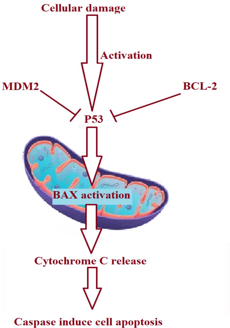
Research in the field of cancer treatment increased significantly in finding a drug or an effective way to stop tumors with no side effects (Azhar et al., 2021), miRNA which is a new class of small non-coding RNA molecules that pair with the 3′ non-coding region of target mRNA, resulting in the deterioration of the mutated mRNA or inhibition of translation, thus controlling and increasing the level of some other people (Fu et al., 2020; Josson et al., 2014), Cancer is usually caused by mutation, and this mutation causes damage to the cellular gene and depresses the tumor protected gene, such as P53, with an increase in other mutagenic proteins (A Al-Hassany et al., 2021; Fu et al., 2020). It is a miRNA with a tumor suppressor effect discovered in recent years and has been found in different types of tumors, such as gastric cancer , bladder and prostate cancer (Yuan et al., 2019; Zhang et al., 2020). Down-regulation of expression targets a variety of genes to regulate tumor proliferation, invasion, and spreading metastases (Bagherian et al., 2021). In the cell, there are several regulatory proteins such as BAX and Bcl2, cyclin D, and P53; P53 works as a tumor suppressor protein regulated by the theTP53 gene, this protein involves controlling cell DNA and preventing cancer development by either apoptosis or modifying and renewing the mutated gene (Alkuraishy et al., 2017;Yudhani et al., 2019) . In the case of DNA mutation P53 forced to increase the expression of the BAX protein, which eventually leads to apoptosis of mutated cells, Bcl2 in the antiapoptotic protein works by suppressing the effect of BAX and increasing the incidence of cancer of several studies such as this regulatory protein with cancer ( protection of propagation) but any defect in cellular DNA can cause a mutation in p53 and BAX expression by decreasing its expression, and this will increase the incidence of cancer (Figure 1), however, limited articles describe this effect of miR 409 3p in this occurrence and propagation of NSCLC (Al-hassany et al., 2021). Influence of target genes on lung cancer and how it works and its effect on the level of the P53 and BAX protein (Alkuraishy HM; et al., 2017; Alzobaidy et al., 2021).
Figure 1.

Shows the Role of P53-BAX-BCl2 in the Apoptosis Pathway
The Aim of this study was to monitoring the result of miR 409 3p and its response to tumor proliferation and its mechanism of action on some types of lung cancer in vitro (A549 cell line
Materials and Methods
Materials
cell line A549 (Cell Resource Center, Iraqi medical and genetic center, Baghdad, this type of cells is one of the best human non-small cell lung cancer cell lines used).
Reagent
DMEM medium (HyClone company); fetal calf serum (FCS) (Hangzhou Sijiqing Bioengineering Co., Ltd.) miR-409-3p as in (Habibzadeh et al., 2022).
Mock and negative control nucleotides (Shanghai Gema Pharmaceutical Technology Co., Ltd.); Luciferase expression vector (Ambion Co.); Dual luciferase reporter gene analysis kit (Promega Co.); -Aldrich Company); Flow flow apoptosis detection kit (Nanjing Keygen Biotechnology Development Co., Ltd.); Mouse anti-human ELF2, rabbit anti-human β-actin (CST Company); Lipoibctamine2000 transfection reagent, RIPA3 lysate, Invitrogne Trizol kit and by RT-qPCR (“real-time quantitative reverse transcription polymerase chain reaction kit”) ; concentration of protein assay method such as (Bradford kits) and ECL chemiluminescence-based immunodetection kit (Shanghai Biyuntian Biotechnology Co., Ltd.).
Methods
Cell grouping and treatment
DMEM medium composed of approximately 10% fetal serum, penicillin-streptomycin 100 units per ml was used, cells were placed in humidity and incubated conditions at 37 ° for a day before transfection, and after which cells were seeded in 6 wells at a concentration of about 1×10⁵ cells/well, and when cell confluence reached 70%-80%, 100 nmol / L MicroRNA-409-3p mimicking this group will be a positive control. The second (- control) was transfected into cells with lipofectamine in a 10 μL system After 6-8 h, it was replaced with cell culture medium containing serum and double antibody. The expression of the target gene was detected after 48 h. (Khan et al., 2019; Yudhani et al., 2019).
RT-qPCR to identify miR 409 3p expression: Total ribonucleic acid (RNA) and extraction using the Trizol technique and Prime Script RT
The mature miRNA converted to cDNA (by reverse transcription) then the quantity evaluated by the ABI7500 quantitative fluorescence PCR instrument. We used the SYBR-Green method to estimate cell expression (MicroRNA-409 3p). Moreover, final sequences of the primer have been as described in the following: upstream of miR-409-3p: 5’-GCGAATGTTGCTCGTGGA-3’, Among them, 5’GTGCAGGGTCCGAGGT-3 ‘. The internal reference U6 upstream: 5 ′ AAGAGCCCTGTGGTCG 3 ′, downstream: 5 ′ CATTTCAAAGCACTTCCCCT 3 ′. The relative expression of ΔmiR-409-3p was calculated using the 2-ΔΔCt method. CtmiR-409-3p-CtU6) experiment-(CtmiR-409-3p-CtU6) control group (Khan et al., 2019;Liu et al., 2022).
Western blot detection of protein expression: RIPA cleavage method
The concentration of total cell protein was measured using the Bradford technique,10 μg total cell protein was separated by SDS polyacrylamide condensation electrophoresis, after western polyvinylidene fluoride Western blot membrane was used, proteins were transferred to this membrane , blocked with 5% bovine serum albumin (BSA) for approximately 1 hour at 25oC (room temperature) on a shaker, and mouse anti-human ELF2 (1:1,000) and BCL2 were added, respectively. (1:1,000), BAX (1:1,000),
CyclinD1 (1:1,000), rabbit anti-human β-actin (1:1,000) was placed at 25°C at room temperature for 1 hour and then the horseradish peroxidase labeled secondary antibody was added Goat anti-rabbit secondary antibody (1:2,000) then we were incubated for a second time at room temperature for one hour prior to detection by chemiluminescence (ECL) (Naji et al., 2022).
To detect cell proliferation, the MTT test was used: Cells were harvested 48 hours after transfection
In relative density, about 3500 cells/well A594 cells have been seeded in 96-well plates, then fresh MTT solution diluted in buffer was added, then this sample was incubated, the Colorimetric assay carried out for each 24, 48, 72, and 96 hours after inoculation. This colorimetric assay calculated the absorbance value (A) at wave length 570 nm was measured to compare the proliferation of cells in each group (Nazarian et al., 2019).
The use of a flow cytometer for the detection and counting of cell cycle and apoptosis
Take A549 cells 48 hours after transfection, adjust cells to 1 × 10⁶ cells/mL, perform apoptosis staining according to the instructions of the apoptosis detection kit, and use flow cytometry (BD FACS Calibur) to detect.
Target gene prediction
To predict target genes, use the target gene prediction websites TargetScan and miRanda, and look for genes with 3 ‘UTR binding sites to miR-409-3p (Nazarian et al., 2019).
Vector construction and detection of dual fluorescent gene reporter: According to the target
Synthetic primers from the ELF2 ‘UTR gene sequences 3’ were considered and the amplified fragment contained the predicted oncogene ELF2 combined with miR-409-3p.
The conserved sequence was amplified by PCR using 293T cell DNA as a template (Wan et al., 2014).
The PCR product and the luciferase expression vector pMIR-REPOR were digested with restriction endonuclease and then ligated, transformed into DH5α competent cells of DH5, and the recombinant plasmid was prepared with the name pMIR-REPOR-ELF2-WT; ELF2 and ELF2 were constructed by a similar method. A549 cells were normally cultured in DMEM, surrounded with 10% fetal calf serum at normal cell growth temperature and stable carbon dioxide approximately 37°C and 5% carbon dioxide (Wan et al., 2014).
The liposome lipofectamine 2000 method was used for transfection and the operation was performed according to the instructions. The dual luciferase reporter was used. Gene analysis kit to detect fluorescent signal (Thangavelu et al., 2022).
Statistical analyses
After collecting the evaluation of the result, the second step is to assess the standard deviation typically found (x ± s) in these two groups using SPSS 16.0. statistical program. Data were also calculated using the t-test for the two groups (Zhang et al., 2020).
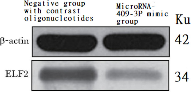
Figure 5.
Effect of miR-409-3P Overexpression in ELF2
Table 4.
Level of Expression of the ELF2 Protein Expression Level of ELF2 by Over-expression miR-409-3p (x±s A value n=96)
| Group | ELF21 | Β- actin |
|---|---|---|
| Contrast oligonucleotide's | 4.35±2.15 | 5.37±6.32 |
| miR-409-3P mimics | 2.18±1.96* | 5.84±2.51* |
*P<0.05, **P<0.01 compared to contrast oligonucleotide
Results
Changes in miR 409 3p expression
The express of transfected cells with miR 409 3p mimic approximately (4.46% ± 1.98%), This result was significantly more than the negative control oligonucleotides 0.52%±0.06% of the acid group (P<0.05).
Effect of miR 409 3p overexpression on A549 cell spread
After increasing miR-409-3p expression, the cell growth and production rate was higher than that of the control grouphis effect show in (Table 1).
Table 1.
Effect of MiR-409-3p Overexpression on A549 Cell Cancer and Propagation in the Cancer Cell Line (x±s, A value, n=96)
| Group | 24 H | 48 H | 72 H | 96 hours |
|---|---|---|---|---|
| Contrast oligonucleotides | 0.24±0.08 | 0.36±0.11 | 0.79±0.2 | 1.08±0.23 |
| miR-409-3p Mimic | 0.22±0.09 | 0.30±0.11 | 0.48±0.13 | 0.79±0.19* |
*P<0.05 compared to contrast oligonucleotides , H-hours
The effect of increased expression of miR-409-3p on apoptosis of NSCLC A549 cells
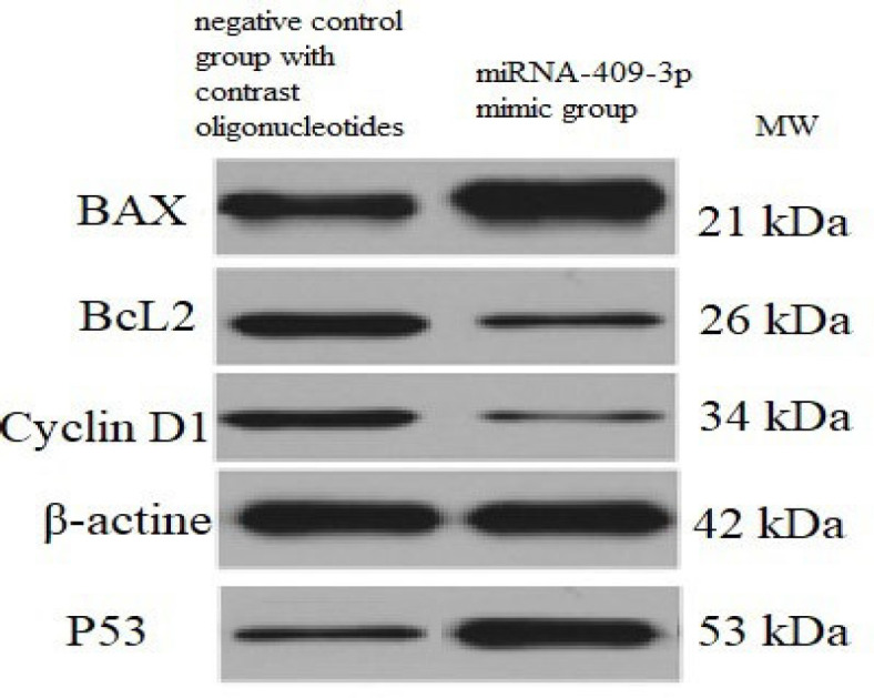
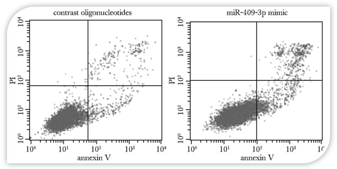
The level of apoptosis has increased significantly as a result of this overexpression of miR-409-3p (P<0.05) (Table 1). This overexpression results in an increase in the level of BAX, which is one of the important regulatory proteins that worked as apoptosis or antitumor (tumor suppressor gene) the in positive controlled cells increasing the level of P53 will increase the level of BAX that eventually leads to apoptosis, as shown in (Figure 2), however, the level of BAX suppresser level (Bcl-2) decreased significantly in the Bcl2 protein that acts as an anti-apoptotic regulator in miR-409-3p mimic treated cells (P<0.05) (Table 2, Figure 1) (Feng et al., 2021).
Figure 2.
Effect of miR-409-3p Overexpression on Apoptosis of A549 Cells
Table 2.
Apoptosis Ratio of miR-409-3P Expression in A549 Cells (x±s, %, n=96)
| Group | Early stage | Late period | Apoptosis |
|---|---|---|---|
| Contrast oligonucleotide's | 4.11±1.94 | 3.67±2.09** | 7.79±1.94** |
| miR-409-P3 mimic | 7.35±2.89 | 15.31±6.54 | 22.68±4.62 |
*P<0.05, **P<0.01 compared to contrast oligonucleotide
Western blot analysis and Cyclin-D1 level
Effect of overexpression of miR-409-3p on the NSCLC A549 cell line cycle / stage (P<0.05) (Table 3), and the level of cyclin cyclinD1 was significantly reduced (P<0.05) (Figure 3,4).
Table 3.
Effect of Overexpression of miR-409-3p on the Expression of Cyclin D1, Bax, Bcl-2 (x±s, A value, n=96)
| Contrast oligonucleotides | 3.47± 0.82 | 2.84±0.23 | 5.26±1.02 |
|---|---|---|---|
| miR-409-3P mimics | 1.31±0.55* | 7.19±1.53** | 2.82±0.73* |
*P<0.05, **P<0.01
Figure 3.

Western Blot Analysis of Cyclin D1, BAX, Bcl-2, and p53 Protein in Both Groups-
Figure 4.
Software Prediction of the miR-409-3p Target Gene ELF2 Sequence
Overexpression of MicroRNA 409 3p
In the ELF2 oncogene The prediction of the impact target gene shows that the ELF2 oncogene is miR-409-3p.
Dual-luciferin reporter assay to verify that miR-409-3p targets ELF2.
When the wild-type plasmid pMIR-REPOR-ELF2-WT and miR-409-3p were co-transfected to 293T cells, the relative luciferase activity was 0.51 When the mutant plasmid pMIR-REPOR-ELF2-Mut and miR-409-3p were transfected to 293T cells, the results were similar to those of the control group. The relative activity of luciferase was 1.07 ± 0.01. The results illustrate that miR-409-3p could suppress the level of ELF2 (Filardi et al., 2022; Al-Kuraishy et al., 2022).
Discussion
miR4093p is a miRNA with a tumor suppressor effect. The expression of miR-409-3p is significantly decreased in tumor cell lines, however, if we induce overexpression of miR-409-3p, it can inhibit and suppress cancer by decreasing the expression level of the finger protein (Al-Kuraishy et al., 2022;Al-Hussaniy et al., 2021).
However, several studies also show that miR-409-3p is highly expressed in bone metastases of prostate tumors and is linked to “progression-free” survival in life in some cases (Al-hussaniy et al., 2022b). Overexpression of miR-409-3p in prostate cancer in vitro promotes tumor growth and bone metastasis and decreases the expression of this microRNA associated with a poor prognosis (Akeel Naji, 2021; Cao et al., 2016). However, miR-409-3p plays an important role in the development of NSCLC.
The role of miR-409-3p in transfected cells
Cells transfected with miR-409-3p mimics showed a significant increase in MicroRNA expression compared to the negative control oligonucleotide group (P<0.20). Furthermore, as a result of this, there is a significant suppression of proliferation ability, indicating that miR-409-3p has the effect of inhibiting tumor growth (Ursu et al., 2020; Rashid et al., 2018). Additionally, flow cytometry along with supplement V/PI double staining showed that after increasing the level of miR-409-3p it significantly suppresses cancer cells, Compared to cells transfected with the control group, the proportion of total cell apoptosis also increased. The above results and literature reports indicate that miR-409-3p mRNA play a significant role in the tumor suppressor gene in (NSCLC). miR-409-3p can inhibit the growth of NSCLC non-small cell lung cancer by regulating tumor cell proliferation and apoptosis. In this investigation, ELF2 was also predicted as the target of miR-409-3p from hundreds of potential target genes (Al-hussaniy et al., 2021; Al-hussaniy et al., 2022 a).
Furthermore, overexpression of ELF2 can also directly promote tumor cell proliferation, indicating that ELF2 plays the role of an oncogene in the process of tumorigenesis and development. The protein expression level was significantly reduced, suggesting that ELF2 is the target gene for miR-409-3p and may inhibit cell proliferation by controlling the transcription level of ELF2. miR-409-3p can suppress the proliferation of (NSCLC) A549 cells and promote their programmed cellular death (Abdulameer et al., 2022). This mechanism is related to the regulation of the expression of the ELF2 oncogene and increases the expression of BAX and P53.
Author Contribution Statement
All work is done by Hany Akeel Al-Hussaniy.
Acknowledgments
Approval
If it was approved by any scientific Body.
Ethical Declaration
This research receives ethical approval number 202112 from Iraqi Medical Research Center - Baghdad - Iraq.
Study Registration
not required
Conflict of Interest
The authors declare that there is no conflict of interest.
References
- Abdulameer AA, Mohammed ZN, Tawfeeq KT. Endoscopic characteristics and management of Subepithelial Lesions in Video-Gastascopie. Med Pharm J. 2022;1:4–13. [Google Scholar]
- Akeel Naji H. The psychosocial and economic impact of uveitis in Iraq. RABMS. 2021;7:207–15. [Google Scholar]
- Al-hassany HA, Albu-rghaif AHA, Naji M. Tumor diagnosis by genetic markers protein P-53, p16, C-MYC, N-MYC, protein K-Ras, and gene her-2 Neu is this possible? Pakistan J Med Health Sci. 2021;15:2350–4. [Google Scholar]
- Al-Hussaniy HA, Alburghaif AH, Naji MA. Leptin hormone and its effectiveness in reproduction, metabolism, immunity, diabetes, hopes and ambitions. J Med Life. 2021;14:600–5. doi: 10.25122/jml-2021-0153. [DOI] [PMC free article] [PubMed] [Google Scholar]
- Al-hussaniy HA, Altalebi RR, Alburagheef A, Abdul-Amir AG. The use of PCR for respiratory virus detection on the diagnosis and treatment decision of respiratory tract infections in Iraq. J Pure Appl Microbiol. 2022;16:201–6. [Google Scholar]
- Al-hussaniy HA, Altalebi RR, Tylor FM, et al. Leptin Hormone: In Brief. Med Pharm J. 2022a;1:1–3. [Google Scholar]
- Al-Kuraishy AA, Jamal H, Mahdi AS, Al-hussaniy HA. Endoscopic characteristics and management of Subepithelial Lesions in Video-Gastascopie. Med Pharm J. 2022;1:24–34. [Google Scholar]
- Alkuraishy HM, Al-Gareeb AI, Al-hussaniy HA. Doxorubicin-induced cardiotoxicity: molecular mechanism and protection by conventional drugs and natural products. Int J Clin Oncol Cancer Res. 2017;2:31–44. [Google Scholar]
- Al-Kuraishy HM, Al-Gareeb AI, Al-Hussaniy HA, et al. Neutrophil Extracellular Traps (NETs) and Covid-19: A new frontiers for therapeutic modality. Int Immunopharmacol. 2022;104:108516. doi: 10.1016/j.intimp.2021.108516. [DOI] [PMC free article] [PubMed] [Google Scholar]
- Alkuraishy HM, Al-Gareeb AI, Ha A-H. Doxorubicin-induced cardiotoxicity: molecular mechanism and protection by conventional drugs and natural products. Int J Clin Oncol Cancer Res. 2017;2:31–44. [Google Scholar]
- ALZobaidy MA, AlbuRghaif AH, Alhasany HA, Naji MA. Angiotensin-converting enzyme inhibitors may increase risk of severe COVID-19 infection. Ann Romanian Soc Cell Biol. 2021;6:17843–9. [Google Scholar]
- Alzobaidy MA, Alburghaif AH, Alhasany HA, Naji MA. Angiotensin-converting enzyme inhibitors may increase risk of severe COVID-19 infection. Ann Romanian Soc Cell Biol. 2021;25:17843–9. [Google Scholar]
- Azhar MA, Chainurridha Aisyi M. Profile of PD-L1 mRNA expression in childhood acute leukemia. Asian Pac J Canc Biol. 2021;6:37–41. [Google Scholar]
- Bagherian T, Tackallou SH, Mohammadgholi A. Quantitative measurement of Bax and Bcl2 genes and protein expression in MCF7 cell-line when treated by Aloe Vera extract. Gene Rep. 2021;23:101123. [Google Scholar]
- Cao G-H, Sun X-L, Wu F, et al. Low expression of miR-409-3p is a prognostic marker for breast cancer. Eur Rev Med Pharmacol Sci. 2016;20:3825–9. [PubMed] [Google Scholar]
- Ding L, Wang R, Shen D, et al. Role of noncoding RNA in drug resistance of prostate cancer. Cell Death Dis. 2021;12 doi: 10.1038/s41419-021-03854-x. [DOI] [PMC free article] [PubMed] [Google Scholar]
- Feng P, Zhitong G, Guo Z, Lin L, Yu Q. A comprehensive analysis of the down-regulation of miRNA-1827 and its prognostic significance by targeting SPTBN2 and BCL2L1 in ovarian cancer. Front Mol Biosciences. 2021:8. doi: 10.3389/fmolb.2021.687576. [DOI] [PMC free article] [PubMed] [Google Scholar]
- Filardi T, Catanzaro G, Grieco GE, et al. Identification and validation of miR-222-3p and miR-409-3p as plasma biomarkers in gestational diabetes mellitus sharing validated target genes involved in metabolic homeostasis. Int J Mol Sci. 2022;23:4276. doi: 10.3390/ijms23084276. [DOI] [PMC free article] [PubMed] [Google Scholar]
- Fu D, Shi Y, Liu J-B, et al. Targeting long non-coding RNA to therapeutically regulate gene expression in cancer. Mol Ther Nucleic Acids. 2020;21:712–24. doi: 10.1016/j.omtn.2020.07.005. [DOI] [PMC free article] [PubMed] [Google Scholar]
- Habibzadeh SZ, Salehzadeh A Moradi-Shoeili Z, Shandiz SA. Iron oxide nanoparticles functionalized with 3-chloropropyltrimethoxysilane and conjugated with thiazole alter the expression of BAX, BCL2, and p53 genes in AGS cell line. Inorganic Nano-Metal Chem. 2022;3:1–8. [Google Scholar]
- Josson S, Gururajan M, Hu P, et al. miR-409-3p/-5p promotes tumorigenesis, epithelial-to-mesenchymal transition, and bone metastasis of human prostate cancer. Clin Cancer Res. 2014;20:4636–46. doi: 10.1158/1078-0432.CCR-14-0305. [DOI] [PMC free article] [PubMed] [Google Scholar]
- Khan AQ, Ahmed EI, Elareer NR, et al. Role of miRNA-regulated cancer stem cells in the pathogenesis of human malignancies. Cells. 2019;8:840. doi: 10.3390/cells8080840. [DOI] [PMC free article] [PubMed] [Google Scholar]
- Liu S, Zhan N, Gao C, et al. Long noncoding RNA CBR3-AS1 mediates tumorigenesis and radiosensitivity of non-small cell lung cancer through redox and DNA repair by CBR3-AS1/miR-409-3p/SOD1 axis. Cancer lett. 2022.:2022. doi: 10.1016/j.canlet.2021.11.009. [DOI] [PubMed] [Google Scholar]
- Naji MA, Alburghaif AH, Saleh NK, Al-hussaniy HA. Patient expectations regarding consultation with a family doctor: a cross-sectional study. Med Pharm J. 2022;1:35–40. [Google Scholar]
- Nazarian A, Mohamadnia A, Danaee E, Bahrami N. Examining the expression of miR-205 and CEA mRNA in peripheral blood of patients with OSCC(oral squamous cell carcinomas) and comparing them with healthy people. Asian Pac J Canc Biol. 2019;4:65–8. [Google Scholar]
- Rashid F, Naji T, Mohamadnia A, Bahrami N. MRNA biomarkers for detection of oral squamous cell cancer. Asian Pac J Canc Care. 2018;3:1. [Google Scholar]
- Thangavelu L, Geetha RV, Devaraj E, et al. Acacia catechu seed extract provokes cytotoxicity via apoptosis by intrinsic pathway in HepG2 cells. Environ Toxicol. 2022;37:446–56. doi: 10.1002/tox.23411. [DOI] [PubMed] [Google Scholar]
- Ursu A, Childs-Disney JL, Andrews RJ, et al. Design of small molecules targeting RNA structure from sequence. Chem Soc Rev. 2020;49:7252–70. doi: 10.1039/d0cs00455c. [DOI] [PMC free article] [PubMed] [Google Scholar]
- Wan L, Zhu L, Xu J, et al. MicroRNA-409-3p functions as a tumor suppressor in human lung adenocarcinoma by targeting c-Met. Cell Physiol Biochem. 2014;34:1273–90. doi: 10.1159/000366337. [DOI] [PubMed] [Google Scholar]
- Yudhani RD, Astuti I, Mustofa M, Indarto D, Muthmainah M. Metformin modulates cyclin D1 and P53 expression to inhibit cell proliferation and to induce apoptosis in cervical cancer cell lines. Asian Pac J Cancer Prev. 2019;20:1667–73. doi: 10.31557/APJCP.2019.20.6.1667. [DOI] [PMC free article] [PubMed] [Google Scholar]
- Yuan C, Zhang Y, Tu W, Guo Y. Integrated miRNA profiling and bioinformatics analyses reveal upregulated miRNAs in gastric cancer. Oncol Lett. 2019;18:1979–88. doi: 10.3892/ol.2019.10495. [DOI] [PMC free article] [PubMed] [Google Scholar]
- Zhang W, Xu C, Guo J, et al. Circ-ELF2 acts as a competing endogenous RNA to facilitate glioma cell proliferation and aggressiveness by targeting MiR-510-5p/MUC15 signaling. Onco Targets Ther. 2020;13:10087–96. doi: 10.2147/OTT.S275218. [DOI] [PMC free article] [PubMed] [Google Scholar]


