Journal of Cardiovascular Echography logoLink to Journal of Cardiovascular Echography
. 2022 Aug;32(Suppl 1):S1–S80.

Abstract

PMCID: PMC9811855
J Cardiovasc Echogr. 2022 Aug;32(Suppl 1):S1–S80.

SEVERE ATRIAL FUNCTIONAL MITRAL REGURGITATION: CLINICAL AND ECHOCARDIOGRAPHIC CHARACTERISTICS AND FOLLOW UP IN MITRAL VALVE ANNULOPLASTY PERFORMED VIA A RIGHT MINITHORACOTOMY APPROACH

Albanese Miriam 1, Nicolardi Salvatore 2, Zaccaria Salvatore 2, Mangia Federica 2, Scotto di Quacquaro Antonio 2, Pano Marco 2, Rocco Domenico 2, De Razza Luigi Pio 3, Cucurachi Marco 3, Panzera Demetrio 3, Miccoli Matteo 3, Casali Giovanni 4, Greco Cosimo Angelo 2

Introduction: Atrial functional mitral regurgitation (AFMR) remains poorly defined clinically. Aim: To compare clinical, echocardiographic characteristics and outcomes of severe AFMR to primary mitral regurgitation (PMR) after mitral valve annuloplasty (MVA) via right minithoracotomy approach (RMA)

Methods: Consecutive patients, who underwent MVA by RMA at our institution between 2016 and 2021 for severe mitral regurgitation with preserved left ventricular function, were screened. We excluded endocarditis, cardiomyopathy, prior mitral intervention. The absence of leaflet pathology defined AFMR. Outcomes included death and heart failure hospitalizations (HFH)

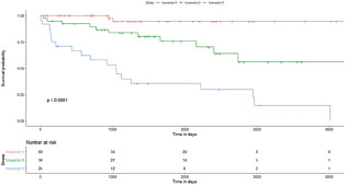
Results: 154 MVA were performed. After excluding 11 patients, among the remaining 143, AFMR were diagnosed in 24 patients, PMR in 119 patients. Compared to PMR, patients with AFMR were older (p 0,002), female (p 0,003), with worst New York Heart Association functional class (p 0,0072), with more comorbidities, including hypertension (p 0.00015) and atrial fibrillation (p 0.00001), higher left atrium volume (p 0,000004), higher average E/e' (p 0.0003) and more frequent severe tricuspid regurgitation (p 0,00001). During the follow up (38±13 mounths), 24 patients were lost and 5 patients died (4,3% in the AFMR group, 4% in PMR group). Patients with AFMR and PMR, treated by annuloplasty, had the same survival rate (log-rank p 0.09). No HFH were registered

Conclusions: AFMR is characterized by an unfavorable cardiovascular background. An early surgery correction could improve survival of these patients.

All (n=143) AFMR (n=24) PMR (n=119) P
    Baseline characteristics
Age, Years 63,7±11,5 70,2±9,2 62,3±11,5 0,02
BMI, Kg/m2 25,8±4,0 26,5±5,0 25,7±3,7 0,43
Female 66 (46,2%) 18(75%) 48 (40,4%) 0,003
Smokers 53 (37,1%) 8 (33,3%) 45 (37,8%) 0,818
NYHA class 0,0072*
    - I 0 0 0 -
    - II 46(32,2%) 2 (8,3%) 44(37,0%) -
    - III 75 (52,4%) 19 (79,2%) 56 (47,0%) -
    - IV 22 (15,4%) 3 (12,5%) 19(16,0%) -
Atrial fibrillation 37 (25,9%) 19(79,2%) 18(15,1%) 0,00001
Arterial Hypertension 77 (53,8%) 20 (83,3%) 57 (47,9%) 0,0015
Dyslipidemia 37 (25,9%) 5 (20,8%) 32 (26,9%) 0,6183
Coronary Artery Disease 52 (36,4%) 9(20,8%) 43(36,1%) 1
Diabetes mellitus 11 (7,7%) 4(16,7%) 7 (5,9%) 0,0892
    Echocardiographic Characteristics
Ejection fraction, % 66,2±6,9 60,7±6,6 67,3±6,4 0,09
End Diastolic Volume, ml 140,6±39,8 125,0±29,7 146,0±41,5 0,02
End systolic Volume, ml 47,5±18,7 54,7±25,4 45,1±15,2 0,065
Severe Tricuspide Regurgitation 20 (14,0%) 17 (70,8%) 3 (2,5%) 0,00001
Anteroposterior Diameter of left atrium, mm 47,4±8,3 51,9±8,6 46,5±7,9 0,002747
Left atrium volume, ml 73,4±31,4 93,3±15,8 69,7±32,2 0,000004
E/e’ 15,2±2,5 18,S±3,0 14,7±2,6 0,0003
J Cardiovasc Echogr. 2022 Aug;32(Suppl 1):S1–S80.

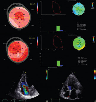
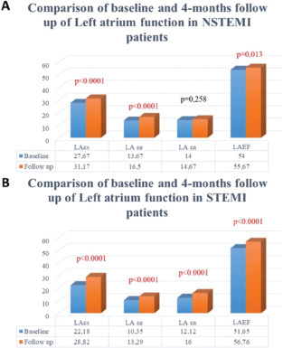
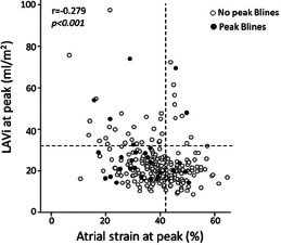
THE ACUTE LEFT ATRIAL RESPONSE IN PATIENTS WITH MYOCARDIAL INFARCTION PREDICTS EARLY ADVERSE OUTCOMES

Albini Alessandro 1, Malagoli Alessandro 2, Benfari Giovanni 3, Torlai Triglia Laura 1, Tondi Stefano 2

Introduction: Little is known about the left atrial (LA) response during acute left ventricular ischemia. Aim To investigate peak atrial longitudinal strain (PALS)-related risk of early cardiovascular (CV) events after acute myocardial infarction and define the pathophysiological and clinical PALS correlates.

Materials and Methods: All consecutive patients with ST-segment elevation myocardial infarction (STEMI) referred for primary percutaneous coronary intervention, between August 2019 and December 2020. We excluded patients with history of coronary artery disease, hemodynamically unstable, or with poor echocardiographic windows. Echocardiography was performed within 48 h of admission. The endpoint was a composite of heart failure hospitalization or CV mortality.

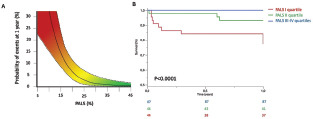
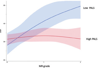
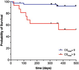
Results: Of 356 consecutive patients screened, 176 patients were included. The 12-months follow-up was completed for 100% of patients, during which the endpoint occurred in 13 (7.4%) patients. We created a spline modeling of the hazard ratio. Excess event-risk appears around the PALS value of 25%, and its double per each 5% decrease. Survival curves according to PALS values started to diverge early after the index event. Patients in quartiles with PALS above the median value were associated with a lower risk of composite endpoint at 1 year (P=0.001). PALS remained independent predictors after adjusting for LVGLS (P=0.02). The major echocardiographic features associated with PALS were LA volume and LV GLS (p=0.0001).

Conclusions: LA function predicts early adverse outcomes in first-STEMI patients. The PALS value is mostly influenced by LA size and LV GLS.

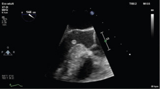
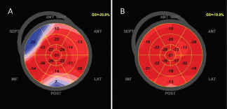
graphic file with name JCE-32-1-g001.jpg

J Cardiovasc Echogr. 2022 Aug;32(Suppl 1):S1–S80.

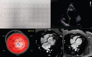
SUSPECTED BIVENTRICULAR ARRHYTHMOGENIC CARDIOMIOPATHY IN A HEALTHY MAN WITH NEW ONSET VENTRICULAR ARRHYTHMIAS AND SYNCOPE

Alderighi Chiara 1, Peluso Diletta 1, Zasso Antonella 1, Simonetto Federico 1, Nguyen Kim Ahn 1, Polo Angela 1, Pasinato Antonio 1, Soldà Elena 1, Iavernaro Antonio 1, Zadro Mirco 1, Carasi Massimo 1, Baccillieri Stella 1, Corletto Anna 1, Cucchini Umberto 1, Chirillo Fabio 1

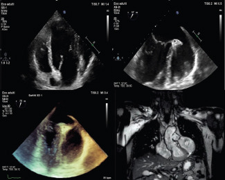
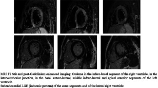
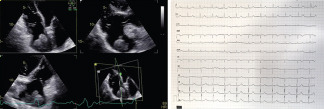
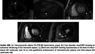
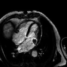
Left-dominant arrhythmogenic cardiomyopathy (ACM) is an emerging entity; despite recently refined diagnostic criteria, differential diagnosis with myocarditis and other cardiomiopathies remains challenging. The fibro-fatty scar is the distinctive histological marker of ACM; morpho-functional and structural abnormalities are sufficient to achieve diagnosis, if both ventricles are involved. We present the case of a 53-year-old man with exertional palpitation and syncope. ECG on admission showed T wave inversion in inferior leads and reduced voltages in limb leads. Laboratory exams were unremarkable, except for mildly increased troponin levels on multiple samples. Echocardiography demonstrated mildly impaired left ventricular ejection fraction (LVEF 53%) with regional wall motion abnormalities (RWMA) in the lateral wall, mildly dilated and hypokinetic right ventricle (TDA 24 cmq, FAC 33%). The patient underwent cardiovascular magnetic resonance demonstrating epicardial fatty infiltration in the lateral wall and sub-epicardial and mid-myocardial fibrosis in the inferior-inferolateral walls of the LV with mild dilatation and RWMA of both ventricles. ECG monitoring revealed frequent polymorphic ventricular ectopic beats (VEBs) and non-sustained ventricular tachycardia, with both left bundle branch block (LBBB) superior axis and RBBB morphology. VEB burden mildly increased during maximal exercise stress test. According to the Padua criteria, an ACM was suspected (one major and two minor criteria for left-dominant ACM); moreover, mildly dilated hypokinetic RV and the presence of LBBB morphology-VEB suggested concomitant

J Cardiovasc Echogr. 2022 Aug;32(Suppl 1):S1–S80.

CLINICAL FEATURES AND MORTALITY RATE OF INFECTIVE ENDOCARDITIS IN INTENSIVE CARE UNIT, A LARGE-SCALE STUDY AND LITERATURE REVIEW

Aloia Elio 1, Cresti Alberto 1, Baratta Pasquale 1

Introduction: Large scale multicentric studies reported that, despite advances in diagnosis, antibiotics and surgical treatment, infective endocarditis in-hospital mortality remains high. Most data have been obtained from patients treated in infective disease wards, internal medicine, cardiology or cardiac surgery departments, and are therefore heterogeneous. The few studies focused on complicated IE patients leading to ICU admission have reported different methodologies and results.

Methods: We conducted a prospective case-series population study from 1 January 1998 to 31 December 2020. Aim of our study was to describe the epidemiological, clinical and microbial features of critically ill patients admitted in ICU with a definite IE diagnosis Patients were divided into two groups: “Ward” (group 1) and “ICU” patients (group 2) and a one-year follow up was performed.

Results and conclusions: After performing a Univariate and Multivariate Logistic Regression analysis we found that the independent predictors of ICU admission were: Vegetation diameter > 10 mm; Abnormal PaO/FiO2 ratio, Acute Heart Failure. During hospitalization 88 patients died with a Total mortality rate 27.07%. In the Ward group 29 (12.55%) and in the ICU group 59 patients died (62.77%) with a highly significant difference. Five independent mortality risk factors were identified: SOFA score >14, Not performing surgery, age > 70 y, Acute heart failure, embolic complications.

Table 2.

Univariate and Multivariate Logistic Regression analysis for predictors of ICU admission

Univariate Multivariate
95% C.I. 95% C.I.
Adjusted HR Lower Limit Upper Limit p-value Adjusted HR Lower Limit Upper Limit
Age (years) 1,000 0,984 1,015 0,964
Previous Endocarditis 0,942 0,326 2,720 0,912
Embolic complication 1.854 1.129 3.046 0.015
Perivalvular extension 2,539 1,449 4,449 0,001
Prosthetic Valve 1,145 0,681 1,927 0,609
Vegetation diameter > 10 mm 2,371 1,447 3,882 0,001 2,150 1,160 3,986
S. Aureus Infection 1,654 0,970 2,821 0,064
Creatinine > 2 mg dl 4,945 2,631 9,296 <0,001
PaO/Fi02 ratio (mmHg%) 0,985 0,979 0,992 <0,001 1,689 1,470 1,940
SOFA score (point) 1,711 1,492 1.962 <0,001
AHF 2,346 1,431 3,845 0,001 2,193 1,182 4,070
Charlson Comorbility Index (point) 1,310 1,166 1,471 <0,001
J Cardiovasc Echogr. 2022 Aug;32(Suppl 1):S1–S80.

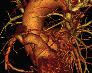
GIANT MEDIASTINAL TERATOMA: A CASE REPORT

Annunziata Roberto 1, Cocchia Rosangela 1, Conte Marianna 1, Marullo Flavio 1, Salzano Andrea 1, Capone Valentina 1, Chianese Salvatore 1, Maramaldi Renato 1, Bossone Eduardo 1

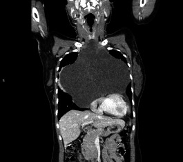
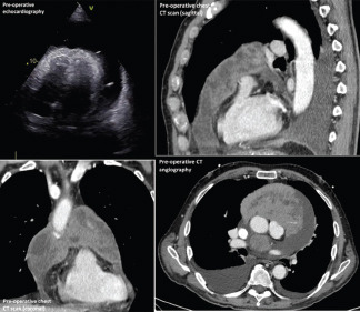
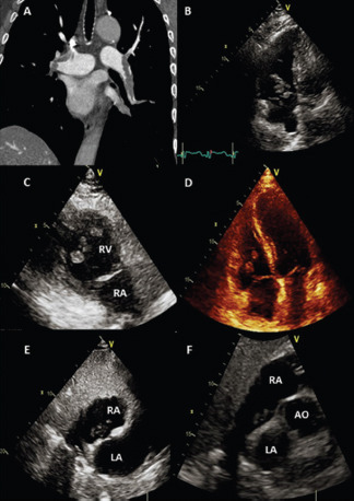
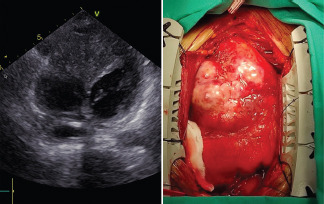
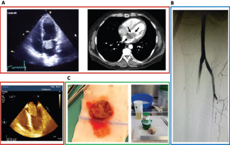
A 28 years old female with no relevant past medical history was admitted to the emergency department with one-month history of exertional dyspnea. At presentation: BP 125/75 mmHg, HR 85 bpm and sPO2 98% at fiO2 21%. Jugular venous distention was present. Chest examination revealed reduced breath sounds intensity. Complete blood count, renal function, BNP, D-Dimer, troponin and liver function tests were within normal limits. The EKG was within normal limits. Transthoracic echocardiography showed a solid extracardiac multicystic mass with dislocation of cardiac chambers towards posterior mediastinum. Apical pericardial effusion was also present, in the absence of signs of hemodynamic compromise. Chest and abdomen computed tomography with radiocontrast detected a giant anterior mediastinal mass, colliquative, with internal solid septa at irregular distribution, extending from jugular region to the base of thorax, with pre-pericardial localization (transversal diameters:8 x 19 cm – longitudinal diameter: 21 cm) suggesting a germinal genesis tumor. The patient underwent complete surgical resection. After median longitudinal sternotomy, the mass appears as a solid formation extending into pleural cavity from right to left with compression and dislocation of cardiovascular structures, strongly adhered to mediastinal pleural in both sides. The histological examination confirmed the diagnosis of mature cystic teratoma and reported concomitant chronic inflammatory aspects with xantogranulomatous degeneration. The 6-months follow-up patient asymptmatic, contrast CT showed a complete restitutio ad integrum.

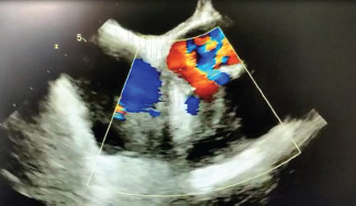
graphic file with name JCE-32-1-g002.jpg

J Cardiovasc Echogr. 2022 Aug;32(Suppl 1):S1–S80.

SACUBITRIL-VALSARTAN IMPROVES POLYGRAPH PARAMETERS IN HEART FAILURE PATIENTS WITH REDUCED EJECTION FRACTION AND SLEEP APNEA

Armentaro Giuseppe 1, Miceli Sofia 1, Condoleo Valentino 1, Monaco Vittoria 1, Clausi Elvira 1, Cassano Velia 1, Barbara Keti 1, Pastura Carlo Alberto 1, Divino Marcello 1, Severini Giandomenico 1, Sesti Giorgio 2, Sciacqua Angela 1

Introduction: Heart failure with reduced ejection fraction (HFrEF) and sleep apnea (SA) frequently coexist. Sacubitril-valsartan (s/v) reduces all-cause mortality in HFrEF patients and can potentially attenuate the development of SA through several pathophysiological mechanisms. The aim of this work is to evaluate the possible improvement of polygraph parameters after the introduction of s/v in a HFrEF and SA patients already treated with continuous positive airway pressure (CPAP).

Materials and Methods: We recruited 132 patients affected by HFrEF and SA, already in treatment with CPAP. Physical examination, echocardiography, nocturnal cardio-respiratory monitoring, and laboratory tests were performed in each patient at baseline and after a 6-month treatment with s/v during a one day CPAP interruption.

Results: S/v induced statistically significant changes in clinical, biohumoral, and echocardiographic parameters. In particular, cardiac index (CI), atrial and ventricular volumes, and global longitudinal strain (GLS) improved, with a significant reduction in global apnea-hypopnea index (AHI) value (p &lt;0.0001), oxygen desaturation index (ODI) (p<0.0001), and percentage time of saturation below 90% (TC90%) (p <0.0001). The changes of CI, estimated glomerular filtration rate (eGFR), NT-proBNP and TAPSE contributed to 23.6%, 7.6%, 7.3% and 4.8% of AHI variability, respectively for a total of 43.3%.

Conclusions: Our results suggest that s/v treatment significantly improves the polygraph parameters of patients with HFrEF and SA, complementing the positive impact of CPAP, suggesting a possible synergistic role.

Table 3.

Echocardiographic parameters at baseline and after 6 months of therapy with sac/val.

Baseline Follow-up P
Variables (n=132) (n=132)
LAVI,mL/m2 49.8 ±13.7 46.1 ± 12.0 0.001
LVEDV/BSA, mL/m2 89.6 ±9.8 87.8 ±8.4 <0.0001
LVESV/BSA, mL/m2 61.0 ±7.1 57.3 ± 5.9 <0.0001
LVEF, % 31.9 ± 1.4 34.7 ±1.6 <0.0001
CI mL/min/m2 1675.6 ±199.9 1856.6 ± 212.9 <0.0001
E/e' ratio 17.4 ±3.5 15.9 ±2.8 <0.0001
GLS, % -7.9 ±1.7 -9.0 ±1.4 <0.0001
RVOT, cm 2.6 ±0.4 2.1 ±0.4 <0.0001
RAA, cm2 20.5 ± 2.8 19.3 ± 2.3 <0.0001
TAPSE, mm 16.3 ±1.1 17.1 ±1.7 <0.0001
S-PAP, mmHg 44.5 ± 6.6 41.5 ±6.6 <0.0001
TAPSE/S-PAP, mm/mmHg 0.37 ± 0.06 0.42 ± 0.08 <0.0001
IVC, mm 20.2 ±1.3 19.1 ±3.3 <0.0001

Abbreviations: LAVI, left atrium volume index; LVEDV, left ventricular end-diastolic volume; BSA, body surface area; LVESV, left ventricular end-systolic volume; LYEF, left ventricular ejection fraction; CI, cardiac index; GLS, left ventricular global longitudinal strain; RVOT, right ventricular outflow tract; RAA, right atrium area; TAPSE, tricuspid annular plane excursion; S-PAP, systolic pulmonary arterial pressure; IVC, inferior vena cava.

J Cardiovasc Echogr. 2022 Aug;32(Suppl 1):S1–S80.

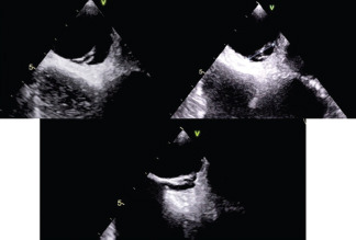
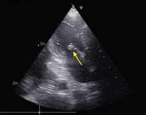
AN UNUSUAL ENDOCARDITIS

Arrigoni Paola 1, Ammendolea Carlo 1, Coscarelli Sebastian 1, Salvador Loris 2, Bilotta Massimo 2, Gobbo Marco 1, Sartor Riccardo 1, Lestuzzi Chiara 3, De Leo Alessandro 1

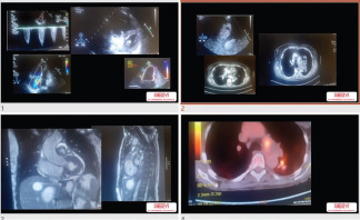
In February 2021 a 28 years old woman presented with effort dyspnea. She refers a history of post caesarean section sepsis treated with antibiotic therapy. At TTE and following TEE: thickening of the interatrial septum, protruding neoformation in the left atrium as well as mitral valve steno-insufficency related to infiltration of the neoformation. The patient was then referred for cardiac surgery in Vicenza. The resection of the neoformation infiltrating the left atrium, interatrial septum, leaflet, annulus and papillary muscles of the mitral valve was performed (minithoracotomy). Finally valve replacement with Hanckock bioprosthesis no. 27 was performed. Extemporaneous histological examination showed a possible intimal sarcoma, which was subsequently confirmed (G3, IV stadium, MDM2+). At CT angiography evidence of vascular involvement (VCI and pulmonary vein) and possible secondary sites. The patient was referred to the regional reference centre (C.R.O. Aviano) where she was treated with six cycles of chemotherapy with EpiADM and Ifofosfamide, resulting in excellent remission of the tumour. At control PET/CT with FDG (01/22) substantial absence of disease. Intimal sarcoma (ISA) is an exceedingly rare undifferentiated sarcoma that arises in main vessels. ISAs behave highly aggressive with a mean patients' survival ranging from 5 to 18 months. Patients with ISA are mostly of middle age at diagnosis and typically present with non-specific symptoms. Thus, patients are often diagnosed in an advanced disease stage. Furthermore, ISAs are often reported to be resistant to conventional chemotherapy.

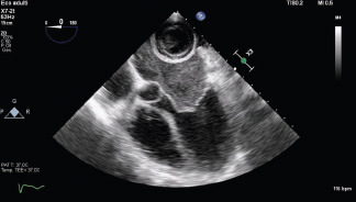
graphic file with name JCE-32-1-g003.jpg

J Cardiovasc Echogr. 2022 Aug;32(Suppl 1):S1–S80.

ADVANCED IMAGING TECHNIQUES FOR THE DIFFERENTIAL DIAGNOSIS BETWEEN IDIOPATHIC AND POST ISCHEMIC DILATED CARDIOMYOPATHY (DCM)

Augusto Florinda Maria 1, Ventura Ettore 1, Purri Ilaria 1, Panuccio Giuseppe 1, Neri Giuseppe 1, Calvelli Pierangelo 1, Mascaro Giuseppina 1, Aquila Iolanda 1, Indolfi Ciro 1

Introduction: despite advances in management of patients with heart failure, morbidity and mortality rates remain high. Common causes of DCM are ischemic heart disease, valvular disease. However, in up to 50% of cases, the cause remains unknown; this condition is called idiopathic DCM. Advances in molecular, immunohistological and genetic analysis have promoted a new era in the diagnosis/classification of DCM.

Aim: little is known about advanced imaging techniques and their discriminating ability and prognostic power.

Materials and Methods: from September 2020 to February 2022 we evaluated echocardiographic data of 68 patients with new diagnosis of DCM with Ejection Fractional <=40% admitted to our department to undergo a coronary examination. We analyzed: the radial strain, global longitudinal strain (GLS); myocardial work (MW); aortic valve (AVC)-means and peak strain dispersion (PSD). We analyzed the independent samples by non-parametric test (Wilcoxon test). Values of P<=0.05 were considered statistically significant.

Results: the etiology was post-ischemic in 53% and idiopathic in 47% of the population. The values of GLS, MW, and PSD did not differ statistically significantly between the two groups. A statistically significant difference was instead observed for AVC-means (336,85 ± 61 vs 365,18 ± 48 p =0,04) for post-ischemic vs idiopathic etiology group respectively.

Conclusions: AVC-means can be an useful parameter to differentiate the ischemic vs idiopathic origin of DCM.

J Cardiovasc Echogr. 2022 Aug;32(Suppl 1):S1–S80.

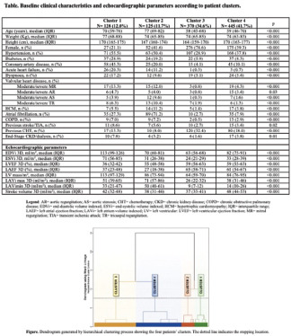
FREQUENCY AND CLINICAL SIGNIFICANCE OF ATRIAL CAVITIES IN-SITU THROMBOSIS. A LARGE SCALE STUDY AND LITERATURE REVIEW

Baratta Pasquale 1, Cresti Alberto 1, De Sensi Francesco 1, Falciani Francesca 1, Aloia Elio 1, Carlotta Sciaccaluga 1, Limbruno Ugo 1

Introduction: Supraventricular tachyarrhythmias are the main causes of atrial thrombosis (AT), usually located inside the left appendage. The prevalence and the causes of atrial body thrombosis have not been recently investigated.

Aim: to describe the epidemiology, the clinical characteristics and predisposing factors of atrial cavity “extra-appendage” thrombosis.

Methods: Consecutive cases of atrial cavity and/or appendage (left and right) thrombosis were collected and analyzed.

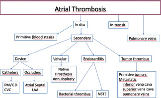
Results: 5,862 consecutive patients referred to a TEE were enrolled in the study. 175 subjects with AT were found with a prevalence of 2.98% (175/5,862). In 24 cases the clot was detected in the atrial cavities, 22 in the left (0.38%) and 2 in the right (0.03%). All the remaining clots lay in the left atrial appendage. Among the 22 patients with extra-appendage” location of left atrial thrombus, 8 were associated with a prosthetic valve, 4 with mitral stenosis whereas in the remaining 9 cases a hypercoagulative condition was present (3 cancer, 4 septic shock, 1 eosinophilic pneumonia, 1 cardiogenic shock). A neoplastic disease was present in one of the two patients with a right atrial clot.

Conclusions: in this large-scale series of patients, we report a prevalence of Atrial Cavity thrombosis of 0.38% (22/5,862) and 0.03% (2/5,862), left and right-sided respectively. Atrial “extra-appendage” thrombosis is a rare condition usually associated to “valvular” atrial fibrillation. Some cases of AT are secondary to a thrombophilia condition. In absence of a valvular heart disease, presence of underlying diseases should be investigated.

graphic file with name JCE-32-1-g004.jpg

J Cardiovasc Echogr. 2022 Aug;32(Suppl 1):S1–S80.

EXTENSIVE LEFT ATRIAL APPENDAGE ANEURYSM - A CASE REPORT OF A 47-YEARS OLD MALE

Barbarossa Alessandro 1, Coraducci Francesca 1, Torselletti Lorenzo 1, Belleggia Sara 1, Coretti Francesca 1, Valeri Yari 1, Stronati Giulia 1, Compagnucci Paolo 1, Ciliberti Giuseuppe 1, Casella Michela 1, Dello Russo Antonio 1, Guerra Federico 1

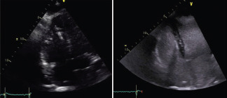
Left atrial appendage aneurysm (LAAA) is a rare condition mostly due to congenital malformations or secondary causes. Since very few cases are described in the literature, there is uncertainty in treatment and prognosis. Diagnosis is achieved by transesophageal echocardiography (TEE), which also allows the detection of thrombus and cardiac magnetic resonance (CMR) that is more specific in describing sizes and relationships with surrounding anatomical structures. Surgical aneurysmectomy could be indicated in the majority of cases, especially if compression of other cardiac chambers or mediastinal structures are present. Medical therapy includes tromboprophylaxys and arrhythmias management. A shared decision making by Heart Team should be considered. We present the case of a 47-year-old male who came to our attention for palpitations and epigastric pain. The ECG showed high-rate atrial fibrillation (AF) with wide QRS. The patient underwent urgent electrical cardioversion and coronary angiography showed patent coronary arteries. He had a giant left auricle appendage diagnosed twelve years before and was on antiarrhythmic prophylaxis for previous AF episodes. A TEE was performed and confirmed the diagnosis of LAAA also showing hypokinetic anterior-apical wall due to the interplay with the giant aneurysm. Subsequent CMR showed no LGE and confirmed the absence of thrombus in the LAAA. After Heart Team consultation surgical treatment was proposed to the patient who refused. Therefore, direct oral anticoagulation and antiarrhythmic therapy with betablockers and flecainide were introduced, and a loop recorder was implant

graphic file with name JCE-32-1-g005.jpg

J Cardiovasc Echogr. 2022 Aug;32(Suppl 1):S1–S80.

INFECTIVE ENDOCARDITIS POST-TRANSCATHETER AORTIC VALVE IMPLANTATION (TAVI) WITH 1 EMBOLIZATION TREATED WITH MEDICAL THERAPY

Barbera Chiara 1, Scandura Salvatore 1, Rizzo Sofia 1, Greco Ylenia 1, Lentini Giuseppe 1, Rugiano Gerardo 1, Gibiino Vincenzo 1, Uccello Salvatore 1, Bentivegna Agnese 1, Milici Anna Lisa 1, Tamburino Corrado 1

Introduction Post-TAVI infective-endocarditis is a rare event. The incidence is 3,25% and it is associate with severe complications and high risk of mortality.

Aim The aim of the study is to focus on the possibility of alternative therapy except surgery in treatment of a complicated endocarditis, choice taken also thanks to TEE, through it is possible analyze the integrity of the prosthesis, evidence/absence of rupture or ulceration and monitoring the evolution during therapy.

Materials and Methods A 76-year-old female patient who went to TAVI, reported fever, increased phlogosis indices and abdominal pain few days after the procedure. Diagnostic imaging resulted in infection of the aortic prosthesis and in thromboembolisms affecting splenic artery and celiac trunk. At TEE an isoechogenic vegetation (14 x 8 mm) was described in correspondence of aortic prothesis and high gradient was detected. Thromboembolic events have occurred and thromboendoarteriectomia of right femoral artery was performed. Staphylococcus Lugdunensis was found in blood cultures and intravenous antibiotic and anticoagulant therapy was undertaken.

Results After one month of medical therapy, at TEE significant vegetation reduction was found. Patient was discharged at home, asymptomatic. After 1 month, at TTE no sign of endocarditis was detected, blood sample was fine and patient was asymptomatic.

Conclusions This case is an example of an early infective process involving biological valve post-TAVI, followed by systemic embolization, in which the only medical therapy was successful. Echocardiography has played a fundamental role.

graphic file with name JCE-32-1-g006.jpg

J Cardiovasc Echogr. 2022 Aug;32(Suppl 1):S1–S80.

FEASIBILITY AND ROLE OF ECHOCONTRAST EVALUATION OF PATIENTS WITH LVAD

Baroni Giulia 1, Pergola Valeria 1, Semeraro Laura 2, Dellino Carlo Maria 1, Mastro Florinda 1, Aruta Patrizia 1, Cecchetto Antonella 1, Previtero Marco 1, Fiorencis Andrea 1, Gerosa Gino 1, Iliceto Sabino 1, Tarzia Vincenzo 1, Mele Donato 1

Introduction: Development of right heart failure in patients with LVAD has a direct effect on mortality. Aims: evaluation of clinical safety and feasibility of echocontrast (EC) in patients implanted with 3 different LVAD; improvement in heart visualization; inter-operator agreement.

Methods: Between 2014 and 2019, 43 patients were implanted with LVAD in Padua Hospital: 7 with Jarvik, 31 with HeartMate, 5 with HVAD. Nine patients refused EC, two lost their follow-up. In 3 patients, echocardiography was technically challenging. Our final population was of 29 (mean age 65±7 y; 100% Male). Doppler echocardiography was performed with an ultrasound transducer system (1.5-4.0 Mhz); a contrast mode was used in the EC phase of the exam (mechanical index 0.20) injecting 0.8 mL of EC. We also assessed the reproducibility of measurements between two different expert operators (blind analysis).

Results: We observed no allergic reaction to EC. Total 329 (64%) of 516 RV wall segments were available for qualitative analysis without contrast vs 451 (87%) with contrast (p&lt;0.001) with a significant improvement of evaluability of regional contractility and FAC (41% vs 90%, p0.86), 2D-baseline derived parameters a good one (ICC = 0.74), showing improvement in regional contractility.

Conclusion: EC is safe with all types of LVAD we examined. A routine use of EC could play a pivotal role improving RV morphologic and functional judgments.

J Cardiovasc Echogr. 2022 Aug;32(Suppl 1):S1–S80.

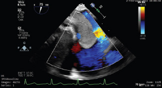
A CASE OF SEVERE MITRAL REGURGITATION WITH COMPLEX ANATOMY IN A PATIENT AWAITING HEART TRANSPLANTATION

Bellettini Matteo 1, Vairo Alessandro 1, Zaccaro Lorenzo 1, Avondo Stefano 1, Montefusco Antonio 1, Alunni Gianluca 1, De Ferrari Gaetano Maria 1

Introduction: Transcatheter edge-to-edge repair (TEER) for secondary mitral regurgitation (MR) showed conflicting results, emphasizing the importance of an adequate candidates selection. Patients with advanced heart failure (HF) represent a subgroup with even greater uncertainties.

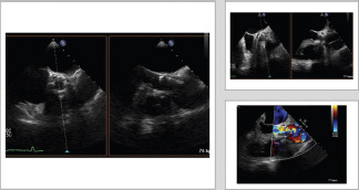
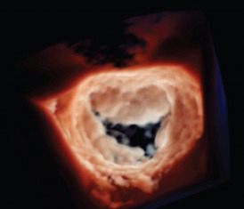
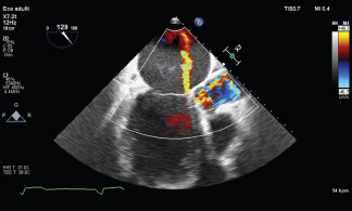
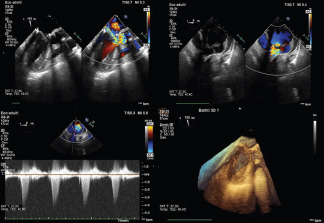
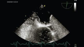
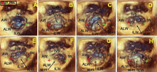
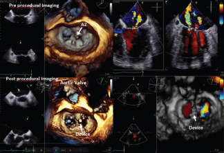
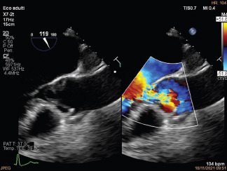
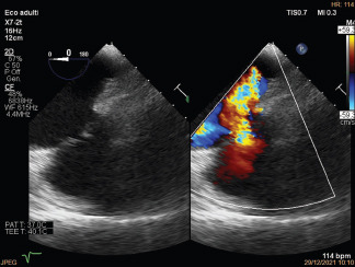
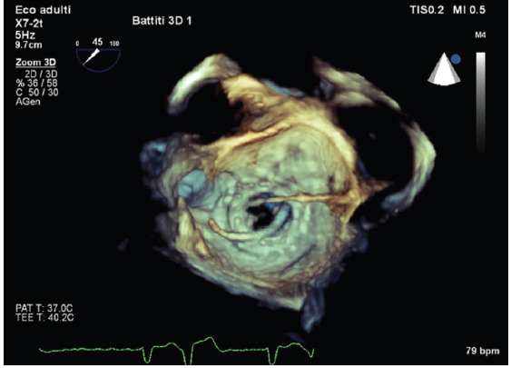
Case: A 40 year old man with non-ischaemic dilated cardiomyopathy was referred to our center for evaluating heart transplantation as during the last year he had 3 hospitalizations for HF. We performed a comprehensive evaluation starting with a transthoracic echocardiography showing a dilated left ventricle, EF 25%, severe MR, dilated right ventricle with mild dysfunction and moderate tricuspid regurgitation. Right heart catherization showed a combined pre-and-post capillary pulmonary hypertension. Patient was listed for heart transplantation and discharged. One month later patient was admitted again due to HF. Heart Team decided to perform a transoesofageal echocardiography (TOE) to evaluate TEER as a bridge to transplantation. TOE confirmed severity of MR and established feasibility of TEER even if in the presence of a complex anatomy due to a cleft-like indentation between P2-P3 (Figure). TEER was performed with a single clip between A2-P2 with a good result. At the follow-up there were no new hospitalization for HF and we had the opportunity to up-titrate vasodilator and reduce diuretic therapy.

graphic file with name JCE-32-1-g007.jpg

Conclusions: TEER may represent a therapeutic option in selected patients with advanced HF awaiting heart transplantation, as it may improve patient's hemodynamic profile alleviating symptoms and reducing HF hospitalizations.

graphic file with name JCE-32-1-g008.jpg

J Cardiovasc Echogr. 2022 Aug;32(Suppl 1):S1–S80.

A DISQUALIFYING T-WAVE INVERSION

Belli Lorenzo 1, Ricci Fabrizio 1, Gallina Sabina 1

Introduction: A 41-year-old man professional tennis player with dyslipidemia presented to our cardiology unit for atypical chest pain, palpitations on effort and chronic troponin leakage. He reported family history of sudden death and previous history of myocarditis.

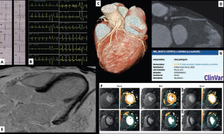
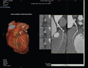
Case summary: ECG(A) revealed T-wave inversion without J point elevation extending beyond lead V4. Resting echocardiogram was unremarkable. Exercise testing showed ventricular ectopy with uncommon right bundle branch block morphology(B). Cardiac CT(C) yielded a diagnosis of high-risk intramyocardial bridge(D) of proximal left anterior descending (LAD) with nonobstructive accelerated atherosclerosis involving left main and LAD ostium. Cardiac magnetic resonance demonstrated sub-epicardial scarring of basal infero-lateral left ventricular wall(E), but normal myocardial perfusion reserve on adenosine challenge(F). Molecular analysis(G) of exon-9 of the plakophillin-2-gene revealed heterozygous nucleotidic deletion c.1840delC with premature termination codon after frameshift mutation p.Leu614Serfs*42. According to the 2020 “Padua criteria”, the patient was diagnosed with left dominant arrhythmogenic cardiomyopathy. The patient was permanently disqualified from competitive sports activity and commenced on beta-blocker, low-dose aspirin and statins. An implantable loop recorder was inserted and screening of first-degree relatives was advised. Two-year follow-up was uneventful.

Conclusions: the final diagnosis was left dominant arrhythmogenic cardiomyopathy and and high-risk LAD myocardial bridging resulting in permanent disqualification from competitions.

graphic file with name JCE-32-1-g009.jpg

J Cardiovasc Echogr. 2022 Aug;32(Suppl 1):S1–S80.

UNEXPLAINED RECURRENT MINOCA AFTER TRANSCATHETER AORTIC VALVE IMPLANTATION (TAVI)

Bellisario Irina 1, Di Fulvio Maria 2, Pirro Davide 1, Foglietta Melissa 1, De Luca Enrico 1, Gallone Anna 1, Appignani Marianna 2, Mantini Cesare 3, Gallina Sabina 1, Ricci Fabrizio 1

Introduction. A 80-years-old woman presented to our cardiology unit for a recurrent acute coronary syndrome without persistent ST-segment elevation.

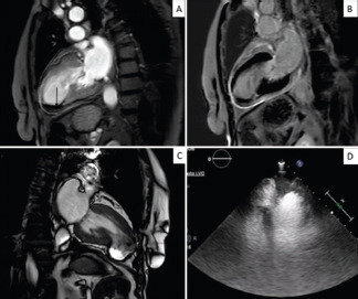
Case summary. The lady underwent TAVI 6 months earlier and reported a MINOCA 3 months later. Cardiovascular magnetic resonance (CMR) showed akinesia of the apical left ventricular (LV) segments, moderate LV systolic dysfunction, LV thrombus (A), transmural late enhancement of the LV apex surrounded by localized, dense, pericardial enhancement (B), a sign of local inflammation harbinger of possible delayed pseudoaneurysm formation. Repeat CMR at 3-months showed resolution of the LV thrombus and a residual apical LV pseudoaneurysm, with abrupt transition of myocardium from normal thickness to a thin layer (C). The Heart Team recommended a conservative management. During current work-up, repeat invasive coronary angiography demonstrated the absence of obstructive coronary lesions. Contrast echocardiography (D) ruled-out myocardial rupture and LV thrombosis. The Heart Team decided to pursue medical treatment with close imaging surveillance, in view of the hemodynamic stability and the overall very high surgical risk. Follow-up at 3 months was uneventful.

Conclusions. This is a case of unexplained recurrent MINOCA after TAVI complicated by LV dysfunction, thrombosis and pseudoaneurysm formation. LV pseudoaneurysm is a potentially life-threatening complication of acute myocardial infarction and timely diagnosis is key to start appropriate treatment and improve the patient's prognosis. Pericardial delayed enhancement by CMR imaging is a helpful imaging feature for predict

graphic file with name JCE-32-1-g010.jpg

J Cardiovasc Echogr. 2022 Aug;32(Suppl 1):S1–S80.

AORTO-RIGHT ATRIUM FISTULA AS A COMPLICATION OF BIOLOGICAL AORTIC PROSTHESIS ENDOCARDITIS

Bernardini Gaetano 1, Barengo Alberto 1, D'Addario Sandra 1, Savino Ketty 1

Introduction: Aorto-cavitary fistula is a rare complication of infective endocarditis (IE), frequently seen in mechanical prostheses and life threatening. AIM: Patient with biological aortic prosthetic valve IE complicated by aortic-right atrium fistula.

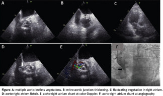
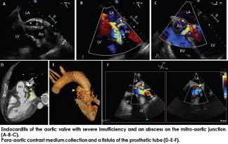
Material and Methods: 86-year-old male with aortic vascular and biological aortic valve prostheses. Hospitalized for sepsis due to Dysgalactiae Streptococcus, ischemic stroke and splinter haemorrhages in nail beds. Clinical diagnosis was IE. Transthoracic echo shows increased aortic transvalvular gradient, free floating mass in right atrium. Transesophageal echo (TEE) demonstrates multiple aortic leaflets vegetations, mitro-aortic junction thickening (1.3 cm) and free-floating right atrium vegetation. Antibiotic therapy was immediately started. Few days later at TEE appears mitro-aortic junction abscess and small periannular detachment point. Clinical conditions remained stable and inflammatory indices (white blood cells, CRP) decreased. Some weeks later TEE confirms periannular detachment with periprosthetic aortic regurgitation, prosthetic aortic valve and right atrial vegetations reduction and revealed presence of aortic-right atrial fistula (confirmed at aortography).

Results: Stable hemodynamics and prohibitive operative risk led to conservative treatment. At 3 months follow-up patient is alive and in good clinical condition.

Conclusion: Aorto-cavitary fistulization is a rare and problematic complication of periannular spread of IE. Appropriate echo diagnosis and clinical evaluation are crucial to lead to proper therapy for this rare condition.

graphic file with name JCE-32-1-g011.jpg

J Cardiovasc Echogr. 2022 Aug;32(Suppl 1):S1–S80.

UNEXPECTED AND CHALLENGING DIAGNOSIS OF RIGHT VENTRICULAR CARDIOMYOPATHY

Bernardini Gaetano 1, Crusco Federico 2, Broccatelli Andrea 3, Savino Ketty 1

Introduction: We discuss about an unexpected diagnosis of ARVC. AIM: Underline the role of cardiac imaging for diagnosis of right ventricular cardiomyopathy (ARVC).

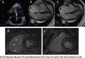
Material and Methods: 63 years old male admitted to hospital for congestive heart failure NYHA III class (progressive dyspnea, orthopnea, and leg edema); chest pain, palpitations and syncope were denied. On admission: normal vital signs, breathing 30 breaths/min, saturating 90% on room air. Blood tests were normal, except for elevated TnHS and NTproBNP levels. ECG showed T wave inversions in V1-V3. Transthoracic echocardiography revealed reduced left ventricular function (EF 30%) and severely impaired (TAPSE 8 mm; S' wave 6 cm/s) and dilated right ventricle (RV). Congestive heart failure therapy was started. During hospital stay ECG monitoring revealed runs of non-sustained and sustained ventricular tachycardia of left bundle branch morphology which became unstable and had to be converted with DC shock. Then antiarrhythmic therapy was started. Coronary angiography was normal. Ischemic etiology excluded, ECG and echo findings were suggestive for ARVC. CMR was performed and revealed dilated RV and medium free wall dyskinesia. LGE showed fibrous replacement of RV free wall and left ventricle inferolateral wall.

Results: Our patient met two major criteria from CMR for ARVC leading to a definitive diagnosis. Patient was candidate to ICD implantation and a screening was advised to his family.

Conclusion: The presentation of AVRC can be non-specific and diagnosis may be challenging. CMR confirmed his primary role in differential diagnosis of cardiomiopathies.

J Cardiovasc Echogr. 2022 Aug;32(Suppl 1):S1–S80.

A RARE CASE OF OVAL MASS IN LEFT VENTRICULAR OUTFLOW TRACT

Boccia Filomena 1, Vetrano Erica 1, Florio Maria Teresa 1, Macrì Angela 1, Borrelli Marco 1, Ascione Raffaele 1, Torracca Lucia 2, Ascione Luigi 1, Palmiero Giuseppe 1

graphic file with name JCE-32-1-g012.jpg

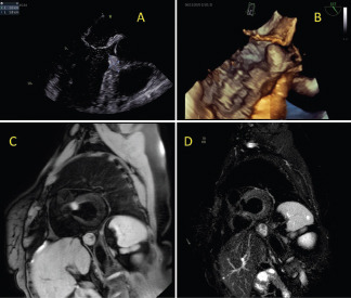
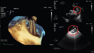
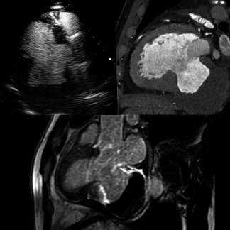
A 62-years-old woman was admitted to our department because of a transient ischaemic attack after a negative neurological study. Transthoracic echocardiography showed an echo dense oval mass appearing in systole in LVOT (Panel A), without modifications when a contrast medium was added. Then, transoesophageal echocardiography with 3D reconstruction was performed: the oval mass showed a maximum diameter of 18x10 mm, was attached to the anterior basal portion of the interventricular septum, in proximity to the left and right aortic cusps, without determining LVOT obstruction (Panel B). Cardiac MRI showed a hypermobile and ill-defined ovoid heterogeneous and slightly hyper-isointense (to the myocardium) mass in CINE sequences. Black blood T1 Weighted MR with Fat suppression showed high signal within the mass (Panel C), while T2 STIR sequences showed complete signal loss (Panel D). With and without fat suppression, LGE sequences showed avid and homogenous enhancement of the lesion. Ignoring the mass's aetiology and being unable to predict the risk of progression (possible LVOT obstruction, arrhythmias, and dissemination if malignant), a total surgical excision was performed through an anterior longitudinal aortotomy. Histological analysis diagnosed a mature cystic teratoma with differentiated thyroid tissue. Cardiac teratomas are extremely rare (&lt;1% of all cardiac tumours in adults). They are diagnosed primarily in children, usually affecting the pericardium. Complete surgical excision is the most effective treatment because they are relatively resistant to chemotherapy and radiotherapy.

graphic file with name JCE-32-1-g013.jpg

J Cardiovasc Echogr. 2022 Aug;32(Suppl 1):S1–S80.

A MATTER OF TIME

Bono Giuseppe 1, Antonazzo Andrea 1, Zema Domenica 1, Scigliano Fabio 1, Scappatura Rosa Maria 1, Pasquale A Fratto 1, Benedetto Frank 1

Introduction: Interventricular septal defect (VSD) is a rare and life-threatening mechanical complication of acute myocardial infarction, with a very high mortality rate. AIM: Optimal timing of surgery in still widely debated: even the two major scientific cardiological (American Heart Association vs European Society of Cardiology) societies disagree on this.

Materials and Methods: A 68-years old man was admitted with symptoms and sign of infero-posterior acute myocardial infarction with ST elevation and hemodynamic instability. The transthoracic echocardiogram revealed a big interventricular septal rupture, about 3 cm, extended up to the basal inferior wall, with left-to-right shunt and moderate pericardial hemorrhagic effusion; these findings were confirmed by the inspection on the operating field. The coronary angiogram showed the total occlusion of the proximal right coronary artery.

Results: After positioning of IABP and multidisciplinary evalutation by Heart Team, indication was given for urgent cardiac repair, successfully performed with endocardial patch with infart exclusion with T. David technique. In the post-operative course the patient needed prolonged inotropic support and mechanical ventilation, during the 28 days of ICU long-stay; after ten more days of hospitalization in cardiac surgery ward, the patient was discharged from the hospital.

Conclusions: In this case, the early surgical approach proved to be beneficial for the patient's survival. The decisive parameter for the decision to perform early surgery is represented by the high risk of cardiogenic shock and multiorgan failure.

graphic file with name JCE-32-1-g014.jpg

J Cardiovasc Echogr. 2022 Aug;32(Suppl 1):S1–S80.

ACUTE RUPTURE OF A SINUS OF VALSALVA ANEURYSM INTO THE RIGHT ATRIUM

Borrelli Marco 1, Vetrano Erica 1, Florio Maria Teresa 1, Macrì Angela 1, Pagnano Gianpiero 1, Caso Ilaria 1, Bancone Ciro 2, Ascione Luigi 1, Palmiero Giuseppe 1, De Feo Marisa 2

A 64-years-old-man with an antecedent history of coronary artery disease (previous PTCA + stenting with DES on the right coronary artery) and known dilation of the sinuses of Valsalva and the ascending aorta in follow-up was admitted to our Department because of acutely decompensated severe congestive heart failure. The transthoracic echocardiography showed a mild left ventricular dilation with akinesia of the apex and anterior wall, severe systolic dysfunction (LVEF 27%), and right atrial and ventricular dilation. At 4-chamber view, a bilobed structure was noticed into the right atrium, in contiguity with the proximal tract of the aorta. The transesophageal echocardiography showed the rupture of the aneurysmal non-coronary sinus of Valsalva in the right atrium with fistulisation and right-sided volume and pressure overload by a shunt at a high-velocity rate. A coronary angiogram showed severe obstruction of the proximal left anterior descending (LAD) artery. Therefore, the patient underwent surgical revascularisation by coronary artery bypass grafting with a left interior mammary artery on LAD, non-coronary sinus surgical repair with patch and concomitant aortic valve replacement with mechanical prosthesis and aortic repair with Bentall's technique. The postoperative course was uneventful. Therefore, six months after a follow-up echocardiographic examination showed excellent surgical results with mild improvement of LV systolic function (LVEF 37%) and significant remodelling of the right-sided chambers.

graphic file with name JCE-32-1-g015.jpg

J Cardiovasc Echogr. 2022 Aug;32(Suppl 1):S1–S80.

TRICUSPID REGURGITATION IN PULMONARY HYPERTENSION: FROM ETIOPATHOGENESIS TO PROGNOSTIC IMPLICATIONS

Botti Andrea 1, Serra Walter 2

Introduction: Pulmonary arterial hypertension (PAH) is a pathological hemodynamic condition characterized by an increase in arterial vascular resistance in pulmonary circulation, related with increased mortality and morbidity. Echocardiography represents the most widely used diagnostic imaging modality in PAH and provides useful information about the right heart-pulmonary circulation unit. Recently, there has been increasing interest in the echocardiographic evaluation of functional tricuspid valve regurgitation (FTVR), whose severity has been related with adverse outcomes and poor quality of life. FTVR could also have an active role in the PAH pathophysiology, representing in some cases the first cause of the disease.

Aim: The purpose of our study was the evaluation of the severity of FTVR as outcome predictor in a selected population of patient affected by PAH.

Materials and Methods: 123 patients with PAH confirmed through right heart catheterization were observed for a mean follow-up period of 3,4 years, collecting data regarding the severity of FTVR and the all-causes mortality rate.

Results: Consistently with the scientific literature, FTVR resulted to be an important prognostic marker in patients with PAH. Among the patients with severe FTVR the mortality rate was 79,16%, while the mortality in moderate and mild FTVR were, respectively, 28,20% and 3,33%, with the degree of FTVR severity confirmed as the main outcome predictor at univariate and multivariate analysis.

Conclusion: Moderate to severe FTVR has a strong relation with adverse outcomes, and it could represent the main prognostic marker in patients with PAH.

graphic file with name JCE-32-1-g016.jpg

J Cardiovasc Echogr. 2022 Aug;32(Suppl 1):S1–S80.

DIAGNOSTIC CHALLENGE OF A MYSTERIOUS MASS

Caiazzo Giancarlo 1, Fusto Antonio 2, Del Giglio Mauro 3, Valente Loredana 2, Quadruccio Raffaella 2, Morrone Sara 2, Durastante Giorgia 2, Spampinato Andrea 2

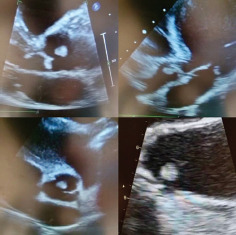
A 71-year-old woman performed a follow-up visit. She performed an echocardiogram control that showed a rounded, pedunculated plus image (diameter 0.7 * 0.7 cm) adhering to the free margin of the non-coronary aortic cusp, mobile and floating in the aortic vascular side. Her past medical history included: drainage surgery for abscess in the right parotid four months earlier, due to which she began and continued empirical broad-spectrum antibiotic therapy and a NSTEMI treated by PCI with implantation of two medicated stents in the anterior descending coronary artery. After suspicion of thrombotic formation, the antibiotic was withdrawn, maximum-dose anticoagulant therapy was initiated and blood cultures were performed. Blood cultures were negative and a month later she underwent a transesophageal echocardiogram which showed no change in the size of the mass and was not conclusive to the diagnosis. This mass does not appear to be a vegetation because the valve is healthy and there are no other signs of endocarditis; it should not be a tumor because there was no mass in the recent checks and it didn't grow in two months of follow up, so the most likely hypothesis would seem to be that of a thrombus, although the diameter has not changed after one month of anticoagulation therapy. Due to the risk of embolization and to have a diagnosis of certainty, at the end of the month the patient will perform a surgery to remove the mass, during which the histological examination will be performed to make a definitive diagnosis.

J Cardiovasc Echogr. 2022 Aug;32(Suppl 1):S1–S80.

MULTYMODALITY IMAGING IN CORONARY VASCULITIS

Calamelli Sara 1, Purita Paola 1, Sacca' Salvatore 1

graphic file with name JCE-32-1-g017.jpg

48-year-old male, heavy active smoker with no other risk factors or comorbidities. First access to the emergency room (PS) for chest and abdominal pain dysphagia with weight loss in the last few days. ECG within limits. Increased CPR levels and chest x-ray demonstration of parenchymal thickening and mild pleural effusion. He was discharged with ibuprofen, levofloxacin, and cefixime. After five days, he was readmitted to the hospital with the same symptoms, but on this occasion, we found troponin hs and PCR 96. The ECG was within limits, and there was a slight pericardial effusion on echocardiography. Three days later, new echocardiography showed the resolution of the pericardial effusion, regional kinetics was normal. Coronary angiography shows uninjured coronary vessels. The cardiac MRI showed hypokinesia of the middle wall of the right ventricle with subendocardial LGE in the same site and subepicardial LGE in the middle part of the lateral wall, with a picture compatible with acute myocarditis. During hospitalization, the patient complained of other episodes of chest pain without electrocardiographic changes and with a gradual decrease in troponin hs. The PCR showed slight fluctuations. He was discharged with colchicine, ibuprofen, bisoprolol, and lansoprazole. The patient returned to the hospital the day following discharge with acute chest pain and st-segment elevation in inferior leads. Coronary angiography demonstrated thrombotic occlusion of a medium caliber postero-lateral branch treated, with thrombus aspiration, by angioplasty and stenting. During the subsequent hospitalization, the patient continued t

graphic file with name JCE-32-1-g018.jpg

J Cardiovasc Echogr. 2022 Aug;32(Suppl 1):S1–S80.

NON-INVASIVE MYOCARDIAL WORK ROLE IN THE PREDICTION OF LEFT VENTRICULAR RECOVERY AFTER ACUTE MYOCARDIAL INFARCTION

Caminiti Rodolfo 1, Vetta Giampaolo 1, Parlavecchio Antonio 1, Pelaggi Giuseppe 1, Lofrumento Francesca 1, Restelli Davide 1, Parisi Francesca 1, Vinciguerra Paolo 1, De Santis Giulia 1, De Ferrari Tommaso 1, Gianluca Di Bella 1, Scipione Carerj 1, Micari Antonio 2, Zito Concetta 1

Introduction: Predicting left ventricular recovery (LV-REC) after acute myocardial infarction (AMI) is both challenging and of prognostic importance. Aim: Our objective was to evaluate the usefulness of noninvasive myocardial work (MW) to predict LV-REC after AMI.

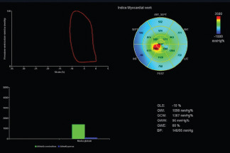
Materials and Methods: We prospectively enrolled all patients admitted with AMI treated with primary percutaneous coronary intervention (PCI) from January to July 2021. Patients underwent transthoracic echocardiography (TTE) within 24-48 hours after PCI. Follow-up included outpatient visit and TTE at 1, 4 and 12 months. MW was derived from the strain-pressure loops, integrating in its calculation the noninvasive arterial pressure. LV-REC was defined as an improvement of LV ejection fraction (EF) ≥ 5% in patients with LVEF ≤50% at baseline.

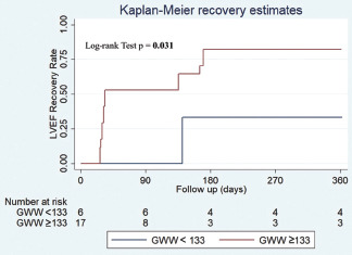
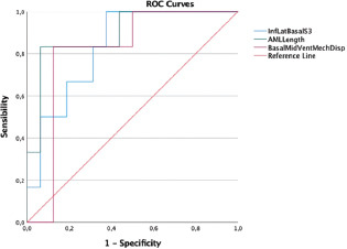
Results: Fifty-two patients (61.96 ± 12.06 yrs) were enrolled. LV-REC at 12 months of follow-up was observed in 30.8 % of patients. Comparing the groups with and without LV-REC, there was a statistically significant difference in the baseline Global Wasted Work (GWW) value (194.0 mmHg/5vs 137.36 mmHg/%; p=0.009). ROC curve analysis identified a cut off value of ≥133 mmHg/% for baseline GWW (Sensitivity 87.50%, Specificity 71.43%, AUC 0.839, p =0.011) to identify patients with LV-REC at 12 months. Kaplan–Meier curve estimate for LV-REC revealed that there was a significant difference between patients with and without baseline GWW ≥ 133 mmHg/% (p&lt; 0.031), as seen in [Figure 1].

Conclusions: In our study GWW was able to predict LV-REC at 12 months after AMI. Further large-scale studies are needed to validate these results

graphic file with name JCE-32-1-g019.jpg

J Cardiovasc Echogr. 2022 Aug;32(Suppl 1):S1–S80.

A NOVEL SPHYGMOMANOMETER FOR CENTRAL VENOUS PRESSURE ASSESSMENT DURING STRESS ECHOCARDIOGRAPHY

Campagnano Ettore 1, Ciampi Quirino 1, Lodi Simone 2, Marcelli Emanuela 2, Plichi Gianni 2, Bombardini Tonino 3, Villari Bruno 1, Picano Eugenio 3

Introduction. Central venous pressure (CVP) is important in the hemodynamic of failing heart, and the invasive assessment can be surrogated by a sphygmomanometer for venous pressure (SVEN). Aim: To assess CVP measurements by SVEN during stress echocardiography (SE).

Materials and methods: We evaluated 22 patients (age 66 ±11 years, 15 men, ejection fraction 60 ±6%) referred for dipyridamole (n=20, for chest pain) or treadmill (n=2, for dyspnea) SE with ABCDE protocol. Step A, regional wall motion abnormalities (RWMA); step B, B-lines (diastolic function); step C, LV contractile reserve based on force; step D, coronary flow velocity reserve (CFVR) in left anterior descending artery; step E, heart rate reserve (HRR). In each patient, the SE data was integrated with CVP measurement by SVEN.

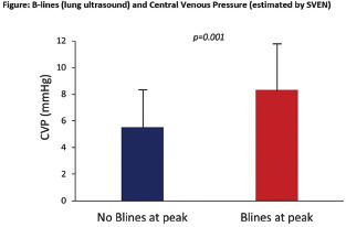
Results. SVEN success rate was 22/22 (100%). Resting CVP was correlated inversely to ejection fraction at rest (r=-0.489, p=0.021) and at peak stress (r=-0.545, p=0.001), CFVR (r=-0.505, p=0.023), HRR (r=-0.503, p=0.017) and directly to RWMA (r=0.431, p=0.045), B-lines at peak stress (r= 0.626, p = 0.002) and left atrial volume index at rest (r= 0.552, p =0.012) and at peak stress (r=0.648, p=0.002). CVP was significantly higher in patients with B-Lines at peak stress (Figure). ABCDE score was directly related to CVP (r=0.560, p=0.007).

Conclusion. SVEN assessment is feasible during SE with high success rate. CVP increase is associated with more B-lines (a sign of pulmonary congestion and elevated pulmonary capillary wedge pressure) and larger left atrium (an integrated barometer of left ventricular end-diastolic pressure).

graphic file with name JCE-32-1-g020.jpg

J Cardiovasc Echogr. 2022 Aug;32(Suppl 1):S1–S80.

PROGNOSTIC STRATIFICATION OF PATIENTS WITH LOW-RISK HYPERTROPHYC CARDIOMYOPATHY: THE ROLE OF MYOCARDIAL DEFORMATION IMAGING AND MYOCARDIAL FIBROSIS

Cannizzo Noemi 1, Madaudo Cristina 1, Di Lisi Daniela 1, Bellavia Diego 2, Falletta Calogero 2, Di Gesaro Gabriele 3, Palmeri Andrea 1, Lo Voi Anna Maria 4, Clemenza Francesco 5, Novo Giuseppina 1

Introduction: Hypertrophic cardiomyopathy (HCM) is associated with high incidence of cardiovascular events (CVE). Current ESC sudden cardiac death (SCD)-risk stratification model doesn't include echocardiographic myocardial deformation and myocardial fibrosis at cardiac magnetic resonance (CMR). Aim:Evaluate echocardiographic myocardial deformation (left ventricular global longitudinal strain GLS and peak atrial longitudinal strain PALS) and CMR parameters (delayed enhancement DE) prognostic role in SCD-low risk HCM patients.

Materials and Methods: 166 enrolled, age 56.25±16. Echocardiogram and CMR performed. After 2.5 years assessed: ICD shocks/ventricular arrhythmias, atrial fibrillation (AF) occurrence, heart failure (HF) symptoms and/or hospitalizations, heart transplant, death.

Results: 7 heart transplants, 8 deaths, 29 developed AF and 6 ventricular arrhythmias/ICD shock, 17 hospitalized, 16 developed HF. Patients with CVE had significantly lower PALS (p&lt;0.0001), GLS and LVEF (p 0.0033); higher left atrial volume LAV (p-value<=0.001) and DE extension (p 0.0082). Patients with ventricular arrhythmias had no significant changes in LVEF, LAV and PALS, but significantly lower GLS (p 0.02) and greater DE extent (p 0.04). AF patients had increased LAV (p 0.0034) and reduced PALS (p<0.0001);LVEF, GLS and DE were not significantly reduced. In HF patients both LVEF, GLS (p 0.035), LAV, PALS and DE (p<0.0001) were altered.

Conclusion: Reduced PALS, LVEF and GLS, higher DE extension and LAV appear as prognostic factors in HCM patients. PALS and LAV predict death, HF and AF.GLS and DE predict ventricular arrhythmias and HF.

J Cardiovasc Echogr. 2022 Aug;32(Suppl 1):S1–S80.

THROMBOSIS OF MITRAL PROSTHETIC VALVE IN A PATIENT WITH CIRRHOSIS AND DUODENAL VARICEAL BLEEDING

Capone Valentina 1, Cocchia Rosangela 1, Chianese Salvatore 1, Conte Marianna 1, Marullo Flavio 1, Annunziata Roberto 1, Maramaldi Renato 1, Salzano Andrea 2, Bossone Eduardo 1

graphic file with name JCE-32-1-g021.jpg

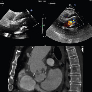
A 45-year-old man with known HCV-related cirrhosis (Child Pugh C) was admitted to the ED for shortness of breath and melena. Vital signs: blood pressure 80/60 mmHg, heart rate 98/minute, respiratory rate 22/minute, body temperature 37°C, saturation oxygen level 95%. Blood tests showed severe anemia treated with red blood cell transfusion, hepatic dysfunction including altered coagulation tests, and impaired renal function. Past medical history included chronic coronary syndrome and mitral valve infective endocarditis requiring mitral valve replacement surgery (St Jude Medical bio prosthesis 29), complicated by dehiscence treated with percutaneous paravalvular leak's closure. The patient was transferred to gastroenterology division where he underwent esophagogastroduodenoscopy revealing duodenal variceal bleeding treated with endoscopic injection sclerotherapy; a transjugular intrahepatic portosystemic shunt (TIPS) was indicated. He was referred to the cardiologist who revealed severe stenosis and thrombosis of mitral valve prosthesis (MVP), right ventricle dilatation/dysfunction along with severe pulmonary hypertension were evident on transthoracic color-Doppler echocardiogram. No pulmonary emboli were detected at the lung computed tomographic angiography, whose imaging allowed for unique views of the MVP dysfunction. Fibrinolysis of MVP thrombosis was contraindicated for high bleeding risk due to duodenal bleeding varices. TIPS was contraindicated too as it could have determined a volume overload that could have provoked right ventricular failure in a patient with preexisting hyperdynamic circulation.

J Cardiovasc Echogr. 2022 Aug;32(Suppl 1):S1–S80.

AN UNEXPECTED CAUSE OF TRICUSPID VALVE REGURGITATION

Caputo Adriano 1, Ilaria Caso 1, Ascione Luigi 1, Palmiero Giuseppe 1, Cacciapuoti Fulvio 1, Merenda Raffaele 1, D' Andrea Antonello 2, Calabrò Paolo 3

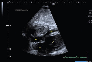
We reported the case of a 72-year-old female, presenting signs and symptoms of heart failure and history of permanent atrial fibrillation, systemic arterial hypertension, diabetes type 2 and chronic pulmonary disease referred to our third-level centre for a transesophageal echocardiographic (TEE) examination of massive tricuspid regurgitation (TR). At TEE, the right heart chambers were dilated with functional massive TR directed towards the interatrial septum and indirect signs of pulmonary hypertension. Furthermore, we described a 5 mm dropout of the atrioventricular portion of the membranous septum generating a predominantly systolic left ventricle-right atrium shunt, coherent with a Gerbode supravalvular defect. TEE 3D realtime of the defect allowed us to have a better anatomical characterization of the origin, the course, the shape and the size of the septal defect, excluding additional shunts. Gerbode defect is a rare abnormal communication between the left ventricle and right atrium resulting from either a congenital defect or prior cardiac insults. The congenital form accounts for less than 1% of all congenital heart disease and for 0.08% of all cardiac shunts, while iatrogenic shunt incidence has been increasing with the growth of structural heart surgeries and interventions. This case cautions us to consider echocardiographic screening of congenital heart lesions in adults with signs and symptoms of right-heart overload. Moreover, it highlights the great value of 2D-3D TEE in defining the anatomy of heart structures, allowing the diagnosis and a precise characterization of this rare cardiac anomaly.

graphic file with name JCE-32-1-g022.jpg

J Cardiovasc Echogr. 2022 Aug;32(Suppl 1):S1–S80.

OXIDATIVE STRESS AND LEFT VENTRICULAR PERFORMANCE IN PATIENTS WITH DIFFERENT GLYCOMETABOLIC PHENOTYPES

Cassano Velia 1, Miceli Sofia 1, Armentaro Giuseppe 1, Mannino Gaia Chiara 1, Fiorentino Vanessa Teresa 1, Perticone Maria 1, Succurro Elena 1, Hribal Marta Letizia 1, Andreozzi Francesco 1, Perticone Francesco 1, Sesti Giorgio 2, Sciacqua Angela 1

Introduction: In normoglucose-tolerant subjects (NGT), glycaemia ≥155 mg/dl 1-h during OGTT identifies a worse cardio-metabolic profile. AIM: We evaluated the correlation between oxidative stress and subclinical myocardial damage, assessed with speckle-tracking echocardiography in NGT≥155 vs NGT&lt;155 subjects, impaired glucose tolerance (IGT) and T2DM patients.

MATERIALS AND Methods: We enrolled 100 Caucasian patients (61/39 M/F, mean age 61.4±10.7). Subjects underwent clinical and laboratory evaluation. 8-isoprostane and NOX-2 serum values were assessed with ELISA-sandwich. Statistical analysis was performed with ANOVA-test, linear correlation analysis and stepwise multivariate linear regression model.

Results: 30 patients were NGT&lt;155, 24 NGT≥155, 28 IGT, 18 T2DM. 8-isoprostane and NOX-2 serum values were higher in NGT≥155 than NGT&lt;155 group, but similar to IGT. GLS appeared lower proceeding from NGT&lt;155 to T2DM group. For similar EF values, NGT≥155 subjects presented lower GLS than NGT&lt;155, but similar to IGT. Endo/epi-ratio was inversely correlated with 1-h glycaemia, NOX-2, 8-isoprostane; GLS was inversely correlated with 1-h glycaemia and directly correlated with 8-isoprostane and NOX-2. NOX-2 resulted the major predictor of endo/epi ratio (40.7% of its variation); 1-h glycaemia was the second predictor (9.2%) and the strongest predictor of the GLS (53.9%).

Conclusions: Our study demonstrated that NGT≥155 present functional alterations of myocardial contractile fibers. This data have a central role in ongoing research on the association between 1-hour post load hyperglycaemia and cardiovascular risk.

J Cardiovasc Echogr. 2022 Aug;32(Suppl 1):S1–S80.

FIRST CASE OF COMBINED TRANSCATHETER AORTIC BIOPROSTHESIS IMPLANTATION “VALVE-IN-VALVEAND PERCUTANEOUS MITRAL VALVE REPAIR IN A HIGH-RISK PATIENT WITH ACUTE HEART FAILURE REFRACTORY TO MEDICAL THERAPY

Ceriello Laura 1, Paparoni Francesco 1, Tomassoni Gianluca 1, Lavorgna Alberto 1, De Rosa Mario 1, Artale Alessia 2, Taraschi Francesco 2, De Remigis Franco 1, Fabiani Donatello 1

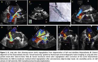
An 84-year-old female was admitted to Coronary Unit for acute pulmonary edema due to severe aortic regurgitation from degeneration of full root stentless bioprosthesis and severe secondary mitral regurgitation. The patient was affected by ischemic heart disease with moderate reduction of left ventricular systolic function, diabetes, atrial fibrillation and chronic kidney disease. Optimized medical therapy and ultrafiltration were performed obtaining a labile hemodynamic stabilization. Given the comorbidities and hemodynamic instability the risk of a surgical redo was deemed prohibitive. Transoesophageal echocardiography, CT scan and coronary angiography were carried out to evaluate the feasibility of percutaneous correction of both valvular diseases. The patient underwent Valve-in-Valve (ViV) implantation of an aortic bioprosthesis and simultaneous percutaneous “edge-to-edge” repair of the mitral valve (TMVR) with mild-to-moderate residual regurgitation. The procedure was guided by fluoroscopic-echocardiographic fusion imaging which allowed the correct positioning of the bioprosthesis at the level of the suture ring despite the absence of radiopaque markers. Post-procedural course was characterized by a significant improvement in haemodynamic parameters. Discussion. The execution of ViV TAVI in patients with full root stentless bioprosthesis poses numerous technical problems and it has never been performed in conjunction with TMVR. This first experience of combined procedure highlights the technical feasibility and the key role of multimodality imaging, despite its prognostic role remains to be defined.

graphic file with name JCE-32-1-g023.jpg

J Cardiovasc Echogr. 2022 Aug;32(Suppl 1):S1–S80.

SUCCESSFUL SURGICAL REPAIR OF LEFT VENTRICULAR PSEUDOANEURYSM IN A PATIENT WITH SUBACUTE ST- ELEVATION MYOCARDIAL INFARCTION

Chianese Salvatore 1, Cocchia Rosangela 1, Capone Valentina 1, Annunziata Roberto 1, Salzano Andrea 2, Maramaldi Renato 1, Marullo Flavio 1, Conte Marianna 1, Bossone Eduardo 1

A 65-year-old man was admitted to the emergency department for abdominal pain begun 4 days before. High-sensitivity cardiac troponin was mildly elevated. Electrocardiogram showed Q waves with ST elevation in antero-lateral leads consistent with subacute STEMI. Transthoracic echocardiogram revealed aneurysmatic expansion of apical left ventricular walls (EF:30%) with a large apical thrombus (35x15 mm). Coronary angiography showed complete occlusion of proximal left anterior descending artery and a critical stenosis of right coronary artery; percutaneous coronary intervention with stent implantation on right coronary artery was performed. He received triple antithrombotic therapy with aspirin, clopidogrel, intravenous heparin first and then oral anticoagulation with warfarin. During coronary care unit stay, the patient was asymptomatic and hemodynamically stable. However, a control TTE in cardiology ward revealed the presence of pseudoaneurysm with an oval out-pouching (23 × 13 mm) from the apical aspect of LV septum, communicating with the LV chamber through a passage measuring 1.6 cm. Partially organized pericardial effusion was evident around right ventricular free wall. The patient was immediately transferred to the cardiac surgery department for urgent surgical repair. The LV anterior wall rupture with pseudoaneurysm was then treated with endoaneurysmectomy of the LV with dacron patch and freewall reconstruction with prolene sutures and teflon felt stripes. The patient was finally discharged 3 weeks after surgery and at one month follow-up visit, the patient was stable with acceptable functional capacity.

J Cardiovasc Echogr. 2022 Aug;32(Suppl 1):S1–S80.

ONE-HOUR POST LOAD GLYCAEMIA AND SUBCLINICAL MYOCARDIAL DAMAGE

Clausi Elvira 1, Miceli Sofia 1, Filicetti Elvira 1, Monaco Vittoria 1, Cassano Velia 1, Armentaro Giuseppe 1, Barbara Keti 1, Scozzafava Aleandra 1, Crescibene Daniele 1, Succurro Elena 1, Perticone Maria 1, Andreozzi Francesco 1, Sesti Giorgio 2, Sciacqua Angela 1

graphic file with name JCE-32-1-g024.jpg

Introduction: Normoglucose-tolerant subjects (NGT) with glycaemia >155 mg/dl 1-h during OGTT present higher risk of developing type-2 Diabetes Mellitus (T2DM) than NGT&lt;155, because they have a worse cardio-metabolic profile.

Aim: We evaluated atrial and ventricular subclinical myocardial damage, assessed with speckle-tracking echocardiography, in NGT≥155 vs NGT<155, impaired glucose tolerance (IGT) and T2DM patients.

Materials and Methods: We enrolled 229 Caucasian patients (117/112 M/F, mean age 58,7±10.7). Main exclusion criteria were cardiovascular and respiratory diseases, cancers, drugs influencing glucose metabolism, alcohol and smoking abuse. All patients underwent blood chemistry analysis, OGTT, advanced Color-Doppler echocardiography with evaluation of main atrial and ventricular parameters. Data were analyzed with ANOVA, post-hoc Bonferroni's test and Chi-quadro. Linear correlation analysis and stepwise multivariate linear regression model were performed for statistical significant variables.

Results: 77 patients were NGT155, 57 IGT and 38 TDM2. NGT>155 had worse GLS than NGT<155, but similiar to IGT, higher LAVI and lower PACS than NGT<155. PALS, LAVI/PALS and LAVI/E/e' were better in NGT<155 than the other groups (p155 present subclinical atrial dysfunctions, which can determine atrial fibrillation, so atrial strain could be used for thromboembolic risk evaluation and therapeutic optimization.

J Cardiovasc Echogr. 2022 Aug;32(Suppl 1):S1–S80.

ANALYSIS OF MYOCARDIAL FUNCTION BY DEFORMATION IMAGING IN PATIENTS WITH TRANSTRIRETIN GENE MUTATION

Comparato Francesco Comparato 1, Di Lisi Daniela 1, Puglisi Cataldo 1, Portelli Maria Cristina 1, Cannizzo Noemi 1, Damerino Giuseppe 1, Di Caccamo Leandro 1, Ortello Antonella 1, Galassi Alfredo Ruggero 1, Novo Giuseppina 1

Introduction. Hereditary transthyretin amyloidosis is a rare disease that can affect various organs. The early identification of cardiac involvement through echocardiography allows to corroborate the diagnosis and to start therapy at an early stage.

Aim: The aim of our study was to detect early signs of cardiac involvement in patients with TTR gene mutation and neurological phenotype or absence of any organ disease (carriers) compared to patients with TTR gene mutation and cardiac amyloidosis (CA).

Materials and Methods. An observational study was conducted on a sample of 31 patients with TTR gene mutation. Patients were divided into 3 groups (23% CA, 42% amyloid neuropathy and 35% carriers). All patients underwent echocardiography with evaluation of left ventricular myocardial deformation indices (apical/basal strain ratio and relative apical sparing).

Results: Carriers and patients with amyloid neuropathy had normal left ventricular systolic and diastolic function. Analyzing left ventricular longitudinal global strain (GLS) we didn't find significant differences between carriers and neuropathy, but we found a significant difference about apical/basal strain ratio and relative apical sparing (respectively 1,33 ± 0,20 vs 1,58 ± 0, 25, p value 0,001 and 0,62 ± 0,07 vs 0,72 ± 0,08, p value 0,039).

Conclusion: relative apical sparing is specific to identify CA; the apical/basal ratio seems to gradually increase from carriers to patients with cardiac amyloidosis and it could be used to monitor patients with TTR mutation, before developing CA.

J Cardiovasc Echogr. 2022 Aug;32(Suppl 1):S1–S80.

SPRING DYSPNEA IN PLASTIC SURGERY

Corapi Antonella 1, Voci Daniela 2, Bullotta Eros 3, Serafino Sergio 3

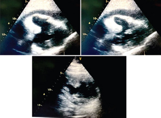
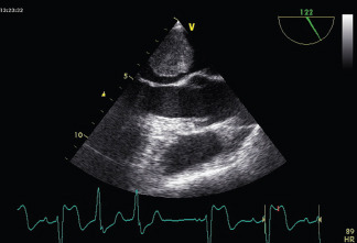
February 18th 2022 there was an Emercency in Plastic Surgery: a man of 71 y.o. with diabetes, cognitive deficit, psychosis, atrial fibrillation without anticoagulant home therapy, was soporous, dyspneic and with high frequency atrial fibrillation (ECG, photo 1). He was hospitalized because affected by necrotic ulcer at 3th middle lowerright leg. After surgery, he became dyspneic, but a chest x ray made evident a pulmunary right effusion, then the patient was subjected at polmonary drainage (thoracic surgical consultation, photo 2), but during the afternoon, his clinical condition became better and the patient became very dispnoic and soporous. So, in the patient's bed I performed an urgent Echocardiogram : it made evident an isohypoechoic rounded lesion, with net margins, its size was 6.23x6 cm, localizzed in left atrial, near Anterior Mitral flap attached at inner atrial septum, it was a mobile lesion and obstructed out flow mitral tract(video 1, 2, 3, photo3). Then the patient was immediately tranferred in Cardiac Surgery and he subjected at the lesion excision: histological analysis was mixoma.

J Cardiovasc Echogr. 2022 Aug;32(Suppl 1):S1–S80.

INFECTIVE ENDOCARDITIS: A SUBTLE PRESENTATION

Corsi Elisabetta Corsi 1, Bosso Alessandra 1, Venturini Elio 2

Background: Infective endocarditis is a serious and deadly disease: the diagnosis is not always easy and it is still associated with high mortality and severe complications, especially in patients with prosthetic valves.

Case Description: A 75-years-old woman presented to the ED for persistent low-grade fever and mild dyspnoea. She had an aortic biologic prosthetic valve implanted the year before for severe aortic stenosis. She also complained a persistent back pain, irresponsive to drugs or physiotherapy. Getting back in time, she had been hospitalized for genitourinary tract infection from multi-resistant E.faecalis, treated with antibiotics. A transthoracic echocardiogram was performed, showing an apparently intraprosthetic moderate aortic regurgitation, never previously described. In the strong suspect of endocarditis, she underwent transoesophageal echocardiography, that showed dehiscence of the prosthetic valve with severe paravalvular abscess and regurgitation, without rocking motion of the prosthesis. A TC scan and a spine MRI also confirmed the presence of infectious spondylodiscitis, responsible for that persistent back pain. The patient was urgently sent to cardiac surgery and a Bentall procedure was performed. Despite the severity of the clinical condition, the patient survived and after 3 weeks of hospitalization was sent back to our Department for Cardiac Rehabilitation.

graphic file with name JCE-32-1-g025.jpg

Conclusion: This case shows the subtle but serious evolution that usually controlled infections can have in patients with high risk of endocarditis, as ones with prosthetic valves, and the role of multimodality imaging in these sets.

graphic file with name JCE-32-1-g026.jpg

J Cardiovasc Echogr. 2022 Aug;32(Suppl 1):S1–S80.

A GIANT MYXOMA IN THE LEFT ATRIUM ADHERENT TO THE INTERATRIAL SEPTUM

Cortese Francesca 1, Costantino Marco Fabio 1, Mazzeo Pietro 1, Lapetina Ornella 1, Prestipino Filippo 1, Manzan Enrico 1, D'Ascoli Riccardo 1, D'addeo Giampaolo 1, Lopizzo Agostino 1, Bochicchio Angela 1, Cristino Angela 1, Luzzi Giampaolo 1

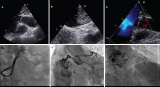
A 73-year-old woman came to our observation to perform a routine echocardiographic examination. The patient had arterial hypertension, dyslipidemia, hypothyroidism, paroxysmal atrial fibrillation. The clinical evaluation was normal, as was the electrocardiogram. Transthoracic echocardiographic evaluation showed the presence of a rounded formation with variable echogenicity, with an irregular surface (4 x 3 cm), adherent to the interatrial septum at the level of the fossa ovalis, which did not hinder the excursion of the mitral valve. No other significant cardiac abnormalities were detected. Tansesophageal echocardiography confirmed the presence of atrial mass adhered to the oval fossa (panels A, B). of coarsely rounded shape, and variable echogenicity, in the absence of interference with the movement of the valve leaflets and of anomalies of the interatrial septum (panel C). Coronary angiography showed a right-dominant coronary circulation (panel D) and a mild stenosis of the mid left anterior descending artery. An abundant arterial perfusion of the atrial mass, with several vessels originating from the circumflex artery was also evident at the coronary angiography (panels E, F). The patient underwent cardiac surgery; the surgical exeresis of the left atrial mass was performed through right mini thoracotomy. The post-operative course was uneventful. Histological analysis confirmed the diagnosis of atrial myxoma. At six-month follow-up the patient was asymptomatic and the echocardiogram was superimposable to discharge.

graphic file with name JCE-32-1-g027.jpg

J Cardiovasc Echogr. 2022 Aug;32(Suppl 1):S1–S80.

SCREENING FOR ANDERSON-FABRY DISEASE IN RELATIVELY YOUNG PATIENTS WITH MILD LEFT VENTRICULAR HYPERTROPHY AND UNEXPLAINED CONDUCTION DISORDERS REQUIRING PACEMAKER IMPLANTATION

Crea Pasquale 1, Allegra Marta 1, Poleggi Cristina 1, Mancinelli Anna 1, Trimarchi Giancarlo 1, Dattilo Giuseppe 1, Licordari Roberto 1, Giuffrida Giulia 1, Carerj Scipione 1, Micari Antonio 1, Di Bella Gianluca 1

Introduction: Anderson-Fabry Disease is a rare X-linked lysosomal storage disorder. Cardiac manifestations include left ventricular hypertrophy (LVH) and arrhythmias. The rate of pacemaker implantation (PMI) in AFD has been described to be 25 times higher than in general population.

Purpose: Our aim is to detect AFD among relatively young patients with LVH and unexplained conduction disturbances requiring PMI.

Methods: Among 650 patients afferent to our ambulatory for routinary pacemaker follow-up, we considered population with sinus node dysfunction or AV block and an age, at the time of PMI, ≥40 and ≤70 yo. Exclusion criteria were: patients with previous myocardial infarction or known cardiac disease. Thus, in 26 adult patients (13 M; 13 F; 63 ± 7 yo) screening for AFD was performed. After clinical evaluation, transthoracic echocardiography and pacemaker check, a dried blood spot sampled in filter paper was analyzed to evaluate the α-galactosidase A enzyme activity in males and for genetic investigation in females.

Results: Analysis revealed 58% (15/26) of patients affected by mild LVH (IVS diameter ranging from 11 to 15mm). No patient had severe LVH or moderate-severe renal dysfunction. In the cohort considered, we found one 69 yo female patient with heterozygosis GLA pathogenic mutation, NM_000169.2:c.638A>C p.(Lys213Thr). She had normal value of liso-Gb3 1,1 ng/ml (n.v. ≤ 1,8 ng/ml). She had mild LVH (IVS 12 mm) and no renal dysfunction. Familiar screening was programmed.

Conclusion: Screening efforts for AFD disease should be increased in the selected population of relatively young patients with LVH and conduction disturbances requiring PMI.

J Cardiovasc Echogr. 2022 Aug;32(Suppl 1):S1–S80.

UNEXPECTED TRIGGER OF RELAPSING ATRIAL TACHYARRHYTHMIA: A HUGE PERICARDIAL CYST

Cucchini Umberto 1, Alderighi Chiara 1, Bedont Silvana 2, Peluso Diletta 1, Polo Angela 1, Soldà Elena 1, Zasso Antonella 1, Nguyen Kim Ahn 1, Pasinato Antonio 1, Simonetto Federico 1, Zadro Mirco 1, Carasi Massimo 1, Corletto Anna 1, Chirillo Fabio 1

Pericardial cysts are benign, often asymptomatic thoracic masses discovered incidentally during routine instrumental imaging. Sometimes, they can determine symptoms following compression of mediastinal structures. Herein, we present the case of a patient admitted to the emergency department because of recurring atrial tachycardia associated with weakness, dizziness and chest pain. An urgent point of care echocardiogram (POCUS) identified an anechoic mass without flow signal at Doppler interrogation, adjacent to the right atrial-ventricular free wall. The patient underwent urgent chest computed tomography that described a bulky mass with pure-liquid density extending from the superior mediastinum, downward to the anterior/ right mid mediastinum. The mass had a close relation with the great vessels and the heart, impinging right atrium and right atrial appendage. The absence of enhanced signal after contrast injection or other local linfadenopathy corroborated the diagnosis of a huge pericardial cyst. The poorly controlled supraventricular arrhythmia, together with persistent symptoms of systemic congestion represented an indication to surgery, despite the presumed benign nature of the lesion. The patient underwent intervention through thoracotomic approach converted to sternal because of challenging detachment of the mass from the venous vessels. The patient had a single episode of atrial fibrillation immediately thereafter without other episodes. Conclusions: Pericardial cysts despite rare and usually asymptomatic may be sometimes so huge that can determine several types of complications needing surgical resect

J Cardiovasc Echogr. 2022 Aug;32(Suppl 1):S1–S80.

AORTIC DISSECTION TYPE A: ROLE OF TTE

D'agostino Simone 1, Gizzi Germana 1, Parato Vito Maurizio 1

Introduction: Aortic dissection is a high-mortality condition requiring immediate diagnostic-therapeutic approach.

Clinical Case: In August 2021 an 89-year-old woman suffering from systemic arterial hypertension, dyslipidemia and Parkinson's disease was admitted to ER for syncopal episode in the absence of prodromes. She reported chest pain. Patient presented alert, oriented, asymptomatic, hemodynamically stable. At the time of cardiologic evaluation, transthoracic echocardiography revealed dissection of the proximal ascending aorta with an intimal flap starting from the aortic root and extending to the proximal tract of the aortic arch, moderate aortic valve regurgitance and moderate circumferential pericardial effusion (2cm) with blood clots. TC angiography confirmed Stanford type A aortic dissection with intimal flap originating in the supravalvular site (max diameter 6cm), extended longitudinally for about 6 cm at the level of the ascending aorta until the emergence of the epiaortic vessels. Because of the patient's advanced age and prohibitive operative risk considered comparable of clinical course of disease, surgery was denied. The patient was admitted to ICU where she remained asymptomatic and hemodinamically stable until death occurred on the third day for cardiocirculatory arrest from PEA.

Conclusions: In emergency conditions such as aortic dissection, bedside transthoracic echocardiography represents the examination of more prompt execution and it not rarely allows the diagnosis before radiological confirmation by angioTC.

graphic file with name JCE-32-1-g028.jpg

J Cardiovasc Echogr. 2022 Aug;32(Suppl 1):S1–S80.

ROLE OF ATRIAL STRAIN FOR EARLY DETECTION OF CHEMOTHERAPY-INDUCED CARDIOTOXICITY

Damerino Giuseppe 1, Bergamini Corinna 2, Cadeddu Dessalvi Christian 3, Casavecchia Grazia 4, Di Lisi Daniela 1, Sprighetti Paolo 5, Niro Lorenzo 5, Madaudo Cristina 1, Sinagra Francesco Paolo 1, Rossetto Ludovico 1, Manganaro Roberta 5, Ricci Sara Jane 3, Zito Concetta 5, Moreo Antonella 6, Novo Giuseppina 1

Introduction: Chemotherapy drugs can give serious cardiovascular side effects, especially left ventricular disfunction. Speckle tracking echocardiography (STE) has emerged as a technique to accurately estimate cardiotoxicity in cancer patients.

Aim: The aim of our study was to evaluate the echocardiographic role of left atrial strain as early marker of subclinical damage in patients with breast cancer receiving antineoplastic treatment with cardiotoxic drugs.

Materials and methods: A prospective multicentric study was conducted on a population of 140 women with newly diagnosed breast cancer aged between 18 and 65 years, who were scheduled to receive potentially cardiotoxic chemotherapy. At the time of enrollment, a complete echocardiogram was performed, including analysis of Global longitudinal Strain (GLS) and atrial function by STE at baseline (T0), at 3 months (T1), at 6 months (T2).

Results: Left ventricular GLS was significantly reduced at T1 and T2 from baseline [GLS -20.62 ± 1.80 at T0 vs -19.38 ± 2.07 at T1 (P &lt; 0,0001) vs -19.28 ± 2.56 at T2 (P &lt; 0,0001)]. Analyzing the atrial function, a significant variation in the reservoir strain and atrial stiffness at follow-up was also found [PALS 36.13 ± 9.1 at T0 vs 32.3 ± 8.4 at T1 (P&lt; 0,0001) vs 29.3 ± 7.1% at T2 (P = 0,0006)]; [LASI 0.19± 0.08 at T0 vs 0.23 ± 0.09 at T1 (P= 0,0098) vs 0.22±0.06 at T2 (P &lt; 0,0001)].

Conclusions: Left atrial function assessed by STE could be considered an early cardiac damage marker in patients with breast cancer undergoing chemotherapy.

J Cardiovasc Echogr. 2022 Aug;32(Suppl 1):S1–S80.

ROLE OF ATRIAL STRAIN FOR EARLY DETECTION OF CHEMOTHERAPY-INDUCED CARDIOTOXICITY

Damerino Giuseppe 1, Bergamini Corinna 2, Cadeddu Dessalvi Christian 3, Casavecchia Grazia 4, Di Lisi Daniela 1, Sprighetti Paolo 2, Niro Lorenzo 2, Madaudo Cristina 1, Sinagra Francesco Paolo 1, Rossetto Ludovico 1, Manganaro Roberta 5, Ricci Sara Jane 3, Zito Concetta 5, Moreo Antonella 6, Novo Giuseppina 1

Introduction: Chemotherapy drugs can give serious cardiovascular side effects, especially left ventricular disfunction. Speckle tracking echocardiography (STE) has emerged as a technique to accurately estimate cardiotoxicity in cancer patients. The aim of our study was to evaluate the echocardiographic role of left atrial strain as early marker of subclinical damage in patients with breast cancer receiving antineoplastic treatment with cardiotoxic drugs.

Methods: A prospective multicentric study was conducted on a population of 140 women with newly diagnosed breast cancer aged between 18 and 65 years, who were scheduled to receive potentially cardiotoxic chemotherapy. At the time of enrollment, a complete echocardiogram was performed, including analysis of Global longitudinal Strain (GLS) and atrial function by STE at baseline (T0), at 3 months (T1), at 6 months (T2).

Results: Left ventricular GLS was significantly reduced at T1 and T2 from baseline [GLS -20.62 ± 1.80 at T0 vs -19.38 ± 2.07 at T1 (P &lt; 0,0001) vs -19.28 ± 2.56 at T2 (P &lt; 0,0001)]. Analyzing the atrial function, a significant variation in the reservoir strain and atrial stiffness at follow-up was also found [PALS 36.13 ± 9.1 at T0 vs 32.3 ± 8.4 at T1 (P&lt; 0,0001) vs 29.3 ± 7.1% at T2 (P = 0,0006)]; [LASI 0.19± 0.08 at T0 vs 0.23 ± 0.09 at T1 (P= 0,0098) vs 0.22±0.06 at T2 (P &lt; 0,0001)].

Conclusions: Left atrial function assessed by STE could be considered an early cardiac damage marker in patients with breast cancer undergoing chemotherapy.

J Cardiovasc Echogr. 2022 Aug;32(Suppl 1):S1–S80.

EARLY ECHOCARDIOGRAPHIC DETECTION OF SYSTOLIC DYSFUNCTION IN CARDIAC AMYLOIDOSIS: THE ADDED VALUE OF MYOCARDIAL WORK

De Gaetano Fabrizio 1, Losi Valentina 1, Consoli Alessio 1, Tamburino Corrado 1, Monte Ines Paola 1

Introduction: Amyloid can seep into many tissues, but the cardiac involvement represents the main determinant of mortality and morbidity for patients with systemic amyloidosis (SA). In this survey we searched for echocardiographic signs of early systolic impairment in cardiac amyloidosis (CA): among them we adopted the analysis of myocardial work (MW), an emerging less load-dependent tool for the evaluation of cardiac performance.

Methods: We chose a cohort of patients (n=11; mean age 67±12 years) with AL or ATTR CA and we compared them with a control group (n=8; mean age 67±8 years) afflicted by multiple myeloma without evident cardiac involvement. We examined their systolic and diastolic function and we calculated the contributors of MW from speckle tracking echocardiography.

Results: We found a statistically relevant difference in cardiac mass (144±28 versus 85±20 g/m2) and in signs of both systolic and diastolic worsening between patients with CA and the control group. In CA we identified a dramatic decrease in Global Work Index (981±327 versus 2050±260 mmHg%) and Global Constructive Work (1107±391 versus 2381±379 mmHg%), with no significant difference between AL and ATTR amyloidosis.

Conclusions: The infiltration by amyloid and the consequent increase in cardiac mass lead to an early and often severe weakening of the intrinsic contractility of the heart in both AL and ATTR amyloidosis, even when ejection fraction is normal or slightly reduced. Therefore, we suggest the analysis of MW starting from the first echocardiographic evaluation in patients with known SA or multiple myeloma.

graphic file with name JCE-32-1-g029.jpg

J Cardiovasc Echogr. 2022 Aug;32(Suppl 1):S1–S80.

A NEURO-CARDIO-ONCOLOGICAL CLINICAL CASE

De Paolis Marcella 1, Bier Nicola 2, Mezzetti Paola 1, Patella Marco Mariano 1, Camilli Giulia 1, Milici Caterina 1, Dominici Marcello 3

Cardiac metastases (CM) are 20 to 40 times more common than primary tumors and occur more often in cases of melanoma, leukemia, mediastinal and renal cell tumors. The right side of the heart is more commonly involved. Clinical manifestations (obstruction, embolism, arrhythmias) depend on tumor site and infiltration. We presented a rare case of cardioembolic complication from left-sided CM of lung cancer. A 61-year-old ex-smoker and dyslipidemic woman, recently undergone pulmonary nodule biopsy, was referred to our hospital because of acute right hemisome motor deficit: left middle cerebral artery ischemia was evidenced and systemic thrombolysis was undertaken. Transthoracic Echocardiography (TTE) revealed normal systolic function of LV with isoechoic mobile masses adhered to a thickening localized on middle lateral wall, without pericardial effusion. Suspecting intraventricular thrombus, in accordance with neurologist, after repeated cerebral computed tomography, anticoagulant therapy was started. Complete resolution of IV was diagnosed 5 days later. Meanwhile, pulmonary biopsy was positive for adenocarcinoma. Afterwards, cardiac magnetic resonance (CMR) showed an oval-shaped mass involving myocardium of LV middle lateral wall, with rough borders, increased signal intensity in T2-weighted and marked heterogeneous enhancement on late gadolinium enhancement imaging. A multimodality imaging approach is often required to diagnose CM, primarily using ETT which is limited for small CM. CMR is the gold standard technique to confirm presence, localization, invasion, functional impact and tissue characterization of CM.

graphic file with name JCE-32-1-g030.jpg

J Cardiovasc Echogr. 2022 Aug;32(Suppl 1):S1–S80.

ATYPICAL PRESENTATION OF INFECTIVE ENDOCARDITIS (IE): A CASE SERIES

De Paolis Marcella 1, Bier Nicola 2, Mezzetti Paola 1, Patella Marco Mariano 1, Camilli Giulia 1, Milici Caterina 1, De Bonis Paolo 1, Dominici Marcello 3

IE is a relatively rare, but deadly cause of sepsis, with an overall mortality ranging from 20 to 25% in most series. We describe 2 cases of IE with unusual presentation: in 1 cancer and in 1 non-CR patients. An apiretic 91-year-old male, with recent surgery for bladder cancer and chronic renal insufficiency, was admitted in hospital for anuria. Dialysis was undertaken. Due to elevated values of C-reactive protein and erythrocyte sedimentation rate and, then, after Enterococcus Faecalis positive blood cultures (PBC), a transthoracic echocardiogram (ETT) was performed. A severe mitral regurgitation (MR) with a large mobile vegetative lesion on posterior leaflet and a moderate aortic valve insufficiency with multiple vegetation on right coronary cuspid were documented. An 81-year-old man, for 3 months in corticosteroids for treating a moderate “no-autoimmune hemolytic anemia”, presented to hospital for dyspnoea and tachycardia: on blood tests presence of normal white blood cell, severe anemia and thrombocytopenia with reduced fibrinogen; on electrocardiogram presence of tachy-arythmia for atrial flutter considered a secondary event. For Streptococcus Cristatus PBC, an ETT was performed with evidence of severe MR due to destructor plus image of 3x3 centimeters on anterior leaflet. The diagnostic doubt of the previous hemolytic anemia caused by mitral valvulopathy was raised. Both patients died after a few days. Inspite of diagnostic and imaging tools evolution, IE still remains a challenging diagnostic problem especially in elderly patients with comorbidities. Diagnostic delay can greatly increase mortality.

J Cardiovasc Echogr. 2022 Aug;32(Suppl 1):S1–S80.

DISTACCO DI MITRACLIP: ESTRAZIONE PERCUTANEA

De Rosa Mario 1, De Remigis Franco 1, Tomassoni Gianluca 1, Fabiani Donatello 1

A 78-year-old man with a history of mitral prolapse came to our observation for heart failure. TTE and TEE showed fibroelastic degeneration of mitral valve, P2 flail and ring dilation associated with severe regurgitation. The case was discussed in Heart Team. In consideration of the high surgical risk, MitraClip was indicated. The procedure was performed in the absence of complications up to the “grasping” phase. Subsequently there was an incomplete separation of the clip from the release system, detachment of the same from the valve and its dislocation at the level of the interatrial septum. The relative stability of the clip in the embolization site led us to attempt percutaneous recovery (Snare catheter). Once the oval fossa was reached, the recovery of the clip was performed using 2-3D echo and fluoroscopic support. Then a new attempt grasping was performed whitout complications. Patient was discharged on day fifth of hospitalization. Percutaneous correction of mitral regurgitation using MitraClip is now a widely performed procedure with excellent results both in the treatment of functional and organic pathology. The complications usually related to the surgery are in most cases not severe. Device embolization is an extremely rare complication (0.1%) and life-threatening for the patient. We have described a rare case of MitraClip embolization due to complications of the release mechanism resolved by percutaneous extraction of the device. The procedure was subsequently completed with correct positioning of the MitraClip in the absence of significant complications and with early discharge of the patient at hom

graphic file with name JCE-32-1-g031.jpg

J Cardiovasc Echogr. 2022 Aug;32(Suppl 1):S1–S80.

MULTIMODALITY IMAGING TO ASSESS SEVERITY AND OUTCOME IN ASYMPTOMATIC PATIENTS WITH AORTIC STENOSIS: A MEDIUM-LONG TERM FOLLOW-UP

De Sarro Rosalba 1, Zito Concetta 1, Manganaro Roberta 1, Licordari Roberto 1, Bursi Francesca 2, Mantovani Francesca 3, Benfari Giovanni 4, Malagoli Alessandro 5, Bertolacelli Ylenia 6, D'Angelo Tommaso 7, Antonini-Canterin Francesco 8, Carerj Scipione 1, Barbieri Andrea 9

Introduction: Multimodality imaging approach is becoming increasingly common in evaluating the severity and outcome of aortic stenosis (AS). To assess the outcome of asymptomatic AS and the usefulness of aortic calcium score (CS) for solving the dilemma of low flow low gradients (LFLG) severe AS.

Methods: 70 (81.4±8.4yrs) prospective asymptomatic patients with AS were followed for 2.77±2.01yrs with a TTE every 6 months. Fig. 1 shows the summary of the study.

Results: At baseline we found a mild AS in 32.9%, moderate in 28.4%, severe in 27.1%; 36.8% of severe AS were LFLG. During FU, 32.8% of pts died (5.7% LFLG) and 18.5% underwent AVR/TAVR. The best cut-off to predict survival was AVA =1cm2 (100% sensitivity and 80% specificity). In 34 pts ending FU we found an overall progression of AS severity (p&lt;0.05 for all parameters vs baseline). Fig.1 shows the main results at the end of FU. All patients with LFLG severe AS underwent CS revealing that AS was not severe in 6 (1233±1123 AU; 622±55AU/m2) and true severe in 12 (3388±1188 AU;1858 ±795 AU/m2; p=0.005 and p=0.002, respectively). Symptomatic severe LFLG AS were all true severe according to CS (Fig.1). Main calcium score correlations were with: LV hypertrophy:r=0.78, p&lt;0.001; pulse pressure; r=0.54,p=0.036;LVEF:r= 0.61;p=0.015;TAPSE:r=0.57,p=0.027;peak gradient:r=0.88, p&lt;0.001;peak velocity:r=0.80,p=0.002; increase in mean gradient (delta gradient):r=0.75, p=0.001.

Conclusions: Asymptomatic AS is associated with high mortality and rapid progression. AVA remains the best predictor of outcome. In severe LFLG AS, CS correlates with AS progression and outcome.

graphic file with name JCE-32-1-g032.jpg

J Cardiovasc Echogr. 2022 Aug;32(Suppl 1):S1–S80.

RIGHT VENTRICULAR FREE WALL LONGITUDINAL STRAIN (RVFWSL) A NEW OUTCOME PREDICTOR IN PATIENTS CANDIDATE FOR TAVI

Dellino Carlo Maria 1, Pergola Valeria 1, Torresan Francesca 1, Cecchetto Antonella 1, Aruta Patrizia 1, Tarantini Giuseppe 1, Fraccaro Chiara 1, Mele Donato 1, Iliceto Sabino 1

Introduction: Right ventricular (RV) systolic dysfunction is an outcome predictor in various cardiovascular diseases. RV dysfunction, assessed by RV free wall longitudinal strain (RVFWSL) in patients candidate for TAVI, has not been extensively explored as an outcome predictor. Purpose: evaluate the prognostic value of pre intervention RVFWSL in patients undergoing TAVI.

Methods: Retrospective analysis of 100 patients who underwent TAVI from 2015 to 2019. Clinical and echocardiographic data before and after TAVI and follow-up data were collected. We considered the value of [23.3]% the cut-off of normality for RVFWSL. The primary end-point was a composite of death from any cause and hospitalization for heart failure.

Results: Median age of the patients was 81 years with a functional status NYHA II-III (81%) before the intervention. EF was preserved in most of the patients (median 56%), while RV dysfunction assessed with RVFWLS was reduced in half of the patients at baseline. At a median follow-up of 1023 days (630-1387), the univariate analysis demonstrated a predictive value for a reduced RVFWSL (&lt;[23.3]%, P=0.015) and EF&lt;50% (P=0.014) before TAVI. Cox regression analysis found that pre-TAVI reduced RVFWSL (HR 2.875, I.C. 95% 1.113-7.425; P=0.03) and EF&lt;50% (HR 2.511, I.C. 95% 1.07-5.892; P=0.03) were independently associated with composite end-point of the study. Moreover, a reduced EF associated with RVFWSL &lt;[23.3]% showed an incremental value in predicting the outcome (P=0.021).

Conclusions: Among patients with severe aortic stenosis undergoing TAVI, a reduced pre-implant RVFWSL is able to predict long-term outcome

J Cardiovasc Echogr. 2022 Aug;32(Suppl 1):S1–S80.

AN UNUSUAL CASE OF ENDOCARDITIS OF EUSTACHIAN VALVE

Dell'Uomo Marco 1, Parise Antonio 2, Castellani Claudia 2, Sforna Stefano 2, Padoan Laura 2, Bernardini Gaetano 2, Notarianni Gianfranco 2, Giuffrè Giuseppe 2, Dominioni Irene 3, Conti Serenella 1

Introduction: Fungal endocarditis is an extremely debilitating disease associated with high mortality that is increasing in modern era due to the raised use of devices, ranging from prosthetic heart valves to central venous catheters. It is most prevalent in patients who are immunosoppressed and intravenous drug users. Eustachian valve endocarditis is a rare underdiagnosed entity.

Clinical Case: A 50-year-old woman was hospitalized for worsening general condition and persistent fever. The patient had active systemic lupus erythematosus and was carrying a central venous catheter. Laboratory tests showed anemia and a state of severe immunosuppression. She performed TTE with evidence of a voluminose mass in the right atrial. The TOE showed the presence of a hyperechoic, highly fluctuating and mobile formation adhering to the inferior posterior wall of the right atrial wall with involvement of the Eustachian valve. Strangely, the central venous catheter was free from vegetations. Blood cultures were positive for Candida Parapsilosis. CVC was removed and antifungal therapy started.

Discussion: The patient presented some of the most important risk factors for the development of fungal endocarditis such as the presence of a severe state of immunocompromised and the presence of a CVC. Endocarditis of the right heart is much less common than those of the left heart, and even less the involvement of the Eustachian Valve. Despite its rarity and the few cases describe, its interest is always to be sought in the case of right heart endocarditis

graphic file with name JCE-32-1-g033.jpg

J Cardiovasc Echogr. 2022 Aug;32(Suppl 1):S1–S80.

INFECTIVE ENDOCARDITIS IN THE 3D TRANSESOPHAGEAL ECHOCARDIOGRAM ERA

Dell'Uomo Marco 1, Conti Serenella 1, Dominioni Irene 2, Bier Nicola 1, Parise Antonio 1, Dominici Marcello 1

A 69-year-old woman was admitted in the emergency room for a sudden pain in left leg. Angio-CT showed occlusion of distal third of left popliteal artery, so she was promptly subjected to revascularization by mechanical removal of thrombus and locoregional fibrinolysis. In following day, she referred pain, hypothermia and “sock-like” pallor of left lower limb; angiography revealed obstruction of femoral-popliteal-tibial axis, so she received another revascularization by mechanical removal of thrombus and locoregional fibrinolysis. In the same evening, in conjunction with a feverish peak, onset a state of drowsiness associated with left facio-brachio-crural pyramidal hemisyndrome with plegia of limbs and deviation of the gaze to the right. An urgent brain CT showed a recent right hemispheric ischemic area. In the suspicion of endocarditis, a cardiological evaluation was carried out with a TEE. TEE 2D documented mobile plus images sub centimetric and soft on the ventricular side of the left and non coronary cusps and a possible perforation of the right coronary cusp; with the 3D modality, the perforation was confirmed and the 3D color sized an area of 2.7 cm2 like a severe regurgitation. Diagnosis: severe aortic regurgitation secondary to perforation of right coronary cusp and vegetations of valve referable to an infective endocarditis. In this case, 2D TEE better characterizes the vegetations, while the 3D TEE allows to understand the mechanism of the regurgitation and the severity of valve insufficiency.

graphic file with name JCE-32-1-g034.jpg

J Cardiovasc Echogr. 2022 Aug;32(Suppl 1):S1–S80.

ECHOCARDIOGRAPHIC DIAGNOSIS OF SEVERE MITRAL REGURGITATION DUE TO RECURRENT CLEFT AFTER 37 YEARS FROM SURGERY CORRECTION OF CONGENITAL HEART DISEASE: ANATOMICAL CONFIRMATION

Demicheli Gloria 1, Ghidella Silvia 1, Fabbrocini Mario 2

A 50 years old woman with dyspnea, declining edema and ascites was referred to us for an echocardiogram. In 1985 she had a correction of partial atrioventricular canal defect (the surgery consisted in closure of the atrial septal defect with a pericardial patch and direct suture of the mitral anterior leaflet cleft; her last cardiological check-up was done in 2004. The echocardiogram (2D, 3D) showed a massive mitral regurgitation with the presence of a cleft in the anterior leaflet, a severe tricuspidalic regurgitation with elevated sistolic pulmonary artery pressure and right pleural effusion. There was no evidence of shunt at the level of the interatrial pericardial patch. We referred the patient to the hospital where she was only partially compensated due to the severity of the valvulopaties. Subsequently she underwent cardiac surgery that consisted in mitral valve replacement and tricuspid valve repair. The surgery confirmed the echocardiographic data that showed a cleft of the anterior leaflet of the mitral valve (Fig.) and the absence of shunt throughout the pericardial patch

graphic file with name JCE-32-1-g035.jpg

J Cardiovasc Echogr. 2022 Aug;32(Suppl 1):S1–S80.

INCIDENTAL DISCOVERY OF A MEMBRANOUS VENTRICULAR SEPTAL ANEURYSM

Demicheli Gloria 1, Ghidella Silvia 1, Costa Piero 2

A 57 years old woman with history of palpitations in the last year, underwent an ECG that showed sinus rhythm with right axial deviation and anomalous R wave in the precordial leads. We decided to perform an echocardiogram in the suspicious of congenital or ischemic heart disease. The echocardiogram showed an aneurysmal - like structure arising in and around the left ventricular outflow tract like sinus of Valsalva aneurysm or aneurysm of the membranous interventricular septum. No shunt was detected. For an accurate diagnosis we decided to perform a CT angiography scan that showed an polylobulated aneurysm measuring 2 x 1 cm arising from the membranous part of interventricular septum. It was found to be extended into the right ventricle without any outflow tract obstruction. No coronary arteries diseases nor anomalies was found. For the referred palpitations we suggested an ECG Holter that did not show any arrhythmias but the patient was asymptomatic during the recording. Now the management consists of a regular follow up with particular attention to the symptoms of the patient.

J Cardiovasc Echogr. 2022 Aug;32(Suppl 1):S1–S80.

A MULTIMODAL APPROACH TO CARDIOVASCULAR RISK STRATIFICATION IN PATIENTS WITH AMYLOIDOSIS

Dentamaro Ilaria 1, Grimaldi Massimo 1, Guida Piero 1, Ciccone Marco Matteo 2, Colonna Paolo 2, Vacca Angelo 3, Racanelli Vito 3, Belahnech Pujol Yassin 4, Evangelista Masip Artur 4, Rodriguez-Palomares Jose 4

Introduction: Amyloidosis is a systemic disease and cardiac affection is presented among 50% patients with worse prognostic. The aim of this study was development of a risk stratification score (the “CAMY-HF Score), using clinical and noninvasive instrumental parameters, in order to identify patients with cardiac amyloidosis and a high risk of hospitalization to heart failure at intermediate follow-up.

graphic file with name JCE-32-1-g036.jpg

Methods: We recruited 100 patients, diagnosed of cardiac amyloidosis between 2018 and 2021, prospectively recruited from three tertiary hospitals. We collected at start clinical condition and we performed an electrocardiogram and transthoracic echocardiogram.

Results: At the presentation, a significant proportion of patients were only mildly symptomatic but during the follow-up the 55% of patients have presented heart failure with the need of hospitalization. The parameters that were tested for independent predictive values and considered eligible for the final inclusion into the score were: low voltage on the ECG, LVEF ≤40% and interventricular septum (SIV) ≥ 14 mm. All of these variables received a specified score ranging from 0 to 4. A range score 3-4 was related to 80% probability of HF-hospitalization, while between 1 and 2 was related to 50% of probability. At three years of follow-up the 84% of the patients with a high score goes to HF hospitalization, the 53%goes to HF death and the 68% goes to all-cause death.

Conclusions: The CAMY-HF score is a simple and rapid score that used in daily clinical practice, can really improve the prognostic information in patient with cardiac amyloidosis

graphic file with name JCE-32-1-g037.jpg

J Cardiovasc Echogr. 2022 Aug;32(Suppl 1):S1–S80.

ECHOCARDIOGRAPHIC PREDICTORS OF MALIGNANT EVENTS IN ARRHYTHMIC MITRAL VALVE PROLAPSE

Desalvo Paolo 1, Vairo Alessandro 1, Piroli Francesco 1, Montali Nicolò 1, Gaiero Lorenzo 1, De Lio Francesca 1, Bellettini Matteo 1, Tribuzio Anna 1, Fioravanti Francesco 1, Alunni Gianluca 1, Gaita Fiorenzo 1, Giustetto Carla 1, De Ferrari Gaetano Maria 1

Introduction: Bi-leaflet mitral valve prolapse (b-MVP) has been linked to major arrhythmic events. Transthoracic echocardiography (TTE) is the best tool to analyze ventricle mechanics and correlation with electrical myocardial activation. The aim of this study was to find new echocardiographic malignant predictors in b-MVP.

Methods: We conducted a retrospective comparative analysis of 22 b-MVP patients with high arrhythmic risk profile. 6 of them had previous major event and received ICD (ICD-MVP), while 16 had no major events (A-MVP). All patients underwent TTE in the last year following a specific 2D and 3D imaging protocol completed with tissue doppler and speckle tracking analysis.

Results: ICD-MVP, compared with A-MVP, presented a longer anterior leaflet (AML) length (IQR: 24,1–31,1 mm vs 20,4–24,0 mm; p= 0,03), larger mitral valve annulus (MVA) indexed area (IQR 6,27–7,87 cm2/m2 vs 4,93–6,15 cm2/m2, p= 0,02), lower MVA anteroposterior diameter/AML length ratio (IQR: 1,21–1,41 vs 1,32–1,62; p= 0,049), higher inferolateral basal S3 velocity (IQR: 20,8–29,6 cm/s vs 10,1–21,3 cm/s; p= 0,02) and a greater mechanical dispersion (MD) of the basal and mid-ventricular segments calculated with speckle tracking (IQR: 125-131 msec vs 45–106 msec; p= 0,03). Logistic bivariate regression confirmed AML length as an independent predictor of malignant events (p= 0,01), while MD of basal and mid-ventricular segments showed a trend toward significancy (p= 0,07).

Conclusions: AML length and MD of the basal and mid-ventricular segments are the best predictors of malignant events in a high-risk MVP patients.

graphic file with name JCE-32-1-g038.jpg

J Cardiovasc Echogr. 2022 Aug;32(Suppl 1):S1–S80.

THE CHALLENGING MANAGEMENT OF VENTRICULAR ARRHYTHMIAS IN PATIENTS WITH AL CARDIAC AMYLOIDOSIS

Di Caccamo Leandro 1, Di Lisi Daniela 1, Damerino Giuseppe 1, Comparato Francesco 1, Ortello Antonella 1, Mineo Violetta 1, Portelli Maria Cristina 1, Galassi Alfredo Ruggero 1, Novo Giuseppina 1

Arrhythmias are common complications of Cardiac amyloidosis (CA) especially in light chains (AL) type. We present a case of AL-CA presenting with heart failure (HF) and subsequently syncope. Case presentation A 52-year-old woman was admitted to hospital for HF. Echocardiography showed: left ventricular concentric hypertrophy with “granular sparkling” appearance of myocardium, mild reduction in left ventricular ejection fraction (LVEF 48%), 2° grade diastolic dysfunction, biatrial dilatation, reduced global longitudinal strain (GLS) with apical sparing pattern. Electrophoresis with serum and urinary immunofixation (positive), a bone scintigraphy with diphosphonates (negative for cardiac uptake) and a cardiac magnetic resonance were performed. Multiple myeloma IgG lambda and AL-CA were diagnosed. Patient started chemotherapy with bortezomib. After 3 cycles, syncope occurred. Chemotherapy was suspended. An ECG-holter showed: frequent PVCs and sporadic, nonsustained, ventricular tachycardia. So, beta-blocker therapy was started. It was not possible to start amiodarone because of Qtc interval elongation. Another ECG-Holter was performed which did not showed arrhythmias. Therefore, chemotherapy was resumed and a loop recorder was implanted. After 6 months, the patient reports no new syncopal episodes. New ECG holter and the loop recorder does not reveal arrhythmic events. Conclusion HFpEF and arrhythmias can be the first signs of CA. Echocardiography is important to suspect CA. Considering that death in CA rarely occurs from ventricular arrhythmias, loop recorder can help to understand the cause of syncope.

J Cardiovasc Echogr. 2022 Aug;32(Suppl 1):S1–S80.

RIGHT ATRIAL MASS AND THIRD-DEGREE ATRIOVENTRICULAR BLOCK: A TRANSIENT CAUSE OR NOT?

Di Fazio Luca 1, Di Lisi Daniela 1, Madaudo Cristina 1, Gambino Grazia 1, Giannola Fulvia 1, Rossetto Ludovico 1, Galassi Alfredo Ruggero 1, Novo Giuseppina 1

Primary cardiac lymphoma is a rare malignant tumor that often manifests itself with atrioventricular block (AVB). Often cardiac pacing is necessary. A 71-years old woman was admitted to the emergency department with syncope. She was hemodynamically unstable. Electrocardiogram showed complete AVB. Isoprenaline infusion was immediately started and the patient hospitalized. Echocardiography showed normal left and right ventricular function, a polylobed mass with regular margins adhering to the roof of the right atrium, thickening and infiltration of interatrial septum. Cardiac magnetic resonance confirmed the atrial mass and also highlighted infiltration at the level of the superior cava vein. Diagnosis of large B cell lymphoma was made after cardiac biopsy. Assessed the technical difficulties of a pace-maker (PM) implantation due to the position of the mass and the hemodynamic stability obtained with medical therapy, we decided to start chemotherapy continuing rhythm monitoring and isoprenaline infusion. 21 days later, we ascertained dimension reduction of the mass and resolution of interatrial septum infiltration, increase of heart rate and restoration of sinus rhythm with first degree AVB. One month after chemotherapy, Holter ECG recorded sinus rhythm. Echocardiography showed disappearance of the atrial mass. This represents one of the few cases in which pacing should not be necessary in clinically stable patients with AVB and cardiac lymphoma responding to chemotherapy. Thus, in clinically stable patients it may be reasonable to delay PM implantation, until the clinical response to chemotherapy is evaluated.

J Cardiovasc Echogr. 2022 Aug;32(Suppl 1):S1–S80.

ATYPICAL CASE OF MITRAL DISEASE: CONGENITAL OR ACQUIRED?

Di Ienno Luca 1, Laterza Anna 1, Passarini Giulia 1, Tonet Elisabetta 1, Pavasini Rita 1

graphic file with name JCE-32-1-g039.jpg

Posterior mitral leaflet hypoplasia is a rare congenital disease. Clinical presentation starts commonly in childhood. However, recent data suggest that the prevalence in asymptomatic elderly patients may be higher than expected. Rheumatic heart disease continues to be a significant public health problem in many developing countries and in some of immigrant populations in developed countries. We report a case of a 42-year-old Nigerian men admitted in hospital for stroke with a history of rheumatic fever in childhood and hypertension. During hospitalization he underwent transthoracic echocardiography, that revealed the presence of a significant mitral stenosis and severe regurgitation. A transesophageal echocardiography (TOE) was performed to establish the entity and the etiology of the mitral disease. TOE showed the presence of thickened large and redundant anterior mitral leaflet dooming in diastole and prolapsing in systole with a calcified hypoplastic posterior mitral leaflet with a restricted movement; it was observed focal calcification of mitral commissures, appearing fused. This features resulted in significant mitral stenosis and severe regurgitation with an extremely eccentric jet. The case was discussed by heart team: it was decided not to proceed with intervention upon mitral valve for the bad stroke outcomes, so the histopathological evaluation on the valve was not performed. In this case we observed features of rheumatic valvular disease with a probable underlying congenital abnormality, possibly contributing both to the clinical scenario.

graphic file with name JCE-32-1-g040.jpg

J Cardiovasc Echogr. 2022 Aug;32(Suppl 1):S1–S80.

CORONARY EMBOLISM IN A YOUNG PATIENT AFFECTED BY NON-BACTERIAL THROMBOTIC ENDOCARDITIS RELATED TO ANTI-PHOSPHOLIPID SYNDROME

Diana Davide 1, Trovato Orazio Christian 1, Vadalà Giuseppe 1, Sucato Vincenzo 1, Evola Salvatore 1, Galassi Alfredo Ruggero 1, Novo Giuseppina 1

Coronary embolism (CE) is a rare and underdiagnosed cause of myocardial infarction (MI). We discuss a case of MI due to CE from Non-bacterial thrombotic endocarditis (NBTE) of the aortic valve related to Antiphospholipid Syndrome (AS). A 29-year-old man without cardiovascular risk factors was admitted for MI. Transthoracic echocardiogram showed apical akinesia and aortic valve cusps thickening associated with regurgitation. Coronary angiography showed a thrombotic occlusion of distal left descendent artery and optical coherence tomography excluded plaque or myointimal lesions suggesting embolism. Thromboaspiration restored a normal flow and stenting was not used. Transesophageal echocardiogram showed a mobile mass adherent to the aortic cusps causing severe regurgitation. Auricular thrombi and interatrial septal defects were excluded. Empiric antibiotic therapy was started and serial blood cultures, resulted negative, were sampled. A total-body computed tomography excluded systemic embolization. After surgical valve replacement with a mechanical prosthesis, valve evaluation confirmed the vegetation. Antiphospholipid antibodies were positive. NBTE related to AS, known as Libman-Sacks endocarditis, was diagnosed. CE is a rare cause of MI, being suspected in young people without atherosclerotic risk factors in the setting of hypercoagulability. AS is an acquired thrombotic disorder affecting heart valves, particularly aortic cusps, showing thickening and sterile round vegetations (NBTE) prone to embolization. Multimodal imaging is useful for definitive diagnosis and treatment

graphic file with name JCE-32-1-g041.jpg

J Cardiovasc Echogr. 2022 Aug;32(Suppl 1):S1–S80.

HEMODYNAMIC FORCES AS PREDICTORS OF CARDIAC REMODELING AND OUTCOME IN HEART FAILURE WITH REDUCED EJECTION FRACTION TREATED WITH SACUBITRIL/VALSARTAN

Fabiani Iacopo 1, Pugliese Nicola Riccardo 1, Castiglione Vincenzo 1, Pedrizzetti Gianni 1, Tonti Giovanni 1, Chubuchny Vladislav 1, Taddei Claudia 1, Gimelli Alessia 1, Del Punta Lavinia 1, Balletti Alessio 1, Masi Stefano 1, Cameli Matteo 1, Emdin Michele 1, Giannoni Alberto 1

Introduction: Sacubitril/Valsartan is a pillar of treatment in heart failure with reduced ejection fraction (HFrEF). Its effectiveness on reverse remodeling may be estimated by the use of advanced echocardiographic imaging. Aim: Evauating the predictive value of echo-derived hemodynamic forces (HDF) compared to other echocardiographic, biohumoral and cardiopulmonary parameters on: a) ARNI-response to 6-months treatment; b) cardiovascular events at follow-up.

Methods: 89 consecutive HFrEF patients from two HF sites performed complete evaluation. Responders to ARNI were considered those patients experiencing no adverse events and showing a ≥ 50% reduction in NT-proBNP level, and/or ≥ 10% increase in LVEF over a 6-month period. Patients were then followed-up for the composite endpoint of HF-related hospitalization, new-onset atrial fibrillation and cardiovascular death.

Results: Out of 89 patients, 45 (51%) were ARNI-responders. Only the whole cardiac cycle left ventricle strength (wLVS) calculated from HDF was the only independent predictor of ARNI-response at multivariate logistic regression analysis (odds ratio 1.36; 95% confidence interval 1.10–1.67; p=0.004), with a good accuracy (optimal cutpoint ≥3.7%, AUC = 0.736, 0.607–0.840; p&lt;0.0001).

Conclusions: During a 33-months median period (IQR 23-41), 6-months wLVS increase (ΔwLVS) showed a high discrimination ability at time-dependent ROC analysis (optimal cut-off: ≥0.5%; AUC=0.811, 0.69–0.90; p&lt;0.0001), stratified prognosis (log-rank p&lt;0.0001) and remained an independent prognostic predictor for the composite endpoint (hazard ratio 0.76, 0.61–0.95; p&lt;0.01).

J Cardiovasc Echogr. 2022 Aug;32(Suppl 1):S1–S80.

MULTI-CHAMBER SPECKLE TRACKING IMAGING AND DIAGNOSTIC VALUE OF LEFT ATRIAL STRAIN IN CARDIAC AMYLOIDOSIS

Fabiani Iacopo 1, Aimo Alberto 1, Giannoni Alberto 1, Mandoli Giulia Elena 1, Vergaro Giuseppe 1, Chubuchny Vladyslav 1, Becherini Francesco 1, Taddei Claudia 1, Castiglione Vincenzo 1, Spini Valentina 1, Passino Claudio 1, Cameli Matteo 1, Emdin Michele 1

Introduction: Amyloid deposits in all cardiac chambers, impairing their function. Aim: Investigating if a speckle-tracking echocardiography (STE) analysis extended to all 4 chambers might hold additive diagnostic value for CA and its subtypes (amyloid transthyretin [ATTR-] and light-chain [AL]-CA).

Methods: We evaluated 423 consecutive patients undergoing a diagnostic workup for CA in 2 referral centres from 2015 to 2020.

Results: CA was diagnosed in 261 patients (62%; ATTR-CA, n=144, 34%; AL-CA, n=117, 28%). Patients with CA had an impaired function of all cardiac chambers, particularly those with ATTR-CA. Peak left atrial longitudinal strain (LA-PALS) was the only STE parameter associated with CA and ATTR-CA independent of laboratory and standard echocardiographic variables (Model 1), and with ATTR-CA among patients with unexplained hypertrophy regardless of a diagnostic score (IWT score). LA-PALS or LA-peak atrial contraction strain (PACS) in the first quartile (LA-PALS &lt;6.65% or LA-PACS &lt;3.62%) had an odds ratio (OR) of 3.60 (95% confidence interval 1.19-10.90) for CA prediction regardless of Model 1, and 3.68 (1.35-10.05) for ATTR-CA. Among patients with unexplained hypertrophy, LA-PALS or LA-PACS in the first quartile had an OR of 8.76 (2.17-35.44) for CA prediction regardless of Model 1, and 2.04 (1.48-2.79) for ATTR-CA independent of the IWT score.

Conclusions: STE measures of all 4 chambers are abnormal in patients with CA, particularly in those with ATTR-CA. LA strain holds independent diagnostic significance. Among patients screened for CA, those with LA-PALS &lt;6.65% and/or LA-PACS &lt;3.6

J Cardiovasc Echogr. 2022 Aug;32(Suppl 1):S1–S80.

THE CMR DIAGNOSTIC STRATEGY AND THERAPEUTIC MANAGEMENT OF MINOCA PATIENTS. THE MISERICORDIA HOSPITAL CASES.

Falciani Francesca 1, Baratta Pasquale 1, Aloia Elio 1, D'Aiello Incoronata 1, Santoro Amato 2, Limbruno Ugo 1, Cresti Alberto 1

Introduction: Acute myocardial infarction with non-obstructive coronary arteries(MINOCA) is a condition caused by a heterogeneous group of coronary and non coronary pathological conditions. These patients are more likely to be younger and female and less likely with cardiovascular risk factor. Absence of a definitive diagnosis can make treatment difficult and worsen prognosis. Aim: Given its ability to evaluate tissue characterization, Cardiac Magnetic Resonance(CMR) can be extremely useful for the differential diagnosis in this setting of patients and has the ability to identify the underlying cause in a high percentage of patients.

Methods: A total of 70 patients with the diagnosis of MINOCA underwent CMR in our hospital. Mean age was 61 years and 58,5%(n.41) were females.

Results: In our population the most frequent diagnosis after CMR was acute myocardial infarction(AMI) with the confirmation of endocardial late gadolinium enhancement(LGE) in 35,7%(n.25) of cases, followed by myocarditis with the evidence of edema or epicardial LGE in 25,7%(n.18), and Tako Tsubo Syndrome in 14,3%(n.10) showing a mid-apical myocardial disfunction without edema or fibrosis. In 24,3%(n.17) CMR were normal, without pathological finding. There were significant changes in therapy prescription, like dual antiplatelet therapy(DAPT) and statins, before and after CMR. We could interrupt DAPT and therefore reduce the inappropriate increase in bleeding risk in 64,3% of cases.

Conclusions: CMR is crucial for a definitive diagnosis and its use can improve the correct clinical management and the best tailored therapy at discharge.

J Cardiovasc Echogr. 2022 Aug;32(Suppl 1):S1–S80.

DIAGNOSIS AND THERAPEUTIC APPROACH IN HEREDITARY TRANSTHYRETIN CARDIAC AMYLOIDOSIS: CLINICAL CASE DESCRIPTION

Fava Antonella 1, Giorgi Mauro 1

Cardiac involvement may be detected in both immunoglobulin light chain and transthyretin (ATTR) amyloidosis. Echocardiography, bone tracer cardiac scintigraphy and MRI plays a fundamental role in the non-invasive diagnosis and assessment of prognosis of cardiac amyloidosis. In detail, transthyretin cardiac amyloidosis (ATTR-CA) is categorized according to the presence or absence of a mutation in transthyretin gene, distinguishing wild-type CA (ATTRwt) and variant CA (ATTRv) in which the neuropathic impairment is relevant. Targeted therapies are available and may modify the natural history. We presented a 76-year-old man with worsening dyspnoea (NYHA III), severe asthenia and postural tremor. NT-pro-BNP and troponin were increased. Transthoracic echocardiogram was suggestive of infiltrative cardiomyopathy with increased left ventricle wall thickness and a sparkling texture of the myocardium (LVEF 55%), grade III of diastolic dysfunction, biatrial enlargement and pericardial effusion. The global longitudinal strain analysis confirmed the typical pattern of “relative apical sparing”. Diphosphonate scintigraphy was Perugini grade 2 and salivar glands biopsy confirmed the diagnosis of ATTR. The neurophysiological study showed a stage 1 predominant-axonal-sensory-motor polyneuropathy. Genetic test revealed a heterozygous mutation (Val142Ile). Treatment with Patisiran (siRNA-inhibiting hepatic synthesis of transthyretin) was started. At one-year-follow-up, both symptoms and echocardiographic parameters were significantly improved. Echocardiography has a relevant role in the assessment in patients with ATTR-CA

graphic file with name JCE-32-1-g042.jpg

J Cardiovasc Echogr. 2022 Aug;32(Suppl 1):S1–S80.

THE HYPERTROPHIC SEPTAL PERFORATING BRANCH MISINTERPRETED AS INTERVENTRICULAR DEFECT

Ferraro Daniel 1, Aquila Iolanda 1, Augusto Florinda 1, Eugenia Pasceri 1, Petullà Maria 1, Mascaro Giuseppina 1, Mongiardo Annalisa 1, Indolfi Ciro 1

The septal perforating arteries of the heart usually branch off from the anterior and inferior interventricular arteries and supply the interventricular septum (IVS) and the conduction system. We present the case of hypertrophic septal perforating branch in a 71-year-old man who experienced dyspnoea in which was initially misinterpreted as interventricular defect. Transthoracic echocardiography without and with contrast, coronary angiography and angioTC with 3D reconstructions were performed. Echocardiography showed dilative cardiopathy with severe reduction of ejection fraction 30%, severe mitral regurgitation and moderate tricuspid regurgitation. Coronary angiography showed severe multivessel atherosclerotic coronary artery disease. At the echocardiographic control was reported the presence of turbulent flows at the level of medio-basal segment of posterior IVS. For persisting suspect of interventricular defect was performed contrast echocardiography that showed no clear signs of interventricular shunts. To complete the diagnostic iter was performed CT-Scan of coronary arteries that reported the presence of hypertrophic septal perforating branch with long intramyocardial course in the interventricular septum, but no interventricular defect. Septal perforating arteries have a large variability in their anatomy. Particular clinical conditions such as chronic coronary syndromes could lead to a reactive hypertrophy. Is important recognise it due to its similarity with interventricular defect or myocardial bridging effect to enhance cardiac care.

graphic file with name JCE-32-1-g043.jpg

J Cardiovasc Echogr. 2022 Aug;32(Suppl 1):S1–S80.

PREVIOUS AORTIC REPLACEMENT SURGERY WITH MECHANICAL PROSTHESIS: WHAT HAPPENED?

Gizzi Germana 1, D'Agostino Simone 1

A 68-year-old male patient with a history of ischemic heart disease treated 10 years earlier with surgical myocardial revascularization (LIMA to LAD) and aortic valve replacement with mechanical prosthesis, came to our observation transferred from another hospital in which, due to heart failure with “de novo” severe left ventricular dysfunction, he underwent a coronary examination with evidence of patency of LIMA to LAD and severe LM-Cx stenosis followed by PTCA-DES stenting. During following hospitalization trans-thoracic echocardiogram documented severe left ventricular dilatation with reduced EF (40%), bileaflet mechanical aortic prosthesis in place with normal opening and absence of pathological trans-prosthetic gradients or regurgitation, moderate mitral valve insufficiency with central jet; right sections of normal and contractility in the absence of signs of increased pulmonary pressures. Color Doppler reported a high-speed jet starting from the prosthesis-LAM interface directed towards the left atrium for which a transesophageal echocardiogram (TEE) was performed which confirmed the correct functioning of the aortic mechanical prosthesis and the absence of periprosthetic leaks and highlighted a jet directed from the prosthesis-mitral ring interface towards the left atrium compatible with perforation of the mitro-aortic continuity tissue. The patient was then sent for cardiac surgery which indicated reoperation. Conclusions: The case demonstrates an infrequent complication of surgery on mechanical prostheses and emphasizes the role of the TEE in the evaluation of prostheses and periprosthetic tissues.

J Cardiovasc Echogr. 2022 Aug;32(Suppl 1):S1–S80.

THREE-DIMENSIONAL ECHOCARDIOGRAPHIC CORONARY EVALUATION IN TWO GIANT CORONARY ARTERY ANEURYSMS COMPLICATED BY ACUTE MYOCARDIAL INFARCTION AND TREATED WITH OPEN CHEST SURGERY

Greco Cosimo Angelo 1, Paraninfi Aurora 1, Gilmanov DAnyar 1, Cafaro Alessandro 1, Mangia Federica 1, De Razza Luigi 1, Colonna Giuseppe 1, Zaccaria Salvatore 1

graphic file with name JCE-32-1-g044.jpg

A 49-year old female with a precordial pain and ECG suggestive of anterior ST-elevation myocardial infarction arrived in our hospital. The patient underwent an urgent coronary angiography showing right and left coronary aneurysms (panel A). We decided to perform emergent surgical revascularization via median sternotomy. On the opening of the pericardium an anomalous mass has been found (panel B). Intraoperative transoesophageal echocardiogram (TEE) revealed a huge left coronary artery (LCA) aneurysm at the distal part of the left main stem (LMS), 5.0x4.5 cm large, and contained enormous amount of stratified thrombi (panel C). The left circumflex coronary artery (LCx) origin was still in the correct anatomic relationship with the distal part of the LMS. The anatomic continuity between LCA and LCx was restored with a thin vascular patch (panel D). The ostium of the left anterior descending coronary artery (LAD) originating from the LCA aneurysm and the remaining cavity of the aneurysm were closed. Double coronary artery bypass grafting to the LAD and obtuse marginal branch was performed in a standard manner. Postoperative three-dimensional TEE has shown LCA patch reconstruction (panel D and E) and patent bypasses (panel F). Obtuse marginal branches were highlighted on 3D TEE, starting at 120° on 2D TEE. Turning the probe leftward counterclockwise allowed imaging of the distal portion of the LCx (panel E). Two-year follow-up demonstrated satisfactory clinical conditions and preserved left ventricular systolic function.

graphic file with name JCE-32-1-g045.jpg

J Cardiovasc Echogr. 2022 Aug;32(Suppl 1):S1–S80.

BAILOUT ADDITIONAL WATCHMAN FLX DEVICE IMPLANTATION FOR LEFT ATRIAL APPENDAGE CLOSURE

Greco Ylenia 1, Rizzo Sofia 1, Barbera Chiara 1, Lentini Giuseppe 1, Rugiano Gerardo 1, Giibino Vincenzo 1, Uccello Salvatore 1, Frazzetto Marco 1, Bentivegna Agnese 1, Milici Anna Lisa 1, Scandura Salvatore 1, Grasso Carmelo 1, Tamburino Corrado 1

Introduction: Left atrial appendage closure (LAAC) is an efficient alternative to oral anticoagulation to prevent stroke in patients with non-valvular atrial fibrillation. Due to complex anatomy of LAA, sometime a complete closure may require more than a single device as reported by the kissing-Watchman technique. Aim:This case highlighted the feasibility of adapting this technique as bailout strategy in case of migration of a first device. Materiale and Methods A 65-year-old man with permanent atrial fibrillation and high bleeding risk (HAS-BLED=5) underwent percutaneous LAAC. Transesophageal echocardiogram (TEE) showed a chicken-wing shaped LAA (17 mm x 30 mm). A 24-mm Watchman FLX device was chosen for LAAC procedure. The device was partially recaptured and repositioned due to an initial too deep and inadequate sealing. During this maneuver the distal anchors of the device remained attached to the LAA tissue and the device dislodged inside the LAA after the release, missing the coverage of the LAA ostium. We performed a bailout implantation of a second 20 mm Watchman FLX device that was placed next to the first one. Results Complete LAA closure was achieved. TEE was used to confirm the excellent position and seal of the kissing device. Follow-up TEE showed as well the absence of peri-device leaks and device-related thrombosis at 45 days. Conclusions: This case is an example of the importance of TTE in guiding the procedure, resolving unpredictable problems, ultimately leading to a successful LAAC.

J Cardiovasc Echogr. 2022 Aug;32(Suppl 1):S1–S80.

CONSTRICTIVE PERICARDITIS DEVELOPING 1 YEAR AFTER CARDIAC SURGERY: A CASE REPORT

Guarino Simona 1, Taddei Tamara 1, De Filippo Valentina 1, Falsini Giovanni 1

Introduction. Constrictive pericarditis is a rare complication of cardiac surgery. Little is known regarding predisposing factors. A 20-year case controlled study by Moreyra et al. in 2021 showed that it occurs more frequently in patients with atrial fibrillation (AF), renal disease, multiple cardiac surgery. Aim. The aim of this study was to try to understand risk factors to allow the clinician to recognize this condition. Materials and Methods. We examined a case of a 72-year-old-male patient with a past medical history of coronary artery bypass grafting in May 2020. In the postoperative period, he presented with pulmonary embolism and low platelet count. He initiated anticoagulant therapy with Edoxaban 30 mg 1 time a day. During follow-up, he reported having dyspnea on mild exertion. Physical exam revealed signs of peripheral congestion; a transthoracic echocardiography showed a mildly reduced ejection fraction (EF) of 50%, septal bounce and a heterogeneous mass behind right atrium. Laboratory tests revealed a low NT-proBNP. Results. Thorax computed tomography (CT) confirmed the presence of a pericardial mass like hematoma. The patient underwent right-heart catheterization, the gold standard technique, which showed a right atrial pressure of 21 mmHg, mean pulmonary artery pressure of 24 mmHg and a dip plateau configuration. So he underwent a pericardiectomy. Conclusions. Anticoagulant therapy for pulmonary embolism could be risk factor for constrictive pericarditis after cardiac surgery. This condition is associated with unfavorable long-term prognosis so it is important to have a high index of suspicion.

J Cardiovasc Echogr. 2022 Aug;32(Suppl 1):S1–S80.

ANOMALOUS LEFT CIRCUMFLEX CORONARY ORIGIN: PREDICTING CLINICAL OUTCOMES BY NON-INVASIVE SELECTIVE CORONARY FLOW RESERVE ASSESSMENT

Guida Giuseppina 1, Tosin Elena 1, Miglierina Emilio 1, Pellitteri Alessia 1, Arnò Carlo 1, Ermacora Niccolò 1, Imporzani Andrea 1, Morello Matteo 1, Croce Francesca 1, Marini Andrea 1, Genoni Paola 1, Dacquino Gian Marco 1, Damiani Giulia 1, Castiglioni Battistina 1, De Ponti Roberto 1

Introduction: Anomalous origin of the left circumflex (LCX) coronary artery from the right sinus of Valsalva is the most frequent congenital anomaly of coronary artery origin and course. It's considered a benign anatomic variant, but rare cases of minor and major cardiovascular events have been described in literature. The aim of our study was to evaluate whether selective non-invasive coronary flow velocity reserve (CFVR) assessed in the anomalous LCX may predict cardiovascular clinical outcomes. There are currently no data on the study of CFVR and its prognostic value.

Methods: Based on specific echocardiographic markers, we were able to identify 12 patients (M=9 [75%], age 56,8± 15,8) with an anomalous origin of LCX. The coronary anomaly was then confirmed by angio-CT or coronary angiography in all patients. CFVR was assessed sampling coronary diastolic flow velocity with a focused PW-Doppler on LCX/obtuse marginal branch (MO), during dipyridamole stress transthoracic echocardiography.

Results: Selective Doppler sampling, at baseline and after dipyridamole infusion, was feasible in 8 patients (66,66%). In 7 patients CFVR was >2 (average 2,35±0,58); in 1 patient CFVR was 2 was lost to follow-up; no major cardiovascular events were documented in the other 7 patients. 1 of the 4 patients without a CFVR value had an acute myocardial infarction with an angiographically significant coronary stenosis of MO.

Conclusions: Our experience confirmed that the anomalous origin of LCX is basically a benign condition and non-invasive selective CFVR does not seem to predict major cardiovascular events.

graphic file with name JCE-32-1-g046.jpg

J Cardiovasc Echogr. 2022 Aug;32(Suppl 1):S1–S80.

FROM VALVULOPATHY TO THE DIAGNOSIS OF A RARE SYSTEMIC DISEASE: IGG4-RELATED DISEASE

Hammad Sara 1, Costanzo Piera 1, Noussan Patrizia 1

Sometimes trivial valvulopathy can hide some surprises. A 48-year-old male patient came to our attention for syncope and heart failure during a high penetrance atrial flutter. He suffered from ischemic heart disease with hypokinetic evolution; we documented a worsening of left ventricular systolic function as well as a worsening of a known aortic regurgitation at echocardiography. Transesophageal echocardiography showed an image of possible cusp rupture and a periaortic sleeve, whose presence was confirmed by CT with the involvement of the right coronary. Finally, PET showed a diffuse uptake of the thoracic and abdominal aortic walls up to the iliac arteries. Immunological blood screening revealed increased immunoglobulins IgG4 subtype, while blood cultures were persistently negative. Our patient underwent aortic valve replacement: during the procedure, an extensive degeneration and retraction of the aortic cusps on an inflammatory basis was documented, and biopsies of the valve and aortic wall were performed, showing the presence of a predominantly plasmacellular inflammatory infiltrate with an increased component of positive IgG4 elements and storiform fibrosis. Therefore, a diagnosis of IgG4-related disease was made, and a specific therapy was started.

graphic file with name JCE-32-1-g047.jpg

J Cardiovasc Echogr. 2022 Aug;32(Suppl 1):S1–S80.

RESILIA TISSUE BIOPROTHESIS OUTCOME IN AORTIC VALVE REPLACEMENT: EXPERIENCE OF A SINGLE REFERRAL CENTER

Iesu Ivana 1, Iesu Severino 2, Mastrogiovanni Generoso 2, Masiello Paolo 2, Chivasso Pierpaolo 2, Miele Mario 2, Triggiani Donato 2, De Martino Matteo 3, Cafarelli Francesco 2, Colombino Mario 2, Peluso Angela Pamela 4, Migliarino Serena 4, Cristiano Mario 4, Bellino Michele 4, Citro Rodolfo 4

Introduction:Aim of the study was to evaluate Resilia tissue bioprothesis (RTB) long-term outcome in patients underwent surgical aortic valve replacement (SAVR) in our referral center.

Methods: From February 2018 to March 2021 all patients underwent SAVR with RTB, were consecutively enrolled. Clinical and echocardiographic evaluation were performed.

Results: Sixty-six patients (mean age 61.6 ± 7.4 years, female 21.2%) were included in study. Compared to aortic insufficiency (AI; n=19,28.8 %) and aortic stenosis (AS)/AI (n=7,10.6%) the prevalence of isolated AS (n=40, 60.6%) was higher. At median follow-up of 17 months two patients died(one for cardiovascular cause). In one patient endocarditis complicated by medically treated perivalvular aortic abscess was reported, while in another patient with atrial fibrillation ischemic stroke occurred. Although at echocardiographic follow-up maximum and mean transprosthetic gradient were higher in AS (30.2 ± 11.4 and 15.9 ± 6.2 mmHg) compared with AI and AS/AI (20.4 ± 6.7 and 11.4 ± 3.4 mmHg and 27.5 ± 13.3 and 13.5 ± 5.6 mmHg respectively) no significant statistical differences were reported among this three groups(p=0.07). In the overall population prosthesis dysfunction consisting of mild leakage was observed in 15 patients (22.7%;intra/periprosthetic leak were 6/3 in isolated AS,2/3 in AI and 1/0 in AS/AI respectively).

Conclusions: In our referral single center experience RTB has demonstrated satisfactory postoperative hemodynamic performance independently of the aortic valve disease etiology. About 25% patients presented mild leak requiring closer echocardiographic follow-up.

J Cardiovasc Echogr. 2022 Aug;32(Suppl 1):S1–S80.

IMPACT OF GLOBAL CONSTRUCTIVE WORK AND GLOBAL WORK INDEX ON PROGNOSIS IN PATIENTS WITH ASYMPTOMATIC MODERATE-TO-SEVERE AORTIC STENOSIS

Ilardi Federica 1, Adriana Postolache 2, Dulgheru Raluca 2, Trung Mai-Linh Nguyen 2, Nils de Marneffe 2, Sugimoto Tadafumi 3, Go Yun Yun 4, Oury Cecile 2, Esposito Giovanni 1, Lancellotti Patrizio 2

Introduction: To evaluate the modification of myocardial work (MW) indices related to aortic stenosis (AS) stages of cardiac damage and their prognostic value.

Methods: Echocardiographic and outcome data of 170 patients, with asymptomatic moderate-to-severe AS and left ventricular ejection fraction (LVEF) ≥50%, and 50 age- and sex-comparable healthy controls were analysed.

Results: Increased values of the global work index (GWI), global constructive work (GCW), and global wasted work (GWW) were observed in AS patients compared to controls (GWI:2528±521 vs 2005±302 mmHg%, GCW:2948±598 vs 2360±353 mmHg%, p&lt;0.001; GWW:139±90 vs 90±49 mmHg%, p=0.005), with no changes in GWE. When patients were stratified according to the stages of cardiac damage, GWI showed lower values in Stage 3-4 as compared to Stage 0 and Stage 2 (p=0.024). During a mean follow-up of 30 months (IQ range 15-48 months), 27 patients died. In multivariable Cox-regression analysis adjusted for confounders, GWI (HR:0.998, CI:0.997–1.000; p=0.024) and GCW (HR:0.998, CI:0.997–0.999; p=0.003) were significantly associated with excess mortality. When used as categorical variables, a GWI&lt;1951 and a GCW&lt;2475 mmHg% accurately predicted all-cause and cardiovascular death at 4-year follow-up.

Conclusion: In asymptomatic patients with moderate to severe AS, advanced stages of cardiac damage are characterized by reduced values of GWI, which are associated with increased mortality. Therefore, the evaluation of MW indices may allow for a better identification of asymptomatic patients at increased risk of developing cardiovascular events during follow-up.

graphic file with name JCE-32-1-g048.jpg

J Cardiovasc Echogr. 2022 Aug;32(Suppl 1):S1–S80.

IMPACT OF SONOGRAPHER IN A HOSPITAL ECHOCARDIOGRAPHIC LABORATORY ROUTINE ACTIVITY

Impeduglia Giulia 1, Giovanni Maria De Matteis 1, Stingone Angela Maria 1, Commisso Cosimo 1, Tocci Giampaolo 1, Rubinace Riccardo 1, Vinciguerra Fernando 1

Introduction: Aim of this review is demonstrate Sonographer efficacy in quantitative and qualitative performance indicators, to emphasize productive and economic convenience of Sonographer introduction in routine activity.

Methods: Echo Lab activity was reviewed in terms of examinations quantity, by examination type and clinical areas. Five exams types were considered: Transthoracic (TTE) Neonatal (PED) Transoesophageal (TEE) Stress (STRESS) and new technologies (TECH). Sonographer was enabled to run TTE, while higher level were performed by an expert physician. We analysed August-December 2020 activity vs same period 2021, to understand Sonographer impact and January-February 2021 vs 2022 to estimate current year increasing trend.

Results: August-December 2020 vs 2021 overall activity increased by 36%; all examination types increased. An impressive increase of higher level echoes was observed (PED 39%, TEE 33%, STRESS 33%, TECH 125%). In January-February 2021 vs 2022 Echo Lab performance further increased by 38%. Internal medical area had major benefit due to organizational model provided by bedside examination. This clinical area trend is further increase of 101% in 2022. That provided other specific clinical settings evaluation introduction, cardioncology, valvulopathies, scleroderma, with an increase of 700%, 96% and 114% respectively.

Conclusions: Cardiac Sonographer provided: increase in overall activity; increase in a higher level activity; introduction of dedicated clinics for specific settings. Cardiac Sonographer recruitment is an irreplaceable choice to expand and modernize Echo Lab management.

Tipo Esame 2020 AGO-DIC 2021 AGO-DIC Variazione
Totali Ricoverati Totali Ricoverati Totali Ricoverati
TTE 899 847 1116 1025 24% 21%
PED 36 34 50 47 39% 38%
TEE 72 68 96 87 33% 28%
STRESS 15 10 20 19 33% 90%
TECH 124 82 279 119 125% 45%
Totale 1146 1041 1561 1297 36% 25%
Aree Cliniche 2020 AGO-DIC 2021 AGO-DIC Variazione
Totali Ricoverati Totali Ricoverati Totali Ricoverati
Cardiologi 647 596 617 526 -5% -12%
Medicina 337 292 683 517 103% 77%
Urgenza 106 106 161 161 52% 52%
Chirurgia 20 13 50 46 150% 254%
Pediatria 36 34 50 47 39% 38%
Totale 1146 1041 1516 1297 32% 25%
Ambulatori dedicati Ambulatori dedicati Ambulatori dedicati
CRIIS 41 74 80%
Oncologia 0 87 #DIV/0!
Amb ded 46 62 35%
Tipo Esame 2021 GEN-FEB 2022 GEN-FEB Variazione
Totali Ricoverati Totali Ricoverati Totali Ricoverati
TTE 240 210 386 356 61% 70%
PED 21 17 27 23 29% 35%
TEE 27 22 33 30 22% 36%
STRESS 5 3 4 4 -20% 33%
TECH 123 107 122 38 -1% -64%
Totale 416 359 572 451 38% 26%
Aree Cliniche 2021 GEN-FEB 2022 GEN-FEB Variazione
Totali Ricoverati Totali Ricoverati Totali Ricoverati
Cardiologi 187 1631 165 137 -12% -16%
Medicina 157 133 315 229 101% 72%
Urgenza 32 32 46 46 44% 44%
Chirurgia 19 14 19 16 0% 14%
Pediatria 21 17 27 23 29% 35%
Totale 416 359 572 451 38% 26%
Ambulatori dedicati Ambulatori dedicati Ambulatori dedicati
CRIIS 14 30 114%
Oncologia 7 56 700%
Amb ded 23 45 96%
J Cardiovasc Echogr. 2022 Aug;32(Suppl 1):S1–S80.

LEFT ATRIAL APPENDAGE CLOSURE WITH “MOTHER AND CHILD” DOUBLE DEVICE: A CASE REPORT, A NEW PERSPECTIVE

Iuliano Giuseppe, Carbonella Marco 2, Eusebio Geppina *, Di Maio Marco 2, Cogliani Francesco 2, Catalano Angelo 2, Bottiglieri Giuseppe 2

Introduction: left atrial appendage (LAA) closure (LAAC) is an invasive procedure to prevent embolic complications in patients with atrial fibrillation (AF) and contraindication to anticoagulant therapy. We report the case of a bilobated LAA with a very large ostium successfully closed with two devices in a “mother and child” fashion. Case presentation: an 81-year-old male with clinical history of permanent AF, hypertension, coronary artery disease and gastrointestinal angiodysplasia who suffered from major bleedings during oral anticoagulation was referred for LAAC. Preprocedural transesophageal echocardiography (TEE) documented an enlarged bilobated LAA with “broccoli” morphology, free from thrombi, with a large ostium which diameter ranged from 24 to 27 mm. Computed tomography (CT) scan was deemed not necessary. Intraprocedural angiography revealed a larger ostium and a bilobated LAA with complex morphology. Therefore, a double device strategy was decided. Under TEE guidance, a 31 mm “mother” device was placed in the major lobe and a 20 mm “child” device was released in the lower lobe. Both devices were successfully implanted using the same delivery sheath. Angiogram and TEE showed good sealing of the ostium and no residual blood flow in LAA (Fig.1). The patient was discharged the third day without complications. Conclusions: a double-device in single-time approach may be a safe therapeutic strategy in patients with large LAA ostium and complex anatomy. Accurate preprocedural planning for LAAC through multimodality imaging is preferable to avoid intraprocedural complication and device failure.

J Cardiovasc Echogr. 2022 Aug;32(Suppl 1):S1–S80.

FINDING A RARE DOUBLE ORIFICE MITRAL VALVE WITH A COEXISTING DILATATION OF ASCENDING AORTA

Labellarte Clio 1

Introduction: DOMV is a rare congenital pathology, which is frequently linked to other heart defects, usually corrected in childhood. According to Trowitzsch, there are three types of DOMV:1.Hole type (the most common) 2. Incomplete bridge type 3. Complete bridge type

Aim: A 73 years old male patient, under follow-up due to dilated ascending aorta, became candidate for surgery because of the progression of ectasia during the implementation of a routine coronary artery CAT (CT). The comorbidities were: High blood pressure, ex-smoker, family history of ischemic heart disease, prostatic hyperplasia. The overall picture of cardiorespiratory fitness showed good results, no alterations found through ECG or chest radiography, related to chronic mitral diseases.

Materials and Methods: During the execution of the intraoperative TEE, it was discovered the presence of a congenital double orifice mitral with tissue bridge extended from the anterior to the posterior anular portion with fusion of A2-P2(DOMV type3) with normal anterograde flow and minimal insufficiency.

Results: Finding DOMV in adults is rare and frequently occasional(the valve remains normal in 50% of cases). This anomaly is often associated with other cardiac structural changes of surgical interest. DOMV could be defined as acquired in the cases in which it is the result of surgery as the Alfieri's plastic of mitral valve, or the placement of MitraClip between A2 andP2.

Conclusions: It may be interesting to collect clinically significant DOMV in a database, for a comparison with clinical and echocardiographic data derived from the study of “acquired”DOMV.

graphic file with name JCE-32-1-g049.jpg

J Cardiovasc Echogr. 2022 Aug;32(Suppl 1):S1–S80.

PRESSURE-STRAIN LOOPS UNVEIL HEMODYNAMICS BEHIND MECHANICAL CIRCULATORY SUPPORT SYSTEMS

Landra Federico 1, Mandoli Giulia Elena 1, Sciaccaluga Carlotta 1, Gallone Guglielmo 2, Pica Andrea 1, Cavigli Luna 1, D'Ascenzi Flavio 1, Focardi Marta 1, Maccherini Massimo 3, Bernazzali Sonia 3, Valente Serafina 1, Cameli Matteo 1

Introduction: Mechanical circulatory support (MCS) systems are increasingly employed in critical hemodynamic states. A thorough understanding of the complex interactions occurring between heart, vasculature and device is essential to optimize patient's management. This study aimed to explore the hemodynamic profile of patients under MCS using an echocardiographic method based on pressure-strain (PS) analysis.

Methods: Consecutive patients admitted to the cardiological intensive care unit from August 2021 to January 2022 undergoing different MCS systems positioning/implantation were retrospectively reviewed. Patients without a useful echocardiographic exam and/or arterial blood pressure invasive measurement at the time of echocardiography were excluded. Myocardial work analysis was performed in these patients.

graphic file with name JCE-32-1-g050.jpg

Results: The use of IABP shifted the PS curve rightward and downward. Global work index (GWI) and global wasted work (GWW) decreased after IABP positioning, whereas global work efficiency (GWE) increased. The use of continuous-flow pumps, whether temporaneous (Impella®) or long-term (dLVAD), induced a change in the PS loop towards a triangular shape. ECMO positioning alone resulted in a narrowing of the PS loop, with a decrease in GWI and GWE and an increase in GWW and mean arterial pressure. The combined used of IABP with ECMO widened the PS loop and improved GWI and GWE.

Conclusions: PS loops unveil hemodynamic variations induced by MCS systems. Thus, myocardial work could be used to monitor ventricular-arterial-device coupling in such patients and therefore guide tailored management.

graphic file with name JCE-32-1-g051.jpg

J Cardiovasc Echogr. 2022 Aug;32(Suppl 1):S1–S80.

“VERSIONE MODIFICATA”: RIGHT VENTRICULAR PSEUDOANEURYSM FOLLOWING PACEMAKER IMPLANTATION

Lanzoni Laura 1, Andreassi Dal Ben Nicoletta 1, Costa Alessandro 1, Linardi Daniele 2, Dalla Chiara Emiliano 3, Marinelli Alessio 4, Urban Michele 4, Bulgari Sara 4, Molon Giulio 4, Ghiselli Luca 4, Bonapace Stefano 4

Right ventricular(RV) psudoaneurysm could be rare complication due pacemaker(PM) implantation. The diagnosis is often challenging because of its rarity and lack of typical clinical features. A 75-year-old woman presented with a hystory of syncope and was found to have a complete atrio-ventricular block. A dual chamber permanent PM implantation was performed at a different hospital. Six months-later she presented to our hospital with sepsis and pleural effusion. On pre- and post-contrast CT angiogram, an out-puoching from the apical ventricular side into the pericardial space was observed, although the correct point of origin was really difficult to recognize due to PM leads artefacts (Panel A). The transthoracis echocardiogram (TTE) confirmed the perforation of the RV apical free wall with psudoaneurysm formation (34 x 23 mm), the relationship with pacing tip, a small amount of pericardial effusion was also noted (Panel B). Contrast TTE showed a persistent flow between RV and the psudoaneurysm throught a small neck and laminated thrombus inside (Panel C). PM interrogation revealed an abrupt increase of t he theshold of ventricular pacing after six months implantation (Panel D). The patient underwent cardiac surgery: the lead was removed by simple traction after incision of psudoaneurysm (Panel E) and a new epicardial lead was positioned. Lead perforation is a potentially fatal complication following PM implantation. Rapid diagnosis and properly managed might play a crucial role for patient survival. Different cardiac imaging techniques including contrast TTE should be considered to obtain adequate menagement.

graphic file with name JCE-32-1-g052.jpg

J Cardiovasc Echogr. 2022 Aug;32(Suppl 1):S1–S80.

VERSIONE MODIFICATA- PREVALENCE AND CORRELATES OF LOW-FLOW STATE BY 3-DIMENSIONAL AUTOMATED ECHOCARDIOGRAPHY IN AORTIC STENOSIS WITH NORMAL EJECTION FRACTION

Laus Vera 1, Barbieri Andrea 1, Albini Alessandro 1, Chiusolo Simona 1, Forzati Nicola 1, Maisano Anna 1, Muto Federico 1, Passiatore Matteo 1, Stuani Marco 1, Torlai Triglia Laura 1, Ziveri Valentina 1, Boriani Giuseppe 1

Introduction. Measurement of left ventricular (LV) stroke volume normalized for the body surface area (SVi) is required in aortic stenosis (AS) to calculate aortic valve area (AVA) and determine flow status. However, the LV outflow tract in most patients is hourglass rather than cylindrical, leading to a significant overestimation of the low-flow state and AS severity. Therefore, an accurate LV volumetric approach may potentially outperform the standard linear method in SVi measurement. Aims. To investigate the prevalence of low-flow state (SVi ≤35 ml/m2) in patients with AS and normal ejection fraction (EF) by automated machine learning 3D echocardiography based on an adaptive analytics algorithm.

Materials and Methods. Consecutive patients with AS (peak velocity>2.5 m/s) and EF>50% underwent Dynamic Heart Model (DHM) using the larger settings of the boundary detection sliders (end-diastolic position = 60/60; end-systolic position = 30/30). Exclusion criteria were >moderate aortic or mitral regurgitation, rheumatic valve disease or endocarditis, and previous valve repair or replacement.

Results. We included 36 patients (mean 78+-12 years; 58% men, mean AVA 1.08+-0.36 cm2). On DHM, 6/36 patients (16%) demonstrated low SVi. The LVEDVi, LVESVi, AVA, LV mass, and EFLA were significantly lesser in patients with low SVi (Table).

Conclusions. When quantified with the modern 3D volumetric method, the prevalence of low-flow state in patients with AS and normal EF may be significantly lower than previously reported. The phenotypic profile of patients with low SVi can be refined when new metrics by DHM are employed.

Variable TOTAL SVi > 35 ml/m2 SVi ≤ 35 ml/im2 P Value
36 (100%) 30 (83%) 6 (16%)
Age (Y) 78.4 [66-84] 77 [64-83] 82 [75-88] NS
Female sex, n (%) 15 (41.6) 11 (36.6) 4 (66.6) NS
BSA (m2) 1.81 [1.71-1.93] 1.84 [1.72-1.94] 1.72 [1.52-1.86] NS
LVEDVi (ml/m2) 81.0 [66.5-89.4] 82.7 [70.9-94.4] 52.0 [46.8-59.9] p<.001
LVESVi (ml/m2) 32.5 [27.6-39.3] 35.7 [30.2-39.9] 21.5 [18.8-28.7] p<.001
EF (%) 58 [54-60] 58 [54-60] 58 [53-62] NS
AVA (cm2) 1.08 [0.80-1.35] 1.16 [0.90-1.40] 0.73 [0.42-0.85] p=.003
LV mass (g/m2) 87.4 [73.7-100.6] 89.6 [74.6-103.5] 70.5 [60.2-85.0] p=.009
EFLA(%) 47 [34-53] 49 [37-55] 22 [13-47] p=.014
iLAVmax (ml/m2) 53.0 [43.9-65.3] 53.0 [46.5-66.2] 53 [37.0-65.0] NS
iLAVmin (ml/m2) 29.8 [21.5-41.3] 28.4 [21.0-38.3] 37.0 [28.2-50.3] NS

Legend: BSA body surface area, LVEDVi left ventricular end-diastolic volume normalized for BSA, LVESVi left ventricular end-systolic volume normalized for BSA, EF ejection fraction, EFLA left atrial ejection fraction, iLAVmax maximum left atrial volume normalized for BSA, iLAVmin minimum left atrial volume normalized BSA.

J Cardiovasc Echogr. 2022 Aug;32(Suppl 1):S1–S80.

A FOUR-LEAF CLOVER DOES NOT ALWAYS BRING LUCK

Lerose Caterina Cecilia 1, Gesuete Valentina 2, Balducci Anna 2, Hasan Tammam 2, Bronzetti Gabriele 2, Ferrè Cristina 1, Fanaro Silvia 1, Solinas Agostina 1, Ragni Luca 2, Donti Andrea 2

Background Despite well-known classification of aortic valve morphology, echocardiographic features of aortic valve anatomy could be challenging to diagnose and could also be incidentally found. We present a case of a four-cusps valve occasionally diagnosed in a newborn. Case summary A six-days old female term-newborn was referred to paediatric cardiologist for an ejective systolic murmur. The electrocardiogram and clinical examination were normal. At transthoracic echocardiography the morphology of aortic valve was challenging to assess because of its quadricuspid shape on diastole with an “X” form with closed cusps (Figure 1a). A deeper investigation in systole highlighted a bicuspid valve, slightly thickened, with double rafe (Figure 1b) without stenosis and with a trivial regurgitation. The explorable aortic segments were age appropriate: aortic anulus 9 mm Z-score + 1.36, aortic root 10 mm Z-score + 0.67. Another incidental finding was a coronary artery anomaly: the right coronary artery born from left aortic sinus with interarterial course (Figure 1 c). Conclusion Bicuspid aortic valve is a congenital aortic valve anomaly that commonly becomes symptomatic in the adulthood and can present as an isolated finding or in association with other congenital disease. Echocardiography is the most used imaging modality to diagnose BAV although with multifaceted phenotypes that are not always easy to classify. Bicuspid valve disease is much more frequent compared to quadricuspid valve (2% vs 0.008%) even if a double rafe type 2 BAV is the most uncommon finding.

graphic file with name JCE-32-1-g053.jpg

J Cardiovasc Echogr. 2022 Aug;32(Suppl 1):S1–S80.

RELATIONSHIP BETWEEN PULSE WAVE VELOCITY AND MYOCARDIAL WORK IN UNTREATED HYPERTENSIVE PATIENTS WITH PRESERVED LVEF

Lofrumento Francesca 1, Mandraffino Giuseppe 1, Trimarchi Giancarlo 1, Mancinelli Anna 1, Restelli Davide 1, De Sarro Rosalba 1, Sinicropi Davide 1, Cinquegrani Maria 1, Cusmà-Piccione Maurizio 1, Manganaro Roberta 1, Recupero Antonino 1, Di Bella Gianluca 1, Zito Concetta 1, Carerj Scipione 1

Introduction: Hemodynamic interaction between left ventricular (LV) and arterial system is a key determinant of cardiovascular performance. The non-invasive carotid-femoral Pulse Wave Velocity (PWV), obtained with applanation tonometry, is considered the most precise way of estimating arterial stiffness. The LV myocardial work (MW), based on non-invasive LV pressure-strain loop (PSL), is a new promising tool to assess LV function. Aims: The aim of the study was to evaluate the correlation between PWV and MW parameters in a population of non-hypertensive and newly diagnosed untreated hypertensive people.

Materials and methods: 50 people (M=30), divided in hypertensive (n=25, 40±8 years) and non-hypertensive (n=25, 38± 9 years), were prospectively enrolled. All underwent conventional transthoracic echocardiography and at the same day carotid femoral PWV was calculated with applanation tonometry (SphygmoCor® XCEL). MW parameters were obtained using 2D Speckle-tracking technique.

Results: Comparing the two groups, PWV and Global Work Wasted (GWW) were significantly higher in the hypertensives group (9,44±2,4 vs 7,56±1,1; p=0,001 and 130±2,5 vs 80,60±1, 5, p=0,002) whereas Global Work Efficiency (GWE) and Global Work Constructive (GWC) were significantly lower (94±2,5 vs 95,8±1, 2, p=0,003 and 2393±20 vs 2166±18, p=0,02). We found a linear and positive correlation of PWV with GWW (r=0,315, p=0,026), as seen in [Figure 1], and a linear and negative correlation of PWV with GWE (r=-0,315, p=0,026).

Conclusions: This study highlights the role of PWV and MW evaluation in hypertensives.

graphic file with name JCE-32-1-g054.jpg

J Cardiovasc Echogr. 2022 Aug;32(Suppl 1):S1–S80.

ROLE OF POST-SURGERY ECHOCARDIOGRAPHIC CONTROL IN PATIENTS WITH HAEMODYNAMIC INSTABILITY

Lombardi Gabriele 1, Carla Manca 1, Perna Gian Piero 1

58-year-old patient suffering from severe mitral insufficiency due to secondary Barlow's Disease bileaflet prolapse and moderate aortic insufficiency, underwent aortic valve replacement with Edwards Resilia Inspiris bioprosthesis n° 25 and mitral valve repair with Memo 4D ring n° 38 implant. Post-surgery TEE control revealed good biventricular kinetics, good aortic prosthesis functioning, absence of intra and periprosthetic leakage. Absence of residual mitral regurgitation. Upon leaving the operating room, the patient was hemodynamically stable in sinus rhythm. Approximately 6 hours after in postoperative intensive care unit: development of hypotension and tachycardia with poor inotropes (norepinephrine and dobutamine) response. A transthoracic echocardiogram was performed despite inadequate acoustic windows; an acceleration of flow was observed in the aortic subvalvular site with Color-Flow and Continous Wave Doppler. This finding is then evaluated with a transesophageal echocardiogram which highlights a SAM with dynamic obstruction to the outflow tract and secondary mitral insufficiency characterized by postero-lateral eccentric jet. Therefore therapy was modified: infusion of standard-dose beta-blocker (esmolol) and low-dose norepinephrine with volemic filling. This resulted in a progressive reduction in tachycardia, increased pressure values and hemodynamic stability. Conclusions: role of echocardiography can be decisive in understanding hemodynamic status underlying mechanism and in choosing the correct therapeutic treatment.

J Cardiovasc Echogr. 2022 Aug;32(Suppl 1):S1–S80.

LEFT ATRIAL DISSECTION AFTER MITRACLIP IMPLANTATION: ECHOCARDIOGRAPHIC FEATURES

Lucchino Alessandro 1, Aquila Iolanda 2, Riverso Vincenzo 2, Pasceri Eugenia 2, Mascaro Giuseppina 2, Mongiardo Annalisa 2, Indolfi Ciro 1

Introduction: The MitraClip system (MC) is the leading transcatheter technique to treat mitral regurgitation (MR), with low adverse event rates. Left atrial(LA) dissection (LAD) is defined as a gap from LA wall. Aim: we report a case of LAD occurring after MC implantation, treated conservatively, focusing on suggestive echocardiographic features.

Materials and methods: transthoracic (TTE) and transoesophageal echocardiography (TOE) with 3D reconstructions were performed.

Results: the patient was a 73-years-old woman with symptomatic severe MR due to flail leaflet, treated with MC. Seven days later, the TTE showed a mass in the LA not visible previously, initially thought to be a thrombus. The TOE showed a large formation with uneven echogenicity at the level of the LA without signs of obstructing mitral valve inflow. 3D-rendering revealed an echolucent area with increase echogenicity portions between the endocardium and the LA free wall, suggestive of being LAD partially thrombosed inside. The patient was treated with unfractionated heparin and after six days the TTE showed complete resolution of thrombotic elements, with residual double-track linear hyperechoic formation within the LA posterior wall, identifying an anechoic space. So, the echocardiographic features suggested the diagnosis of LA intramural hematoma, initially thrombosed, then dissolved.

Conclusions: LAD is a rare complication either of cardiosurgical and transcatheter procedures. Both operator awareness and imaging features are important for diagnosis. 3D rendering can be useful in the early differential diagnosis, facilitating an optimal management.

graphic file with name JCE-32-1-g055.jpg

J Cardiovasc Echogr. 2022 Aug;32(Suppl 1):S1–S80.

INFECTIVE ENDOCARDITIS: ROLE OF TRANSESOPHAGEAL ECHOCARDIOGRAPHY TO PREDICT OUTCOME

Luongo Alfredo 1, Lo Savio Armando 1, Restelli Davide 1, Mancinelli Anna 1, Poleggi Cristina 1, Molinero Agustin Ezequiel 1, Perfetti Silvia 1, Pelaggi Giuseppe 1, Pantano Maria Jasmine 1, Patanè Francesco 2, Cusmà-Piccione Maurizio 1, Scipione Carerj 1, Di Bella Gianluca 1, Zito Concetta 1

Introduction: Risk factors for poor prognosis in infective endocarditis (IE) are only partially known. AIM To evaluate clinical and echocardiographic findings in patients with IE trying to identify possible predictors of worse prognosis such as occurrence of systemic embolism/stroke or uncontrolled infection requiring surgery.

Methods: 43 patients (65±14 yrs) with a diagnosis of IE according to Duke's criteria were enrolled. All underwent transesophageal echocardiography(TEE). Patients with suspect of cerebral and/or peripheral embolism underwent brain magnetic resonance or total-body computed tomography. We correlated clinical and echocardiographic data with outcome by using T-test, chi-squared test and logistic regression analysis (Tab I).

Results: Out of 43 IEs, S. aureus was isolated in 23.2% of blood cultures. IE affected native valves in 32(74%) (mitral valve 39%), prosthesis in 7(16.3%) and cardiac leads in 4(9.3%) of cases. Embolism occurred in 15 patients (34%) and 34 patients (79%) had indication to valve replacement: 9 due to uncontrolled infection (9.3scess;6.9%pseudoaneurysm;0.2%fistula)but only 17(39%) underwent surgery owing to prohibitive risk. Male gender (p=0.025), mitral valve (p=0.017), higher LVEF (p=0.002) and comorbidity (0.003) were associated with embolism occurrence. Valvular prosthesis (p&lt;0.001), left-side IE (p&lt;0.0001), severe valve regurgitation (p&lt;0.006), pseudoaneurysm (p=0.004) were associated with surgery.

Conclusions TEE plays a key role to identify IE patients at higher risk of dangerous complications as well as those needing surgery that still remains a challenging.

graphic file with name JCE-32-1-g056.jpg

Table I.

Logistic regression analysis to identify predictors of embolism/stroke and surgery

Embolism/stroke O.R. I.C. P
Age, yrs 0,958 0,916-1 0,057
Ejection Fraction, % 1,047 1,013-1.082 0,002
Mitral valve 0,976 0.954-1 0,003
Surgical treatment O.R. I.C. P
Age, yrs 0,97 0.95-0,99 0,008
Vegetation lenght, cm 0,59 0,35-0,98 0,044
Ejection fraction, % 1,047 1,013-1,082 0.007
Embolism 0,200 0,047-0.856 0,030
Severevalve regurgitation 1,11 0,92-1,23 0,044
J Cardiovasc Echogr. 2022 Aug;32(Suppl 1):S1–S80.

TWO PARADIGMATIC CASES OF CARCINOID-RELATED VALVULAR HEART DISEASE

Mabritto Barbara Maria Teresa 1, Peddis Martina 2, Radano Ilaria 1, Zappia Luca 1, Delponte Stefano 1, Parisi Francesco 1, Pizzuti Alfredo Rodolfo Aldo 1, Casula Matteo 1, Pietrangiolillo Flavio 2, Centofanti Paolo 1, Musumeci Giuseppe 1

Carcinoid syndrome is a rare disease caused by malignant neuroendocrine tumors (NET). The triad of cutaneous flushing, bronchospasm, and diarrhea characterize the syndrome, due to vasoactive substances released in the systemic circulation. Carcinoid heart disease (CHD) is a frequent occurrence in those patients, affecting above all right-sided valve and being responsible for substantial morbidity and mortality. We present two cases of malignant carcinoid syndrome with associated CHD. The first patient is a 61-year-old male. The diagnosis of his dual regurgitant and stenotic tricuspid disease is detailed: 2- and 3-dimensional echocardiographic images demonstrate the patient's complex tricuspid dysfunction. The second patient is a 44-year-old male, operated for tricuspid carcinoid disfunction six months before, who quickly develops also pulmonary valve disfunction, with right heart failure and bad evolution. Both cases were managed in Heart Team, with cardiac surgeons and cardiac anesthesiologists, but also endocrinologists and gastroenterologists. CHD encompasses a rare but important subset of valvular dysfunction caused by circulating vasoactive substances, with development of tipical plaque-like, fibrous, endocardial thickening. Echocardiography is the diagnostic gold standard. It can help expedite the diagnosis and treatment and assist in the avoidance of complications. Despite its relatively well-recognized clinical presentation, carcinoid syndrome and its associated heart disease still remain a challenging condition to and treat, often requiring the input of several subspecialties.

J Cardiovasc Echogr. 2022 Aug;32(Suppl 1):S1–S80.

A COMPLEX CASE OF CORONARY ARTERY DISEASE

Macrì Angela 1, Borrelli Marco 1, Florio Maria Teresa 1, Vetrano Erica 1, Boccia Filomena 1, Pagnano Gianpiero 1, Dongiglio Francesca 1, Caso Ilaria 1, Ascione Luigi 1, Palmiero Giuseppe 1

A 38-years-old man was admitted to our Department because of acutely decompensated severe heart failure. He had a history of Hodgkin's lymphoma treated with chemo and radiotherapy at 18-years-old. Transthoracic echocardiography showed a dilated cardiomyopathy with severe systolic biventricular dysfunction (LVEF 30%, GLS -7.4%, FAC 22%), restrictive physiology (E/E' 23), severe functional mitral regurgitation (EROA 0.2 cm2, RVol 35 ml/beat) and severe pulmonary pressures (sPAP 75 mmHg). Right catheterization showed combined (pre-and post-capillary) severe pulmonary hypertension (mPAP 45 mmHg, PCWP 23 mmHg, PVR 3.6 WU). Coronary angiogram showed radiation-induced triple vessel coronary artery disease with an anatomical indication for CABG. Meanwhile HF-therapy, including high-dose furosemide and levosimendan, was introduced. After an adequate time of optimal medical therapy, echocardiography showed a significant improvement in biventricular systolic function (LVEF 45%, GLS -10.3%, FAC 33%), LV filling pressures (E/E' 14), functional MR (EROA 0.1 cm2, RVol 18 ml/beat) and pulmonary pressures (sPAP 40 mmHg). A cardiac stress MRI showed areas of myocardial necrosis at the apical level and in the middle segment of the anterior wall with viable tissue in the remaining segments. The CABG was then scheduled but postponed due to the onset of fever and night sweats. After chest CT imaging and bacteriological examinations, pulmonary tuberculosis due to Mycobacterium tuberculosis infection was diagnosed. The patient was then treated with antibiotic therapy and after his recovery underwent CABG with LIMA to LAD, SVG to OM and

graphic file with name JCE-32-1-g057.jpg

J Cardiovasc Echogr. 2022 Aug;32(Suppl 1):S1–S80.

PATIENTS WITH ACUTE MYOCARDITIS: USEFULNESS OF GLS AND DE IN DEPICTING RISK OF SUPRAVENTRICULAR ARRHYTHMIAS

Madaudo Cristina 1, Di Lisi Daniela 1, La Franca Eluisa 2, Carmina Maria Gabriella 3, Trovato Rosaria Linda 1, Romano Giuseppe 3, Novo Salvatore 1, Clemenza Francesco 2, Di Bella Gianluca 4, Bellavia Diego 3, Novo Giuseppina 1

Introduction: Myocarditis have variable clinical presentation, evolution and prognosis. Aim of our study was to evaluate the value of speckle tracking echocardiography and cardiac magnetic resonance (CMR) in the short-term prediction of supraventricular arrhythmias (SVA) in patients with acute myocarditis.

Methods: Seventy patients (mean age 31±14 years old) with acute myocarditis and preserved left ventricular ejection fraction (LVEF) were enrolled. Longitudinal systolic strain (LS) of the left ventricle (LV), mechanical dispersion (MD) and CMR with quantitative measurement of delayed enhancement (DE) were performed in a subset of 43 patients. Logistic regression and ROC analysis were used to identify predictors of SVA

Results: Only LS measured at sup-epicardial, mid-wall and sub-endocardial level of the apical 4-chamber view was significantly lower in patients with SVA, while MD was marginally prolonged in this setting. A value of LS > - 16.1% measured at LV mid-wall in the apical 4-chamber view (ROC-AUC .75, Sensitivity 63%, Specificity 85%) was the most accurate measure to identify patients with SVA. DE mass was also helpful with a ROC-AUC .76; a DE-Mass > 18.9 gr. had a Sensitivity 63% and a Specificity 77%, to identify patients at risk of SVA.

Conclusions: Both DE mass and LS were associated with higher risk of SVA in patients with acute myocarditis and preserved LVEF. However, LS measured at the mid-wall level and limited to LV segments included in the apical 4-chamber view was the most accurate measure and did not show interaction with DE mass.

J Cardiovasc Echogr. 2022 Aug;32(Suppl 1):S1–S80.

ULTRASOUND-GUIDED AXILLARY VEIN PUNCTURE FOR CARDIAC DEVICE IMPLANTATION: A SAFE AND EFFECTIVE APPROACH

Maffè Stefano 1, Paffoni Paola 1, Bergamasco Luca 1, Prenna Eleonora 1, Facchini Emanuela 1, Careri Giulia 1, Paino Anna Maria 1, Franchetti Pardo Nicolo' 1, Di Nardo Francesco 2, Dellavesa Pierfranco 1

Introduction: The most frequently used accesses in cardiac lead implantation are the subclavian vein and the cephalic vein, each of which carries risks of complications (pneumothorax, brachial plexus damage, long–term electrode fracture, difficult lead progression). The axillary vein, is today a valid alternative: we can approach the axillary vein under ultrasound guidance; this latter allows various complications to be overcome. Aim: compare the safety, efficacy, and radiation exposure data of the ultrasound–guided axillary approach with those of the other conventional accesses in a population of patients undergoing cardiac device implantation.

Methods. A retrospective analysis of consecutive patients who had undergone either ultrasound-guided axillary vein puncture (study group) or any other approach (control group) was performed. The ultrasound probe was aligned longitudinally to the vein, in order to track the needle during all the steps of cannulation.

Results. 130 consecutive patients were enrolled (age 49-94 years): 65 (50%) in the study group, 65 in the control group. Statistically significant differences were observed in terms of procedure time (p 0.007), and radiation exposure: fluoroscopy (p&lt;0.001), Air–Kerma (p&lt;0.001) and dose–area product (p&lt;0.001). Contrast medium was used in 16 (24.6%) patients in the control group.

Conclusions. The ultrasound–guided axillary venous approach is a feasible and safe technique for cardiac lead implantation. It allows radiological exposure to be reduced, and it can be useful in obese patients, with renal insufficiency or in anticoagulant therapy

graphic file with name JCE-32-1-g058.jpg

J Cardiovasc Echogr. 2022 Aug;32(Suppl 1):S1–S80.

KEARNS-SAYRE SYNDROME: A CHALLENGING MANAGEMENT

Maiellaro Francesco 1, Radano Ilaria 2, Iuliano Giuseppe 2, Benvenga Rossella Maria 2, Bellino Michele 2, Esposito Cristina 2, Ciccarelli Michele 2, Galasso Gennaro 2, Vecchione Carmine 2, Citro Rodolfo 2

Introduction Kearns–Sayre syndrome (KSS) is a mitochondrial cytopathy characterized by ophthalmoplegia, pigmentary retinopathy. Cardiac involvement is reported in 50% of cases, including dilated cardiomyopathy, conduction defects and life-threatening ventricular arrhythmias. Case presentation A 39 years-old woman, was admitted to our outpatient clinic. diagnosed with KSS at 25 years old, by muscle biopsy and genetic test. Hemodynamic and physical parameters were not remarkable. Electrocardiogram (ECG) showed sinus rhythm with left bundle branch block and atrium-ventricular block type I. Echocardiography revealed preserved ejection fraction and intra-ventricular dyssynchrony. Cardiac magnetic resonance revealed patchy non-ischemic late gadolinium enhancement pattern. Subsequently, patient was hospitalized for atrial fibrillation. ECG Holter monitoring revealed high degree atrium-ventricular block, leading to pacemaker implantation. Being KSS patients at high risk for arrhythmic sudden cardiac death (SCD), a bicameral pacing/defibrillation device was implanted. At discharge, oral anticoagulation therapy was prescribed despite low CHA2DS2VASc score. After 4 months, patient experienced a sustained ventricular tachycardia treated by device shock, furthermore of subclinical paroxysm of AF were recorded. Conclusion Owing to the variety of clinical manifestations and lack of large case series in literature, the management of KSS patients is challenging. Cardioverter defibrillator implantation in primary prevention should be considered, due to the elevated SCD risk.

graphic file with name JCE-32-1-g059.jpg

J Cardiovasc Echogr. 2022 Aug;32(Suppl 1):S1–S80.

CHRONIC THROMBOEMBOLIC PULMONARY HYPERTENSION COMPLICATED BY RIGHT VENTRICULAR THROMBOSIS

Manca Fabrizio 1, Giusti Martina 1, Isola Umberto 1, Nocco Silvio 2, Aste Andrea 2, Manunza Antonio 2, D'armini Andrea Maria 3, Cadeddu Dessalvi Christian 1

Chronic thromboembolic pulmonary hypertension (CTEPH) is defined by a mean pulmonary artery pressure equal or greater than 25 mmHg, that persists over 6 months after pulmonary embolism (PE) is diagnosed. It can complicate about 2-4% of PE. We describe the case of a 35-year-old woman who arrived at the Emergency Department with acute respiratory failure and systemic venous congestion. She complained fatigue for the last year. A marked increase in D-dimer and just a slight increase in troponin was observed. ECG showed sinus tachycardia and right axial deviation. At the echocardiogram it was evidenced an hypertrophic, dilated and hypokinetic right ventricle with an internal ovoid mass of 29 * 10 mm markedly hypermobile, with normal left ventricular function. A CT angiography of the chest showed bilateral pulmonary embolism with partial right pulmonary artery's recanalization . A Doppler ultrasound showed deep vein thrombosis of the right saphenous-femoral ostium. CTEPH complicated by right ventricular thrombosis was diagnosed. At the ICU it was established a therapy with unfractionated heparin and furosemide infusion. After ten days, due to the absence of clinical improvement, the patient was transferred to the center of reference for CTEPH in Pavia where urgent thromboendarterectomy was performed. The case presented is interesting because a correct diagnosis and excellent timing in directing the patient towards treatment has allowed to improve the prognosis (CTEPH without treatment has a average survival of less than three years), besides the evidence of right ventricular thrombosis, a rare complication of CTEPH.

J Cardiovasc Echogr. 2022 Aug;32(Suppl 1):S1–S80.

A STRANGE CASE OF SYNCOPE

Manca Carla 1, Lombardi Gabriele 1, Perna Gian Piero 1

52-years-old female patient was brought to the emergency department because of syncope; a similar episode occurred a year earlier with a particular extended head posture and arms raised. Vascular syncopal episode hyphothesis was posed: subclavian steal syndrome. She underwent brain CT examination. It showed bilateral cerebral ischemic lesions and nucleus caudate ischemic outcome. She underwent TSA EcoDoppler. It found diffuse myiontimal thickening in absence of significant lesions. She underwent intracranial circulation AngioTC. It showed fetal posterior vertebral artery and right vertebral artery intramural tract stenosis. Doubtful genesis: marked focal constitutional hypoplasia or atheromatous stenosis. Physical examination: exertional dyspnea and apical systolic ejection murmur. She underwent transthoracic echocardiography which excluded heart valve desease but it highlighted small thickenings on both mitral leaflet free margins. She also underwent TEE echo which confirmed presence of isoechogenic, edunculated, mobile masses on the atrial side of the mitral leaflet. There was family history for SLE. She underwent anti-ENA, antiphospholipid antibodies, ANA and LAC, dosage. Conclusions: final diagnosis of SLE + antiphospholipid syndrome (APS) and CNS ischemic lesions without reliquates, Liebman-Sachs endocarditis, vertebrobasilar insufficiency (congenital vascular anomalies of the posterior circulation).

graphic file with name JCE-32-1-g060.jpg

J Cardiovasc Echogr. 2022 Aug;32(Suppl 1):S1–S80.

ROLE OF THE THREE-DIMENSIONAL ECHOCARDIOGRAM IN MITRAL REGURGITATION ASSESSING AFTER MITRAL VALVULOPLASTY

Manca Carla 1, Lombardi Gabriele 1, Perna Gian Piero 1

Patient undergoing mitral valve surgery: quadrangular resection of the posterior mitral flap, triangular resection of P1, plication of the mitral annulus at the levels of P2 and implantation of a 33 mm Taylor ring, myectomy of the LVOT, aortic valve replacement with a biological prosthesis CE Perimount MAGNA 21 mm, tricuspid annuloplasty with CE Physio 36 mm ring. Appearance of dyspnoea and apical systolic murmur after three years. Patient underwent transthoracic echocardiogram which showed severe mitral regurgitation recurrence. He performs transesophageal echocardiography for a better definition of mitral regurgitation mechanism. Two-dimensional transesophageal echocardiographic study confirms the presence of severe mitral regurgitation characterized by multiple jets: first jet in the central site (A2-P2 anatomical site), second greater eccentric jet directed antero-laterally and third jet directed in opposition to the previous. P2 and P3 anatomical site (residual cleft possible). Mechanism underlying genesis of the described regurgitation jets cannot be identified with the two-dimensional ultrasound. Conclusions: only previously acquired 3D video crop made it possible to identify the underlying mitral regurgitation mechanism: detachment of the prosthetic ring for 2/3 of the circumference in its central portion. It was confirmed by cardiac surgeon feedback in the operating room.

graphic file with name JCE-32-1-g061.jpg

J Cardiovasc Echogr. 2022 Aug;32(Suppl 1):S1–S80.

PROCEDURAL RESULTS OF PATENT FORAMEN OVALE CLOSURE WITH TEE GUIDANCE UNDER MODERATE CONSCIOUS SEDATION

Mancini Nastasia 1, Vizzari Giampiero 1, Cusmà Piccione Maurizio 1, Sanfilippo Maria 2, Ceratti Simona 1, Manganaro Roberta 1, Scimone Ignazio Massimo 3, Carerj Scipione 1, Di Bella Gianluca 1, Micari Antonio 4, Zito Concetta 1

Introduction: Patent Foramen Ovale (PFO) closure is currently performed in patients with cryptogenic cerebral ischemic events. AIM To assess outcome after PFO closure other than feasibility, effectiveness and safety of an innovative moderate conscious sedation (MCS) protocol.

Methods: 72 patients (52.6±12.3yrs) underwent PFO closure for stroke (33%), transient ischemic attack (32%) and migraine (35%). MCS consisted of i.v. infusion of dexmedetomidine (1.0 mcg/in bolus followed by continuous infusion of 0.2-1.0 mg/kg/h) combined with i.v. bolus of midazolam to induce sedation and facilitate TEE probe introduction. After procedure both patients and operators were questioned, with a satisfaction scale (0-4),about the quality of overall procedure (level of sedation, comfort etc.). Procedural results and outcomes after 6 months were collected (Tab. I)

Results: 78% of patients were treated under sedation and TEE guidance and 22% with ICE and any sedation. MCS was used to sedate 48.6% of patients and GA 29.1%. MCS facilitated TEE probe insertion in 93.7% of cases; 85-90% of MCS procedures reached the higher grade of satisfaction scale. Mean procedural time was slightly longer in MCS than in GA (28.9±18.7vs33.7±17.1min, p=0.27). Independently of the type of sedation, time was longer in cases of atrial septal aneurysm (r=0.21; p=0.036) and when larger devices were implanted (r=0.21; p=0.043).

Conclusions: PFO closure is an effective and safe procedure. MCS provides valuable results, allowing the use of TEE guidance, without the need of endotracheal intubation that affects patient's recovery and overall procedural costs.

J Cardiovasc Echogr. 2022 Aug;32(Suppl 1):S1–S80.

LEFT ATRIAL STRAIN MEASURED WITH THE NEW DEDICATED SPECKLE TRACKING SOFTWARE: FEASIBILITY AND REPRODUCIBILITY ANALYSIS

Mandoli Giulia Elena 1, Pastore Maria Concetta 1, Procopio Maria Cristina 2, Pica Andrea 1, Benfari Giovanni 1, Cavigli Luna 1, D'Ascenzi Flavio 1, Focardi Marta 1, Valente Serafina 1, Cameli Matteo 1

Introduction: Left atrial (LA) strain by speckle tracking echocardiography (STE) is considered a useful tool in several clinical settings. However, the lack of a dedicated software was considered one of its main limitations. Of note, software tools dedicated to the LA have been recently developed. Aim:to compare LA strain measures by the new fully automated dedicated software with the “traditional” semi-automated ventricular-based one in different patients.

Methods: Healthy subjects, patients with pressure overload and pressure-volume overload were analyzed. STE analysis was performed on 2D echocardiographic images (Vivid E9,GE) by two independent experienced operators, using both the ventricular-based and the dedicated Echopac software, compared by matched-pairs analysis. Both operators repeated the same measurement after 10 days. Exclusion criteria were prosthetic valves, heart transplant, atrial fibrillation.

Results: overall,78 patients were analyzed (29 healthy,25 pressure overload,24 pressure-volume overload). Peak atrial longitudinal strain (PALS) showed high reproducibility with both methods. The dedicated method had slightly higher inter-operator reproducibility (mean difference(MD) -3.3; 95%CI=[-19.37;-12.77]; vs -5.6; 95%CI=[-21.66;11.26]) and intra-operator reproducibility (MD=-0.6; 95%CI=[4.31;5.51] vs. 0.8; 95%CI [-5.7; 7.3]) and lower time consumption (90s vs.105s) than the ventricular-based one.

Conclusions: Both the automated and the semi-automated software provided optimal reproducibility and time-consumption and could be equally chosen for LA strain calculation in clinical practice.

Table 1.

Intraproccdural outcome and 6-month follow-up

Major complication, n=0 (0%)
Intraprocedural complications n=5(6.9%) °
  • Pericardial effusion, n=2*

  • Vascular damage, n=3

°unrelated to the type of sedation. * without hemodynamic instability;
Atrial fibrillation at 6 months, n-6 (8.3%) §
§Related to device size (r=0.226; p-0.024) and atrial septal aneurym (r=0.22; p=0.029)
Residual shunt at 6 months (n=9#)
# unrelated to any tested variables

graphic file with name JCE-32-1-g062.jpg

graphic file with name JCE-32-1-g063.jpg

J Cardiovasc Echogr. 2022 Aug;32(Suppl 1):S1–S80.

MITRAL ANNULUS DISJUNCTION ASSESSMENT IN PATIENTS WITH MITRAL VALVE PROLAPSE AND SEVERE MITRAL REGURGITATION

Mantegazza Valentina 1, Tamborini Gloria 1, Muratori Manuela 1, Gripari Paola 1, Ghulam Ali Sarah 1, Fusini Laura 1, Pontone Gianluca 1, Pepi Mauro 1

Introduction. Mitral annulus disjunction (MAD) is linked to mitral valve prolapse (MVP). Different MAD prevalence rates were reported, and several imaging techniques were used to assess MAD. Aim. To evaluate MV morphology and MAD by transthoracic echocardiography (TTE) in a large cohort of MVP patients with severe mitral regurgitation. To assess in a subgroup of patients, the agreement among TTE, transesophageal echocardiography (TEE) and cardiac magnetic resonance (CMR) on MAD localization and measurement.

Methods. We enrolled 979 patients eligible for surgery: 637 Barlow's disease (BD); 342 fibroelastic deficiency (FED). TTE long-axis views were retrospectively analyzed; we assessed MAD at end-systole, MV annulus (MVA) end-diastolic diameters, and MV morphology. Respectively in 106 and 131 patients, TEE and CMR were available, and comparable to TTE for MAD evaluation.

Results. By TTE, MAD was identified in 159/979 patients (BD: 22%, FED: 6%; p&lt;0.001). MAD+ had significantly different valvular features vs. MAD- (Figure). Agreement on MAD localization and measurement was higher between TEE and CMR, than between TTE and CMR (Figure). Assuming CMR as reference, diagnostic accuracy of TTE and TEE changed according to MAD cut-off values (Figure).

Conclusions. Being linked with larger MVA and higher prevalence of bileaflet MVP, MAD may identify patients at major risk for complex MV lesions. The ability to detect MAD varies among imaging techniques. If TTE/TEE is fundamental for morphological assessment of MVP and hemodynamics, CMR may be useful to identify small-length MAD and myocardial fibrosis.

graphic file with name JCE-32-1-g064.jpg

J Cardiovasc Echogr. 2022 Aug;32(Suppl 1):S1–S80.

UTILITY AND FUTILITY OF MITRACLIP IMPLANTATION IN SECONDARY MITRAL REGURGITATION IN A REAL-WORLD POPULATION

Mantegazza Valentina 1, Muratori Manuela 1, Ghulam Ali Sarah 1, Garlaschè Anna 1, Gripari Paola 1, Fusini Laura 1, Vignati Carlo 1, De Martino Fabiana 1, Agostoni Piergiuseppe 1, Ferrari Cristina 1, Bartorelli Antonio Luca 1, Pontone Gianluca 1, Pepi Mauro 1, Tamborini Gloria 1

Introduction. A ratio between effective regurgitant orifice area (EROA) and left ventricular end-diastolic volume (LVEDV)≥0.150 was proposed to identify patients with secondary mitral regurgitation (SMR), benefiting from MitraClip procedure (MCp). Aim. To assess the prognostic role of clinical and echocardiographic parameters in a real-world population of SMR patients undergoing MCp.

Methods. We retrospectively reviewed clinical, and laboratory data, 2D and 3D transthoracic echocardiography (TTE), and intraoperative transesophageal echocardiography in 92 patients (Figure). The endpoint was a composite of cardiovascular death and/or hospitalization for heart failure within 12-months follow-up.

Results. 31 patients reached the endpoint (EP+). Anti-remodeling drugs were similar in EP+ and EP-. Among comorbidities and laboratory data, EP+ significantly differed from EP- in extracardiac artery disease prevalence (39% vs 16%), EuroScoreII (12.2% vs 5.2%), NYHA class ≥3 (94% vs 69%), hemoglobin (12±2 vs 13±2 g/dL), and brain natriuretic peptide levels (855[426-1500] vs 357[170-902] pg/mL). At 2DTTE no significant difference emerged, including SMR grade (Figure), except for TAPSE (18±4 vs 20±4 mm, p=0.010). Biventricular 3D ejection fraction was significantly lower in EP+ vs EP- (Figure). Intraoperative SMR grade after MCp was 1.9±0.6 in EP+ vs 1.3±0.5 in EP-(p&lt;0.001).

Conclusions. EROA/LVEDV ratio may be suboptimal for predicting MCp utility in real-world populations. Rather, prognosis may be more influenced by MCp success, preoperative clinical status, and biventricular function, as assessed by 3DTTE.

graphic file with name JCE-32-1-g065.jpg

J Cardiovasc Echogr. 2022 Aug;32(Suppl 1):S1–S80.

MULTIMODAL APPROACH TO SUSPECTED ATYPICAL CARDIAC MYXOMA

Marchi Enrico 1, Solari Marco 2, Tozzetti Valentina 1

A 43-years old woman affected by multiple sclerosis was referred to cardiologist for evaluation before starting a sphingosine-1-phosphate receptor modulator. The TTE showed a left atrial mass (12 mm), so, for a better definition of the lesion, a TEE was performed. It showed bi-leaflet mitral valve prolapse in the context of Barlow disease associated with a mild regurgitation and a mobile pedunculated mass (14x11 mm) with microcalcification and multiple excrescences rising from the inferolateral wall of the left atrium reaching the mitral valve without obstructing it. The patient had a recent diagnosis of multiple sclerosis with symptoms onset &lt; 1 year. The most frequent cardiac masses that can be found in the left atrium are thrombus, myxoma, sarcoma, metastasis, and lipoma. Most primary cardiac tumors are benign with a 30-day mortality of 1% and their presentation is heterogeneous and related to localization, however from 3.3% to 27.7% of tumors are detected incidentally in asymptomatic individuals. Due to location and characteristic of the mass in our patient we suspected an atrial myxoma. Its manifestations are tumor plop heard during cardiac auscultation, embolic events, and constitutional symptoms. Based on one case report in literature we wondered if her symptoms of multiple sclerosis (diffuse paresthesia, hyposthenia and Lhermitte sign) could be relate to cerebral embolization but the typical aspect of the demyelinating plaques at the MRI and the non-focal neurological presentation wasn't coherent with our hypothesis. A cardiac CT scan was scheduled, and the patient was referred to the cardiac surgeon.

graphic file with name JCE-32-1-g066.jpg

J Cardiovasc Echogr. 2022 Aug;32(Suppl 1):S1–S80.

UNUSUAL ENDING OF AN OLD ANEURYSM

Marocco Maria Crstina 1

A 71-year-old man, with an history of acute extensive myocardial infarction of the the anterior wall (known aneurismatic apical evolution with thrombus which required long-term OAT), and ICD carrier in primary prevention, entered our emergency room for sudden loss of consciousness and fall to the ground with transient strength loss in the right arm. After neurological causes of syncope were ruled out, he underwent cardiological evaluation. The ecofast performed in the ER suspected rupture of apical aneurysm. The patient was immediately sent to perform chest CT which indorsed presence of two huge aneurysmal cavities at the level of the apex of the LV, one of the inferior wall and the other one, even bigger, attached to the antero-septal wall with an external profile delimited by thrombotic material, protruding into the LV cavity. A new transthoracic ecocardiography confirmed the unusual finding. The patient was transferred to cardiac surgery department. In the operating room, extensive heart rupture, buffered by pericardium, was substantiated in the presence of extensive thrombotic formation. The patient underwent left ventricular aneurymectomy (according to Dor technique) and reconstruction of the cavity using an autologous pericardium patch. The operation was successful and the patient is still in good condition, regularly presenting himself to ICD checks.

graphic file with name JCE-32-1-g067.jpg

J Cardiovasc Echogr. 2022 Aug;32(Suppl 1):S1–S80.

ADVANCED SPECKLE-TRACKING ECHOCARDIOGRAPHY ANALYSIS IN PATIENTS WITH SEVERE LEFT VENTRICULAR DYSFUNCTION AND IMPLANTABLE-CARDIOVERTER DEFIBRILLATOR

Migliarino Serena 1, Ferraioli Donatella 1, Iuliano Giuseppe 1, Esposito Cristina 1, Manzo Michele 1, Cristiano Mario 1, Ruggiero Artemisia 1, Lionetti Noemi 1, Ciccarelli Michele 1, Galasso Gennaro 1, Vecchione Carmine 1, Citro Rodolfo 1

Introduction: left ventricular (LV) ejection fraction (EF) is the main echocardiographic parameter used to evaluate candidates for ICD or CRT-D implantation and to estimate prognostic risk. However, the role of LV EF has been challenged due to technical limitations. Aim: to evaluate the impact of new indices derived from speckle-tracking echocardiography (STE) in the prognostic stratification of patients with heart failure (HF) with reduced EF candidates for ICD or CRT-D.

Methods: from October 2017 to June 2020, patients candidate for ICD or CRT-D implantation were consecutively enrolled. At preoperative evaluation, STE was performed obtaining LV global longitudinal strain, mechanical dispersion (MD), and myocardial work (Figure 1). We considered a composite endpoint including all-cause death, rehospitalization for HF and ventricular tachycardia.

Results: 50 patients were included in the study, 32 (64%) of which were referred for CRT-D implantation, and followed for 20 ± 7 months. The composite outcome was significantly more frequent in patients with CRT-D compared with those with ICD (p=0.003). Patients with CRT-D had significantly higher MD values (88.8 ± 37.4 ms vs 69.0 ± 22.5 ms; p=0.047), lower global work efficiency (75.1 ± 11.0 % vs 81.5 ± 8.7 %; p=0.042) and higher global wasted work values (243.8 ± 140.4 mmHg% vs 165.7 ± 105.8 mmHg%; p=0.046) compared with ICD patients.

Conclusions: STE may provide additional prognostic information compared to traditional parameters assessing LV systolic function in predicting major cardiac events in patients who are candidates for CRT-D instead of ICD.

graphic file with name JCE-32-1-g068.jpg

J Cardiovasc Echogr. 2022 Aug;32(Suppl 1):S1–S80.

EFFECTS ON MYOCARDIAL FUNCTION OF THE NEW ANTIDIABETIC DRUGS SGLT2 INHIBITORS

Milazzo Salvatore 1, Guarino Tommaso 1, Storniolo Salvatore 1, Balasus Fabio 1, Rossetto Ludovico 1, Di Lisi Daniela 1, Puccio Danilo 1, Manzullo Nilla 1, Vizzini Maria Chiara 1, Gargano Marta 1, Corrado Egle 1, Galassi Alfredo Ruggero 1, Novo Giuseppina 1

Introduction SGLT2 inhibitors have been shown to reduce cardiovascular events and the risk of death in patients with heart failure (HF). It is currently unclear whether these drugs can improve myocardial function. Aim The objective of the study was to evaluate cardiac effects of SGLT2-i in patients with DM-II and HF, compared to patients with the same characteristics not treated with SGLT2-i.

Methods A case-control study was conducted enrolling patients with HF and diabetes treated with SGLT2-I according to current guidelines and patients with diabetes and HF not treated with SGLT2-I. All patients underwent clinical, electrocardiographic and echocardiographic evaluation before starting treatment (T0) and after 6 months (T1). Left ventricular global longitudinal strain (GLS) and myocardial work (MW) indices were assessed.

Results 20 patients (mean age 64.9±7.43 years) and 20 controls (mean age 64.4±8.4 years) were enrolled. We found a significant improvement of the NYHA class in cases compared to controls. We found only in cases a significant increase in left ventricular ejection fraction (p value 0.0001) and in GLS (p value 0,0001). All MW parameters have improved significantly at T1 in cases: GWI (1102±477,8 mmHg% at T0 vs 1202±552,5 mmHg% at T1; p 0,0034), GCW (p 0,0006), GWW (p 0,001), GWE (p 0,0001). No significant changes at T1 have been found in controls.

Conclusions In patients with DM-II and HF SGLT2-i therapy seems to improve clinical and echocardiographic parameters.

J Cardiovasc Echogr. 2022 Aug;32(Suppl 1):S1–S80.

INTRAPROCEDURAL THREE-DIMENSIONAL ECHOCARDIOGRAPHIC GUIDE TO PERCUTANEOUS RETRIEVAL OF A DISLOCATED LEFT ATRIAL APPENDAGE OCCLUDER

Millesimo Michele 1, Vairo Alessandro 1, Scudeler Luca 1, Garberoglio Lucia 1, Stelitano Maria 1, Lanfranchi Antonio 1, Russo Caterina 1, Castagno Davide 1, Anselmino Matteo 1, Ferraris Federico 1, Alunni Gianluca 1, Gaetano Maria De Ferrari 1

A 75-year-old male, with previous mitral valve repair surgery, had noticed the onset of dyspnea and fatigue one week after a successful percutaneous left atrial appendage occlusion. Three-months trans-esophageal echocardiography (TEE) showed dislocation of the LAA occluder, which was found in the left atrium, immediately above the mitral valve plane, attached to the internal border of prosthetic ring, causing valvular obstruction (mean gradient 8 mmHg). The patient was then planned for an attempt of percutaneous device retrieval using a double snaring technique. Three-dimensional (3D) TEE is a fundamental tool, in this setting, to guide the interventionalist. The embolized LAA occluder was immobilized with a bioptome and encircled along its transversal axis by a snare catheter. The tightening of the snare catheter her gave the device an “hourglass” shape, as can be appreciated in the 3D rendering, with its atrial side pointing upwards. The bioptome was swapped with a second snare catheter, that was able to catch the device close to its proximal end. The first snare catheter was then loosened, and the second one was tractioned. Being pulled from a site close to the former allocation of the screw, the device collapsed almost entirely in the introducer and successfully removed. This case shows the 3D TEE strengths in intraprocedural setting, allowing better real time spatial localization of the devices, permitting complex maneuverers, and avoiding, in selected cases, the necessity of open surgery. Dyspnea and fatigued resolved immediately after the procedure and no other complications were reported at follow-up.

graphic file with name JCE-32-1-g069.jpg

J Cardiovasc Echogr. 2022 Aug;32(Suppl 1):S1–S80.

PAPILLARY FIBROELASTOMA ON THE TRICUSPID VALVE

Molfese Maria 1, Bernabò Paola 1, De Benedictis Mauro 1

Papillary fibroelastoma (PFE) on the tricuspid valve is uncommon heart benign tumor, only 15% of PFE described in the literature. We present the case of a 55-year-old man who was hospitalized with presyncope and chest CT scan documentation of small subsegmental defects on perfusion lung of distal branches in the left upper lobe. On transthoracic and transesophageal echocardiographic examination, an extremely mobile mass measuring 1.2 x 1.2 cm and attached with a pedicle to the tricuspid septal leaflet was identified in the right atrium. This mass was compatible with PFE. The patient was treated with fondaparinux and then he underwent cardiac surgery for mass excision and septal leaflet repair with an autologous pericardial patch. On histological examination the mass was found to be papillary fibroelastoma. Surgical resection of papillary fibroelastoma has been proved safe and curative in patients with no particular contraindications to surgery, especially considering the embolic risk of masses with increased mobility.

J Cardiovasc Echogr. 2022 Aug;32(Suppl 1):S1–S80.

AN UNCOMMON CLINICAL PRESENTATION IN PROSTHETIC VALVE ENDOCARDITIS: ALWAYS GIVE A LOOK TO THE TREMORS

Molisana Michela 1, Gizzi Germana 1, Selimi Adelina 2, Ianni Umberto 1, D'Agostino Simone 1, Traini Federica 1, Scarano Michele 1, Pelliccioni Simona 1, Parato Vito Maurizio 3

A 67-year-old woman who underwent aortic valve replacement in September 2021 with a biological Perimount Magna n. 21 started complaining about upper limbs tremor since February 2022, especially during the night. No fever, neither chills were in the initial clinical presentation. Hence the persistence and intensity of the symptom she decided to go to the emergency department on February the 18th, but she abandoned the hospital before first medical contact. On March the 3rd, the sudden onset of high fever, chills and oppressive chest pain made her come to the emergency department. Echocardiography findings showed a mobile, big, friable vegetation adherent on the right coronary cusp causing obstruction of the valve opening and resulting in severe aortic stenosis (MG 45 mmHg, PG 75 mmHg). The transesophageal echocardiography confirmed these findings and showed a partial detachment of the prosthesis. Urgent operation was executed under the diagnosis of prosthetic valve endocarditis with aortic root abscess. Intraoperative findings showed a huge vegetation on one of the bio- prosthetic leaflets and abscess formation under the left sinus of Valsalva. Even if a complete debridement of the infected tissue and a reconstruction of the aortic root with a aortic bioprosthesis implantation was performed, the general condition of the patient was too compromised and the postoperative course was unfavourable resulting in death after a long standing in Intensive Care Unit.

graphic file with name JCE-32-1-g070.jpg

J Cardiovasc Echogr. 2022 Aug;32(Suppl 1):S1–S80.

AN UNCOMMON CASE OF LEFT VENTRICULAR APICAL OBLITERATION: LöEFFLER'S ENDOCARDITIS

Musci Rita leonarda 1, Bartolomucci Francesco 1

Löeffler's Endocarditis (LE) is a rare restrictive cardiomyopathy associated with eosinophilia in which there is abnormal infiltration of eosinophils into the endomyocardium, with subsequent tissue damage and diastolic dysfunction due to endomyocardial fibrosis. We report a rare case of a 51-year-old male with LE diagnosed at cardiac magnetic resonance imaging (cMRI) in the absence of peripheral eosinophilia. An initial transthoracic echocardiogram (TTE) showed normal left ventricular (LV) systolic function and a large mass, with different echogenicity, in the LV apex that raised the suspicion of a thrombus (Fig.1). However, using cMRI we were able to clearly demonstrate diffuse subendocardial thickening with obliteration of LV apical lumen, subendocardial hyperintensity signal in the medium-apical LV segments, diffuse subendocardial late gadolinium enhancement (LGE) involving LV apex and an apical LV thrombus. These findings strongly suggested LE of the LV, despite the absence of blood eosinophilia, and the patient started on high-dose steroid therapy, oral anticoagulation and diuretics. In this case report we show that eosinophilic endomyocarditis may even occur in the absence of peripheral hypereosinophilia in contrast to current paradigm that LE is an organ manifestation of hypereosinophilic syndrome. Therefore, cMRI is more useful than TTE in the diagnosis and management of LE and ultimately may obviate the need for cardiac biopsy to confirm the diagnosis.

graphic file with name JCE-32-1-g071.jpg

J Cardiovasc Echogr. 2022 Aug;32(Suppl 1):S1–S80.

CLINICAL VALIDATION OF A CONVOLUTIONAL NEURAL NETWORK BASED ALGORITHM FOR AUTOMATED CORONARY ARTERY CALCIUM SCORE QUANTIFICATION USING AN HETEROGENOUS MULTIVENDOR CT DATASET

Muscogiuri Emanuele 1, Marly van Assen 1, Tessarin Giovanni 1, Fusaro Michele 2, Morana Giovanni 3, Carlo N De Cecco 1

Introduction CAC is a well-established risk factor for adverse cardiovascular events and its measurement has been included in multiple international guidelines. Aim To validate a convolutional neural network(CNN) algorithm for coronary artery calcium(CAC) quantification in comparison with expert readers using a multivendor dataset. Materials and Methods This single-center retrospective study included 432patients who underwent CAC scans with computed tomography(CT) scanners from multiple vendors. CAC was measured manually and with an CNN algorithm. The performance of the CNN was compared with an expert reader(R1). Variability analysis was performed with a second reader(R2) on a subset of patients. Statistical analysis was performed per-patient and per-vessel and on per patient risk categories, recording time of analysis for CNN and R1. Results There was a strong correlation and agreement between the CNN and R1 for per patient CAC measurement (ρ=0.963,R2=0.989,ICC=0.994, p&lt;0.001) and risk classification (κ=0.859, ICC=0.965, p&lt;0.001). Variability analysis also showed excellent agreement and correlation comparing the CNN, R1 and R2 for both CAC measurements (ρ=0.955,ICC=0.979, ρ=0.969, ICC=1, p&lt;0.001, for CNNvs.R1 and R2vs.R1, respectively) and risk classification (κ=0.831, ICC=0.961, κ=0.903, ICC=0.979, p&lt;0.001, for CNNvsR1 and R2vsR1, respectively). Time of analysis was significantly lower for CNN compared with R1(p&lt;0.001). Conclusions Automated CNN algorithm for CAC quantification showed excellent agreement with expert reader analysis in a multivendor CT data-set with significantly reduced image analysis time.

J Cardiovasc Echogr. 2022 Aug;32(Suppl 1):S1–S80.

BIOPROSTHESIS DEGENERATION: THIRD CARDIAC SURGERY OR TRANSCATHETER MITRAL VALVE-IN-VALVE REPLACEMENT?

Rugiano Gerardo 1, Milici Annalisa 1, Bentivegna Agnese 1, Gibiino Vincenzo 1, Uccello Salvatore 1, Barbera Chiara 1, Greco Ylenia 1, Lentini Giuseppe 1, Rizzo Sofia 1, De Santis Jessica 1, Sanfilippo Maria 1, Castania Giuseppe 1, Barbanti Marco 1, Scandura Salvatore 1, Tamburino Corrado 1

Introduction: Transcatheter Mitral Valve Replacement (TMVR) represents a new treatment option for inoperable or high-risk patients with degenerated or failed bioprosthetic valves (valve-in-valve) or failed repairs (valve-in-ring).

Aim: These procedures are particularly challenging given the complex anatomy of the mitral valve apparatus and potentially causing left ventricular outflow tract (LVOT) obstruction. Multimodal imaging is crucial.

Methods: We present a case of a 78 year-old female patient undergoing TMVR for degeneration of a mitral valve bioprosthesis. In 2007, she underwent surgical mitral and aortic valve replacement (Mitral Valve Replacement with mechanical valve-29 mm; Aortic Valve Replacement with mechanical valve-23 mm) for mitral and aortic valve stenosis. After a month, for prosthetic valve thrombosis, the patient required a redo surgery with replacement of the mitral mechanical valve with a bioprosthesis (27 mm). In 2019, she presented with recurring dyspnea and progressive degeneration of the mitral valve bioprosthesis. After Heart Team evaluation we have decided to perform a TMVR after transesophageal echocardiography (TOE) e computed tomography scan evaluation.

Results:The patient underwent TMVR (balloon-expandable-26 mm) using a standard transseptal approach and TOE and fluoroscopic guidance. TOE evaluation immediately post implantation showed excellent valve position and function.

Conclusions: TMVR in degenerated bioprostheses or valvular rings shows promise as an alternative to cardiac surgery in selected high-risk patients, but preprocedural planning is crucial.

graphic file with name JCE-32-1-g072.jpg

J Cardiovasc Echogr. 2022 Aug;32(Suppl 1):S1–S80.

DIFFERENT RIGHT VENTRICULAR MALADAPTATION IN HEART FAILURE PATIENTS WITH REDUCED OR PRESERVED EJECTION FRACTION: DIAGNOSTIC AND PROGNOSTIC IMPLICATIONS

Ruocco Gaetano 1, Pirrotta Filippo 2, Palazzuoli Alberto 3

Introduction: The prognostic impact of right ventricle (RV) dysfunction and pulmonary hypertension (PH) in patients affected by chronic heart failure (HF) has been well described by several studies in both patients affected by HF with preserved ejection fraction (HFpEF) and HF with reduced ejection fraction (HFrEF).

Aims: In this study we would like to evaluate: 1. Different RV adaptation in hospitalized HF patients with reduced or preserved EF 2. prognostic significance of an early echocardiographic assessment of RV structure comparing TAPSE/PASP vs s'/PASP in HFrEF and HFpEF.

Methods: we included 381 patients included in the study, 209 had HFrEF and 172 had HFpEF who were studied by echocardiography. Patients were followed for 6 months after discharge for the composite outcome of cardiovascular death and re-hospitalization.

Results-Patients with HFrEF demonstrated a larger RV end diastolic diameter (EDD) compared to HFpEF (43 [37-45] vs 39 [36-44] mm; p=0.009) and more reduced TAPSE (19 [16-21] vs 20 [17-22] mm; p=0.04). Conversely, s' wave was much more reduced in HFpEF (9 [7-11] vs 12 [9-13] cm/sec; p=0.008) than in HFrEF. TAPSE /PAPS was associated with adverse event in HFrEF(p=0.003) but not in HFpEF (p=0.55). Whereas, s/PAPS was associated with more increased risk in HFpEF(p&lt;0.001) than in HFrEF(p=0.03).

Conclusions: Right ventricular dysfunction and maladaptation are associated with poor outcome in either HFrEF and HFpEF. Different adaptations and echo measurements may be accounted during the acute evaluation of HFrEF and HFpEF.

J Cardiovasc Echogr. 2022 Aug;32(Suppl 1):S1–S80.

SUDDEN CARDIAC DEATH IN A 41-YEAR-OLD MAN WITH MITRAL VALVE PROLAPSE

Saladini Francesca 1, Bettella Natascia 1, Angela Susana 1, De Gasperi Monica 1, Basso Cristina 2, Verlato Roberto 1

A 39-year-old male came to our attention for a relapse of non-sustained ventricular tachycardia (VT). He experienced the same disorder 3 years before, no coronary stenosis was observed, he underwent a cardiac magnetic resonance (CMR) that concluded for dilated cardiomyopathy probably due to a previous silent myocarditis. ICD implantation for primary prevention was proposed, but the patient refused. Amiodarone was started but subsequently stopped due to iatrogenic hyperthyroidism, methimazole and steroids were started. These drugs went on for 12 months, after that, due to normalization of thyroid hormone levels, they were discontinued. At entry he was asymptomatic, heart sounds were rhythmic, with mild mitral click. Rest electrocardiogram (ECG) was normal; blood chemistry highlighted again severe hyperthyroidism, transthoracic echocardiography (TTE) showed left ventricle (LV) dilation with mild reduction of ejection fraction (46.16 %), mild mitral valve prolapse (MVP) with mild regurgitation. Methimazole, prednisolone, bisoprolol and ramipril were started. Cardiac magnetic resonance (CMR) showed late gadolinium enhancement in the LV inferior wall. A strictly follow-up was planned: clinical visits, 24h ECG recordings at 3 month intervals, TTE every 6 months and a CMR after 1 year. At 24h recordings, no relapse of sustained VT was observed left ventricle (LV) function remained mildly reduced. In 2019 the patient suddenly died at rest. Autoptic heart examination confirmed the myxoid thickening of the MV leaflets coupled with replacement-type fibrosis of the LV inferobasal wall and papillary muscles.

graphic file with name JCE-32-1-g073.jpg

graphic file with name JCE-32-1-g074.jpg

J Cardiovasc Echogr. 2022 Aug;32(Suppl 1):S1–S80.

A CHALLENGING CASE OF HFREF: THE MISSING PIECE

Salvatore Tanya 1, Angelozzi Andrea 1, Angeramo Francesca 1, Notaristefani Camilla 1, Restauri Luigia 1, De Remigis Franco 1, Giancola Raffaele 1, Fabiani Donatello 1

A 67 year old man with no cardiological history was admitted to our department for worsening dyspnea. Echocardiogram showed concentric left ventricular (LV) hypertrophy with hypokinesia of inferior wall and moderate-severe functional mitral regurgitation (MR) with q waves and t waves inversion on ecg inferior leads. Coronary angiography showed severe three vessel disease and cabg with mitral valve repair with annuloplasty ring were performed. After one month patient developed acute pulmonary edema; in acute setting moderate LV disfunction with moderate residual MR were documented and intravenous diuretics were administered. In the following period patient was hospitalized every month becoming a frequent flyer. Echo showed severe LV dysfunction and severe residual MR with thickened and retracted posterior leaflet. Coronary angiography was repeated; venous bypass was occluded and native right coronary artery was recanalized. But something strange was noticed: one year after the first hospitalization qrs voltage were significantly reduced in spite of an increase in thickness of left ventricle and levels of NT-proBNP had increased from about 3000 to about 10.000 pg/ml. Bone scintigraphy was performed and a diagnosis of cardiac ATTR amyloidosis was made (fig.1). After Heart Team discussion, patient underwent to percutaneously correction of mitral regurgitation with mitraclip system; however leaflet grasping failed and severe regurgitation persisted so surgical mitral valve replacement was performed. To date patient was in NYHA class II, LVEF is about 40% and he's waiting to start therapy with tafamidis.

graphic file with name JCE-32-1-g075.jpg

J Cardiovasc Echogr. 2022 Aug;32(Suppl 1):S1–S80.

ACCIDENTAL FINDING OF A PAPILLARY FIBROELASTOMA WITH UNCOMMON LOCATION: CASE REPORT

Salvatore Tanya 1, Cardinali Alfredo 1, Ruggieri Benedetta 1, Angeramo Francesca 1, Minuti Ugo 1, Giancola Raffaele 1, Fabiani Donatello 1

A 64-years old man with no cardiological history came to our laboratory to perform a transthoracic echocardiogram because of increased blood pressure. Patient was asymptomatic. Echocardiogram revealed the presence of a small mass attached to the proximal part of the interventricular septum (IVS), characterized by high mobility within the left ventricular outflow tract (LVOT). Transesophageal echocardiogram confirmed the presence of a 8 mm pedunculated mass with implant base on IVS, 2 cm below the aortic valve and oscillating in LVOT (fig.1). The high risk of peripheral embolization led the Heart team to propose cardiac surgery. The operation was conducted through a J-ministernotomy on a fourth intercostal space, cannulation of ascending aorta and right atrium, transverse aortotomy and identification of the mass via the transaortic route. The appearance of the mass, glossy brown and with a gelatinous consistency, first appeared to be attributable to papillary fibroelastoma. The histological investigation confirmed the diagnosis. Postoperative course was regular and the patient was discharged on fifth postoperative day.

graphic file with name JCE-32-1-g076.jpg

J Cardiovasc Echogr. 2022 Aug;32(Suppl 1):S1–S80.

AN UNEXPECTED SURPRISE

Santopietro Mario 1, Masiello Paolo 1, Soriente Lucia 1

Aortic Pseudoaneurysms often represent a complication of reconstructive surgery of the aortic valve and ascending aorta due to dehiscence of the surgical suture, especially in correspondence of reimplanted coronary ostia. We present the case of a 74-year-old patient with a recent feverish episode and positive blood cultures for Serratia marcescens, who came to our attention to perform a transesophageal echocardiogram for suspected endocarditis. In history, previous aortic valve replacement with biological prosthesis about 6 months before, complicated by pseudoaneurysm of the ascending aorta, treated by pseudoaneurismectomy and reconstruction of aortic continuity. Transesophageal echo showed a normopositioned bioprosthesis in the aortic location and absence of formations compatible with vegetations. During the examination, a subtle drop-out of echoes in the anterior wall of the ascending aorta was observed at approximately 2 cm from the aortic valve plane, raising suspicion of communication with a large pseudoaneurysmal sac. The Colordoppler showed a jet of discrete turbulence between the two cavities; for these reasons it was indicated to perform an urgent angioTC, which documented the presence of proximal ascending aorta fissuring with active overflow of contrast media with a considerable blood collection in the anterior mediastinum of about 51x38x60mm. The patient underwent false lumen exclusion surgery by percutaneous amplatzer device placement. A careful echocardiographic evaluation sometimes allows the diagnosis of “unexpected surprises” and to treat them before it is too late.

J Cardiovasc Echogr. 2022 Aug;32(Suppl 1):S1–S80.

THE ROLE OF GLOBAL AND REGIONAL LONGITUDINAL STRAIN AS AN EARLY MARKER OF MYOCARDIAL DAMAGE IN PATIENTS AFFECTED BY ANOREXIA NERVOSA. A PROSPECTIVE STUDY

Scheggi Valentina 1, Menale Silvia 1, Vanni Francesco 1, Giovacchini Jacopo 1, Filardo Concetta 1, Rinaldi Anna 1, D'Anna Giulio 1, Castellini Giovanni 1, Giammetti Simone 1, Alterini Brunetto 1, Marchionni Niccolò 1, Ricca Valdo 1

Introduction: Anorexia Nervosa (AN) is a psychiatric disorder that can lead to cardiac complications, especially in severely malnourished patients. A few studies have examined the echocardiographic abnormalities secondary to AN, reporting mainly a decreased cardiac mass and an increased incidence of pericardial effusion.

Aims: the objective of this study was to evaluate global and regional longitudinal strain changes in patients affected by anorexia nervosa (AN) as an early marker of myocardial damage.

Methods and results: We prospectively enrolled 42 consecutive AN patients and 34 age- and sex-matched healthy controls. In all subjects, we performed echocardiography, including GLS measurement. A subset of 26 AN patients had further echocardiographic examinations during the follow-up. Compared with healthy controls, AN patients had a greater prevalence of pericardial effusion (9/42 vs 0/34, p = 0.008), a smaller left ventricular mass (62 ± 15 vs 99 ± 29 g, p &lt; 0.001), a lower absolute value of GLS (-18.8 ± 2.8 vs -20.3 ± 2.0%, p = 0.014) and of basal LS (-15.4 ± 6.1 vs -19.6 ± 2.8%, p &lt; 0.001). The bull's eye mapping showed a plot pattern with blue basal areas in 17/42 AN patients vs 1/34 controls (p &lt; 0.001). During the follow-up, of 13 patients with blue areas in the first bull's eye mapping, 11 recovered completely, and of 13 patients with a red bull's eye at the first examination, none presented blue areas at the second one.

Conclusion: GLS is significantly altered in AN patients, and a basal blue pattern on bull's eye mapping identifies more severe cases. These changes seem to be reversible.

graphic file with name JCE-32-1-g077.jpg

graphic file with name JCE-32-1-g078.jpg

J Cardiovasc Echogr. 2022 Aug;32(Suppl 1):S1–S80.

A “TRUCK TIREINTO THE HEART: A GIANT AND DOUBLE-WALLED BALL VALVE THROMBUS COMPLICATING A RHEUMATIC MITRAL STENOSIS

Selimi Adelina 1,2, Ianni Umberto 1,2, Gizzi Germana 1,2, D'Agostino Simone 1,2, Molisana Michela 1,2, Pelliccioni Simona 1,2, Traini Federica 1,2, Scarano Michele 1,2, Parato Vito Maurizio 1,2

High thromboembolic risk associated with rheumatic mitral stenosis (MS) has been well established, especially in concomitant atrial fibrillation (AF), but the presence of free-floating ball thrombus in the left atrium is rare. Case Summary; a 75-year old woman with a vague history of rheumatic MS was admitted in emergency department with cardiogenic shock (CS), severe metabolic acidosis with high lactates and AF. Emergency electrical cardioversion restored sinus rhythm. Due to the persistence of hemodynamic instability associated with severe pulmonary edema, she was intubated and mechanically ventilated and received infusion of inotropes, as well as diuretics and systemic anticoagulation. Transthoracic echocardiography (TTE) and transesophageal echocardiography (TEE) revealed severe reduction of left ventricular ejection fraction (LVEF 15%), severe rheumatic MS and left atrial ball thrombus. The case was discussed in Heart Team and considering the poor hemodynamics in the contest of refractory CS with multiorgan failure, an emergency surgical procedure was deemed prohibitive. Patient developed cardiac arrest and TTE showed left atrial mass engaged into the mitral valve totally obstructing the left ventricle inflow tract (LVIT). The autopsy and histologic examination confirmed the thrombotic nature of the mass. Discussion: a free-floating ball thrombus in the left atrium is rare in rheumatic MS and it may cause systemic embolization or acute LVIT obstruction, resulting in syncope, pulmonary edema and sudden death. When possible, emergency surgical thrombectomy and mitral valve replacement can be life-saving.

graphic file with name JCE-32-1-g079.jpg

J Cardiovasc Echogr. 2022 Aug;32(Suppl 1):S1–S80.

AN ATYPICAL CASE OF PULMONARY HYPERTENSION

Serra Walter 1, Napolitano Fiorenza 1, Palumbo Alessandro 1, Cacciola Giovanna 1, Cattabiani Alberta 1, Suma Sergio 1, Ruffini Laura 1

For the onset of persistent worsening dyspnea, a 76-year-old female, performed a trans-thoracic echocardiogram, which highlighted severe tricuspid insufficiency, severe pulmonary hypertension (PAPs 100 mmHg), dilation of the right ventricle and a mass in the outflow tract of the right ventricle. The patient underwent chest CT angiography which showed a massive pulmonary thromboembolism involving the pulmonary artery, its main branches; a mass was also found in the outflow tract of the right ventricle measuring 30x20 mm. The patient was treated with LMWH at an anticoagulant dosage. On the third day of hospitalization there was a syncopal episode. Cardiac MRI showed a confluent formations involving the trunk of the pulmonary artery and the outflow tract of the right ventricle, the largest of which 34x23 mm, compatible with angio-sarcomatous tumor. Dilation of the right ventricle with depression of the RVEF (26%) and severe tricuspid insufficiency, was detected. A total body PET-CT was performed with 18-FDG, which showed intense and inhomogeneous uptake of the tracer at the level of the trunk of the pulmonary artery extended to the right ventricle, with diffusely enhancing walls. The case was discussed in heart-oncologic team and it was agreed not to starting specific chemotherapy for the high risk of heart failure and therapy with radiopharmaceuticals due to lack of receptor positivity after Gallium-68 DOTATATE, a tracer for evaluating neuroendocrine tumors. Cardiac tumors have nonspecific and late onset symptoms. Multi-modality Imaging is the best approach to guide us from clinical suspicion to the diagnosis.

graphic file with name JCE-32-1-g080.jpg

J Cardiovasc Echogr. 2022 Aug;32(Suppl 1):S1–S80.

PERCUTANEOUS TREATMENT OF A GIANT SAPHENOUS VEIN GRAFT ANEURYSM (SVGA)

Signorile Vincenzo 1, D'Amato Nicola 1, Pestrichella Vincenzo 1

Introduction: large saphenous vein graft aneurysm (SVGA) is a rare, late complication following coronary artery bypass surgery, but its real incidence may be underestimated. Usually asymptomatic and incidentally detected, SVGA may present with angina, symptoms due to compression on neighboring structures, dyspnea and even the initial presentation could be rupture leading to sudden death. Aimof the case report: utility of multimodality imaging in the study of mediastinal mass and its best treatment.

Description: we report the case of a 67-years-old Caucasian man with a history of cardiac bypass surgery 24 years prior and exertional dyspnea (NYHA class 2). The patient underwent a first Transthoracic Echocardiogram (TTE) which detected a mediastinal mass and an increased flow gradient through the tricuspid valve (15 mmHg mean gradient in inspiration). Chest contrast computed tomography revealed a 10 x 8 cm aneurysm arising from the SVG to the right coronary artery, compressing the right atrioventricular groove and right ventricular inflow tract. Despite the clinical presentation and aneurysm's size, a less invasive percutaneous approach was preferred to surgery. The aneurysm exclusion procedure was successfully performed using two vascular plugs and the patient was transferred to cardiological rehabilitation to monitor his clinical progress.

Conclusions: There is no consensus in published reports about the preferred treatment but due to the significant impact that percutaneous treatment has had in the last decade, we decided to use vascular plugs, which provided a precise placement and optimal graft occlusion.

J Cardiovasc Echogr. 2022 Aug;32(Suppl 1):S1–S80.

A CASE OF PARADOXICAL EMBOLISM: FROM TIBIAL DEEP VEIN TO MIDDLE CEREBRAL ARTERY

Sorvillo Graziano 1, Ciccarelli Michele 1, Vecchione Carmine 1, Galasso Gennaro 1, Soriente Lucia 1

graphic file with name JCE-32-1-g081.jpg

A 64 years woman presented to emergency department referring syncope. At initial clinical assessment patient was awake and aphasic with right hemiparesis (National Institutes of Health Stroke Scale NIHSS 16) and stable vital signs. Urgent brain computed tomography(CT) scan was performed with evidence of ischemia in left internal capsule and lenticular nucleus, with total occlusion of left internal carotid and middle cerebral artery. Therefore patient was carried in angiography room and mechanical thrombectomy was successfully performed, with following hospitalization in stroke unit. On the same day, control CT scan showed acute pulmonary embolism with laboratory evidence of increased D-dimer(1280 ng/L), hs Troponin I(64 ng/L) and LDH(948 U/I). Anticoagulant therapy with fondaparinux was then initiated. Hereditary thrombophilia and tumor markers were tested with normal results. 24 hours ECG Holter monitoring showed sinus rhythm without significant arrhythmias. Transthoracic echocardiogram showed normal biventricular function, systolic pulmonary artery hypertension (PAPs 55 mmHg) and atrial septal aneurysm with systolic diastolic bulging. Transesophageal echocardiogram was performed and patent foramen ovale(PFO) was detected with evidence of inter-atrial shunting of microbubbles when agitated saline was injected intravenously. Compression ultrasound(CUS) of lower limbs was then performed with evidence of deep vein thrombosis in right tibial region. In next days fondaparinux was interrupted, therapy with novel oral anticoagulant was initiated and patient was then discharged home.

graphic file with name JCE-32-1-g082.jpg

J Cardiovasc Echogr. 2022 Aug;32(Suppl 1):S1–S80.

RELATIONSHIP OF RIGHT VENTRICULAR FUNCTION AND EXERCISE TOLERANCE IN PULMONARY ARTERIAL HYPERTENSION

Sorvillo Graziano 1,2, Ferruzzi Germano 1,2, Vigorito Francesco 1,2, Ciccarelli Michele 1,2, Galasso Gennaro 1,2, Vecchione Carmine 1,2, Aliberti Chiara 1,2, Soriente Lucia 1,2

Introduction: Pulmonary arterial hypertension(PAH) is a rarely disease that is characterized by a progressive increase in pulmonary vascular resistance and arterial pressure. The main characteristic of pulmonary hypertension(PH) is exercise intolerance. Aim: The purpose of our study is to explore the relationship of right ventricular dysfunction(RVD) and poor exercise tolerance.

Methods: Patients with PAH were prospectively enrolled from 2018 to 2021 at baseline assessment in the PH center. RVD was defined as Tricuspid Annular Plane Systolic Excursion(TAPSE)&lt;17 mm in apical four chamber view. Patients were divided in two subgroups according to the presence or absence of RVD. Finally, all subjects underwent exercise tolerance evaluation with six minute walking test (6MWT).

Results: A total of 33 PAH patients (66,33±12,74 years; 42% male) were included in the study. Most frequent comorbidities were dyslipidemia(n=15;44%), diabetes(n=7;20%), hypertension(n=23;67%). Concerning echocardiographyc evaluation, mean Left Ventricular Ejection Fraction(LVEF), PAPs and TAPSE was 56,64±5,37%, 57,88±25,85 mmHg, 20,93±5,20 respectively. Mean 6MWT was 318,91±11,61 meters. When compared patients with RVD with those without, statistically significant differences of right ventricular basal diameter (p 0,012), right atrial area (p 0,002), LVEF (p 0,015) and 6MWT (p 0,008) were detected. In the overall population a statistically significant linear regression was observed between RVD and 6MWT (p 0,029).

Conclusions: RVD is a predictor of poor functional capacity as assessed with 6MWT in patients with PAH at baseline.

J Cardiovasc Echogr. 2022 Aug;32(Suppl 1):S1–S80.

VENTRICULAR SEPTAL CRYPTS: REMNANTS OF SPONTANEOUS INTERVENTRICULAR DEFECT CLOSURE?

Sparla Stefania 1, Cresti Alberto 1, Stefanelli Stefania 1, Limbruno Ugo 1

Introduction Ventricular septal crypts are small invaginations penetrating more than 50% of myocardial thickness and are frequently observed in patients affected by hypertrophic cardiomyopathy. They are a relatively common finding during echocardiographic or cardiac magnetic resonance exams but their etiology is still a matter of debate. A possible link between ventricular septal defects (VSD) and crypts has been hypothesized but never demonstrated. The aim of this study is to assess the prevalence of septal crypts in patients who underwent spontaneous VSD closure. Methods From January 1997 to December 2020 consecutive newborns referring to the echocardiography laboratory of the Cardiological Department of Grosseto Misericordia Hospital with a suspicion of congenital heart disease were registered into our database. Patients affected by a VSD were selected and followed up until spontaneous closure to visualize the possible occurrence of a myocardial crypt Results A total of 11.707 newborns were screened, 370 of whom showed isolated VSD (315 muscular, 51 peri-membranous, 4 inlet and 1 outlet). Muscular location was central 187 (59.5%), apical 87 (27.7%), marginal 27 (8.6%), and multiple 13 (4.14%). Among a long follow up, spontaneous closure occurred in 284 (90,1%), 20 of these evolved to a septal crypt (7.04%), 26 did not close (8.25%), 2 required surgical intervention (0.63%) and 3 were lost at follow up (0.95%) Conclusions VSD may evolve to a septal crypt in 7.04% of the cases. This study demonstrates for the first time that a septal crypt may represent the evolution of the spontaneous closure of a VSD

J Cardiovasc Echogr. 2022 Aug;32(Suppl 1):S1–S80.

ATRIAL ELECTRO-MECHANICAL COUPLING AS PREDICTOR OF CATHETER ABLATION EFFICIENCY

Spatafora Davide 1, Barletta Valentina 2, Gentile Francesco 2, Parollo Matteo 2, Canu Antonio 2, Segreti Luca 2, Di Cori Andrea 2, De Lucia Raffaele 2, Maria Grazia Bongiorni 2, Zucchelli Giulio 2

Introduction: atrial fibrillation results in electrical and structural remodeling of the atria, and the extent of remodeling has already been found to be associated with higher AF recurrence rate after catheter ablation. Aim: The aim of the study was to investigate the role of PA-TDI interval (echocardiographic parameter derived from TDI, introduced to assess the total atrial activation time) as predictor of AF ablation efficacy.

Methods: Consecutive patients with persistent or paroxysmal symptomatic AF referred to our Center to perform catheter ablation were prospectively enrolled. In these patients, a complete transthoracic echocardiography was performed before and after the ablation procedure, including assessment of the PA-TDI interval.

Results: From October 2018 to May 2020, 221 consecutive patients with symptomatic AF, undergoing catheter ablation were enrolled. 25% of patients experienced recurrence of arrhythmia during the follow-up (mean 16 months). Compared to patients who did not relapse, patients with AF recurrence have a generally longer post-procedural PA-TDI interval (139.6±22.1 ms vs 153.9±33 ms, respectively). In the multivariable analysis only post-procedure PA-TDI and re-do interventions were found to be independent predictors of AF recurrence. A PA-TDI cut-off > 144 ms identifies patients at risk of post ablation AF recurrence with sensitivity of 58.7% and specificity 73.5% (AUC 0.697).

Conclusions: The PA-TDI interval is an independent predictors of AF recurrence after catheter ablation. This echo parameter is easily obtainable, low-cost, reproducible, and accessible.

J Cardiovasc Echogr. 2022 Aug;32(Suppl 1):S1–S80.

LEFT ATRIAL MECHANICS IN SEVERE AORTIC STENOSIS. A PROSPECTIVE STUDY USING AUTOMATED SPECKLE TRACKING EVALUATIONS

Springhetti Paolo 1, Urbani Giulia 1, Ciceri Luca 1, Dotto Alberto 1, Fanti Diego 1, Quer Laura 1, Tafciu Elvin 1, Bergamini Corinna 1, Maffeis Caterina 1, Ribichini Flavio 1, Rossi Andrea 1, Benfari Giovanni 1

Introduction: Speckle tracking echocardiography (STE) provides useful parameters to the clinicians as LA and LV longitudinal function. However, the physiological correlates of LA function in patients with aortic valve stenosis (AS) are not yet clarified. AIM Evaluating the LA function by an automated STE tool in AS setting and assess its relationship with conventional echocardiographic measurements.

MATERIALS AND Methods: We prospectively analyzed echocardiograms of consecutive patients with severe AS in 2021. Exclusion criteria were poor acoustic window, severe AR or severe MS.

Results: A total of 109 patients formed the study cohort. PALS presented a good correlation with GLS (R= 0.70), MAPSE (R=0.65), LACI (R=0.58), LAVI (R=0.49), E/A (R=0.49), SVi (R=0.49), LAEF (R=0.48) and DT (R=0.37) (p&lt;0.001). At multivariable analysis, the major PALS determinants were age, LA size, and LV longitudinal function (R2 0.57 using GLS and 0,56 using MAPSE, p&lt;0.001), the inclusion of DTE and AV severity measurements increased the prediction (R2 0.65 using AV mean gradient and R2 0.63 using AVA, p&lt;0.001). In a third model we included MR grade and SVI (R2 0.65 p&lt;0.001). Lower PALS was associated with symptoms (NYHA I-II mean PALS 26,6±10,7 and NYHA III-IV 16,3±8,5 p&lt;0.001 and worse RV and RA function (RVFW strain R=0.61, TAPSE R=0.55, FAC R=0.39, and S' TDI R=0.45) (p&lt;0.001).

Conclusions: Our study confirms that determinants of LA function in patients with severe AS are age, LV longitudinal function, and LA size; AS severity and MR grade also play a role. LA strain is strongly associated with symptomatic status and RV function.

J Cardiovasc Echogr. 2022 Aug;32(Suppl 1):S1–S80.

A 2D-SPECKLE TRACKING ANALYSIS IN PATIENTS UNDERGOING TRASTUZUMAB IN NON-METASTATIC BREAST CANCER

Springhetti Paolo 1, Bergamini Corinna 1, Niro Lorenzo 1, Ferri Luisa 1, Minnucci Ilaria 1, Trento Laura 1, Tafciu Elvin 1, Maffeis Caterina 1, Benfari Giovanni 1, Rossi Andrea 1, Elena Fiorio 1, Flavio Ribichini 1

Introduction: Trastuzumab (TZ) is widely used in HER+ breast cancer, and the most concerning cardiovascular complication is cardiotoxicity. However, there are only few studies investigating a possible atrial damage.

Aim: to analyze the modification of GLS and peak atrial systolic longitudinal strain (PALS) in patients undergoing therapy with TZ in a follow-up period of 12 months.

Methods: 105 women were enrolled in our single center prospective study. Each patient underwent a complete echocardiography at baseline and every 3 months. An off-line 2D-Speckle tracking analysis was performed using Tomtec software. Subclinical LV disfunction was defined as a GLS reduction of >= 15% and left atrial impairment was defined as a PALS reduction of >=25% compared to the baseline value.

Results: 48.9% patients developed subclinical LV dysfunction. Similarly, 48.3% patients showed a left atrial impairment. Interestingly a significant reduction in GLS and PALS (p=0.0001 for both) was observed during the follow-up, particularly in the first 6 months of treatment. 6 patients presented a diagnosis of diabetes at baseline, and showed lower PALS compared to the non-diabetic counterpart (37,6+-9,9% vs 48,7+-12,2%), similarly 2 patients presenting a significant renal impairment (eGFR ≤30 ml/min) showed lower PALS at baseline compared to the rest of the population (32+-7 and 48+-7; p=0.05).

Conclusions: In patients treated with TZ development of left atrial impairment is frequent and PALS modifications follow a similar pattern to GLS variations during the treatment.

J Cardiovasc Echogr. 2022 Aug;32(Suppl 1):S1–S80.

MEDIASTINAL SYNDROME REVEALED HEART LOCALIZATION OF A PRIMARY MEDIASTINAL B-CELL LYMPHOMA

Susca nicola 1, Branca Erika 1, Longo Lidia 1, Magistro Arianna 1, Albanese Federica 1, Pappagallo Fabrizio 1, Giliberti Tiziana 1, Morelli Claudia 2, Dell'Atti Cristian 3, Ingravallo Giuseppe 1, Prete Marcella 1, Lauletta Gianfranco 1, Ria Roberto 1, Vacca Angelo 1, Cicco Sebastiano 1

Introduction: Primary mediastinal B-cell lymphoma (PMBCL) is an aggressive B-cell lymphoma that represents 2-3% of cases and typically affects young adult Caucasian women. Diagnosis can be difficult and often need a multidisciplinary approach. Aim of this abstract is to describe a unique presentation of PMBCL

Methods and Results: a 75-yr-old hypertensive female came to ER for severe dyspnea. She complains also neck and left arm oedema. Transthoracic ecocardiography was inconclusive, describing a hypoechoic right atrium (RA) dilation while the other three chambers were reduced in dimensions. Chest TC revealed a huge mediastinal mass next to a thrombosis of superior cava vein and RA and a diffuse subsegmental pulmonary embolism. Heparin twice a day and HFNC treatments were performed. PET revealed FDG uptake in antero-superior mediastinum, but there was the same uptake in heart RA without a connection with the previous described. Therefore, this was no more considered as thrombus but as suspected heart sarcoma. The micro-bubble test revealed right to left shunt. She refused heart MRI. The patient was not suitable for an open-surgery biopsy, intravascular biopsy of atrial mass by femoral vein was performed. The patient did not present any sequelae after procedure and she continued anticoagulation. The subsequent histological analysis revealed a PMBCL with a primary localization in RA.

Conclusion: a PMBCL is a rare and aggressive disease. Mediastinal syndrome is often associated. Intracardiac localization is even rarer. In our patient, open-surgery was not suitable for multiple comorbidities. Intravascular approach wa

J Cardiovasc Echogr. 2022 Aug;32(Suppl 1):S1–S80.

CRITICAL LEFT ANTERIOR DESCENDING CORONARY STENOSIS DETECTED WITH TRANSTHORACIC DOPPLER ECHOCARDIOGRAPHY: COMPARISON WITH CORONARY ANGIOGRAPHY

Tosin Elena 1, Guida Giuseppina 1, Di Paolantonio Diana 1, Miglierina Emilio 1, Pace Manuela 1, Seganfreddo Francesca 1, Galli Andrea 1, Carpani Alessia 1, Ghirimoldi Elena 1, Cadonati Cristina 1, Oliva Carlo Agostino 1, Blasi Federico 1, Refugjati Taulant 1, Castiglioni Battistina 1, De Ponti Roberto 1

Coronary angiography is the gold standard to detect a critical stenosis of left anterior descending (LAD) coronary. However, with transthoracic Doppler echocardiography (TTDE) is possible to sample coronary flow signal and suspect significant stenosis. The aim of our study was to evaluate feasibility and capability of TTDE to detect critical stenosis (≥70%) in three LAD segments, compared to the detection of angiographically significant coronary stenosis (ASCS). 26 consecutive patients (age 64±8.6) were analyzed with a TTDE, before undergoing coronary angiography. Standard and an “extreme apical 5-chambers” windows were used to visualize LAD in resting conditions. We sampled coronary flow in 63 coronary segments out of 78 total segments (feasibility 81%) and we evaluated: proximal, mid and distal LAD diastolic peak flow velocity (pDV; mDV; dDV); minimum/maximum diastolic LAD velocity ratio (mMDVR); distal diastolic/systolic LAD velocity ratio (DSVR); fixing a cut-off for suspecting critical stenosis respectively ≥0.7 m/sec, ≤0.45 and ≤1.7. We evaluated feasibility, sensitivity, specificity and accuracy of our parameters to identify an ASCS: pDV and mDV have respectively 85%, 100%, 100%, 100% for proximal segment and 71%, 100%, 75%, 96% for mid segment; only 1 patient has ASCS in distal segment with dDV under cut-off. In order to find ASCS in one or more segments, mMDVR has respectively 100%, 94%, 50%, 80%, while distal DSVR has 88%, 93%, 75% and 87%. Coronary flow sampling by TTDE has high accuracy in identifying angiographic significant LAD stenosis in a non-invasive way, especially for proximal and mid segments

J Cardiovasc Echogr. 2022 Aug;32(Suppl 1):S1–S80.

MYOCARDIAL FIBROSIS IN AORTIC STENOSIS: COMPARISON BETWEEN CLINICAL DATA, LABORATORY, ECHOCARDIOGRAPHY AND CARDIAC MAGNETIC RESONANCE

Tosto Giuseppe Giovanni 1, Deste Wanda 1, Gibiino Fortunata Alessandra 1, Cannizzaro Maria Teresa 1, Bottaro Giuseppe 1, Passaniti Giulia 1, Barbanti Marco 1, Sgroi Carmelo 1, Indelicato Antonino 1, Valenti Noemi 1, Agnello Federica 1, Barbagallo Andrea 1, Monte Ines Paola 1, Tamburino Corrado 1

graphic file with name JCE-32-1-g083.jpg

Introduction: Patients with aortic stenosis often develop hypertrophy and fibrosis, regardless of symptoms. Cardiac magnetic resonance (CMR) represents the gold standard for the evaluation of fibrosis despite numerous limitations: cost, availability, atrial fibrillation, claustrophobia, kidney failure or inability to apnea.

Aim: The aim is to validate the role of echocardiographic parameters, such as Global Longitudinal Strain (GLS), as early marker of fibrosis. Clinical and laboratory data, particularly BNP, were also analyzed.

Methods: In our study we recruited 33 patients with severe aortic stenosis, comparing echocardiographic values of GLS with those of Late Gadolinium Enhancement (LGE) and T1 mapping of CMR.

Results: 70% of patients with an alteration of GLS had LGE+. Univariate logistic regression shows that the factors associated with the presence of LGE on CMR are hypertension (p = 0.043), GLS (p = 0.032) and elevated Pro-BNP values (p = 0.021); for GLS, odds ratio (OR) is 5 so the chance of finding fibrosis on CMR increases 5 times in presence of an altered GLS. The multivariate analysis confirms the association with impaired GLS values (p = 0.033 =) and hypertension (p = 0.025), but not with elevated Pro-BNP values.

Conclusions: In patients with severe aortic stenosis, association between GLS and LGE can help identify earlier those with structural changes caused by the disease, who could benefit from early intervention. It remains to be established, through a longer follow-up, how the presence of these alterations can influence the outcome of patients suffering from aortic stenosis.

graphic file with name JCE-32-1-g084.jpg

J Cardiovasc Echogr. 2022 Aug;32(Suppl 1):S1–S80.

CARDIAC TAMPONADE AS ACUTE PRESENTATION OF ADVANCED STAGE THYMOMA

Tozzetti Valentina 1, Marchi Enrico 1, Scheggi Valentina 1

Thymomas are rare epithelial cancers but represent the most common primary tumours in the anterior mediastinum. We report a case of a 69-year-old male who presented at the emergency for recent onset of dyspnea, orthopnea and edema of the lower limbs. Echocardiography showed large pericardial effusion (maximum diameter of 4 cm) and significant thickening of the right ventricle free wall. A CT angiography of the thoraco-abdominal aorta showed a large vascularized pathological mass (maximum diameter of 12 cm) in the pericardial sac, pericardial and bilateral pleural effusions, sub-occlusive thrombus in the right internal jugular vein and mild perfusion defects in both lungs. The patient underwent urgent pericardiocentesis with removal of 1300 ml of serous-hematic fluid and immediate symptoms improvement. The case was discussed in a multidisciplinary team and the patient underwent surgical resection of the mass (macroscopically a white and translucent heteroplastic tissue with hemorrhagic, cystic and necrotic areas), adherent to aorta, pulmonary artery, and brachiocephalic vein; surgeons also performed ante-phrenic pericardiectomy to achieve the most radical result; histological evaluation revealed a thymoma B3. A CT scan 53 days post-surgery showed residual pathological tissue on ascending aorta and no evidence of metastatic disease; therefore, it was decided to start a first-line chemotherapy. Cardiac involvement and large pericardial effusion leading to cardiac tamponade are rare manifestations of advanced stage thymoma. Surgical excision is the mainstay of diagnosis and treatment, followed by chemio-radiotherapy.

graphic file with name JCE-32-1-g085.jpg

J Cardiovasc Echogr. 2022 Aug;32(Suppl 1):S1–S80.

IMPACT OF CENTRAL PRESSURE AND WAVE REFLECTIONS ON LEFT ATRIAL AND LEFT VENTRICULAR FUNCTION IN INDIVIDUALS WITH NORMAL EJECTION FRACTION

Trimarchi Giancarlo 1, Lofrumento Francesca 1, Messineo Angela 1, De Sarro Rosalba 1, Mancinelli Anna 1, Spanò Federico 1, Certo Giuseppe 1, Cusmà-Piccione Maurizio 1, Scoglio Riccardo 1, Manganaro Roberta 1, Di Bella Gianluca 1, Carerj Scipione 1, Mandraffino Giuseppe 1, Zito Concetta 1

Introduction: Central systolic pressure (CSP), augmentation pressure (AP), defined as the height above the first systolic shoulder of the aortic waveform, and augmentation index (AIx), defined as the ratio of AP to central pulse pressure, are thought to relate to pressure wave reflection from the periphery that appears to be an independent predictor of cardiovascular events.

Aim: To investigate whether changes in central pressure parameters are associated with changes in left atrial (LA) function and left ventricular (LV) diastolic and systolic function. Materials and Methods: 50 young individuals (male 31; mean age 37 ± 12 years) were prospectively enrolled. All underwent pulse wave analysis measurement through a standard brachial cuff for measuring CSP (mmHg), AP (mmHg) and Aix (%). At the same time a complete transthoracic echocardiogram was performed and global longitudinal LA strain and LV myocardial work parameters were calculated.

Results: Higher central AP (r=-0,44; p=0,007) and higher Aix (r=-0,47; p=0,004) are associated with lower LA reservoir. On the other hand, LA reservoir is inversely correlated with diastolic dysfunction degree (r=-0,41; p= 0,01) and higher degree of diastolic dysfunction, in turn, correlates with higher CSP (r= 0,57; p&lt; 0,001). Moreover, CSP is inversely correlated with LV global work efficiency (r=-0,52; p &lt;0.001) and directly correlated with LV wasted work (r= -0,54; p&lt;0.001). Finally, Aix correlates with LV mass (r=0,44; p=0,007) and systolic pulmonary artery pressure (r=0,52; p=0,012).

Conclusions: Higher central pressure impacts LA and LV function leading to an early damage.

graphic file with name JCE-32-1-g086.jpg

J Cardiovasc Echogr. 2022 Aug;32(Suppl 1):S1–S80.

EARLY CHANGES OF GLOBAL CARDIOVASCULAR PERFORMANCE IN HYPERTENSION AS ASSESSED BY ADVANCED ECHOCARDIOGRAPHY AND PULSE WAVE VELOCITY

Trimarchi Giancarlo 1, Lofrumento Francesca 1, Toscano Arianna 1, Cusmà-Piccione Maurizio 1, De Sarro Rosalba 1, Mancinelli Anna 1, Restelli Davide 1, Luongo Alfredo 1, Manganaro Roberta 1, Hamel Alessio 1, Di Bella Gianluca 1, Mandraffino Giuseppe 1, Zito Concetta 1, Carerj Scipione 1

Introduction: Hypertension is a leading cause of left ventricular diastolic dysfunction (LVDD) and decreased longitudinal function, which contribute to the development of heart failure with preserved ejection fraction (HFPEF). Pulse wave velocity (PWV) is evaluated in these cases to further assess the impact of hypertension on large arteries stiffness (AS)

Aim: To investigate whether increased AS is associated with LVDD and lower cardiac performance in hypertensives.

Methods: 50 young individuals were prospectively enrolled, distinguishing between newly diagnosed untreated hypertensive patients (n=25;40±8 years) and healthy controls (n=25; mean age 38±9). All underwent carotid-femoral PWV measurement through a tonometer. At the same time a complete echocardiogram was performed and diastolic function degree, global longitudinal strain (GLS) of both LV and left atrium (LA), and myocardial work parameters were derived.

Results: a higher prevalence of first degree LVDD was found in hypertensives than in controls (42,3%vs 5,3%; p&lt;0,001). LVDD was directly correlated with PWV (r=0,52; p=0,001). Moreover, LVDD was associated with lower global work efficiency (GWE) (r=-0,47; p=0,002), increased global wasted work (GWW) (r=0,47; p=0,002) and lower LA reservoir (r=-0,41; p&lt; 0,02), suggesting that increased stiffness could be an underlying mechanism of LVDD, ultimately leading to a global cardiac performance impairment.

Conclusions: PWV is a useful tool enabling a fast study of arterial stiffness who plays a role in the pathogenesis of HFPEF. Early changes of GWE, GWW and LA reservoir can be markers of HFPEF.

J Cardiovasc Echogr. 2022 Aug;32(Suppl 1):S1–S80.

A NEW WAY TO EXPLORE VENTRICULAR-ARTERIAL COUPLING IN YOUNG PATIENTS WITH UNTREATED HYPERTENSION

Trimarchi Giancarlo 1, Lofrumento Francesca 1, Mandraffino Giuseppe 1, Verachtert Sabrina 1, Cusmà-Piccione Maurizio 1, De Sarro Rosalba 1, Mancinelli Anna 1, Certo Giuseppe 1, Spanò Federico 2, Morabito Antonio 3, Scimone Ignazio Massimo 1, Di Bella Gianluca 1, Carerj Scipione 1, Zito Concetta 1

Introduction: Commonly assessed as arterial elastance (Ea) to ventricular end-systolic elastance (Ees) ratio, ventricular-arterial coupling (VAC) has independent prognostic value in hypertension.

Aim: To investigate if new VAC indices are able to identify left ventricular (LV) performance changes induced by pressure (BP) overload earlier than conventional one.

Methods: 50 individuals were prospectively enrolled:25(40±8yrs) newly diagnosed untreated hypertensives and 25 ctrls (38±9yrs). All underwent at the same day carotid-femoral pulse wave velocity (PWV) analisys through a tonometer and an echocardiogram to calculate global longitudinal strain (GLS) and myocardial work efficiency (GWE). Two new indices of VAC were derived: PWV/GLS and PWV/GWE.

Results ESV/SV ratio was lower in hypertensives (0,48±0,17) than in ctrls (0,57±0,14) but without significant difference. PWV/GLS was significantly lower in hypertensives than in ctrls(-0,45±0,19vs-0,35±0,09m/sec%;p=0,02). It correlated with Ea/Ees (r=0,52; p<0,01),diastolic dysfunction degree (r=-0,59; p<0,001),age(r=-0,64; p<0,001), systolic BP (r=-0,41; p=0,002), diastolic BP (r=-0,39;p=0,005) and mean arterial pressure (MAP) (r=-0,40; p=0,004). PWV/GWE was higher in hypertensives than in ctrls (0,10± 0,02 vs 0,08±0,01 m/sec%;p=0,001) and it correlated with Ea/Ees (r=-0,55; p=0,006),age (r= 0,59; p<0,001), diastolic dysfunction degree (r=0,55;p<0,001), systolic BP (r=0,8; p< 0,001), diastolic BP (r=0,5;p< 0,001) and MAP (r=0,52; p<0,001).

Conclusions Innovative indices of VAC could be more sensitive for investigating the impact of hypertension on LV performance.

graphic file with name JCE-32-1-g087.jpg

J Cardiovasc Echogr. 2022 Aug;32(Suppl 1):S1–S80.

RIGHT HEART THROMBUS IN TRANSIT: A SERIES OF THREE CASES

Urban Michele 1, Andreassi Dal Ben Nicoletta 1, Bulgari Sara 1, Chiampan Andrea 1, Cicciò Carmelo 2, Ghiselli Luca 1, Anselmi Anna 1, Molon Giulio 1, Bonapace Stefano 1, Lanzoni Laura 1

Thrombus in transit (TIT) is a life-threatening, rare condition associated with pulmonary embolism (PE). We describe three cases, two of them in the contest of SARS-COV 2 infection. CASE A: A 67-year-old man presented to the emergency department with shortness of breath. A CT showed hypodense endoluminal filling defects, in both pulmonary branches compatible with acute PE (Panel A). Thransthoracic echocardiography (TTE) showed across the tricuspid valve, a hypermobile and serpiginous structure, compatible with free floating right heart thrombus (TIT), type A (Panel B). Other echocardiographic signs of PE were confirmed: RV was severely enlarged, Mc Connell's sign was present, right ventricular systolic pressure (RVSP) elevated (48 mmHg), evidence of D-shape's paradoxical septal motion and dilatated inferior vena cava. Deep venous thrombosis was also noted. CASE B: multiple mobile clot formations appeared to be attached to the ventricular wall or chordae (type B thrombus) and extended into the right ventricle cavity, (Panel C, D). In case C, TTE revealed a long, hypermobile thrombus in the right atrium close to the interatrial septum may be trapped within the Chiari network (Panel E, F). The last two cases are a brief description of this rare complication occuring in patients with Covid-19 related ARDS in whom hypercoagulability and myocardial damage could play an important role. Right heart clot in transit is a medical emergency with high mortality that requires immediate evaluation and treatment. TTE is preferred diagnostic method to evaluate clot's morphology, mobility and hemodynamic consequences of PE.

graphic file with name JCE-32-1-g088.jpg

J Cardiovasc Echogr. 2022 Aug;32(Suppl 1):S1–S80.

LA BUFFER EFFECT PRESERVES RIGHT CHAMBERS FUNCTION IN PATIENTS WITH SEVERE AS AND CONCOMITANT MR

Urbani Giulia 1, Springhetti Paolo 1, Ciceri Luca 1, Dotto Alberto 1, Fanti Diego 1, Quer Laura 1, Tafciu Elvin 1, Bergamini Corinna 1, Maffeis Caterina 1, Ribichini Flavio 1, Rossi Andrea 1, Benfari Giovanni 1

Introduction LA performance is strongly linked with right chambers hemodynamics in the context of MR. However, this physiological effect has not yet been clarified in patients with severe AS. AIM To assess the association between LA function and right chambers performance and pulmonary non-invasive hemodynamics status using an advanced automated echocardiographic approach.

Methods We analyzed echocardiograms of patients with a previous diagnosis of severe AS. We featured 3 patients groups based on MR grade and atrial function: (a) no/mild MR and high PALS (above the median); (b) >mild MR and low PALS or >mild MR and high PALS; (c) >mild MR and low PALS, and analyzed the right ventricular parameters across them.

Results In the cohort, composed of 102 patients (mean age 82±9, 53% male,47% female), the right ventricular function significantly worsened moving from group (a) to (c), as shown by the identified relevant values: sPAP (mmHg) increasing from (a) 34,4±8,5 to (b) 39,2±12,1 to (c) 46,8±13,4 - p &lt; 0.001; RVFW strain (%) decreasing from (a) 24,6±4,5 to (b) 19,3±6,6 to (c) 16,7±4,7 - p &lt; 0.001; RA strain (%) decreasing from (a) 33,1±7,1 to (b) 22,4±9,8 to (c) 18,1±10,0 - p 0.01; RVFW/sPAP decreasing from (a) 0,76±0,21 to (b) 0,54±0,23 to (c) 0,39±0,11 - p &lt; 0.001.

Conclusions In this study the combination of MR and stiff LA holds dismal pathophysiological consequences in patients with AS being associated with symptoms and RV impairment. It is speculated that LA function in the context of AS prevents the development of elevated pulmonary pressures and RV impairment and dilatation, regardless the degree of MR.

graphic file with name JCE-32-1-g089.jpg

J Cardiovasc Echogr. 2022 Aug;32(Suppl 1):S1–S80.

THREE-DIMENSIONAL ECHOCARDIOGRAPHIC METHOD TO LOCATE THE BEST ACCESS SITE DURING TRANS-VENTRICULAR HEART-BEATING MITRAL VALVE REPAIR

Vairo Alessandro 1, Alunni Gianluca 1, Gaiero Lorenzo 1, Desalvo Paolo 1, Bellettini Matteo 1, Avondo Stefano 1, Zaccaro Lorenzo 1, Sebastiano Viviana 1, Marro Matteo 1, Cura Stura Erik 1, Barbero Cristina 1, Gaetano Maria De Ferrari 1, Rinaldi Mauro 1, Salizzoni Stefano 1

Introduction: Trans-ventricular Heart-beating Mitral valve Repair (THMVR) with artificial chordae implantation is a technique to treat mitral valve prolapse. Bi-dimensional (2D) echocardiography completed with simultaneous biplane view during surgeon finger pushing on the left ventricular (LV) wall (finger test [FT]) is currently used to localize the LV desired access, on the inferior-lateral wall, between the papillary muscles (PMs). AIM: We aimed to compare a new three-dimensional (3D) method with conventional FT in terms of safety and better localization of LV access.

Methods: During THMVR conventional FT has been completed using 3D transoesophageal-echocardiography placing the sample box in the bi-commissural view of the LV including the PMs and the apex. 3D volume was subsequently edited to visualize the LV from above (surgical view) to localize the bulge of the operator finger pushing on the LV. We asked the first operator, the second operator and the cardiac surgery fellow, separately, to evaluate location of their finger pushing, both with 2D method and with 3D method, to estimate the inter-operator concordance.

Results: From 2019 to 2021 42 THMVR have been performed without complications related to access using FT completed with 3D method. Regarding the choice of the right and safe entry site, the operator's agreement was higher using 3D rendering compared to conventional FT [82 +/- 21% Vs 59% +/- 29%, IC 95%, p: 0,04].

Conclusions: 3D FT is easy to perform and facilitate the surgeons to choose the best access for THMVR in term of anatomical localization and safety.

J Cardiovasc Echogr. 2022 Aug;32(Suppl 1):S1–S80.

ISOLATED CARDIAC METASTASIS FROM CERVICAL CANCER: RARE CASE OF ACS-NSTE

Ventura Ettore 1, Augusto Florinda 1, Neri Giuseppe 1, Serraino Filiberto 1, Petulla' Maria 1, Mascaro Giuseppina 1, Indolfi Ciro 1, Aquila Iolanda 1

Introduction: The heart is a rare site of tumor metastasis. Although rarely, cardiac metastases may be secondary to a primary intra-abdominal tumor.

Aim: we report the case of a 60-years-old woman, ante-mortem finding of an isolated cardiac metastasis from cervical carcinoma admitted as ACS-NSTE, who was evaluated in our Division of Cardiology.

Methods: transthoracic echocardiography, angioTC, cardiac surgery and histopathologic analysis were performed.

graphic file with name JCE-32-1-g090.jpg

Results: The echocardiogram showed a voluminous hypo-isoechoic formation occupying almost completely the cavity of the right ventricle (RV) which appeared dilated and dysfunctional; D-shape aspect of the left ventricle as from the overload of the right sections and mildly reduced ejection fraction (EF 50%). An urgent Angio-CT was performed and revealed a voluminous solid neoformation with inhomogeneous content and progressive contrast enhancement in the right ventricle. The patient's hemodynamic status gradually worsened. Therefore an urgent cardiac surgery was performed. After drainage of the pericardial effusion, the neoplastic infiltration of RV was appreciated. Therefore, given the extension, the surgeon decided to perform only multiple biopsies. Histopathological examination revealed moderately differentiated squamous cell carcinoma, as from metastasis of cervical cancer.

Conclusions: There is no standardized therapy for the treatment of cardiac metastases, it is necessary to evaluate on a case-by-case basis. One of the most important prognostic factors is the obstructive effect of the intracardiac mass and the extent of the pericardial effusion.

graphic file with name JCE-32-1-g091.jpg

J Cardiovasc Echogr. 2022 Aug;32(Suppl 1):S1–S80.

AN EXTREMELY RARE CASE OF PULMONARY HYPERTENSION AND LEFT VENTRICULAR SYSTOLIC DYSFUNCTION

Vetrano Erica 1, Borrelli Marco 1, Florio Maria Teresa 1, Macrì Angela 1, Boccia Filomena 1, Ascione Luigi 1, Palmiero Giuseppe 1

A 48-years-old African man was admitted to the cardiac ICU with acute decompensated severe heart failure. He had a history of hepatitis-B related chronic liver disease. Transthoracic echocardiogram (TTE) showed dilated left ventricle (LV) with global hypokinesia and severe systolic dysfunction [LVEF 30%, GLS -8.9%; global work efficiency (GWE) 83%], dilated right ventricle with enlarged pulmonary trunk and arteries, significant tricuspid leaflets malcoaptation and secondary torrential tricuspid regurgitation with severe pulmonary hypertension (sPAP 80 mmHg). Coronary angiogram showed multivessel coronary artery diseases treated with percutaneous revascularization by multiple stenting (PTCA + DES on LCX, LAD-diagonal, OM). Right catheterization showed combined (pre and post-capillary) severe pulmonary hypertension (mPAP 35 mmHg, PCWP 17 mmHg, PVR 3.2 WU). The appearance of clinical manifestations such as fever, myalgia, fatigue and hematuria and the findings of hypereosinophilia in the blood raised the diagnostic suspicion of pulmonary hypertension secondary to systemic parasitosis. Schistosome eggs were then detected in urine samples and identified on microscopy. Therefore, medical therapy with corticosteroids and Praziquantel was started, and urine microscopy resulted in negative results four 4-weeks after the treatment. 6-months after TTE documented a significant improvement of LV function (LVEF 45%, GLS -13%, GWE 88%), with an accompanying resolution of tricuspid regurgitation and pulmonary hypertension. Schistosomiasis is the most common parasitic disease associated with pulmonary arterial hypertension (PAH)

graphic file with name JCE-32-1-g092.jpg

J Cardiovasc Echogr. 2022 Aug;32(Suppl 1):S1–S80.

AORTIC NATIVE VALVE ENDOCARDITIS MIMICKING ACUTE CORONARY SYNDROME: A CASE REPORT

Vigolo Stefania 1, Barchitta Agatella 1, Rossitto Giacomo 1, Ruzza Luisa 1, Latella Raffaele 1, Rossi Gian Paolo 1

The challenges posed by infective endocarditis (IE) are significant. It is various in etiology, clinical manifestations and prognosis. IE is associated with significant morbidity and mortality, with heterogeneus embolic events. Here we describe a case of aortic native valve endocarditis in a 65-years-old man without any other pathologic history or risk factors. At the Emergency Department's presentation, the patient reported onset of asthenia, loss of appetite, drowsiness and dyspnoea due to moderate exertion for about 10 days. At the ECG he presented a transient ST-segment elevation in anterior leads (V1-V2) associated with relevant troponin elevation (peak TnI 3368). Before the patient underwent to coronary angiography (CCA), an emergency transthoracic echocardiography (TTE) showed a dilatated left ventricle with normal systolic function (EF 70%), septal hypokinesia and relevant aortic regurgitation with mobile mass suspicious for vegetation which protruded into the outflow tract of the left ventricle, and it seemed to be attached to the left coronary cusp. An emergency TEE confirmed the relevant aortic regurgitation secondary to left coronary cusp prolapse and perforation with a vegetation (dimension 8 mm x 8 mm), without periannular involvement. Instead of a CCA, suspecting a coronary embolism, the patient underwent to coronary CT which confirmed a partial embolic obstruction in the first tract of IVA. After few days was implanted a prostethic C-E Inspiris Resilia valve without any other complication. This case emphasized the need to an emergency TTE in the decision making of patients suspected for acute coronary syndrome.

J Cardiovasc Echogr. 2022 Aug;32(Suppl 1):S1–S80.

THE POTENTIAL ROLE OF ECHOCARDIOGRAPHY IN PREDICTING SACUBITRIL/VALSARTAN MAXIMUM DOSE TOLERABILITY

Visco Valeria 1, Radano Ilaria 1, Campanile Alfonso 2, Masarone Daniele 3, Pacileo Giuseppe 3, Correale Michele 4, Mazzeo Pietro 4, Dattilo Giuseppe 5, Giallauria Francesco 6, Cuomo Alessandra 6, Mercurio Valentina 6, Tocchetti Carlo Gabriele 6, Citro Rodolfo 7, Vecchione Carmine 1, Ciccarelli Michele 1

Introduction: Sacubitril/valsartan(Sac/Val) demonstrated to be superior to enalapril in reducing hospitalizations and mortality in patients with HFrEF, in particular when it is maximally up-titrated. Unfortunately, the target dose is achieved in less than50% of HFrEF patients.

Aim: In this study, we aimed to evaluate the role of Sac/Val and its titration dose on reverse cardiac remodeling and determine which echocardiographic index best predicts the up-titration success.

Methods: We retrospectively identified 95 patients(65.6[59.1-72.8]y;15.8% females) with chronic HFrEF who were prescribed Sac/Val from 5 Italyn University Hospitals and evaluated the tolerability of Sac/Val high dose as the primary endpoint in the cohort.

Results: A significant improvement in NYHA functional class was reported (NYHA II from58% to74%; NYHA III from 41%to14%); After 6 months, LVEF significantly increased from 28.8%[22.2-33] to 35%[29-40]; LVESV significantly decreased from 135[108-180] to 114[83-166]; (p&lt;0.001), and sPAP decreased from 35[28.7-48.5] to 31.5 mmHg[23-42.2]. In relation to the primary endpoint, three continuous variables (age, systolic blood pressure, and TAPSE) resulted significantly associated with the study outcome variable with a strong discriminatory capacity (area under the curve 0.876, 95% CI 0.803–0.949).

Conclusions: From a clinical point of view, a low-dose of Sac/Val may be still effective, therefore it could be argued that patients with TAPSE &lt; 16 mm still need to be considered for Sac/Val treatment and, even, for possible up-titration, maybe with a different follow-up timing and dose modification pattern.

J Cardiovasc Echogr. 2022 Aug;32(Suppl 1):S1–S80.

PHENOTYPIC CLUSTERING OF LEFT CHAMBERS METRICS BY 3-DIMENSIONAL AUTOMATED, MACHINE LEARNING ECHOCARDIOGRAPHY: PATTERNS AND CLINICAL RELEVANCE

Vitolo Marco 1, Barbieri Andrea 1, Albini Alessandro 1, Chiusolo Simona 1, Forzati Nicola 1, Laus Vera 1, Maisano Anna 1, Muto Federico 1, Passiatore Matteo 1, Stuani Marco 1, Torlai Triglia Laura 1, ZIveri Valentina 1, Boriani Giuseppe 1

Introduction. Data-driven approaches such as cluster analysis (CA) may help segregate similar cases without an a priori algorithm for remodeling classification.

Aim. Explore the natural clustering of three-dimensional (3D) echocardiography variables used for assessing left atrial (LA) and left ventricular (LV) structure and function to isolate different phenotypic patterns.

Methods. 1068 unselected patients underwent a 3D automated Dynamic Heart Model (DHM) echocardiography using the default larger settings of the boundary detection sliders. We performed a hierarchical CA based on DHM metrics using Ward's method and squared Euclidean distance after variable standardization. The cluster number was identified by examining the distances between coefficients.

Results. The algorithm identified 4 clusters (Table and Figure) with different phenotypic and clinical characteristics. Cluster 1 (12.0%) was characterized by a higher burden of cardiovascular diseases (CVDs) and risk factors (CVRFs) and severe left chambers remodeling; Cluster 2 (11.7%) depicted older patients with atrial fibrillation, higher LA volume and lower LA ejection fraction; Cluster 3 (34.6%) consisted of younger patients with a high rate of previous chemotherapy and lower burden of CVDs and CVRFs; Cluster 4 (41.7%) described subjects with mild left chamber remodeling.

Conclusions. An unsupervised assessment of DHM metrics revealed unique grouping patterns. These clustering patterns may better identify patients with different risks, and the incorporation into clinical practice may help reduce indeterminate phenotypic patterns classification.

graphic file with name JCE-32-1-g093.jpg

J Cardiovasc Echogr. 2022 Aug;32(Suppl 1):S1–S80.

ADVANCED ECHOCARDIOGRAPHIC EXAM IN MITRAL VALVE PROLAPSE: CAN BE USEFULL IN THE STRATIFICATION OF THE ARRHYTHMOGENIC

Vriz Olga 1, Eltayeb Abdulla 1, Kashif Anwar Muhammad 1, Fawzy Nader 1, Alenazy Ali 1

Introduction. Mitral valve prolapse (MVP) is common with a prevalence that is estimated to be 2- 3% but with an incidence of 0.2-0.4%/year of cardiac sudden death (SCD) related to complex ventricular arrhythmias (AMVP). Myocardial fibrosis on CMR at the level of papillary muscle (PM) and infero-basal wall and to be the possible cause in the pathogenesis of arrhythmia in MVP patients. The aim of our study was to describe longitudinal strain (GLS) and myocardial work (MW) in patients with MVP with and without MAD.

Methods: Patients with MVP were prospectively enrolled. All of them underwent clinical evaluation, 12-lead ECG, 24-hour Holter, comprehensive transthoracic echocardiography included advanced evaluation such as GLS of the LV and myocardial MW and cardiac CMR with contrast.

Results The analysis was performed on 88 patients (mean age 34.89 ± 14, 43.8 % females). The group of patients was classified according the presence (MAD+) or absence (MAD-) of MAD. Females (30) were having more MAD than males (27), p= 0.0276 and MAD+ patients had higher SBP in comparison to MAD- group. Those with MAD+ had higher burden of PVCs (p= 0.044) and higher GLS (mean -21.5%±3% vs -20%±2.7%, p=0.0153) with notable difference in the inferior wall GLS (-19.4%±8% vs -16.4%±8%, p= 0.045).

Conclusion: GLS and MW showed increased longitudinal deformation and increased work for those LV segments traditionally subjected to tension/stress by prolapsing leaflets and MAD+ patients.

J Cardiovasc Echogr. 2022 Aug;32(Suppl 1):S1–S80.

A “FINGERIN THE HEART

Zema Domenica 1, Benedetto Frank Antonio 1

Introduction: Recently several anatomic variants of atrial septal morphology have been identified. In addition to well-known patent foramen ovale (PFO), other anomalies resulting from the incomplete fusion between the septum primum and septum secundum have been described, as atrial pouch and ridge, but their clinical relevance is not yet completely understood. AIM: Not all anatomical variants are benign.

Methods: A forty-years old man with a cryptogenic stroke underwent to transthoracic echocardiogram with microbubble contrast agent and to contrast-enhanced transcranial Doppler ultrasonography, resulted positive for early significant right-to-left shunt. Considering patient's risk of paradoxical embolism (ROPE score 8), clinical and stroke characteristic, the man was subjected to transesophageal echocardiogram (TEE).

Results: The TEE revealed the presence of a long mobile “finger-like pocket” on the left atrial side of the fossa ovalis, with a lenght of 15 mm and a base of 5 mm, associated with a tunnel-type PFO with a long overlap between septum primum and septum secundum. After the injection in right antecubital vein of agitated saline contrast agent, we observed the intence filling of the finger-like pouch and the subsequent significant microbubble shunt via the tunnel, already on normal breath.

Conclusions: In our patient, the finger pouch represent a nest for thrombus formation and the tunnel PFO the means for paradoxical embolization. The potential trombotic role of these atrial variants depends on an accurate anatomical and functional evaluation, on which also depends a tailored management.

graphic file with name JCE-32-1-g094.jpg

J Cardiovasc Echogr. 2022 Aug;32(Suppl 1):S1–S80.

BEDSIDE ECHOCARDIOGRAPHIC EVALUATION OF RIGHT HEART IN COVID-19 PATIENTS

Zema Domenica 1, Bava Annita 1, Frank A Benedetto 1

Introduction: The pathophysiological changes caused by SARS-CoV2 infection affect the heart-lung interaction, with a higher incidence of pulmonary thromboembolism (PE). AIM: Identify echocardiographic typical features of pulmonary embolism and to identify a correlation between advanced clinical, radiological and biohumoral stages with the estimated lung pressures and resistances, and with dimension and function of the right ventricle.

Methods: Echocardiographic bedside evaluation of right ventricular dimensions and function, tricuspid and pulmonary artery flow velocimetry (AcT) has been obtained with a portable ultrasound machine on 31 patients of hospitalized patients in “COVID ward” in April 2020 and the data have been related to clinical and radiological features, ECG signs of right ventricular strain and biohumoral parameters of vascular and cardiac disease.

Results: Patients with a more serious clinical and radiologic disease, had lower AcT values (p 0.031 – p 0.06) and higher level of D-dimer. No increase of PAP estimated values has been detected. This reminds the so-called “60/60 sign” in acute PE. No correlation has been seen between right ventricular dimension and function, ECG signs of RV strain neither with highest level of biohumoral parameters of vascular and cardiac disease.

Conclusions: Despite of several limitations, the study suggests the utility of bedside echocardiografic evaluation in Sars-Cov2 patients, as integration of prognostic evaluation, to suspect a PE, and to guide follow-up for early detection of signs of chronic pulmonary hypertension.

J Cardiovasc Echogr. 2022 Aug;32(Suppl 1):S1–S80.

PROGNOSTIC VALUE OF LEFT VENTRICULAR MYOCARDIAL WORK INDICES IN PATIENTS UNDERGOING TRASCATHETER AORTIC VALVE REPLACEMENT

Fortuni Federico 1, Wu Yoska 2, Butcher Steele 2, Frank Van der Kley 2, Lustosa Rodolfo 2, Tjahjadi Catherina 2, Arend de Weger 2, Delgado Victoria 2, Bax Jeroen 2, Ajmone Marsan Nina 2

Introduction Left ventricular myocardial work (LVMW) is a novel echocardiographic-based method to assess left ventricular (LV) function using pressure-strain loops that takes into account LV afterload.

Aim To evaluate the prognostic value of LVMW indices in patients undergoing trascatheter aortic valve replacement (TAVR). Materials and Methods LVMW Indices (including: global constructive work [GCW], wasted work [GWW], work index [GWI] and work efficiency) were calculated in 281 patients with severe aortic stenosis who underwent TAVR. As previously validated, LV systolic pressure was derived non-invasively by adding the mean aortic gradient to the brachial systolic pressure. LV global longitudinal strain and echocardiography-derived LV systolic pressure were then incorporated to construct pressure-strain loops.

Results While LVGCW, GWW and work efficiency did not show a significant association with all-cause mortality; LVGWI was independently associated with all-cause death (HR per-tertile-increase 0.639;95%CI 0.463-0.883;P=0.007). Moreover, when added to a basal model, LVGWI yielded a higher increase in predictivity compared to other parameters of LV systolic function (Figure). The population was therefore divided according to tertiles of LVGWI. During a median follow-up of 52(IQR,41–67) months, 64 patients died and the patients in the lowest GWI tertile had the worst survival rates (figure).

Conclusions LVGWI is independently associated with all-cause mortality in patients undergoing TAVR and has a higher prognostic value compared to both conventional and advanced parameters of LV systolic function.

https://www.siec.it/zxcontent/uploads/wpcf7-submissions/23217/Figura-SIECVI.jpg

J Cardiovasc Echogr. 2022 Aug;32(Suppl 1):S1–S80.

PEAK GLOBAL LONGITUDINAL STRAIN DURING DIPYRIDAMOLE STRESS ECHO: AN EXCELLENT PREDICTOR CORONARY DISEASE AND ALL-MORTALITY

Moderato Luca 1, Greco Alessandro 2, Pastorini Guido 3, Magnani Giulia 2, Lazzeroni Davide 4, Aschieri Daniela 1, Sticozzi Concetta 1, Binno Simone 1

Introduction: Multiparametric evaluation during stress echocardiography is crucial as wall motion abnormalities are becoming infrequent.

Aim: Aim of our study was to investigate peak Global Longitudinal Strain (GLS) as a diagnostic and prognostic tool.

Methods: We enrolled patients with suspected Coronary Artery Disease (CAD) referred to our echo lab for Dipyridamole Stress Echo (DSE) from 2017 to 2019. 0,84mg/kg during 6 minutes infusion protocol was performed. GLS was calculated at rest and at peak of the stress (after 2-4 minutes after peak) with dedicated software, as average of 4-ch, 3-ch and 2-ch values. Delta GLS was calculated as the difference between stress and rest average GLS. Coronary angiography was performed according to clinical decision. 75% or greater coronary angiography stenosis were considered clinically significant. Mean follow-up was 38 ±20 months.

Results 495 patients were enrolled, mean age was 67±11 years, with male sex predominant (65%). 30 patients died (6,1%) during follow-up. Patients without peak GLS improving at peak and consequently with a Delta GLS > 0, had a greater probability of having coronary artery stenosis (AUC 0,883, Sensitivity 77%, Specificity 86%). Moreover, abnormal Delta GLS was correlated with all-cause death risk (OR 3.94, CI 1.82-8.5, p<0,001), independently from the presence of CAD. Death rate was 12% for patients with abnormal delta GLS (n = 159) compared with 3% for patients with normal GLS (n=322, p < 0.0001).

Conclusions Delta GLS during DSE could significantly improve accuracy in defining coronary stenosis and stratifying prognosis in CAD patients.

J Cardiovasc Echogr. 2022 Aug;32(Suppl 1):S1–S80.

THE ROLE OF ECHOCARDIOGRAPHY IN STROKE. INTEGRATED HOSPITAL-TERRITORY DIAGNOSTIC PATHWAY. LOCAL CONSENSUS OF THE HEART-BRAIN TEAM

Greco Cosimo Angelo 1, Paraninfi Aurora 2, Colonna Giuseppe 1, Caggiula Marcella 1, Trianni Massimo 1, Rizzo Annalisa 1, Tondo Antonio 1, Garzya Massimiliano 1, Donateo Mario 1, Zaccaria Salvatore 1, Leonardo Barbarini 1

More than half of ischemic stroke is related to heart or great vessel disease while cardiovascular risk factors engage in the most of remaining causes. This has resulted in a high need for cardiological evaluation in neurology departments. Echocardiogram is a highly requested exam in clinical practice because of its availability and non-invasiveness; however the resources does not allow to perform it in all cases. Clinicians delay discharge waiting for the exam and this increase hospitalization time by about 3 days, despite the results obtained can change therapy in a minority of cases. In addition, American Heart Association and American Stroke Association guidelines advise performing echocardiography in ischemic stroke with a recommendation class II only. Therefore, we elaborated a consensus between cardiologists and neurologists by drawing up an integrated hospital-territory pathway in a continuity of care. The echocardiographic network (consisting of an archive of reports and images shared between hospital and territory) is the infrastructure used for this goal. The in-hospital imaging will define the pathogenetic classification of ischemic stroke searching for cardioembolic sources. Patients with hemorrhagic, lacunar or established pathogenesis stroke will be entrusted to territory for the control of cardiovascular risk factors (see table). This will improve care efficiency by reducing hospitalization times and appropriateness.

J Cardiovasc Echogr. 2022 Aug;32(Suppl 1):S1–S80.

THE ASSOCIATION BETWEEN NT-PROBNP AND LEFT ATRIAL STRAIN AND ITS PROGNOSTIC IMPACT IN ACUTE AND CHRONIC HEART FAILURE

Pastore Maria Concetta 1, Mandoli Giulia Elena 1, Ghionzoli Nicolò 1, Stefanini Andrea 1, D'Ascenzi Flavio 1, Lisi Matteo 1, Cavigli Luna 1, Focardi Marta 1, Valente Serafina 1, Patti Giuseppe 2, Cameli Matteo 1

Introduction: in acute and chronic heart failure (HF), the relief of congestion is pivotal for clinical outcome. NT-proBNP is a well-known marker of congestion, though with limited specificity. Peak atrial longitudinal strain (PALS) by speckle tracking echocardiography (STE) is as an index of left ventricular filling pressure and prognosis in HF, however, its role as a marker of congestion should be clarified.

Aim: to determine the association between NT-proBNP and PALS and their prognostic implications in patients acute and chronic HF.

Methods: patients hospitalized for de-novo or recurrent acute HF and ambulatory patients with chronic HF were included in this retrospective study. Patients with missing data and previous cardiac surgery were excluded. Clinical, biochemical, echocardiographic and STE data were collected. Primary endpoint was a combination of all-cause death and HF hospitalization.

Results: overall, 388 patients were included,172 with acute HF and 216 with chronic HF (mean age 65±12, 37% female). Global PALS had a significant inverse correlation with NT-proBNP both in acute and chronic HF (Fig.; all p&lt;0.001). During a median follow-up of 1 year, 98 patients reached the combined endpoint. With ROC curves, NTproBNP (AUC=0.87) and PALS (AUC=0.82) showed to be good predictors of the combined endpoint, and Kaplan Meier-curves showed a good risk stratification for NTproBNP ≥ 874.5 and PALS, even higher for their combination.

Conclusions: global PALS is associated with NTproBNP in acute and chronic HF and their association could enhance the prognostic evaluation and management of HF.

J Cardiovasc Echogr. 2022 Aug;32(Suppl 1):S1–S80.

IN-HOSPITAL COURSE OF PATIENTS WITH PRIMARY AND SECONDARY TAKOTSUBO SYNDROME: SINGLE CENTER EXPERIENCE

Radano Ilaria 1, Prota Costantina 1, Silverio Angelo 1, Ferraioli Donatella 1, Benvenga Rossella Maria 1, Iuliano Giuseppe 1, Bellino Michele 1, Provenza Gennaro 1, Bottiglieri Pompea 1, Baldi Cesare 1, Ciccarelli Michele 1, Galasso Gennaro 1, Vecchione Carmine 1, Citro Rodolfo 1

Introduction Takotsubo syndrome (TTS) is an acute reversible heart failure syndrome, frequently experienced by post-menopausal women. TTS can be classified in primary (with psychological/emotional trigger or without identifiable stressors) and secondary (with physical stressors or critical illnesses).

Aim To compare clinical features and in-hospital outcome of primary or secondary TTS patients'.

Methods Study population included 210 patients: 165 and 45 with primary and secondary TTS, respectively. Clinical, echo, laboratory data and in-hospital events were recorded in both groups.

Results Patients with secondary TTS were older and more frequently man. Several comorbidities and atypical presentation were prevalent in secondary TTS patients. Prolonged QT interval was detected in a majority of secondary TTS. Echocardiography revealed larger left ventricular end-diastolic and end-systolic volumes (62.7±25.3 vs 50.6±14.3 ml/mq; p= 0.024 and 35.1±14.5 vs 28.7±9 ml/mq; p= 0.048) in secondary TTS. Secondary TTS patients experienced more frequently acute heart failure (40.5% vs 23.1%; p=0.024), cardiogenic shock (15.9% vs 3.2%; p=0.002), hyperkinetic arrhythmia (9.1% vs 1.9%; p=0.022), mechanical ventilation use (9.3% vs 1.3%; p= 0.006) and in-hospital stay (10.8 ± 6.9 vs 7.4 ± 6.2 days, p= 0.004) was longer.

Conclusion Our results demonstrate the higher incidence of acute complications in secondary TTS, confirming the importance of classifying TTS patients according to etiology. Because of the increased morbidity and mortality a more intensive and cause-direct treatment approach should be adopted in this cohort.

J Cardiovasc Echogr. 2022 Aug;32(Suppl 1):S1–S80.

MITRAL ANULUS CALCIFICATION EFFECT ON ANULUS DYNAMICITY AND MITRAL REGURGITATION SEVERITY IN TAVR PATIENTS

Tafciu Elvin 1, Lia Micaela 1, Niro Lorenzo 1, Maffeis Caterina 1, Rossi Andrea 1, Ribichini Flavio Luciano 1

Introduction: Mitral anulus calcification(MAC) is frequent in patients with severe aortic stenosis undergoing TAVR but its effect on mitral anulus(MA) dynamicity and mitral regurgitation(MR) is not clear.

Aim: to assess the pattern of MAC distribution, its effect on MA dynamicity, change in MR after TAVR and clinical outcome.

Methods:135 patients which underwent TAVR have been prospectively assessed between 2017 and 2019. Calcium severity and distribution analysis, and mitral anulus dimensions in systole and diastole were assessed using offline reconstruction of CT scan of pre TAVR patients.

Results:75% of patients presented some degree of MAC, involving only the anterior mitral leaflet(AML) in 10% of patients, the posterior mitral leaflet(PML) in 33% and both leaflets in 32%. Risk factors related to MAC in univariate analysis were female sex, age, and renal insufficiency (p&lt;0.04 for all). The most dynamic parameter of mitral anulus was the antero-posterior(APd) diameter (28.5 mm ± 4.1 in systole vs 24.5 mm ± 4.2 in diastole, p&lt;0.001) and mitral anulus area (MAA) (10.1 cm2 ± 2.2 vs 8.8 cm2 ± 1.9, p&lt;0.001). The severity of MAC negatively affects the dynamic change of MAA, perimeter and APd (all p&lt;0.003) but not the inter-commissural diameter (ICd). Moderate or severe MAC predicts MR worsening or unchanging post TAVR(OR 2.2, 95% CI 1-4.9). Both leaflet involvement predicts PM implant after TAVR (OR 3.6; 95% CI 0.9-14.7) in multivariate analysis corrected for risk factors.

Conclusions: MAC is frequent in TAVR patients and its severity has a negative effect on mitral anulus dynamicity, MR and clinical outcomes post TAVR.

J Cardiovasc Echogr. 2022 Aug;32(Suppl 1):S1–S80.

HTA OF PATIENT MANAGEMENT WITH AND WITHOUT TRIPLE RULE OUT CT PROTOCOL

Tessarin Giovanni 1, Fusaro Michele 1, Milana Marzio 1, Bortolanza Carlo 1, Balestriero Giovanni 1, Emanuela Foglia 2, Ferrario Lucrezia 2, Morana Giovanni 1

Introduction: Triple Rule Out Coronary CT Angiography is an ECG-gated CT protocols that allows to rule out the three major causes of acute chest pain: Acute Coronary Syndrome, Pulmonary Embolism and Acute Aortic Syndrome.

Aim: To perform an Health technology assessment (HTA) of Triple Rule Out (TRO) CT protocol vs standard of care for the management of Patients with acute chest pain, in the clinical setting of a tertiary referral hospital.

Methods: We evaluated 8 parameters derived from the HTA hospital-based model: general relevance, safety, effectiveness in literature and real life, economic impact, equity, social and ethical impact, management impact. The study consists of 3 phases: parameter prioritization, analysis and conclusions.

Results: We identified 1420 expected cases/year for NSTEMI, 426 for pulmonary embolism, 9676 for aortic aneurysm. Both safety and social impact fostered TRO protocol (Likert scale 1 vs 0). Effectiveness, quoted in terms of length of stay in the hospital, was 23.22 hours for TRO vs 30.8 hours for standard of care. Estimated costs were 349.23 € for TRO and 336.65 € for standard of care per Patient. Cost-effectiveness analysis showed a Incremental cost-effectiveness Ratio of -1.66 for the TRO. Both protocols had same equity, legal and organisational impact. Final score was of 0.89 for CT vs 0.74 for standard of care, showing superiority of TRO.

Conclusions: HTA analysis showed a better score for TRO, thanks to his safety, effectiveness and good social impact.

J Cardiovasc Echogr. 2022 Aug;32(Suppl 1):S1–S80.

PROGNOSTIC VALUE OF RIGHT VENTRICULAR FREE WALL LONGITUDINAL STRAIN IN PATIENTS WITH FUNCTIONAL MITRAL REGURGITATION UNDERGOING TRANSCATHETER EDGE-TO-EDGE REPAIR

Iuliano Giuseppe 1, Migliarino Serena 1, Silverio Angelo 1, Baldi Cesare 1, Bellino Michele 1, Di Maio Marco 1, Esposito Luca 1, Radano Ilaria 1, Cristiano Mario 1, Palumbo Rossana 1, Provenza Gennaro 1, Ciccarelli Michele 1, Galasso Gennaro 1, Vecchione Carmine 1, Citro Rodolfo 1

Introduction: although right ventricular (RV) systolic dysfunction seems to be associated with adverse outcome after transcatheter edge-to-edge mitral valve repair (TEER), the prognostic value of RV free wall longitudinal strain (RVfws) in this setting has not been investigated.

Aim: to evaluate RVfws as predictor of outcome in patients with severe functional mitral regurgitation (FMR) undergoing TEER and its prognostic role compared with TAPSE.

Methods: from March 2012 to February 2021, patients with functional MR candidate to TEER were enrolled in a prospective registry. Values of RVfws >-20% were recognized as abnormal. We considered a composite endpoint including rehospitalization for heart failure and overall death.

Results: 102 patients were included in the study and followed for 22.1 (9.7-49.3) months. Kaplan-Meier analysis showed a lower survival free from the composite outcome in patients with RV dysfunction according to both TAPSE (Log-Rank = 0.030) and RVfws (Log-Rank = 0.004). After balancing for potential clinical confounders, at multivariable models both TAPSE and RVfws were independently associated with the primary outcome along with left ventricular ejection fraction and left atrial volume index. Moreover, among patients with TAPSE ≥ 17 mm, RVfws was able to further stratify the probability of the composite outcome (Log-Rank = 0.008, [Figure 1]).

Conclusions: RV dysfunction assessed by TAPSE and RVfws was independently associated with poor outcome in patients with FMR undergoing TEER. RVfws was able to further stratify prognosis in patients with normal RV function according to TAPSE

J Cardiovasc Echogr. 2022 Aug;32(Suppl 1):S1–S80.

HETEROGENEOUS MECHANISMS OF PULMONARY CONGESTION IN HYPERTROPHIC CARDIOMYOPATHY UNMASKED BY COMPREHENSIVE EXERCISE STRESS ECHOCARDIOGRAPHY

Palinkas Eszter Dalma 1, Re Federica 2, Peteiro Jesus 3, Tesic Milorad 4, Palinkas Attila 5, Torres Marco Antonio Rodrigues 6, Djordjevic Dikic Ana 5, Beleslin Branco 5, Caroline M Van De Heyning 7, D'Alfonso Maria Grazia 8, Mori Fabio 8, Ciampi Quirino 9, Sepp Robert 10, Olivotto Iacopo 11, Picano Eugenio 12

Introduction: B-lines detected by lung ultrasound (LUS) during exercise stress echocardiography (ESE).

Aim: To assess the functional and anatomical correlates of exercise B-lines in hypertrophic cardiomyopathy (HCM).

Methods: We enrolled 191 HCM patients (age 53±15 y., 123 males) consecutively referred for ESE in 8 countries. ESE assessment at rest and peak stress included: left ventricular (LV) outflow tract gradient, left atrial (LAVi) and LV end-diastolic volume index (EDVi), mitral regurgitation (MR, score from 0 to 3); E/e'; systolic pulmonary arterial pressure (SPAP) and LV force (LV outflow tract gradient+systolic blood pressure/LV end-systolic volume). B-lines were assessed by LUS with the 4-site simplified scan, positivity criterion was a B-lines score ≥2.

Results: LUS was feasible in all subjects. B-lines were present in 55 (29 %) patients during stress. When compared to patients without stress B-lines (n=136), patients with B-lines (n=55) at peak exercise had lower peak EDVi (43±17 vs 52±18 ml/m2, p=0.003) higher peak E/e' (16±6 vs 12±5, p&lt;0.001), increase in MR (34 vs 12 %, p=0.001), greater stress LAVi (43±14 vs 37±14 ml/m2, p=0.003) and stress SPAP (56±18 vs 40±12 mm Hg p&lt;0.0001). Among baseline parameters, the number of B-lines (OR:7.53, 95%CI 1.21–46.72 p=0.03), LAVi (OR:1.05, 95%CI 1.00–1.09 p=0.04), and LV force (OR:1.36, 95%CI 1.04–1.79 p=0.03) were the independent predictors of exercise pulmonary congestion.

Conclusions: HCM patients with pulmonary congestion on exercise show different, and not mutually exclusive mechanisms of diastolic dysfunction and worsening mitral regurgitation.

J Cardiovasc Echogr. 2022 Aug;32(Suppl 1):S1–S80.

LEFT HEART CHAMBERS METRICS BY 3-DIMENSIONAL AUTOMATED ECHOCARDIOGRAPHY: ASSOCIATIONS WITH PREVALENT VASCULAR RISK FACTORS AND CARDIOVASCULAR DISEASES

Albini Alessandro 1, Barbieri Andrea 1, Chiusolo Simona 1, Forzati Nicola 1, Laus Vera 1, Maisano Anna 1, Muto Federico 1, Passiatore Matteo 1, Stuani Marco 1, Torlai Triglia Laura 1, Ziveri Valentina 1, Boriani Giuseppe 1

Introduction. Artificial intelligence-based echocardiography is changing our daily practice, providing more accurate volume quantification with good agreement with cardiac magnetic resonance.

Aims. To assess the association between left atrial (LA) and left ventricular (LV) structure and function assessed by 3D Dynamic Heart Model (DHM) echocardiography using the larger default boundary detection sliders (end-diastolic position = 60/60; end-systolic position = 30/30) with prevalent vascular risk factors (VRFs) and cardiovascular diseases (CVDs) in unselected patients.

Methods. We estimated the association of DHM metrics with VRFs (hypertension, diabetes, dyslipidemia) and CVDs (atrial fibrillation, stroke, ischemic heart disease, cardiomyopathies, >moderate valvular disease/prosthesis) in patients stratified by disease status: participants without VRFs or CVDs (healthy), with at least one VRFs without CVDs, and with CVDs.

Results. We included 1069 consecutive subjects (60.2 +/- 16.8 years old; 50.6% women). LAEF, iLAVmax, EF, M/V, and LVGFI were associated with VRFs and CVDs (Figure). In multivariable logistic regression models that simultaneously modeled all DHM metrics and adjusted for age, sex, and BSA, LAEF and iLAVmax showed the most consistent associations with prevalent CVDs (OR 0.94 [95% CI, 0.93-0.96]; p&lt; 0.001 and OR 1.03 [95% CI, 1.02-1.05]; p&lt; 0.001, respectively).

Conclusions. Of all new LA and LV metrics by DHM, LAEF and iLAVmax demonstrated the strongest association with key prevalent CVDs. They may have potential clinical utility for disease discrimination and outcome prediction.

J Cardiovasc Echogr. 2022 Aug;32(Suppl 1):S1–S80.

ACQUIRED CORONARY FISTULA IN A PATIENT WITH CONGENITAL AORTIC STENOSIS TREATED WITH PERCUTANEOUS VALVULOPLASTY

Calvelli Pierangelo 1, Bianco Francesco 2, Giamundo Angelo 1, Pasceri Eugenia 1, Canepa Annalisa 1, Mascaro Giuseppina 1, Aquila Iolanda 1, Indolfi Ciro 1

Introduction: coronary fistulas are abnormal communications between coronary arteries and an adjacent structure (vessel or cardiac chamber). They can be congenital or acquired and have traumatic, spontaneous or iatrogenic origin.

Aim: we report the case of a coronaro-cameral fistula in an asymptomatic 23-year-old male, with a congenital aortic stenosis treated with two percutaneous valvuloplasties in the first year of life, who was evaluated in our Division of Cardiology.

Methods: transthoracic echocardiography and angioTC with 3D reconstructions were performed.

Results: echocardiography showed a dilated ventricle with ipertrabeculated aspect and a mildly reduced ejection fraction (47% Simpson), a moderate mitral regurgitation, a moderate/severe aortic insufficiency (PHT 242 ms) and a moderate aortic stenosis (valvular area 1.1 cm2; Gr max/mid 98/61 mmHg). In parasternal long axis view we appreciated a non-echogenic space between the aortic root and the right ventricle, communicating with left ventricular cavity. Angio-TC allowed us to appreciate a fistula connecting left ventricular outflow tract and right coronary artery, which was markedly dilated and tortuous. Surgical correction was deemed necessary for the patient, because of the severity of coronary dilatation and aortic and ventricular disfunction.

Conclusions: acquired fistulas are rare findings which can complicate surgical and non-surgical interventions. They are more often right-sided, but left fistulas have also been described. Integrated imaging is particularly useful for correct definition of anatomy and for surgical planning.

graphic file with name JCE-32-1-g095.jpg

J Cardiovasc Echogr. 2022 Aug;32(Suppl 1):S1–S80.

“CARDIAC” RICHTER SYNDROME: A RARE CASE OF CHRONIC LYMPHOCYTIC LEUKEMIA TRANSFORMATION

Guarnieri Eleonora 1, Ridolfi Lorenzo 1, Giovanni D Aquaro 2, De Caterina Raffaele 1

Introduction Herein we present a clinical case about a 71-year-old patient already diagnosed with Chronic Lymphocytic Leukemia (CLL) who presented with recurrent chest pain. The diagnostic process led us to detect a rare case of “cardiac” Richter Syndrome (RS).

Aim A review of the literature on this topic showed that only two other case reports of “cardiac” RS have been published until now, so that we deemed it useful to describe our experience. Even though it is rare to find out and difficult to recognize, such a disease should be considered when a patient with CLL presents with cardiac symptoms.

Methods and Results Pain features, clinical examination as well as necrosis and inflammation markers elevation made us suspect a form of myopericarditis. Transthoracic echocardiography revealed circumferential pericardial effusion that reached 7 mm at the level of the inferior left ventricular wall. In order to confirm our diagnosis, the patient underwent second-level imaging examinations such as thoracic CT and cardiac MRI (figure). Several atrial and ventricular neoformations and a voluminous mediastinal mass were found. The latter was approached by means of transthoracic biopsy and was characterized as Diffuse Large B-Cell Lymphoma deriving from CLL. This condition is known as Richter Syndrome, a fearsome CLL evolution which affects less than 1% patients/year.

Conclusions Even though cardiac involvement of RS is an uncommon evolution of CLL, it could explain sudden cardiac symptoms in CLL patients. An early diagnosis and a prompt treatment have a considerable impact on patient's prognosis.

graphic file with name JCE-32-1-g096.jpg

J Cardiovasc Echogr. 2022 Aug;32(Suppl 1):S1–S80.

CHALLENGING DIFFERENTIAL DIAGNOSIS OF A LEFT INTRACARDIAC MASS IN A YOUNG ADULT

Guasti Serena 1, Musca Francesco 1, Moreo Antonella 1

A 34-year-old man came to the E.R. for hemoptysis. He complained asthenia, low-grade fever, night sweats and weight loss in recent months. Past medical history was silent. A diastolic murmur was detected at the cardiac apex. Blood tests revealed anemia and increased inflammation indices. Echocardiography showed isoechogenic thickening of the posterior mitral leaflet extending to the subvalvular apparatus and left atrial wall resulting in moderate to severe mitral stenosis with mean PG of 10-15 mmHg and mild to moderate insufficiency with eccentric jet. The differential diagnosis of left intracardiac mass considers endocarditis, thrombosis, tumours, aseptic vegetation and inflammatory or rheumatic valvulitis as possible etiologies. Labelled leucocyte scintigraphy was negative. Cardiac CT scan was performed for better anatomic definition and disease extension. TOE confirmed the thickening of mitral posterior leaflet, with semicircumferential extension from commissure to commissure, that extends both at the atrial and ventricular side. Cardiac MRI confirmed the presence of pathological tissue with patchy LGE. PET showed, in addition to increased cardiac uptake, an area of high concentration of the metabolic tracer in the stomach. So the patient underwent EGDS with evidence of ulcerated lesion on the gastric fundus which is biopsied. Histological diagnosis was “undifferentiated pleomorphic sarcoma”. Sarcoma is the most frequent primary malignant heart tumour. It's very rare and has a poor prognosis. In conclusion, echocardiogram was essential to document the left intracardiac mass and integrated imaging was fundame

J Cardiovasc Echogr. 2022 Aug;32(Suppl 1):S1–S80.

TRANSESOPHAGEAL AND TRANSTHORACIC ECHO INTEGRATED APPROACH IN PERCUTANEOUS PFO CLOSURE PROCEDURE COMPLICATION MONITORING

Manca Carla 1, Lombardi Gabriele 1, Perna Gian Piero 1

Patient with anatomy characterized by extreme mobility of the septum primum with fluctuating kinetics and foramen ovale canal wide diastasis. A first Amplatzer Multifenestrated Septal Occluder 25 mm “Cribiform” device initially appeared as correctly positioned. A following transesophageal echo control device revealed to be not adequately anchored to the septum secundum. So it was removed and replaced with the Amplatzer PFO Occluder 30 mm device. Second device appeared correctly positioned at initial echo check, so it was released. Subsequently device appeared to have lost the connection with the septum secundum due to the large excursions of the septal aneurysm. It was highlighted by significant residual shunt at control bubble test. It was therefore decided to recover the device with goose neck. During the recovery attempts, there was a sudden disappearance of the device from the region of the oval fossa and from the left atrial cavity. Transthoracic echo with suprasternal section was performed in the hypothesis of a migration of the device in the aortic arch. Using the best spatial resolution of the transthoracic method, the device was located near the emergence of the subclavian artery. The right femoral artery was then cannulated, permitting the recovery of the embolized device through goose neck, in the absence of complications of the atrial and ventricular walls and of the aortic wall which appeared intact. Conclusions: a transthoracic echoscopy and transesophageal echocardiography integrated approach made it possible to effectively monitor a hazardous complication.

graphic file with name JCE-32-1-g097.jpg

J Cardiovasc Echogr. 2022 Aug;32(Suppl 1):S1–S80.

A RARE CASE OF LOEFFLER'S ENDOCARDITIS

Padoan Laura 1, Coiro Stefano 2, Sforna Stefano 1, Del Pinto Maurizio 2, Savino Ketty 1

Introduction: Loeffler's endocarditis is a rare disease caused by cardiac infiltration by eosinophiles, with endocardial inflammation, thrombosis and fibrosis. Isolated right ventricular involvement is rare.

Case Report: A 22-year-old male without previous history presented for fever and epistaxis. Eosinophilic leukocytosis (51.2%), anemia and thrombocytopenia were found. Infectious, immunologic, toxicologic tests were negative. Bone marrow aspiration showed eosinophil precursors' hyperplasia and FISH found FIP1L1-PDGFRA fusion variant so primary hypereosinophilic syndrome was diagnosed. TEE showed dilatation, marked thickening and systolic dysfunction of right ventricle. CMR revealed extensive right ventricular infiltration and thrombotic apposition. All these findings indicated a right ventricular Loeffer's Endocarditis. Speckle tracking was performed and showed reduced right ventricular free wall longitudinal strain (-8%), dyssynchrony of strain curves and early systolic lengthening of basal segment. Treatment with targeted therapy brought to significant improvements. After three months reduction of ventricular thickening was observed, together with increase of free wall longitudinal strain (-13%), resynchronization of strain curves and normalization of basal early systolic lengthening.

Conclusions: This is a rare case of isolated right ventricular Loeffler's endocarditis in the context of primary hypereosinophilic syndrome. Imaging techniques are indispensable for diagnosi and follow-up. Moreover, speckle tracking may be useful for systolic function and ventricular mechanical efficiency evaluation.

J Cardiovasc Echogr. 2022 Aug;32(Suppl 1):S1–S80.

A SUPRAVALVULAR DYNAMIC OBSTRUCTION: WHAT'S BEHIND IT?

Piscitelli Laura 1, Pedone Chiara 1, Dattolo Giacomo 1, Coutsoumbas Gloria 1, Riva Letizia 1, Perugini Enrica 1, Iannopollo Gianmarco 1, Casella Gianni 1

Introduction: Aortic prosthetic valve endocarditis represents a life-threatening condition, especially when complicated by perivalvular extension, like pseudoaneurysm of the aortic structures. A multimodality imaging support is essential to promptly recognize this rare and dangerous complication for a correct therapeutic management

Case Report: a 76-year-old man underwent a Bentall surgical procedure for severe stenotic bicuspid aortic valve with a dilated ascending aorta. After five months he got to the hospital complaining fever and a precordial pain with mild dyspnea. Angio-CT revealed a mediastinal blood collection anteriorly to the thoracic aorta and surrounding the bioprosthetic valve together with a splenic infarct. Blood cultures were positive for methicillin-resistant Staphylococcus Aureus. Transesophageal echo documented an endocarditic vegetation involving right prosthetic cusp, a large abscess in the anterior periaortic area and mild paravalvular leakage. A targeted antibiotic therapy was started but after two days echocardiogram showed a paradox systolic movement of the aortic tube causing a supravalvular dynamic obstruction. Angio-CT revealed a large pseudoaneurysm of the aortic structure with an almost complete detachment of the prosthetic valve and aortic tube. Urgent surgical Redo Bentall was performed.

Conclusions: The pseudoaneurysm of the aortic root is a rare, dangerous complication of Bentall procedure. It is usually caused by endocarditis and is associated with severe valvular and perivalvular damage. For its bad prognosis, surgical redo intervention is usually the first therapeutic choise.

J Cardiovasc Echogr. 2022 Aug;32(Suppl 1):S1–S80.

MATCHPOINT IN THE HEART”: AN UNUSUAL CASE OF MYOCARDIAL INFARCTION IN A VOLLEYBALL PLAYER

Ferrua Stefania 1, Cerrato Enrico 2, Meynet Ilaria 1, Chinaglia Alessandra 2, Varbella Ferdinando 1

graphic file with name JCE-32-1-g098.jpg

After his weekly volleyball match, a 44-year old man with no previous cardiac disease experienced acute chest pain with ST elevation in the inferior leads. Emergent coronary angiography documented complete occlusion with blunt stump of the periapical tract of left anterior descending artery and of the terminal tract of an obtuse marginal branch. Balloon angioplasty was attempted in both vessels unsuccessfully. Transthoracic echocardiography showed a large ovoid mass measuring 30x32 mm with a wide stalk attached to the left atrial roof. Transesophageal echocardiography better identified a friable mass not interfering with mitral valve, suggestive of myxoma rather than thrombus. Deeply enquiring about other symptoms, the patient described two episodes of temporary and self-resolving sight loss in the past year. Few days before admission, he performed cerebral MRI documenting multiple small gliotic areas in the subcortical and deep white matter, supporting the hypothesis of multi districts embolization. The atrial mass, measuring approximately 30x40 mm and having a wide implantation area on the interatrial septum close to the output of right inferior pulmonary vein, was surgically removed with a right mini-thoracotomic approach. The histologic examination confirmed the diagnosis of cardiac myxoma.

graphic file with name JCE-32-1-g099.jpg

J Cardiovasc Echogr. 2022 Aug;32(Suppl 1):S1–S80.

MITRAL REGURGITATION AND IN-HOSPITAL MORTALITY IN PATIENTS WITH LOW-FLOW, LOW GRADIENT AORTIC STENOSIS

Ferruzzi Germano Junior 1, Peluso Angela Pamela 1, Attisano Tiziana 1, Migliarino Serena 1, Bellino Michele 1, Iuliano Giuseppe 1, Lionetti Noemi 1, Silverio Angelo 1, Vigorito Francesco 1, Cristiano Mario 1, Baldi Cesare 1, Galasso Gennaro 1, Ciccarelli Michele 1, Vecchione Carmine 1, Citro Rodolfo 1

Aim: this study assessed the prevalence, clinical impact, and in-hospital outcome of moderate/severe mitral regurgitation (MR) in patients with low-flow, low-gradient aortic stenosis (LF-LG AS) hospitalized for heart failure (HF).

Methods: The patients with LF-LG AS were prospectively enrolled in a single referral centre. The patients were divided into two subgroups according to MR severity: no/mild MR vs moderate/severe. In hospital all cause death has been considered as the primary outcome.

Results: 136 patients (78 ± 9 yy; 68 (50%) male) with LF-LG AS were included in the study. Concerning echocardiographic evaluation, the mean gradient of the aortic valve was 26±7 mmHg, the mean index aortic valve area was 0.42±0.10 cm2/m2. The mean left ventricular ejection fraction (LV EF) was 46±13%. LF-LG AS with a preserved and low LV EF were detected in 73 and 63 patients, respectively. In-hospital death occurred in 17 patients. Moderate/severe MR was detected in 44 patients. When comparing the two subgroups statistically significant differences between age (p 0,035), male sex (p 0,028), atrial fibrillation/flutter (p 0,003), obesity (p 0,040) and in-hospital mortality (p 0,013) were detected. In the overall population the multivariate regression analysis showed that only the presence of moderate/severe MR was a significant independent predictor of all-cause in-hospital death (p 0,017; OR 3.571; IC 1.257-10.151).

Conclusions: Moderate/severe MR is frequently detected in patients with LF-LG AS and HF. In this cohort significant MR has a negative impact on outcome and is independently associated with in-hospital mortality.

J Cardiovasc Echogr. 2022 Aug;32(Suppl 1):S1–S80.

A RARE CASE OF REVERSIBLE MYOCARDIAL DYSFUNCTION PRESENTING WITH DILATED CARDIOMYOPATHY

Florio Maria Teresa 1, Vetrano Erica 1, Boccia Filomena 1, Macrì Angela 1, Borrelli Marco 1, Dongiglio Francesca 1, Pagnano Gianpiero 1, Caso Ilaria 1, Ascione Luigi 1, Palmiero Giuseppe 1

A 42-years-old woman was admitted to the cardiac ICU with congestive heart failure. A dilated left ventricle (LV) with global hypokinesia and severe systolic dysfunction [LV ejection fraction (LVEF) 28% by Simpson's biplane method; ventriculo-arterial coupling (VAC) 3.86 by single-beat method; global longitudinal strain (GLS) -7.7% by two-dimensional speckle-tracking (2D-ST); global work efficiency (GWE) 83% by Myocardial Work] was observed by echocardiography. Coronary artery disease was excluded by subsequent coronary angiogram. History tacking revealed no familial cardiovascular (CV) diseases or CV risk factors. In the last four months, the patient noticed sinus tachycardia and paroxysmal arterial hypertension but no medical treatment was started. An iodine-131 metaiodobenzylguanidine (123I-MIBG) scintigraphy revealed an adrenal phaeochromocytoma in the left adrenal medulla. Two weeks after the surgical removal, LV function was significantly improved [LVEF 51%; VAC 1.35; GLS -18.6%; GWE 92%]. Phaeochromocytomas are benign catecholamine-producing tumours of the chromaffin cells of the adrenal medulla, and dilated cardiomyopathy (DCM) is a rare manifestation associated with catecholamine excess. Myocardial dysfunction is multifactorial: sustained catecholamine exposure determines cardiomyocytes' apoptosis and necrosis by oxidative stress and mitochondrial permeability due to intracellular calcium overload and derangement of the β-AR signalling desensitization of β-adrenoceptors, with consequent progression towards heart failure. Catecholamine-induced cardiomyopathy is a rare but p

graphic file with name JCE-32-1-g100.jpg

J Cardiovasc Echogr. 2022 Aug;32(Suppl 1):S1–S80.

PROGNOSTIC VALUE OF RIGHT VENTRICULAR FREE WALL LONGITUDINAL STRAIN IN CARDIAC AMYLOIDOSIS

Fortuni Federico 1, Tjahjadi Catherina 2, Stassen Jan 2, Debonaire Philippe 2, Lustosa Rodolfo 2, Delgado Victoria 2, Bax Jeroen 2, Ajmone Marsan Nina 2

Introduction While the prognostic implications of left ventricular (LV) systolic dysfunction in cardiac amyloidosis (CA) are well established, there are only few studies looking into the association between right ventricular (RV) systolic function and survival. Aim To investigate the prognostic value of RV systolic dysfunction in CA.

Methods 93 patients diagnosed with CA who underwent standard and speckle-tracking echocardiography were retrospectively included.

Results During a median follow-up of 17 (5–38) months, 42 (45%) patients died. RV free wall strain was independently associated with all-cause mortality and had incremental prognostic value over conventional parameters of RV function (figure). Based on spline curve analysis (figure) and Youden index, a value of 16% for RV free wall strain was identified as the optimal cut-off to predict outcome and patients with RV free wall strain <16% had a significantly worse short- and long-term survival during follow-up (1-year and 3-years cumulative survival: 81% vs 31% and 67% vs 20%, respectively; p<0.001).

Conclusions RV systolic dysfunction is independently associated with poor outcome in patients with CA and the use of advanced echocardiographic parameters, such as RV free wall strain, may be of aid for better risk stratification.

graphic file with name JCE-32-1-g101.jpg

J Cardiovasc Echogr. 2022 Aug;32(Suppl 1):S1–S80.

IMPROVING ASSESSMENT OF DIFFERENT FLOW STATE OF AORTIC STENOSIS: IMPLICATION FOR PROGNOSIS IN TAVR PATIENTS

Fusini Laura 1, Muratori Manuela 1, Tamborini Gloria 1, Ghulam Ali Sarah 1, Gripari Paola 1, Mantegazza Valentina 1, Roberto Maurizio 1, Trabattoni Piero 1, Agrifoglio Marco 1, Bartorelli Antonio 1, Pontone Gianluca 1, Pepi Mauro 1

Introduction.Low-flow low-gradient (LF-LG) aortic stenosis (AS) may occur with preserved or depressed EF. Both situations represent the most challenging subset of patients to manage and generally have a poor prognosis. Few and controversial data exist on the outcomes of these patients compared to normal-flow high-gradient (NF-HG) AS following TAVR.

Aim. We sought to characterize different transvalvular flow-gradient patterns and to examine their prognostic value after TAVR.

Methods.1208 patients with severe AS were categorized as follow: 976 patients NF-HG (DPmean>40 mmHg), 107 paradoxical LF-LG (pLF-LG: DP mean50%, Svi<35 mL/m2), and 125 LF-LG (DP mean<40 mmHg, EF<50%, Svi<35 mL/m2).

Results. When compared with NF-HG and pLF-LG, LF-LG had a worse symptomatic status (NYHA III-IV 86% vs 62% and 67%, p<0.001), a higher prevalence of eccentric hypertrophy and a higher level of LV global afterload reflected by a higher valvuloarterial impedance (Figure). Valvular function after TAVR was excellent over time in all patients. While 30-day mortality (p=0.911) did not differ significantly among groups, LF-LG had a lower 5-year survival (LF-LG 50%, pLF-LG 65%, NF-HG 84%, p<0.001). LF-LG AS was associated with a hazard ratio for mortality of 2.41 (95% CI: 1.65-3.52, p<0.001).

Conclusions.TAVR is an effective procedure regardless of transvalvular flow-gradient patterns. However, special care should be given to characterized hemodynamic of AS, as patients with pLF-LG had similar survival rate than patients with NF-HG, whereas survival in LF-LG patients was 2-fold higher.

graphic file with name JCE-32-1-g102.jpg

J Cardiovasc Echogr. 2022 Aug;32(Suppl 1):S1–S80.

DO VALVE TYPE AND POST-BALLOONING AFFECT TRANSPROSTHETIC GRADIENTS IN PATIENTS UNDERGOING TRANSCATHETER AORTIC VALVE-IN-VALVE PROCEDURE?

Fusini Laura 1, Muratori Manuela 1, Tamborini Gloria 1, Gripari Paola 1, Ghulam Ali Sarah 1, Cefalù Claudia 1, Fabbiocchi Franco 1, Galli Stefano 1, Roberto Maurizio 1, Agrifoglio Marco 1, Pontone Gianluca 1, Bartorelli Antonio 1, Pepi Mauro 1

Introduction. Valve-in-Valve transcatheter aortic valve implantation (ViV-TAVI) is an appealing treatment option for patients with degenerated aortic bioprosthesis. However, high post-procedural transprosthetic gradients (DP) are very common.

Aim. We sought to evaluate DP and hemodynamic outcome in ViV-TAVI patients according to valve type and balloon post-dilation. Materials and Methods. We analyzed 111 ViV-TAVI patients. A balloon-expandable valve was used in 35 patients (Group1), a self-expandable valve without balloon post-dilation in 39 (Group2) and with balloon post-dilation in 37 (Group3). TTE was performed at baseline, discharge and 6-months.

Results. Baseline DP, LV volumes, EF, and PASP were similar among groups. A significant improvement in all echocardiographic parameters was observed in all groups over time (Table). A significant reduction in postprocedural DP was observed at discharge and at 6-months compared to baseline in all groups. Immediately after ViV-TAVI, the lowest value of mean DP was observed in Group3 compared to both Group1 and Group2 (p=0.001). This result was confirmed at 6-months (p=0.012). Rate of small valve size (≤23 mm) implanted was similar among groups (p=0.123). Similar 1-year mortality was observed among groups (9%, 13%, 0%, respectively, p=0.135).

Conclusions. In patients with failed surgical aortic prosthesis, ViV-TAVI is an effective option and is associated with sustained improved hemodynamics. The choice of prosthetic valve type and implantation technique are relevant on residual transprosthetic DP and should be taken into account for a better long-term outcome.

J Cardiovasc Echogr. 2022 Aug;32(Suppl 1):S1–S80.

NEW THREE-DIMENSIONAL ECHOCARDIOGRAPHIC PREDICTING PARAMETERS IN TRANS-VENTRICULAR HEART-BEATING MITRAL VALVE REPAIR

Gaiero Lorenzo 1, Vairo Alessandro 1, Gallone Guglielmo 1, D'Ascenzo Fabrizio 1, Fioravanti Francesco 1, Piroli Francesco 1, Desalvo Paolo 1, Marro Matteo 1, Sebastiano Viviana 1, Pocar Marco 1, Alunni Gianluca 1, Gaetano Maria De Ferrari 1, Rinaldi Mauro 1, Salizzoni Stefano 1

graphic file with name JCE-32-1-g103.jpg

Introduction: Trans-ventricular Heart-beating Mitral Malve Repair (THMVR) with artificial chordae implantation is a surgical technique to correct mitral regurgitation (MR) due to prolapse or flail. AIM: The aim of this study is to evaluate results of patients underwent to THMVR in our center for find new parameters to predict MR recurrence.

Methods: We retrospectively analyzed 72 consecutive patients with severe MR treated with THMVR from 2015 to 2021. Mitral Valve (MV) parameters were assessed using bi-dimensional (2D), three-dimensional (3D) trans-esophageal echocardiography (TEE) and post-processing analysis with specific semi-automatic 3D software. Trans-Thoracic Echocardiography were performed at 3, 6, 12 months and then annually.

Results: Success at discharge was 94.5%. Mean follow-up was 30 months. Prevalence of mild or trace MR at 3 years was 70%. End-systolic annulus area (12.5 cm2 vs 14.1 cm2; p=0.038) and end-systolic annulus perimeter (13.2 vs 14 cm; p=0.042) were lower in patients with residual MR less than moderate (MR<3+/4+). 3D annulus measures alone and combined to leaflet, were the best predictors of MR&lt;3+/4+ at follow-up, in particular higher values of [sum of the leaflets/end-systolic annulus area] (AUC 0.74; p=0.029) and the absence of 3D annular disfunction [reduced systo-diastolic annulus area fractional change] (AUC 0.743; p=0.035). Each of these 3D annular measures, were predictive of residual MR, whereas annular 2D dimensions were not.

Conclusions: 3D analysis focused on mitral valve apparatus predicts better MR relapse after THMVR than 2D parameters.

graphic file with name JCE-32-1-g104.jpg

J Cardiovasc Echogr. 2022 Aug;32(Suppl 1):S1–S80.

EVERY CLOUD HAS A SILVER LINING

Gallone Anna 1, Appignani Marianna 1, Sciartilli Adolfo 1, Di Fulvio Maria 1, Caputo Marcello 1, Bellisario Irina 1, Pirro Davide 1, Foglietta Melissa 1, De Luca Enrico 1, Rossi Serena 1, Giuliani Livio 1, Zimarino Marco 1, Ricci Fabrizio 1, Gallina Sabrina 1

Mitral valve (MV) transcatheter edge-to-edge repair (TEER) has long been established as an alternative to open surgery in prohibitive-risk patients with severe MV regurgitation. Complications such as iatrogenic interatrial septal defect (iASD) and clip detachment are becoming apparent as indications for minimally invasive approach extend. We report a peculiar case of MV TEER including both aforementioned complications. Despite sufficient reduction of MV regurgitation, intraprocedural iASD together with significant left-to-right shunt occurred. Right heart catheterization was performed to evaluate hemodynamic impact of the defect. No signs of pulmonary hypertension were detected as assessed by normal values of mean arterial pressure, pulmonary arterial wedge pressure and pulmonary and systemic vascular resistances. On the other hand, thermodilution method showed a significant difference between left and right cardiac output measurements (Qp/Qs ~2). Since there was no overt acute hemodynamic impairment, Heart Team opted for initial watchful waiting. Unfortunately close echocardiographic follow-up showed subsequent severe MV regurgitation relapse. Late detachment of the clip from the posterior leaflet occurred while firm grip to the anterior leaflet prevented it from migration. MV surgery along with iASD repair were ruled out due to elevated EuroSCORE II. Patient NYHA class remained unpredictably unchanged possibly due to the eccentric MV regurgitant jet heading exactly towards the iASD. Right heart chambers likely provided relief to the high left-atrial pressure allowing us a more conservative strategy.

graphic file with name JCE-32-1-g105.jpg

J Cardiovasc Echogr. 2022 Aug;32(Suppl 1):S1–S80.

AN UNUSUAL LEFT VENTRICULAR MASS: CARDIAC HEMANGIOMA

Galzerano Domenico 1, Alhamshari Ahmad 2, Vriz Olga 1, Al Amri Mohammed 3, Al Admawi Mohammed 2, Al Sergani Abdullah 2, Di Michele Sara 4, Alsanei Aly 1, Pergola Valeria 5, Mohammed Shamayel 1

A 21-year-old male, with recent onset of palpitation and shortness of breath on exertion (SOB), was found to have, by transthoracic echocardiography (E), a left ventricular, highly mobile with smooth and well delineated borders and cystic appearance, attached to the mid septum with stalk, 3 X 2.2 cm mass was diagnosed. (panel A). Further two dimensional and three dimensional transesophageal E allowed to better visualize a short stalk, its attachment to the mid septum and a detailed imaging of cystic structure(panel, B,C). By Cardiac magnetic resonance sizing and the morphologic features were confirmed. Pattern of signal enhancement in T1 and T2 was homogeneous with mild heterogeneity in the periphery suggestive of cystic tumor. With fat suppression using STIR, there was no signal, therefore excluding possibility of lipoma. Late gadolinium enhancement showed circumferential hyper-enhancement of the mass (panel D). Echinococcus work up was negative. At surgery a large protruding mass like a cyst containing fluid attached to the interventricular septum with very solid short stalk was found. Pathology diagnosis was hemangioma. At six months follow up no recurrence was found. Discussion Our case is unusual for the location, one of the rarest. For the symptoms, SOB that can be linked to a dynamic partial LVOT obstruction during effort due to the large size and high mobility. Differential diagnosis, mainly with Echinonococcus' cyst, was challenging in the clinical arena. The detection of the classical features on multimodality imaging can drive to the diagnosis.

graphic file with name JCE-32-1-g106.jpg

J Cardiovasc Echogr. 2022 Aug;32(Suppl 1):S1–S80.

ECHOCARDIOGRAPHIC GUIDANCE FOR TRANSCATHETER NATIVE MITRAL VALVE REPLACEMENT WITH THE TENDYNE SYSTEM: PRELIMINARY EXPERIENCE

Galzerano Domenico 1, Vriz Olga 1, Moreo Antonella 2, Bossone Edoardo 3, Alenazy Ali 1, Alsheri Ahmed 1, Al Amri Mohammed 1, Alhamshari Ahmad 1, Aisanei Ali 1, Al Sergani Hani 1

Aim of our study is to describe the echocardiographic (E) imaging key steps in the procedural guidance of Transcatheter native mitral valve replacement (TMVR) with the Tendyne system (Abbott, Menlo Park, California) in a preliminary experience on 6 patients, 55–80 years, 1 male. Step by step E TMVR guidance: – Apical access assessment : Two dimensional (2D) transesophageal (TE) E assessment of matching the site for optimal left ventricular apical access (figure panel C, D). It has to allow a catheter trajectory that bisects the MV in both the commissural and septal-lateral planes as planned by the preprocedural computed tomography (figure panel A,B) - Support for catheter/sheath localization, trajectory and positioning –Valve positioning and clocking: alignment of the outer frame of the bioprosthesis (BP) with the straight edge anteriorly oriented against the aorto–mitral continuity. The anterior straight edge is the highest in comparison with the other edges and this feature allows the echocardiographers its detection; 2D TEE X-plane and three dimensional TEE are able to identify the higher anterior edge making the interventional imager able to guide the implanter to rotate the BP in the correct position (figure panel E-H). Thereafter the prosthesis is withdrawn toward the LV and deployed intraannularly; -LV device positioning: LV outflow tract (OT) obstruction assessment; -Post deployment: -Correct clocking; -Hemodynamic assessment; -Perivalvular leakage; -LVOT obstruction. The knowledge of E multimodality use is key for the interventional imagers and crucial in the success of the procedure.

graphic file with name JCE-32-1-g107.jpg

J Cardiovasc Echogr. 2022 Aug;32(Suppl 1):S1–S80.

MULTIMODALITY IMAGING ASSESSMENT IN HIV PATIENT WITH COMBINED ISCHEMIC AND INFLAMMATORY CARDIOMYOPATHY

Giordana Francesca 1, Tribuzio Anna 2, Coppini Lucia 1, Desalvo Paolo 1, Ruffino Enrico 1, Rolfo Fabrizio 1, Cinconze Sebastian 1, Baralis Giorgio 1, Fabrizi Mauro De Benedetto 1, Rossini Roberta 1

Introduction. Patients affected by Human Immunodeficiency Virus (HIV) may develop hypokinetic cardiomyopathy (CMP). They present higher risk of coronary artery disease (CAD), due to increased atherosclerotic burden and chronic inflammation; furthermore they can develop inflammatory-CMP, both virus and antiretroviral drugs (HAART) toxicity mediated.

Aim. To show the need of omni-comprehensive imaging assessment in HIV-patients with hyopkinetic CMP.

Methods. Trans-thoracic echocardiography (TTE), cardiac-MRI and coronary angiogram (CAG) were performed.

Results. Fifty-seven year-old male with HIV on HAART for 21 years, diabetic, dyslipidemic, presented with acute decompensated biventricular heart failure (HF). TTE showed mildly dilated left ventricle with severely reduced ejection fraction (EF=25%), granular myocardial pattern with inferior and septal akinesia, restrictive diastolic pattern, dilated and hypokinetic right ventricle, mild circumferential pericardial effusion. CAG showed three vessel CAD with chronic total occlusion of circumflex and right coronary (RCA). Cardiac-MRI revealed suggestive pattern for inflammatory-CMP with subepicardial and patchy fibrosis. On the basis of high surgical risk, enhanced by concomitant CMP, percutaneous revascularization with mechanical support (Impella) was performed on left anterior descendent coronary and RCA. Patient was discharged on optimal medical therapy. At 12 months LVEF restored to 50% and follow up was uneventful.

Conclusion. In HIV patients, CAD and inflammatory-CMP can coexist. Multimodality imaging evaluation is fundamental in guiding therapeutic decisions.

graphic file with name JCE-32-1-g108.jpg

J Cardiovasc Echogr. 2022 Aug;32(Suppl 1):S1–S80.

THE DIAGNOSTIC ROLE OF TRANSESOPHAGEAL ECHOCARDIOGRAPHY IN LIBMAN-SACKS ENDOCARDITIS: A CASE REPORT

Giorgi Mauro 1, Fava Antonella 1

Background Libman-Sacks endocarditis (LBE) is a non-bacterial thrombotic endocarditis characterized by thickened cardiac valves with sterile vegetation. It is observed in Pts with solid tumors as adenocarcinoma, antiphospholipid antibodies syndrome and LES. We present a 53y old man admitted with progressive exertional dyspnoea and thoracic discomfort and an apical systolic murmur radiating to the left axilla. TTE and TEE showed thickened mitral valve leaflets and two homogeneously hyperreflectant nodularities sized of 7 x 11 mm (0,55 cm2) and 8.5 x 7.7 mm (0,45 cm2), respectively on the ant and post valvular leaflets. The Color Doppler analysis confirmed a severe mitral regurgitation (two divergent jets). Blood cultures were negative and there was no evidence of infective endocarditis. Rheumatological screening documented persistent antiphospholipid antibodies. Anticoagulant therapy was started. Due to episode of transient aphasia and confusional state, brain MRI was performed and revealed multiple, bilateral and variable size periventricular and deep white matter infarcts. The Pt underwent mitral valve repair with leaflet shaving followed by implantation of a 30 mm annuloplastic ring. Intraoperative inspection showed multiple verrucous formations on the atrial side of the edges of both mitral valve leaflets.

Conclusion This case report highlights the major role of echocardiography, particularly TEE, in the diagnostic and prognostic work-up of patients with suspected LBE as well as in the assessment of the need of surgical therapy and in the decision of the optimal surgical approach

J Cardiovasc Echogr. 2022 Aug;32(Suppl 1):S1–S80.

MID-VENTRICULAR TAKOTSUBO CARDIOMYOPATHY SECONDARY TO EXOGENOUS ADMINISTRATION OF CATECHOLAMINES

Giusti Martina 1, Manca Fabrizio 1, Campana Nicola 1, Gioi Alessandra 1, Marchetti Maria Francesca 1, Biddau Mattia 1, Cadeddu Dessalvi Christian 1, Montisci Roberta 1

Takotsubo syndrome (TTS) is a physical and/or emotional stress-induced cardiomyopathy characterized by transient alterations in regional kinetics that generally extend beyond the distribution territory of a single coronary vessel. Among the less common forms we have the mid-ventricular variant (10-20% of cases). We report a case of a 78 years old man affected by arterial hypertension and parossistic atrial fibrillation, which was indicated for surgery by our hospital otolaryngologist due to oral cancer .A preoperative cardiological evaluation showed normal biventricular function with a FEbp of 70%. During anaesthesia induction for surgery of neoplasia exeresis, he presented marked hypotension with AP 40/10 mmHg. An efedrine bolus plus epinephrine and norepinephrine boli followed by norepinephrine continuous infusion was administered. Post operative electrocardiography rithm was atrial fibrillation, pharmacologically cardioverted with amiodarone. A phasic increase in the values of troponin HS (max value 1119 ng/ L), was observed in blood chemistry tests. An echo had showed hypokinesis of the midventricular section, basal and apical segments spared, with an ejection fraction of 49%. ECG shows hyperacute T waves in leads V3 and V4, prolonged QTc of 495 ms. A Coronary angiography evidenced coronary arteries free from significant stenosis. Therapy with sartan and beta blocker was started; at subsequent serial echocardiographic checks, progressive normalization of the kinetics was observed. TTS was then diagnosed, according to the 2018 InterTAK diagnostic criteria. This case highlighs mid-ventricular TTS develope.

J Cardiovasc Echogr. 2022 Aug;32(Suppl 1):S1–S80.

AT THE “HEARTOF THE PROBLEM: A RARE CAUSE OF ACUTE LIMB ISCHEMIA. A CASE REPORT

Giusti Paola 1, Besola Laura 1, Falcetta Giosuè 1, Ghiadoni Lorenzo 1, De Marco Salvatore 1, Nesti Lorenzo 1

S.C., a 44-years-old male, overweight, without previous or ongoing disease, presented to the emergency department with progressive pain in the left gluteus and lower limb with cold extremity. Suspecting acute limb ischemia, an angioCT was performed, revealing thrombotic occlusion of proximal internal iliac and proximal superficial femoral arteries; urgent treatment commenced with low-molecular-weight heparin and antiplatelet agent, and the patient was referred to the Internal Medicine ward for further investigation. Differential diagnosis was discussed, and it was decided to perform emergent echocardiography for excluding embolic sources. The exam revealed a hyperechoic, round, smooth mass of 44x35x38 mm, adhered to the left side of the interatrial septum through a peduncle (A). The mass was highly mobile with autonomous movement and impingement in the mitral annulus during the diastole, without significant hemodynamic impact. The patient was promptly transferred to the Heart Surgery Unit and subjected to open-heart surgery with the removal of an intracardiac mass compatible with left atrial myxoma (B), while also undergoing invasive thrombectomy and thromboaspiration of the occluded arteries showing flesh-like material occluding the whole arterial bed up to posterior tibial artery (C). Histological examinations are ongoing. The patient had no postoperative complications. Conclusions. Unusual sites and/or characteristics of arterial embolism should always prompt the timely search for embolic sources. Echocardiography is a safe, inexpensive, and readily available technique that allows a timely diagnosis.

graphic file with name JCE-32-1-g109.jpg

J Cardiovasc Echogr. 2022 Aug;32(Suppl 1):S1–S80.

ROLE OF MYOCARDIAL WORK IN THE DETECTION OF MYOCARDIAL DAMAGE IN BREAST CANCER WOMEN TREATED WITH ANTINEOPLASTIC DRUGS

Giusti Michele 1, D Di Lisi 1, C Madaudo 1, Di Palermo Antonio 1, T Guarino 1, E Castelluccio 1, OF Triolo 1, L Rossetto 1, FP Sinagra 1, F Manfre' 1, AR Galassi 1, G Novo 1

Introduction: Antineoplastic treatment are often responsible for cardiac side effects. The role of left ventricular longitudinal strain (GLS) in the diagnosis of subclinical cardiac damage induced by anticancer drugs is now consolidated.

Aims: Considering some strain disadvantages, like the dependence on the haemodynamic loading conditions, the aim of our study was to investigate the usefulness of non-invasive myocardial work indices (MWI) derived from pressure-strain analysis, in the early diagnosis of cardiotoxicity.

Methods: we enrolled 61 patients with breast cancer undergoing adjuvant treatment with anthracycline-containing chemotherapy followed by taxane + trastuzumab. Patients underwent a cardiological evaluation with 2D echocardiography including measurement of left ventricular ejection fraction (LVEF) and other conventional parameters of systolic and diastolic function, GLS and MWI at baseline (T0), 3 (T1) and 6 months (T2) following cancer treatment.

Results: At T1 and T2, we did not find a significant reduction in LVEF but we found a reduction in GLS and MWI (p value < 0,05). In addition, at T2, 31% of patients developed subclinical cardiac dysfunction defined as a relative decrease ≥12% of GLS from baseline. Global work index (GWI), global constructive work (GCW), global waste work (GWW), global work efficiency (GWE) decreased significantly in both patients with subclinical dysfunction and in those without subclinical dysfunction (p value < 0,05).

Conclusions: MWI allows early detection of chemotherapy induced cardiomyopathy and appears to alter more precociously compared to left ventricular GLS, thus these parameters could be included in the multiparametric evaluation of patients undergoing cardiotoxic antineoplastic treatment.

J Cardiovasc Echogr. 2022 Aug;32(Suppl 1):S1–S80.

AN UNUSUAL CASE OF MITRAL BIOPROSTHESIS DYSFUNCTION

Gizzi Germana 1, D'Agostino Simone 1, Parato Vito Maurizio 1

An 83-year-old woman who was symptomatic of progressively worsening dyspnea entered the emergency department of our hospital. In medical history: mitral valve replacement surgery bioprostheses in 2016. At the entrance, the patient was tachycardic, tachypnoic and dyspnoic at rest. Objectively was present sistolic murmur 3/6 on mitral focus, 2/6 on tricuspid focus. Signs of pulmonary congestion. Laboratory tests showed increased NTproBNP values. The echocardiogram performed during the cardiological consultation showed normal biventricular size and contractility. The mitral biological prosthesis appeared in place with an increased transprosthetic gradient (mean gradient 7mmHg) and evidence of severe intraprosthetic regurgitation. There was also moderate to severe tricuspid insufficiency with PAPs estimated at 38 mmHg; the VCI was dilated and hypocollassing. Admitted to cardiac care unit, trans-esophageal echocardiogram was performed which confirmed the finding of bioprosthesis dysfunction caused by severe mitral valve insufficiency in relation to eversion of the posterior flap. The patient remained in conditions of haemodynamic instability despite medical therapy with worsening respiratory failure for which after consulting cardiac surgery she was intubated and transferred for trans-catheter intervention of valve-in valve in emergency. Conclusions: Transesophageal echocardiography represents the reference method in the evaluation of valve bioprostheses: in this case it allowed to highlight an unusual mechanism of mitral bioprosthesis dysfunction due to complete eversion of a flap.

graphic file with name JCE-32-1-g110.jpg

J Cardiovasc Echogr. 2022 Aug;32(Suppl 1):S1–S80.

HYPERTROPHIC CARDIOMYOPATHY AND MITRAL REGURGITATION: NOT ONLY SAM (VERSIONE MODIFICATA)

Gizzi Germana 1, D'Agostino Simone 1, Molisana Michela 1, Ianni Umberto 1, Selimi Adelina 1, Parato Vito Maurizio 1

Introduction: Abnormalities of the mitral valve (MV) apparatus are features of Hypertrophic Cardiomyopathy (HCM). These abnormalities include leaflet elongation, thick leaflets, displacement of papillary muscle, systolic anterior motion (SAM) of the MV not only from the anterior leaflet (AML) but also from the posterior mitral leaflet (PML). MV chordal rupture associated with HCM is a rare entity and predominantly involves the PML.

Case presentation: A 57-year-old lady with history of diabetes, dyslipidemia and a previous episode of atrial fibrillation treated with pharmacological cardioversion, presented to the emergency department for progressive dyspnea (NYHA IV). A transthoracic echocardiogram showed asymmetric left ventricular hypertrophy (basal anteroseptal wall of 19 mm) with normal systolic function. There was SAM of the AML and a left ventricular outflow tract (LVOT) gradient of 56 mmHg at rest rising to 136 mmHg during Valsalva maneuver. There was evidence of moderate to severe mitral regurgitation (MR) with an anteriorly directed jet, not very typical of MR related to SAM. A transesophageal echocardiogram was performed and revealed combined MR mechanism showing PML prolapse with P2-flail from ruptured chordae with consequential eccentric anteriorly directed regurgitant jet and a second MR jet posteriorly directed, related to SAM of AML.

Conclusions: Preoperative echocardiography can discover MV abnormalities in patients with HCM. These patients should never be treated by septal reduction alone. Surgical mitral valve repair or replacement in association with septal myectomy is the preferred approach.

J Cardiovasc Echogr. 2022 Aug;32(Suppl 1):S1–S80.

BICUSPID AORTIC VALVE AND MITRAL ENDOCARDITIS COMPLICATED BY SUBARACHNOID HEMORRHAGE: A MULTIDISCIPLINARY APPROACH

Nepitella Alessandro Alberto 1, Cocco Daniele 2, Cadeddu Dessalvi Christian 1, Cirio Emiliano Maria 3, Pistis Luisella 2, Serra Emanuela 2, Melis Marco 4, Destro Francesco 5, Pilleri Annarita 2

Infective endocarditis (IE) is a rare disease with a high in-hospital mortality, despite early diagnosis and advances in surgical and antibiotic treatments. Patients with bicuspid aortic valve (BAV) show a higher incidence of IE than the general population. A 53 y.o. healthy man was admitted for recurrent fever and paresthesias in the right hemisome. An urgent brain CT showed a left frontal subarachnoid hemorrhage, later confirmed by a MRI. A transthoracic and a transesophageal echocardiogram revealed a BAV with thickened and dysmorphic leaflets, a severe aortic valve regurgitation due to the prolapse of the right cusp and a vegetation on the atrial side of mitral valve. Blood cultures were positive for Streptococcus Anginosus, hence a diagnosis of IE was made and a targeted antibiotic therapy was started. After 7 weeks a progressive normalization of inflammatory biomarkers, blood cultures negativization and resolution of the cerebral hemorrhagic area were observed. The patient underwent successful aortic valve replacement and mitral valve repair and was safely discharged two weeks later. After 1 month he is in good clinical status and is attending a cardiac rehabilitation program. This case singularly shows the association of a BAV with IE and a life-threatening complication. Multimodality imaging was key for the correct diagnosis and a multidisciplinary approach was needed for the appropriate management. As recommended by current guidelines, the presence of an Endocarditis Team is associated with better outcomes.

graphic file with name JCE-32-1-g111.jpg

J Cardiovasc Echogr. 2022 Aug;32(Suppl 1):S1–S80.

EPICARDIAL ADIPOSE TISSUE THICKNESS ASSOCIATES WITH SYSTOLIC RESERVE AND CARDIOPULMONARY FITNESS IN TYPE 2 DIABETES

Nesti Lorenzo 1, Pugliese Nicola Riccardo 1, Chiriacò Martina 1, Tricò Domenico 1, Natali Andrea 1

Introduction: Patients with type 2 diabetes (T2D) suffer from thicker epicardial adipose tissue (EAT) and increased risk of heart failure (HF), wherein EAT greatly influences cardiopulmonary fitness. Nonetheless, the role of EAT in cardiopulmonary performance in T2D without HF is unknown. Aim. To evaluate cardiopulmonary and exercise echocardiography anomalies associated with increased EAT thickness in patients with T2D and normal left ventricular function.

Materials and methods: We analyzed EAT thickness with echocardiography in subjects with T2D without HF undergoing a maximal cardiopulmonary exercise test combined with echocardiography.

Results: We prospectively enrolled 72 volunteers, divided in two groups based on EAT thickness above or below the median value of 5 mm. Thicker EAT was associated with reduced systolic reserve (ΔS' 4.6±1.6 vs 5.8±2.5 m/sec, p=0.02) and cardiac power output reserve (ΔCPO 2.5 ± 1.0 vs 3.0 ± 1.2%, p=0.04), higher natriuretic peptides (NT-proBNP 64 [29–165] vs 31 [26–139] pg/mL, p=0.04), lower peak oxygen uptake (VO2peak 17.1±3.6 vs 21.0±5.7 mL/min/kg, p=0.001), as well as chronotropic insufficiency and impaired heart rate recovery. Ventilation and arterovenous difference did not differ between groups. EAT was an independent predictor of VO2 peak and linearly and negatively correlated with peak heart rate, workload, VO2 at the anaerobic threshold and at peak, cardiac power output, while directly correlating with natriuretic peptides.

Conclusions. Higher EAT thickness in T2D associates with worse cardiopulmonary performance and multiple traits of subclinical systolic dysfunction.

J Cardiovasc Echogr. 2022 Aug;32(Suppl 1):S1–S80.

RIGHT ATRIAL FUNCTION PREDICTS VENOUS CONGESTION IN PATIENTS WITH TRICUSPID REGURGITATION

Niro Lorenzo 1, Manuela Iseppi 1, Fanti Diego 1, Maffeis Caterina 1, Bergamini Corinna 1, Rossi Andrea 1, Tafciu Elvin 1, Ribichini Flavio Luciano 1

Introduction:Tricuspid regurgitation (TR) can lead to systemic venous congestion. Little is known about the role of the right atrium (RA) which acts as an intermediate player between TR and systemic veins. Aim:assess the influence of RA size and function on venous congestion in TR patients.

Material and methods:101 stable patients with TR were enrolled (age 74±13 years). Systemic congestion was assessed by inferior vena cava (IVC) diameter and right atrial pressure (RAP). TR severity was quantified by means of PISA derived EROA and regurgitant volume(RVol). RA and right ventricular (RV) function were assessed by longitudinal strain and indexed RA volume (RAVi) by Simpson's method.

Results:TR was quantified mild or moderate in 52 patients and more than moderate in 49. Mean RAVi was 58±31 ml/mq; mean RA strain was -18±11%. Estimated RAP was ≤5 mmHg in 32 patients, 6-10 mmHg in 26, 11-15 mmHg in 26 and greater than 15 in 17 patients. In univariate analysis both IVC diameter and RAP correlate significantly with EROA, RVol, RAVi and RA strain (p&lt;0.0001 for all); only RAP correlates with RV strain. In linear multivariate analysis only RAVi and RA strain were independent predictors of IVC diameter (p=0.01 and p &lt;0.0001), and only RVol and RA strain were independent predictors of RAP (p=0.001 and p=0.002). We found a RA strain cut-off of -15% to have a sensitivity of 82% and specificity of 70% to identify a RAP greater than 15 mmHg (AUC 0.842).

Conclusions: RA size and function together with the TR-related volume overload were independent predictors of venous congestion; however only RA strain predicted both IVC and RAP.

J Cardiovasc Echogr. 2022 Aug;32(Suppl 1):S1–S80.

RIGHT VENTRICULAR PERFORMANCE IS ASSOCIATED WITH LEFT VENTRICULAR IMPAIRMENT IN PATIENTS TREATED WITH TRASTUZUMAB

Niro Lorenzo 1, Bergamini Corinna 1, Springhetti Paolo 1, Ferri Luisa 1, Trento Laura 1, Minnucci Ilaria 1, Benfari Giovanni 1, Rossi Andrea 1, Tafciu Elvin 1, Maffeis Caterina 1, Fiorio Elena 1, Ribichini Flavio Luciano 1

Introduction: Trastuzumab (TZ) is widely used in HER2+ breast cancer patients and its impact on left ventricular (LV) function is well known. However, the role of the right chamber performance in the context of LV cardiotoxicity has not been yet clarified. Purpose: Evaluating the role of right ventricular (RV) performance in the development of LV cardiotoxicity.

Methods:38 patients affected by HER2+ breast cancer treated with TZ were enrolled in our single centre prospective study during 2020 and underwent a complete echocardiogram every 3 months during follow up. 27 were suitable for an accurate right chamber's evaluation at baseline. LV clinical cardiotoxicity was defined as a decrease in ejection fraction (EF) during follow up < 55%. Offline analysis was performed and both RV conventional parameters (S' TDI, TAPSE) and advanced 2D speckle tracking measurements (RV free wall strain–RVFW) have been evaluated at baseline.

Results: Routinary right chambers echocardiographic assessment did not significantly differ between the two groups at baseline (no CT group: mean TAPSE 24+-3 mm, mean S' TDI 13,2+-2,1 cm/s; CT group: mean TAPSE 24+-2 mm, mean S' TDI 13,7+-2,8 cm/s; p value >0.6). Conversely, advanced RV speckle tracking values were significantly worse in patients developing LV clinical cardiotoxicity (no CT group mean RVFW strain – 27+-5%, CT group mean RVFW strain - 19+-6%; p value=0,01).

Conclusions: A baseline subclinical RV disfunction in patients treated with TZ could possibly identify subjects at higher risk of developing a clinical LV disfunction, allowing a stricter follow up in such group of patients.

J Cardiovasc Echogr. 2022 Aug;32(Suppl 1):S1–S80.

TRANS-THORACIC ECHOCARDIOGRAPHY IN PRONE POSITION DURING COVID-19 PANDEMICS

Nisi Fulvio 1, Giustiniano Enrico 1, Fazzari Fabio 1, Bragato Renato Maria 1, Curzi Mirko 1, Cecconi Maurizio 1

Introduction.During SARS-CoV-2 pandemic there was a surge in number of patients requiring ICU admission, monitoring devices, mechanical ventilation and prone positioning. In such conditions, proper hemodynamic assessment resulted challenging, whilst the need to evaluate right ventricle (RV) performance and pulmonary resistances in prone position ventilation was impellent. Aim. We explored the feasibility of a novel approach to assess both hemodynamics and cardiac function by trans-thoracic echocardiography (TTE) during mechanical ventilation before and after prone positioning.

Materials and Methods.TTE was performed in eight patients before and 1 hour after prone positioning (TTEp), alongside standard hemodynamic monitoring. In order to obtain enough physical space to position the TTE-probe, we deflated the lower-thoracic section of the air-mattress, and placed the probe between the mattress surface and the thorax of the patient. Both apical-4-chambers and apical-5-chambers views were obtained.

Results.We observed an overall improvement in the RV function parameters after pronation, although not statistically significant. In one case, prone position showed a reduction in TAPSE by 43% and an increase in PAPs by 9%, compared to the supine values. The same case showed a negative outcome.

Conclusions.Despite trans-esophageal echocardiography remains the gold standard in patients in prone position, limited availability and the need for skilled sonographers limit its feasibility during pandemics. Though, TTEp guarantees resource-saving and time-effectiveness since multiple information can be drawn even on a single view.

Patient TAPSE variation (%) RVEDD variation (%) P/F variation (%) Crs variation (%) PAPs variation (%) ICU length of stay (days) Outcome
1 -5 -26 27 39 -17 90 Alive
2 -43 -3 -2 -26 9 12 Dead
3 53 -15 30 -26 -42 64 Alive
4 16 -3 22 22 -19 35 Alive
5 -22 3 9 68 -17 64 Alive
6 -9 0 7 14 -17 49 Alive
7 -13 9 4 -20 -23 24 Alive
8 42 -3 -24 0 0 66 Alive

TAPSE, tricuspid annular plane systolic excursion; RVEDD, right ventricle end-diastolic diameter; P/F, pO2/FiO2 ratio; Crs, compliance of the respiratory system; PAPs, systolic pulmonary artery pressure

J Cardiovasc Echogr. 2022 Aug;32(Suppl 1):S1–S80.

A CASE OF MARANTIC ENDOCARDITIS WITH CORONARY EMBOLIZATION

Padoan Laura 1, Sforna Stefano 1, Bernardini Gaetano 1, Parise Antonio 1, Barengo Alberto 1, Notarianni Gianfranco 1, Sardone Maria Grazia 2, Castellani Claudia 2, Del Pinto Maurizio 2, Cavallini Claudio 2

Introduction: Marantic endocarditis is characterized by valves vegetations without a bloodstream infection. It's associated with hypercoagulability or malignancy and is burdened by high risk of systemic embolization. Case report: A 64-year-old man presented for epigastralgy and weigh loss. He was hypertensive and diabetic. ECG showed inferior negative T waves. Laboratory tests revealed anemia, troponin elevation, transaminases and CRP increase. Coronary angiography showed thrombotic occlusion of circumflex artery. However, given patient's anemia and the small caliber of vessel, revascularization was deferred. TTE revealed hypokinesia of infero-posterior wall and severe mitral regurgitation with a neoformation on posterior leaflet. TEE showed two floating vegetations on free edge of both mitral leaflets and perforation of the posterior one. Hemocultures were taken, empiric antibiotic therapy was started. Abdominal ultrasound and CT discovered splenic, hepatic, renal embolization of uncertain nature. Brain CT showed occipital and cerebellar lesions. Despite antibiotic therapy vegetations dimensions didn't change, hemocultures were negative. Suspecting a non-infective endocarditis, autoimmune diseases tests were taken but resulted negative, while tumoral markers, (CEA, AFP) were markedly positive. Instrumental exams failed to discover the primary tumoral lesion, so liver biopsy was made and revealed infiltration from a cholangiocarcinoma. Unfortunately, there were no therapeutic options. Conclusion: Our patient developed an acute coronary syndrome due to embolization of a marantic endocarditis in a malignancy contex.

graphic file with name JCE-32-1-g112.jpg

J Cardiovasc Echogr. 2022 Aug;32(Suppl 1):S1–S80.

PAPILLARY MUSCLE RUPTURE AND VENTRICULAR SEPTAL DEFECT AFTER ACUTE MYOCARDIAL INFARCTION

Padoan Laura 1, Sforna Stefano 1, Bernardini Gaetano 1, Parise Antonio 1, Notarianni Gianfranco 1, Barengo Alberto 1, Ceciclia Alessandro 1, Coiro Stefano 2, Cavallini Claudio 2

Introduction: patients with large infarcts or those who do not receive timely revascularization are at risk for mechanical complications. The most commonly encountered are acute mitral regurgitation secondary to papillary muscle rupture, ventricular septal defect, pseudoaneurysm and free wall rupture. Case report: a 90-years-old woman presented with chest pain. History was negative for cardiovascular risk factors. The ECG showed infero-postero-lateral STEMI, so primary PCI was performed on right coronary artery. She was admitted to cardiac intensive care unit, where TTE showed posteromedial papillary muscle partial rupture causing severe mitral regurgitation and ventricular septal defect in postero-apical region determining left-to-right shunt. Due to the prohibitive surgical risk, the patient was treated conservatively with IABP, vasodilator and diuretic therapy with progressive hemodynamic stabilization. Once reached a fair clinical status, in VII post procedural day she was transferred to spoke Hospital for palliative care. Conclusion: mechanical complications of acute myocardial infarction are high-acuity and time-sensitive conditions associated with high morbidity and mortality rates. Early diagnosis and multidisciplinary involvement in medical resuscitation and stabilization together with tailored planning of appropriate surgical or percutaneous intervention, mechanical circulatory support and palliative care can improve disease and patient-centered outcomes.

J Cardiovasc Echogr. 2022 Aug;32(Suppl 1):S1–S80.

CLINICAL, FUNCTIONAL AND PROGNOSTIC CORRELATES OF EXCESS LEFT VENTRICULAR FORCE IN HYPERTROPHIC CARDIOMYOPATHY

Palinkas Eszter Dalma 1, Marchi Alberto 2, Milazzo Alessandra 2, Tassetti Luigi 1, Zampieri Mattia 1, D'Alfonso Maria Grazia 3, Mori Fabio 3, Palinkas Attila 4, Ciampi Quirino 5, Sepp Robert 6, Olivotto Iacopo 2, Picano Eugenio 7

Introduction: Excess force generation during myocardial contraction represents a cardinal feature of hypertrophic cardiomyopathy (HCM). Aim: To assess the correlates of left ventricular (LV) force (LVf) in HCM.

Methods: We prospectively recruited 408 consecutive HCM patients referred for baseline transthoracic echocardiography in 2 primary HCM centers in Hungary and Italy between 1999-2021. LVf was calculated as LV outflow tract gradient+systolic blood pressure/LV end-systolic volume. Patients were followed for a median of 107 months (IQR 58-158 months). The study endpoint was all-cause mortality.

Results: Mean LVf was 6.0±5.0 mm Hg/ml. ROC analysis identified 7.5 mm Hg/ml the best cut-off value to predict mortality. LVf >7.5 mm Hg/ml was present in 86 patients (21%), more frequently in women, more often in patients with diabetes, beta-blocker, calcium channel-blocker and diuretics compared to patients with LVf &lt;7.5 mm Hg/ml. Patients with excess LVf had higher NYHA class, left atrial diameter (LAd), LV maximal wall thickness, LV EF, mitral regurgitation grade and E/e':see Table. During follow-up 43 deaths occurred. All-cause death was more frequent in patients with excess LVf (21 vs 8%, p&lt;0.001). At multivariable Cox analysis, excess LVf was an independent predictor of mortality (HR 2.9, 95% CI 1.14-7.26, p=0.025) independent of age (HR 1.03, 95% CI 1.00-1.05, p=0.022) and LAd (HR 1.07, 95% CI 1.02-1.14, p=0.005).

Conclusions: LVf with a threshold of 7.5 mm Hg/ml, independently predicts adverse outcome in patients with HCM. LVf is associated with sex, medications, NYHA class and several echocardiographic indices.

graphic file with name JCE-32-1-g113.jpg

Table.

Clinical and baseline echocardiographic characteristics of the 408 HCM patients.

HCM patients with LVf >7.5 mm Hg/ml (n=86) HCM patients with LVf ≤7.5 mm Hg/ml (n=322) p-value
Age (years) 48±15 46±15 0.297
Females 50 (58%) 87(27%) <0.0001
Diabetes 14(16%) 20 (6%) 0.003
NYHA class 1.8±0.7 1.4±0.7 0.0001
Beta-blocker 70 (81%) 186 (58%) <0.0001
Calcium-channel blocker 16(19%) 29 (9%) 0.012
Diuretics 16(19%) 26 (8%) 0.004
Left atrial diameter (mm) 47±7 43±7 <0.0001
LV MWT (mm) 23±6 22±6 <0.0001
LV ejection fraction (%) 75±6 67±7 <0.0001
Mitral regurgitation grade 1.2±0.7 0 . 7 ± 0.7 <0.0001
E/e' 10±5 8±5 0.005

Data are expressed as mean value ±SD or number (%) of patients.

Abbreviations: E: early mitral inflow velocity; e': early diastolic mitral annular velocity; HCM: hypertrophic cardiomyopathy; LV: left ventricular; MWT: maximal wall thickness; NYHA: New York Heart Association

J Cardiovasc Echogr. 2022 Aug;32(Suppl 1):S1–S80.

TOTAL ARTIFICIAL HEART IMPLANTATION AS A BRIDGE-TO-CANDIDACY TO HEART TRANSPLANTATION IN A YOUNG PATIENT WITH AL CARDIAC AMYLOIDOSIS

Palmiero Giuseppe 1, Mattucci Irene 2, Maiello Ciro 2, Amarelli Cristiano 2, Palmieri Vittorio 2, Cerciello Giuseppe 3, Pane Fabrizio 3, Merenda Raffaele 1, Limongelli Giuseppe 4, De Feo Marisa 2

A 42-year-old woman was referred to our Cardiac Intensive Care Unit for possible acute coronary syndrome (acute heart failure and elevated serum cardiac troponin levels). Urgent coronary angiogram was unremarkable. Transthoracic echocardiography revealed severe concentric biventricular hypertrophy, systolic dysfunction (LVEF 26%, FAC 20%), and restrictive physiology (E/E' 27). LV strain analysis showed an apical sparing pattern with severely reduced GLS (-6%) and raised the suspicion of cardiac amyloidosis (CA). The endomyocardial biopsy established the diagnosis of light-chains CA. The patient's prognosis was very poor at the diagnosis, with a median survival of 4 months based on Mayo Clinic's revised staging system. Combination chemotherapy with CyBorD scheme (Cyclofosfamide/Bortezomib/Dexamethasone) was promptly started, but prematurely stopped because of the development of rapidly progressive biventricular failure. Therefore, the patient received a total artificial heart (TAH) as a bridge-to-candidacy to orthotopic heart transplantation (OHT). The CyBorD therapy was then restarted, and complete haematological remission was achieved six months later. Therefore, the patient underwent effective monoclonal antibody therapy for nosocomial SARS-CoV-2 infection. Subsequently, the patient was placed on the urgent transplant list because of the bacterial device's driveline infection. Two months later, she underwent OHT. The patients died three days for multiple reasons: difficult TAH explant with prolonge extracorporeal circulation time, the necessity of central V-A ECMO, graft failure.

J Cardiovasc Echogr. 2022 Aug;32(Suppl 1):S1–S80.

AN UNUSUAL PRESENTATION OF MITRAL VALVE ENDOCARDITIS

Paparoni Francesco 1, Paparoni Saro 2, De Rosa Mario 1, Giancola Raffaele 1, Algeri Emanuela 1, Marrangoni Alberto 1, Bernardini Antonio 1, Taraschi Francesco 1, De Remigis Franco 1, Fabiani Donatello 1

graphic file with name JCE-32-1-g114.jpg

Introduction: A 65 years old patient with an history of mitral valve prolapse presented to our emergency departement for seizures in the absence of fever and with negative CT scan and MRI. Aim: The patient was dismissed with antiepileptic medical therapy but had several recurrences. Despite the absence of fever and the negative results of neuroimaging for embolic events we speculated about a possible slow growing endocarditis complicated by cerebral embolization. Materials and Methods: The patient was hospitalised and underwent multimodality imaging with transthoracic and transesofageal echocardiography, total body CT scan and coroCT. 3 series of blood coltures were driven. Results: We confirmed mitral valve endocarditis with large vegetations on posterior leaflet, all blood coltures finally were positives for streptococcus viridans slowly growing and the new CT scan put in evidence tiny embolic lesions in right frontoparietal lobe. Coronary arteries were normal. The patient underwent surgical replacement of mitral valve with 33 mm bioprosthesis with rapid recovery of good functional status. Conclusions: Seizures can indeed be the first and unique clinical sign of cerebral embolization by infective endocarditis even with initial negative neuroimaging tests and should not be overlooked in patients with risk factors as chronic valvular diseases.

graphic file with name JCE-32-1-g115.jpg

J Cardiovasc Echogr. 2022 Aug;32(Suppl 1):S1–S80.

THREE-DIMENSIONAL ECHOCARDIOGRAPHIC FINDING OF ATRIAL SEPTAL ANEURYSM IN A YOUNG WOMAN WITH ISCHEMIC STROKE

Paraninfi Aurora 1, Barbarini Leonardo 1, Colonna Giuseppe 1, Zaccaria Salvatore 1, Greco Cosimo Angelo 1

A 49-year-old woman went to emergency for left hypovisus. In medical history the patient had hypertension. Brain Magnetic Resonance Imaging (MRI) showed an ischemia in the right occipital area. Electrocardiographic telemetry monitoring excluded atrial fibrillation. Three-dimensional transthoracic echocardiography showed a dilated left ventricle with moderately compromised systolic function with akinesia and expansion of the apex and dilated left atrium with large right-convex atrial septal aneurysm (ASA) with no left-to-right Color-Doppler shunt (Panel A). The voluminous ASA could have been the cause of stoke. In fact, ASA is related to a higher incidence of embolic stroke not only due to the frequent association with patent foramen ovale but also to the possibility of thrombus formation within the aneurysmal cavity. However, the patient's cardiac abnormalities prompted us to search for a more probable source of cardioembolism not detectable through classical echocardiographic projections. We used off-axis projections to better visualize the apex of the left ventricle highlighting a thrombus. A coronary study showed a chronic occlusion of the anterior descendant. Cardiac MRI evidenced thinned apical segments without vitality. The patient underwent cardiac surgery of aneurysmectomy with thrombus excision. In a young woman with ischemic stroke ASA (present in healthy population and rare cause of stroke) is not always the responsible for the event but sometimes it represents an accidental finding and an innocent witness. The diagnostic evaluation of the patient with stroke should consider clinical history, risk fact.

graphic file with name JCE-32-1-g116.jpg

J Cardiovasc Echogr. 2022 Aug;32(Suppl 1):S1–S80.

IMAGING OF CARDIAC MASSES. AN UPDATED OVERVIEW

Parato Vito Maurizio 1, Silvio Nocco 2, Alunni Gianluca 3, Becherini Francesco 4, Conti Serenella 5, Cucchini Umberto 6, Di Gianuario Giovanna 7, Di Nora Concetta 8, Fabiani Donatello 9, Salvatore La Carrubba 10, Leonetti Stefania 11, Montericcio Vincenzo 12, Tota Antonio 13, Petrella Licia 14

Studying cardiac masses is one of the most challenging task for cardiac imagers. The aim of this review article is to focus on the modern imaging of cardiac masses proceeding through the most frequent ones. Cardiac benign masses such as myxoma, cardiac papillary fibroelastoma, rabdomyoma, fibroma, hemangioma are browsed considering the usefulness of most common cardiovascular imaging tools, such as ultrasound techniques, cardiac computed tomography (CT), cardiac magnetic resonance (CMR), in the diagnostic process. In the same way, the most frequent malignant cardiac masses, such as angiosarcoma and metastases, are highlighted. Then, article browses through different types of masses such as cysts, mitral caseous degenerative formations, thrombi and vegetations, highlighting the differential diagnosis between them. In addition, the article helps in recognizing anatomic normal variants that should be not misdiagnosed as pathological entities. Key words: tumoral cardiac masses, non-tumoral cardiac masses, non-invasive cardiovascular imaging.

J Cardiovasc Echogr. 2022 Aug;32(Suppl 1):S1–S80.

MYOCARDIAL WORK IN PREDICTING CORONARY ARTERY DESEASE: SYSTEMATIC META-ANALYSIS

Parlavecchio Antonio 1, Vetta Giampaolo 1, Caminiti Rodolfo 1, Ajello Manuela 1, Magnocavallo Michele 2, Della Rocca Domenico 3, Lofrumento Francesca 1, Crea Pasquale 1, Cusma Maurizio 1, Recupero Antonio 1, Di Bella Gianluca 1, Micari Antonio 1, Zito Concetta 1, Carerj Scipione 1

Introduction: The use of noninvasive tools to detect coronary artery disease (CAD) is a challenge. Myocardial work (MW) is a novel echocardiographic tool that, through the construction of an LV pressure-strain loop, aims to replicate real cardiac workup non-invasively and to derive the temporal relationships between the work performed by different myocardial segments. Aim: Our objective was to evaluate the accuracy of non-invasive myocardial work indices to predict CAD through a meta-analysis.

Materials and Methods: A systematic search in Excerpta Medica Database (EMBASE) and PubMed/MEDLINE was performed to include in the meta-analysis relevant studies that evaluated the use of Myocardial Work indices to predict CAD.

Results: In the meta-analysis we included a total of 5 studies enrolling 802 patients (251 patients with CAD). Global Constructive Work had the best pooled sensitivity (87.9%; 95% CI: 78.60- 93.5) followed by Global Work Index (83.1%; 95% CI: 62.2 – 93.6), Global Longitudinal Strain (82.6%; 95% CI: 72.1-89.7), Global Work Efficiency (79.1%; 95% CI: 51.7- 93.0) and Global Wasted Work (75.6%; 95% CI: 62.8-85.0). GWE had the best pooled specificity (78.1%;95% CI: 73.2- 82.3) followed by GWI (73.2%; 95% CI: 62.0-82.0), GCW (69.8%; 95% CI: 57.5-79.8), GLS (67.5%; 95% CI: 60.5-73.8) and GWW (60.9%; 95% CI 38.9-79.2).

Conclusions: Myocardial Work indices showed good diagnostic accuracy in predicting the presence of CAD.

J Cardiovasc Echogr. 2022 Aug;32(Suppl 1):S1–S80.

LEFT ATRIAL STRAIN PATTERN IN STEMI AND NSTEMI PATIENTS AFTER PRIMARY PERCUTANEOUS CORONARY INTERVENTION: A SINGLE CENTER STUDY

Parlavecchio Antonio 1, Vetta Giampaolo 1, Caminiti Rodolfo 1, Licordari Roberto 1, Demurtas Elisabetta 1, Procopio Cristina 1, Pelaggi Giuseppe 1, Lofrumento Francesca 1, Restelli Davide 1, Allegra Marta 1, Recupero Antonino 1, Gianluca Di Bella 1, Micari Antonio 2, Zito Concetta 1, Carerj Scipione 1

Introduction: It is still unknown how LA function performs in the acute and post-acute phases of myocardial infarction (AMI) Aim: To evaluate LA strain in STEMI and NSTEMI patients in both the acute and post-acute phase and follow its change during follow-up

Methods: We prospectively enrolled all patients admitted with AMI treated with primary percutaneous coronary intervention (PCI) from January to July 2021. Patients underwent transthoracic echocardiography (TTE) within 24-48 hours after PCI. Follow-up included outpatient visits and TTE at 1 and 4 months. LA longitudinal strain was obtained from an optimized apical 4-chamber view of the LA. LA reservoir (εs), conduit (εe), booster pump (εa) function and LA ejection fraction (LAEF) were recorded.

Results: Fifty-two patients (61.96±12.06 yrs) were enrolled (73.1% STEMI; 26.9% NSTEMI). At baseline, NSTEMI patients had significantly higher LA reservoir function (27.67 vs 22.18; p=0.006) and LA active emptying fraction (14.00 vs 12.12; p=0.033) than STEMI patients. In the acute-AMI group LAεs, LAεe and LAEF was negatively correlated with Wall Motion Score Index (WMSI) (r=−0.419 p&lt;0.01; r=−0.355 p&lt;0.05; r = − 0.403 p &lt; 0.01). At the 4-month follow-up there was a statistically significant increase from baseline in LAεs, LA εe and LAEF in NSTEMI patients (Figure 1A) and in LAεs, LA εe, LA εa and LAEF in STEMI patients (Figure 1B).

Conclusions: In our study we showed that baseline LA strain was lower in STEMI than in NSTEMI patients with a recovery in both groups at a short term follow-up.

graphic file with name JCE-32-1-g117.jpg

J Cardiovasc Echogr. 2022 Aug;32(Suppl 1):S1–S80.

AN UNUSUAL CASE OF “POST-CAPILLARY” PULMONARY HYPERTENSION

Passaniti Giulia 1, Mulè Massimiliano 2, Barbanti Marco 1, Tamburino Corrado 1

We present the case of a 60 years-old woman with an unusual post-capillary pulmonary hypertension(PH). In 2009, she presented to our division with lower legs edema, peevish cough and dyspnea for mild efforts. Transthoracic Echocardiogram (TTE) showed a dilatation of the left atrium and of the right heart. Right Heart Catheterization (RHC) was performed and showed post-capillary PH. She was treated with furosemide and then discharged. Her symptoms and her annual cardiological follow ups where stable until 2017, when her dyspnea got worst and she underwent a new RHC, that showed no differences with the previous one. During this hospitalization, while performing a clinical examination, one doctor of our team heard a murmur in her inguinal area: patient underwent a total body CT scan, that showed an arteriovenous fistula between the common iliac artery and the left iliac artery. Therefore, the fistula was treated percutaneously and, in the next days, all cardiac symptoms were resolved, BNP was normalized and sPAP was back to normal. High output cardiac failure (HOCF) is a condition associated with arteriovenous fistula. We should think about it in cases with a post-capillary PH, with no or mild cardiac symptoms and no signs of left heart disease. We discovered that the cause of the diastolic dysfunction was HOCF due to an undiagnosed fistula. Removing the fistula, led to a complete resolution of patient's symptoms and diastolic dysfunction. This case demonstrates that sometimes imaging is not enough and it always has to be associated with clinical evaluation.

graphic file with name JCE-32-1-g118.jpg

J Cardiovasc Echogr. 2022 Aug;32(Suppl 1):S1–S80.

AORTIC BIOLOGICAL PROSTHETIC VALVE DYSFUNCTION SECONDARY TO ENDOCARDITIS: IS PERCUTANEOUS VALVE-IN-VALVE AN OPTION?

Passaniti Giulia 1, Deste Wanda 1, Zappulla Paolo 1, Bottaro Giuseppe 1, Gibino Fortunata Alessandra 1, Tosto Giuseppe 1, Barbanti Marco 1, Di Grazia Angelo 1, Indelicato Antonino 1, Castania Giuseppe 1, Monte Ines Paola 1, Tamburino Corrado 1

Introduction: We present the case of a 69 years old man with an aortic biological prosthetic valve implanted in 2004 with Bentall procedure. In May 2019, the patient experienced dyspnea and fatigue: a diagnosis of prosthetic aortic valve dysfunction was made, leading to severe valvular insufficiency. The dysfunction of the prosthetic valve was linked to a previous infective endocarditis. Therefore, after ruling out active endocarditis, the patient was treated with an off-label ViV TAVI. Aim: ViV TAVI should be considered as an alternative to surgery, in high-risk surgical patients with a previous infectious endocarditis.

Materials and Methods: We evaluated our patient and discovered prosthetic dysfunction with transthoracic echocardiogram. Active endocarditis was ruled out performing blood chemistry tests, 3 sets of blood cultures, Thoracic CT Scan. Before undergoing the procedure, the patient underwent a Thoracic angio CT scan. Under the guidance of transesophageal echocardiogram, the implant of an Edwards SAPIEN ultra 23 mm valve was performed. Since then, the patient underwent periodical follow ups with transthoracic echocardiograms.

Results: Patient was discharged four days after the procedure, he did not present any major or minor complications and he had no cardiac symptoms.

Conclusion: The increased experience and excellent outcomes seen with TAVI in the last few years, ensure this technique in cases of bio-prothesis aortic dysfunction in high-risk patients, assuring great clinical outcomes in terms of low rate of mortality and major complications, improvement of symptoms and NYHA functional capacity.

J Cardiovasc Echogr. 2022 Aug;32(Suppl 1):S1–S80.

ULTRASOUND EVALUATION OF THE ATRIAL CHAMBERS IN THE PATIENT WITH LIGHT CHAIN AMYLOIDOSIS

Patane' Laura 1,3, Mangiafico Sarah 2,3, Bellofiore Claudia 3,3, Del Fabro Vittorio 1,3, Orofino Alessandra 1,3, Romano Alessandra 1,3, La Morella Matteo 1,3, Di Raimondo Francesco 1,3, Conticello Concetta 1,3, Tamburino Corrado 1,3, Monte Ines Paola 1,3

Introduction: Immunoglobulin light chain amyloidosis(AL)is due to a small B-cell clone producing monoclonal light chains that could deposit in different tissues leading to organ failure. Heart is the most important in terms of prognosis. Indices of cardiac involvement are serological(NT-proBnp and Troponin)and instrumental(echocardium and cardiac MRI). Aim:to evaluate right atrial(RA)and left atrial (LA)chambers and their function in AL Amyloidosis by two-dimensional and three-dimensional speckle-tracking echocardiography (2DSTE and 3DSTE).

Materials and Methods: 24 patients(pt)with AL Amyloidosis and 13 healthy subjects were studied. Serum pre-and post-therapy evaluation of ProBNP and Troponin were performed. All cases have undergone complete two-dimensional Doppler Echocardiography,2DSTE and 3DSTE. We tried to correlate serological and instrumental parameters to find some significant correlation.

Results: Among evaluate parameters we have found a significant correlation(p&lt;0.05)among pre-ProBNP value and RA volume and among Troponin value and several RA parameters. Significant differences(p&lt;0.05)could be demonstrated in RA strain and LA strain by 2DSTE between pt and healthy subjects, while statistical significance was not reached with 3D STE.

Conclusions: These preliminary data demonstrate a significant correlation between parameters known to define the amyloidogenic cardiac involvement of AL and parameters concerning the atrial chambers, especially RA, on a limited group of AL pt. These data should be extended to a greater number of pt in order to understand whether atrial involvement can have a prognostic impact in the disease.

J Cardiovasc Echogr. 2022 Aug;32(Suppl 1):S1–S80.

TELE-ECHOCARDIOGRAPHY: A PILOT EXPERIENCE WITHIN A LARGE HEALTHCARE COMPANY

Pedone Chiara 1, Gloria Coutsoumbas 1, Corbo Paola 1, Perugini Enrica 1, Riva Letizia 1, Dattolo Giacomo 1, Piscitelli Laura 1, Casella Gianni 1

Introduction: Tele-echocardiography potentially answers increased need for echocardiography, improves exam quality, provides resources optimization and equity of care in complex healthcare environment. Aim: To test feasibility and impact of tele-echocardiography workflow (T-EW) in high volume hospital echocardiography laboratory within a large healthcare company.

Materials: Since 2019 we have started applying a T-EW with transthoracic echocardiograms (TTEs) done by 2 sonographers supported by real-time interpretation via telemedicine by 2 experienced cardiologists. The transesophageal echocardiograms (TOEs) and complex echocardiographic exams (e.g. stress echo, contrast echo) continued to be performed by cardiologists. To achieve T-E workflow 2 sonographers were hired and an image management solution was implemented; medical time and equipment were unchanged. We compared echocardiogram production in 2016-2018 (P1) and 2019-2021 (P2); on a sample (100 exams) we also tested T-EW feasibility, quality (as T-EW and standard approach agreement) and time used for TTEs.

graphic file with name JCE-32-1-g119.jpg

Results: After introduction of T-EW in our laboratory TTEs increased by 22% (P1 15266; P2 19447) with a progressive trend; TOEs increased by 48% (P1 647; P2 1035) and complex echocardiograms by 99% (P1 2; P2 201) (fig 1). T-E approach was feasible (94%) and accurate (96% agreement). The TTEs mean duration was 28 minutes.

Conclusions: in our experience T-EW was feasible and resulted in a substantial increase of high-quality echocardiographic performance and resource optimization. T-EW could be extended in the context of large healthcare company.

graphic file with name JCE-32-1-g120.jpg

J Cardiovasc Echogr. 2022 Aug;32(Suppl 1):S1–S80.

RIGHT VENTRICULAR DYSFUNCTION IS INDEPENDENT PREDICTOR OF IN-HOSPITAL MORTALITY IN PATIENTS WITH LOW FLOW, LOW GRADIENT AORTIC STENOSIS

Peluso Angela Pamela 1, Ferruzzi Germano junior 1, Attisano Tiziana 2, Migliarino Serena 1, Bellino Michele 1, Iuliano Giuseppe 1, Silverio Angelo 1, Ruggiero Artemisia 1, Cristiano Mario 1, Baldi Cesare 2, Galasso Gennaro 1, Ciccarelli Michele 1, Vecchione Carmine 1, Citro Rodolfo 2

Introduction: Aim of the study is to assess the prevalence and in-hospital death in patients with low flow, low gradient aortic stenosis (LF-LG AS) and right ventricular dysfunction (RVD) hospitalized for heart failure.

Methods: Patients with LF-LG AS hospitalized for heart failure were prospectively enrolled from 2013 to 2021. LF-LG AS was defined according to current guidelines. RVD was defined as tricuspid annular plane systolic excursion (TAPSE) &lt; 16 mm at baseline. Patients were divided into two subgroups according to the presence or absence of RVD. In hospitals all cause death has been considered as the primary outcome.

Results: 130 patients [78±10 yy; 67 (51%) males] with LF-LG AS were included in the study. Regarding echocardiographic evaluation, the mean transaortic gradient was 25.81±7.42 mmHg and the mean index aortic valve area was 0.42±0.10 cm/m2. The mean left ventricular ejection fraction (LV EF) was 46±13%. LF-LG AS with a preserved and low LV EF were detected in 69 and 61 patients, respectively. In-hospital death occurred in 16 patients. When compared patients with RVD with those without a higher prevalence of atrial fibrillation/flutter (n 21, 36%; p 0,042) and in hospital death was observed (n 8; 28%; n 8, 8%; p 0,026). In the overall population at multivariate regression analysis only RVD was a significant independent predictor of all-cause in-hospital death (p 0,028; OR 3.44; IC 1.146-10.334).

Conclusion: RVD can be detected in more than one quarter of patient with LF-LG AS and is an independent predictor of all-cause in-hospital death.

J Cardiovasc Echogr. 2022 Aug;32(Suppl 1):S1–S80.

USEFULNESS AND CLINICAL IMPLICATIONS OF PLAQUE ANALYSIS AND PFAI FOR THE EVALUATION OF CARDIOVASCULAR RISK

Pergola Valeria 1, Cabrelle Giulio 2, Cattarin Simone 3, Dellino Carlo Maria 2, Continisio Saverio 2, Montonati Carolina 2, Giorgino Adelaide 2, De Conti Giorgio 1, Mele Donato 2, Iliceto Sabino 2, Motta Raffaella 2

Introduction: Coronary-Computed-Tomographic-angiography (CCTA) represents a non-invasive approach to assess coronary plaques, whose characteristics correlate better than severity of lumen stenosis to cardiovascular events. Aim: to compare clinical characteristics and outcomes (death, admission for percutaneous angioplasty or by-pass procedures) of patients with different plaques composition and to analyze the relationship between plaque density and peri-coronary-fat-attenuation-index (pFAI).

Methods: 372 patients underwent to CCTA between 03/2016 and 06/2021 (237 male, 57±15 years). Exclusion criteria: age 60HU), 137 (36.8%) with low attenuation plaques- LAP (52 mm3) and 198 (53.2%) without plaques.

Results: LAPs were significantly higher in elderly, male patients. Dyslipidemia and diabetes positively correlated with LAPs (p&lt;0.001). Patients with LAPs showed higher pFAI (p=0.005) and more plaques than patients with HAPs. The overall volume of LAPs was greater than HAPs (p=0.009). It was noted a favorite localization of LAPs in anterior descendant arteries with higher stenosis (p &lt;0.001). Follow-up demonstrated that LAPs independently (p=0.04) correlate to outcomes.

Conclusions: plaque analysis is effective in identifying “at risk” plaques. LAPs are related to higher pFAI values, supporting the hypothesis that inflammation plays a role in the plaques' composition.

J Cardiovasc Echogr. 2022 Aug;32(Suppl 1):S1–S80.

A SPECIAL CASE OF ACUTE MYOCARDITIS WITH LEFT VENTRICULAR THROMBUS

Pesce Federica 1, Castellani Claudia 2, Fabbri Marta 2, Broccatelli Andrea 2, Padoan Laura 1, Freschini Manuel 1, Cavallini Claudio 2

Introduction. Acute myocarditis is a potential life-threatening disease and left ventricular thrombus could be a complication. Nowadays, myocarditis has been observed after SARS-CoV-2 vaccination. We report the case of a young adult female with left ventricular thrombus secondary to COVID19 mRNA vaccine-related acute myocarditis. Case report A 33-year-old afro-descendant female was admitted to the ED with dyspnea, heart-pounding and chest pain. The EKG showed sinus tachycardia with diffuse changes in repolarization. The lab tests showed: TnHS 8437 ng/mL, C-reactive protein 3,8 mg/dL, normal white blood cell count, NTproBNP 3800 pg/mL, GOT 171 UI/l, GPT 57 UI/l. She received the first dose of mRNA SARS-CoV-2 vaccine 21 days earlier. The PCR swab test for SARS-CoV-2 was negative. Clinical examination revealed HR 110 bpm, BP 105/75 mmHg, normal lung sounds, tachycardic heart sounds. Transthoracic echocardiogram showed EF 25%, massive apical adherent thrombus and mild diffuse pericardial effusion. Serological analysis for viral and bacterial infection were negative. Coronary angiography, cardiac-MRI, myocardial biopsy were not performed because of patient refuse and claustrophobia. LMWH 8000 UI bid, Ibuprofen 600 mg tid and Colchicine 0,5 mg/die were administered. The patient was discharged after 14 days with no symptoms, no residual thrombus and total EF recovery. The alleged adverse event was reported to the National Pharmacovigilance Network. Conclusions Myocarditis is a potential consequence of both COVID-19 and its mRNA vaccines. Post-vaccine myocarditis is usually self-limiting with a low rate of consequences.

graphic file with name JCE-32-1-g121.jpg

J Cardiovasc Echogr. 2022 Aug;32(Suppl 1):S1–S80.

COMPLEX CASE OF ENDOCARDITIS POST-COVID INFECTION

Pietrangelo Carla 1, De Rosa Mario 1, Ceriello Laura 1, Ruggieri Benedetta 1, Giancola Raffaele 1, Benvenuto Manuela 1, Lavorgna Alberto 1, Fabiani Donatello 1

A 42-year-old man presented to hospital with fever, dyspnea and fatigue. In the previous days he reported illness and cough and he had not received the vaccination for COVID-19. Moreover, he had a bicuspid aortic valve with mild insufficiency. Blood tests showed raised inflammatory markers and leukocytosis; high-sensitivity cardiac troponin T and natriuretic peptides were also elevated. Serological tests showed a recent COVID-19 infection, but the nasopharyngeal test was negative. A transesophageal echocardiogram was performed showing moderate reduction in the systolic function of the left ventricle, an aneurysm of the aortic root (52mm) and endocarditis of the aortic valve with severe insufficiency and an abscess on the mitro-aortic junction. So, he was started on empirical antibiotic therapy. For the development of multiorgan failure it was decided to replace the aortic valve, the root and the ascending aorta according to Bentall technique. The bacteriological research on blood and surgical material was negative. During rehabilitation there was a new clinical worsening with raised inflammatory markers and fever. An aortic CT angiography and a transesophageal echocardiogram were performed with evidence of a para-aortic contrast medium collection and a fistula of the prosthetic tube. The patient was then transferred to another cardiac center for a new surgery. It is important to be vigilant for the possible cardiovascular complications of COVID-19, including infective endocarditis. Early diagnosis is the corner stone for early treatment and multimodality imaging has a central role.

graphic file with name JCE-32-1-g122.jpg

J Cardiovasc Echogr. 2022 Aug;32(Suppl 1):S1–S80.

PERCUTANEOUS TRANSCATHETER MITRAL VALVE-IN RING REPLACEMENT (TMVIR) IN HIGH RISK PATIENTES: A CASE REPORT

Pilato Giuseppe 1, Cerillo Alfredo 1, Stefano Pierluigi 1, Valenti Renato 1, Del Bene Maria Riccarda 1, Carrabba Nazario 1

Vers modificata An 84-y.o. female with past history of hypertension, diabetes mellitus, dyslipidaemia, and ovarian cancer, presented with progressive exertional breathlessness (NYHA III). In 2016 she had undergone surgical CABG, valvular aortic replacement with bioprothesis for severe aortic stenosis, and mitral valve (MV) repair with a 28 mm C.Edwards II annuloplasty ring for moderate mitral regurgitation. The operation was complicated by ictus cerebri at 48 h with residual left upper arm hyposthenia. Transthoracic echocardiography revealed severe mitral stenosis (MS) with medium gradient>10 mmHg, MVA&lt;1 cm2, systolic pulmonary arterial pressure of 65 mmHg, dilated right heart chambers and a normal systolic left ventricular function. The patient was considered at prohibitive risk for re-operation with traditional heart surgery, so the Heart Team proposed a percutaneous Transcatheter Mitral Valve-in ring Replacement (TMViR) with bioprothesis. Under general anaesthesia and transoesophageal echocardiography (TOE) guide, a TMViR was performed with a balloon-expandable Edwards Sapien 3 ultra n. 26. At the end of the intervention, TOE showed the correct placement of the prothesis, no residual regurgitation and significative reduction of the trans-ring high medium gradient (final gradient 3 mmHg). The procedure was uncomplicated by LVOTO and the patient was discharged after 3 days. As the life expectancy improved in the last years, the risks of heart re-operation are higher because of the patient comorbidities. A valve-in-ring transcatheter mitral valve replacement can be considered in patients at high risk.

graphic file with name JCE-32-1-g123.jpg

J Cardiovasc Echogr. 2022 Aug;32(Suppl 1):S1–S80.

CALCIFIED AMORPHOUS TUMOR OF THE HEART AS A POSSIBLE CAUSE OF EMBOLIC STROKE? A CASE REPORT

Pilato Giuseppe 1, Cesarano Elisabetta 1, Del Bene Maria Riccarda 1

An 80-year-old female patient, with a previous history of stroke, presented to the emergency with intense and progressive lumbar pain, which worsened during the night, associated with hyposthenia and hypoesthesia. While previously searching the causes of her minor stroke, a transthoracic echocardiography was performed, showing a dilated left atrium with a partially calcified, motionless mass adherent to the interatrial septum. A TOE was executed for further study, revealing a calcific membrane of the fossa ovalis and a patent foramen ovale with mild interatrial spontaneous shunt. In doubt of a neoplasm, a CT of the heart was performed. This exam revealed a mass of about 3 x 1.8 cm at the level of the left atrium adhering to the atrial septum in the upper and lower side with hypodense content without significant post-contrast impregnation. The lesion was interpreted as caseotic degeneration / caseoma, compatible with amorphous tumor. A new onset atrial fibrillation was recorded at the 24-Hour Holter Monitoring: the patient was chronically under warfarin and with a recent TOE that excluded any blood knot, so an electrical cardioversion was attempted but was unsuccessful. Anticoagulant treatment was restored, and the patient was dismissed with serial follow-ups to be done. Calcified Amorphous Tumors (CAT) are rarest, benign, intracavitary masses, made of nodular calcium deposits within fibrinous material. As they tend to adhere to valvular leaflets, thus causing obstruction, surgical removal can be suggested.

graphic file with name JCE-32-1-g124.jpg

J Cardiovasc Echogr. 2022 Aug;32(Suppl 1):S1–S80.

VENTRICULAR ANEURYSM AFTER MYOCARDIAL INFARCTION: TRUE O FALSE? THE ROLE OF MULTIMODALITY IMAGING

Piscitelli Laura 1, Pedone Chiara 1, Dattolo Giacomo 1, Perugini Enrica 1, Verardi Roberto 1, Coutsoumbas Gloria 1, Riva Letizia 1, Canovi Luca 1, Casella Gianni 1

BACKGROUND: Left ventricle (LV) pseudoaneurysm is a rare but life-threatening complication of myocardial infarction that require urgent surgery. Differential diagnosis between LV aneurysm and pseudoaneurysm could be difficult but it is an essential task to carry out timing for surgery. Multimodality imaging is recommended. CASE SUMMARY: A 52-year-old man was admitted due to acute myocardial infarction with ST elevation in the anterior leads and underwent primary angioplasty of the left anterior descending (LAD) artery. Echocardiogram showed normal left ventricular volume and a mild reduction of the ejection fraction, akinesia of the apex and the anterior septum. Unexpectedly, a large outpouching of the inferior basal wall with thrombotic stratification-highly suspicious for pseudoaneurysm-was documented. However, EKG did not show any inferior q waves. Echo with Sonovue echocontrast and CT scan were unable to show clear continuity in the myocardium. Despite inferior location and maximal internal width to maximal orifice ratio >1 were suggestive of pseudoaneurysm, Cardiac Magnetic Resonance (CMR) documented a thin myocardial layer with Late Gadolinium Enhancement and small pericardial effusion, demonstrating the presence of a very large aneurysm (maximal diameter 61 mm) with a relatively small neck (50 x 40 mm) and thrombotic apposition. CONCLUSION: Differential diagnosis between true and false aneurysm can be challenging; in our case, CMR was essential in diagnosing an atypical true ventricular aneurysm, due to the detection and characterization of a very thin myocardial layer surrounding the aneurysm.

graphic file with name JCE-32-1-g125.jpg

J Cardiovasc Echogr. 2022 Aug;32(Suppl 1):S1–S80.

ECHOCARDIOGRAPHIC EVALUATION OF RAPID ONSET IDIOPATHIC PULMONARY HYPERTENSION: A CASE REPORT

Pistelli Lorenzo, Vitulo Patrizio 2, Licordari Roberto 1, Cusmà Maurizio 1, Bracco Antonio 2, Carerj Scipione 1, Zito Concetta 1

Introduction: Idiopathic Pulmonary Arterial Hypertension (IPAH) is an emerging, yet still unrecognized, entity. Very little is known about IPAH pathogenesis and particularly its onset time. Case Report: A 64 y-o woman was referred to our department for increscent fatigue, mild dyspnea at rest and pitting oedema at lower limbs since 2 months. On admission, NT-proBNP was 4524 pg/mL. She had history of Cystic Fibrosis with low pulmonary involvement, reason why she was routinely followed with spirometry (FEV1 95%), chest-CT and transthoracic echocardiogram (TTE). Recently (7 months before) she underwent percutaneous PFO closure and last TTE (3 months before) showed normal biventricular function with PAPs 30 mmHg and TAPSE 20 mm. We immediately performed TTE enlightening dilated and dysfunctional right ventricle (TAPSE 11 mm, FAC 29%, RV strain -7.3%) with clinical signs of acute heart failure. Estimated pulmonary pressure showed PAPs > 70 mmHg and PAPm >45 mmHg (Chemla and Mahan methods) with signs of high pulmonary vascular resistance (AccT 15 mmHg while LAP was estimated normal, making pre-capillary Pulmonary hypertension the more likely diagnosis (ePLAR >0.5, BCI >1.9 and D'Alto score 5). Right Heart catheterization confirmed severe pre-capillary PHT irresponsive to inhaled NO and other causes then IPAH were successively ruled out. Conclusions: Very little is known about IPAH in general and literature describing its exact time of onset is almost absent. We documented that IPAH may develop in a relatively short time lapse (3 months).

J Cardiovasc Echogr. 2022 Aug;32(Suppl 1):S1–S80.

LEFT ATRIAL ANOMALOUS BANDS: ROLE OF 3D-ECHOCARDIOGRAPHY

Pizzuti Alfredo 1, Casula Matteo 1, Mabrittobarbara 1, Luceristefania 1, Parriniiris 1, Tomaselloantonino 1, Musumecigiuseppe 1

graphic file with name JCE-32-1-g126.jpg

Abnormal fibro-muscular bands of the left atrium were already described in the19thcentury. Recently, the increased attention to the anatomy of the left atrium and the technological improvement have made their finding more frequent. Here we present six cases, out of approximately 30,000 unselected echocardiograms, in which the use of 3D echo allowed a better definition of their anatomy, course and motility. 1. Male, 71y: A filiform mass originates near the right superior pulmonary vein. 2. Male, 84y: A thin, mobile band attached to the atrial wall near the lateral mitral commissure enters the valvular rim during diastole. 3. Male, 18y: A thin and bright tendon connects the anterior-superior wall of the left atrium to the free edge of the anterior mitral valve leaflet, resulting in valve insufficiency. 4. Male, 77y: two filamentous masses are visible: the first, thin and mobile, originates near the left atrial appendage; the second is fixed, originates at the level of the fibroaortic membrane and is connected to the posterior portion of the mitral annulus. 5. Male, 36y: A filamentous band crosses the atrial cavity between the fossa ovalis and Marshall's ligament. 6. Female, 79y: a band originates from the atrial wall near the lateral mitral commissure. The prevalence of the atrial bands is very low (0.02%), they have no clinical significance, except in case 3. It is important to be aware of their existence because misinterpretation of the images could have dangerous consequences. Their presence could interfere with intracavitary procedures. The association between atrial fibrillation and atrial bands is not clear.

graphic file with name JCE-32-1-g127.jpg

J Cardiovasc Echogr. 2022 Aug;32(Suppl 1):S1–S80.

THREE-DIMENSIONAL ECHOCARDIOGRAPHY EVALUATION OF MITRAL VALVE ANATOMY AFTER PERCUTANEOUS EDGE TO EDGE REPAIR

Polizzi Vincenzo 1, Chianta Vania 2, Russo Marco 1, Pergolini Amedeo 1, Manzara Carla 1, Ranocchi Federico 1, Musumeci Francesco 1

Introduction. Transcatheter edge-to-edge repair (TEER) is a safe strategy for high-risk patients with significant mitral regurgitation (MR). Aim. We aimed to characterize by three-dimensional echocardiography (3D-E) acute reshaping of mitral valve apparatus, with specific reference to the underlying MR mechanism (functional (FMR) and degenerative (DMR)).

Materials and Methods. We prospectively enrolled 15 patients (November 2020 to September 2021), (median age 81 y.o, range 79-84, 50% male, 1 urgent procedure) with severe mitral valve regurgitation who underwent intra-procedural 3D-E before and after device deployment. Using a dedicated semiautomatic software, we obtain parametric quantification of mitral valve anatomy to describe acute changes in FMR and DMR. Eight patients (53%) were affected by FMR of whom one case was performed as bridge to heart transplantation candidacy. In the remaining 7 DMR cases, P2 prolapse was present in 5 (71%), commissural flail and A2 flail in 2 cases. Procedural success (MR&lt;2) was achieved in 14 cases (93%). 30-day survival was 100% in elective cases. A second clip was necessary in 8 patients (53%).

Results. After TEER, the FMR group experienced an immediate annular reshaping, with reduction of antero-posterior diameter (p 0.05), next to a recovery of physiological saddle-shape. The DMR group showed a trend of decrease of maximum annular velocity, addressing a stabilizing effect of the device.

Conclusions. TEER causes multiple effects on mitral valve geometry which varies according to MR mechanism. FMR exhibited pronounced modification. This “annuloplasty-like” effec

graphic file with name JCE-32-1-g128.jpg

J Cardiovasc Echogr. 2022 Aug;32(Suppl 1):S1–S80.

LEFT ATRIAL VOLUME, FUNCTION AND B-LINES AT REST AND DURING VASODILATOR STRESS ECHOCARDIOGRAPHY

Prota Costantina 1, Ciampi Quirino 2, Cortigiani Lauro 3, Campagnano Ettore 2, Wierzbowska-Drabik Karina 4, Jaroslaw D Kasprzak 4, Djordjevic-Dikic Ana 5, Merli Elisa 6, Arbucci Rosina 7, Gaibazzi Nicola 8, D'Andrea Antonello 9, Citro Rodolfo 1, Villari Bruno 2, Picano Eugenio 10

Introduction: Left atrial volume index (LAVi), left atrial reservoir function assessed with global peak amplitude longitudinal strain (PALS), B-lines at lung ultrasound are supplementary markers of left ventricular filling pressures. Aim: To assess the relationship between LAVi, PALS and B-lines at rest and peak vasodilator stress.

Methods: Dipyridamole stress echo (SE) was completed in 266 pts (187 male, age 65±10 yrs) with chronic coronary syndromes (CCS). LAVi and PALS were measured with biplane disk summation and 2-dimensional speckle tracking method, respectively. B-lines were assessed with simplified 4-site scan in third intercostal space (score from 0 to 40, significant ≥2 units).

Results: During SE, LAVi decreased (rest= 26±14 ml/m2 vs stress= 24±12 ml/m2, p&lt;0.001), PALS increased (rest= 33±8 vs stress= 38±10%, p&lt;0.001), and B-lines were more frequent (rest=0.4, median IQR 0-30, vs stress=0.7, median IQR 0-30, units, p&lt;0.001). There was a significant, linear, inverse correlation between LAVi and PALS both at rest (r=-.301, p&lt;0.001) and at peak stress (r=-.279, p&lt;0.001). At group analysis, peak B-lines were directly correlated with peak LAVi (r=.151, p=0.017) and inversely correlated with peak PALS (r=-.234, p&lt;0.001). At individual patient analysis, 4/93 patients (4.3%) showed stress B-lines with normal LAVi (42%).

Conclusion: Vasodilator SE with combined assessment of left atrial volume, function and pulmonary congestion is highly feasible in patients with CCS. Pulmonary congestion is more frequent with dilated LA and reduced atrial contractile reserve.

graphic file with name JCE-32-1-g129.jpg

J Cardiovasc Echogr. 2022 Aug;32(Suppl 1):S1–S80.

INTRAMYOCARDIAL CALCIFICATION IN APICAL HYPERTROPHIC CARDIOMYOPATHY ASSESSED USING MULTIMODALITY IMAGING: OUR CASE SERIES

Radano Ilaria 1, Mabritto Barbara 2, Luceri Stefania 2, Bongioanni Sergio 2, Maiellaro Francesco 2, Zappia Luca 2, Lario Chiara 2, Macera Annalisa 2, Cirillo Stefano 2, Pizzuti Alfredo 2, Musumeci Giuseppe 2

Introduction: Apical Hypertrophic Cardiomyopathy (ApHCM) may result in dynamic apical small-vessel obstruction with microvascular ischemia. Of note, endomyocardial fibrosis (EMF) and calcification are described only in few patients. Case presentation: We report 5 cases of ApHCM with apical intramyocardial calcification. They presented characteristic ECG pattern and fibrosis at echocardiogram. Four patients presented a preserved ejection fraction. Global longitudinal strain was reduced in 3 patients. Diastolic dysfunction was evidenced in 3 patients. Right ventricle involvement was detected in one patient only. On cardiac magnetic resonance, a superficial hypo-intense component, compatible with calcium and a deep layer featured by late gadolinium enhancement (LGE) related to fibrotic tissue, were revealed. LGE was present in all of patients in the apex. One patient presented an apical aneurysm, with high ESC-SCD risk score. Conclusion The EMF pathologic hallmarks were the endocardium and myocardium scarring, evolving to dystrophic calcification. In clinical practice, only a minority of ApHCM patients develops EMF and calcifications. Our clinical series is the largest one in literature. Analyzing patients' history, a microvascular inflammatory trigger was evident in all of them, particularly severe chronic kidney disease, diabetes, high degree obesity, malaria infection, peripheral microangiopathy and form of thalassemia. This series could demonstrate the pathophysiological relation between apical fibrosis, calcification and microvascular ischemia due to hypertrophy and inflammatory conditions coexistence.

graphic file with name JCE-32-1-g130.jpg

J Cardiovasc Echogr. 2022 Aug;32(Suppl 1):S1–S80.

ATRIAL SEPTAL DEFECT CLOSURE-DEVICE THROMBOSIS AFTER SARS-COV2 INFECTION: DESCRIPTION OF A CLINICAL CASE

Raffaelli Elena 1, Surace Chiara 2, Bianco Francesco 2, Baldinelli Alessandra 2, Baldoni Monica 2, Berton Emanuela 2, Bucciarelli Valentina 2, Capestro Alessandro 2, Concetti Teresa 2, Dottori Melissa 3, Arcieri Luigi 2, Merlino Ettore 2, Santoro Gaetano 2, Pozzi Marco 2

INTRODUCTION. We describe the case of a patient who underwent a percutaneous ostium secundum atrial septal defect (OS-ASD) closure and developed a SARS-CoV2 infection a few days later, with the early onset of intracardiac thrombosis on the closure-device. CLINICAL CASEInline graphicAn 11 year-old girl, who underwent an OS-ASD percutaneous closure with a 32 mm occluder, was diagnosed with a mild COVID19 a week after the procedure. After the recovery, at 1-month medical evaluation, transthoracic echocardiography (TTE) showed a large mobile mass attached to the right side of the well-positioned device, impinging on the tricuspid orifice, with no evidence of significant rigurgitation or functional stenosis. The patient was asymptomatic and on DAPT with aspirin and clopidogrel. She was hospitalized, clopidogrel was interrupted and warfarin was started. We excluded endocarditis. Transesophageal echocardiography (TEE) confirmed the diagnosis of intracardiac thrombosis, without evidence of residual interatrial shunt. Angio-pulmonary computed tomography excluded thromboembolism. Thrombophilic screening documented 3 heterozygote mutations (Factor V Leiden, MTHFR C677T/A1298C) and no pathogenetic CYP2C19 polymorphisms emerged. Because of clinical stability, we opted for a conservative approach, continuing warfarin (INR target 2-3) for at least 3 months and close follow-up. CONCLUSIONInline graphicThrombosis on ASD-closure devices is uncommon (&lt;1%). This case suggests that the COVID19-triggered hypercoagulability state may lead to severe complications in patients who already have a prothrombotic predisposition. Thus, a vigilant workup is advisable.

graphic file with name JCE-32-1-g132.jpg

J Cardiovasc Echogr. 2022 Aug;32(Suppl 1):S1–S80.

NON-BACTERIAL THROMBOTIC ENDOCARDITIS WITH ATYPICAL PRESENTATION AS OVERT CONGESTIVE HEART FAILURE

Restelli Davide 1, Trio Olimpia 1, Cusmà Piccione Maurizio 1, Manganaro Roberta 1, Certo Giuseppe 1, Zito Concetta 1, Andò Giuseppe 1

Introduction Non-Bacterial Thrombotic Endocarditis (NBTE) is associated with malignancy or autoimmune disorders. Diagnosis remains a challenge as patients are often asymptomatic up to embolic events or, rarely, valve dysfunction. We report a case of NBTE with uncommon clinical presentation and identified with multimodal echocardiography. Case presentation An 82-year-old man presented reporting dyspnea. Past medical history included hypertension, diabetes, kidney disease, and unprovoked deep vein thrombosis. On physical examination, he was apyretic, mildly hypotensive and hypoxemic, with systolic murmur and lower limbs edema. Transthoracic echocardiography revealed severe mitral regurgitation due to verrucous thickening of leaflets, increased pulmonary pressure, and dilated inferior vena cava. Multiple blood cultures were negative. Transesophageal echocardiography confirmed “thrombotic” thickening of mitral leaflets. Nuclear investigations were suggestive of multi-metastatic pulmonary cancer. We did not proceed in the diagnostic workup and prescribed palliative care. Discussion Echocardiographic findings were suggestive of NBTE: lesions involved both sides of mitral leaflets, close to the edges, had irregular shape and echo-density, a broad base, and no independent motion. Criteria for infective endocarditis were not met and final diagnosis was paraneoplastic NBTE due to lung cancer. Conclusions We report a case of NBTE with atypical symptoms and related to the prothrombotic state induced by lung cancer. Provided the unconclusive microbiological tests, multimodal imaging played a crucial role for diagnosis.

J Cardiovasc Echogr. 2022 Aug;32(Suppl 1):S1–S80.

RENAL CARCINOMA WITH TUMOR-THROMBUS EXTENSION INTO THE RIGHT ATRIUM: ROLE OF TRANSESOPHAGEAL ECHOCARDIOGRAPHY

Rizzo Sofia 1, Deste Wanda 1, Mignosa Carmelo 1, Cimino Sebastiano 1, Grasso Domenico 1, Meduri Rocco 1, Passaniti Giulia 1, Tosto Giuseppe 1, Bottaro Giuseppe 1, Monte Ines Paola 1, Tamburino Corrado 1

graphic file with name JCE-32-1-g133.jpg

Introduction Renal cell carcinoma can invade through the renal vein and the inferior vena cava (IVC) with tumor-thrombus up to the right cardiac chambers in 1% of cases. In such cases radical nephrectomy with en bloc excision of the cavoventricle tumor-thrombus extension results in the complete remission of symptoms and in a substantial improvement in late survival. AIM The aim of the study is to show transesophageal echocardiography (TEE) as an essential tool to define cardiac involvement by an extra-cardiac tumor and to guide the surgery.

Material and Methods A 75-year-old female patient reported weight loss and fever since one year. Thoraco-abdominal CT revealed a mass in the right kidney that affected the upper renal pole and infiltrated the IVC, showing also an endoluminal neoplastic thrombus, which treaded up to involve part of the right atrium. Patient underwent to combined urological and cardiac surgery. First, the right kidney was removed. Then right atriotomy was performed. Intraprocedural TEE showed the involvement of IVC and right atrium, but not of their wall. This allowed the complete excision of tumor-thrombus, preserving cardiac anatomy.

Results At the end at TEE no sign of tumor-thrombus was detected in the heart, neither in IVC. Patient was discharged at home, asymptomatic.

Conclusion This is an example of a rare case of cardiac involvement by a renal cell carcinoma in which TEE played a fundamental role to guide surgery and to establish the success of result.

graphic file with name JCE-32-1-g134.jpg

J Cardiovasc Echogr. 2022 Aug;32(Suppl 1):S1–S80.

CASEOUS NECROSIS OF MITRAL ANNULUS: A CASE REPORT

Rossi Stefania 1, Villa Alessandro 1

This report describes a rare case of a caseous necrosis of mitral annulus, presenting as a pseudotumor in the left ventricle. Medical therapy was preferred, taking into account the absence of symptoms and the decaying nature of surgery. 1. Case report A 79-year-old man with clinical history of dyslipidemia and kidney calcifications; no hypertension, diabetes, absence of chest pain and dyspnea. Occasional finding of atrial fibrillation, so a transthoracic echocardiogram was required. Transthoracic echocardiography (TTE) showed a preserved left ventricular ejection fraction and an increased left atrial size due to a chronic mitral regurgitation (MR), quantified as moderate. Moreover, the TTE showed a large hyper-echogenic, immobile growth, fixed to the posterolateral wall of the left ventricle, in close continuity with the posterior mitral leaflet. The patient was referred to our department for further dyagnosis. Permanent atrial fibrillation at ECG. Slight increase of creatinine (1,2 mg/dl) and dyslipidemia (LDL 157 ml/dl). Magnetic resonance imaging (MRI) and Computed Tomography (CT) confirmed the presence of a large calcified mass, inserted to the mitral annulus, regulary edges, maximum diameters 24 mm AP × 27 mm LL. The diagnosis was made accoridng to the contrasting behavior. Surgical excision was discussed, however considering the absence of clinical symptoms, as well as significant valvulopathy, surgery was contraindicated. Medical therapy with prolonged anticoagulation was preferred because of atrial fibrillation together with lipid-lowering therapy in order to achieve LDL target &lt; 70 ml/dl.

graphic file with name JCE-32-1-g135.jpg

J Cardiovasc Echogr. 2022 Aug;32(Suppl 1):S1–S80.

ECHOGRAPHIC ASSESSMENT OF THE CORONARY SINUS FOR THE ESTIMATION OF CONGESTION

Rossitto Giacomo 1, Ruzza Luisa 1, Maio Daniele 2, Scaparrotta Giuseppe 3, Antonini-Canterin Francesco 4, Piovesana Piergiuseppe 5, Nalesso Federico 3, Calò Lorenzo 3, Seccia Teresa Maria 1, Rossi Gian Paolo 1, Barchitta Agata 1

Introduction: The echographic assessment of the inferior vena cava (IVC) diameter and inspiratory collapsibility is widely used to non-invasively estimate congestion, but can be unfeasible or unreliable. The coronary sinus (CS) could be an alternative. Aim: to explore the effect of congestion and decongestion on CS diameter and collapsibility in comparison to the established IVC standards, and their prognostic value, in patients with kidney-disease on hemodialysis (HD), as models of rapid fluid unloading.

Methods: We obtained echocardiographic CS and IVC measures in 60 patients (age 76 [57-81] years, 40% female, LVEF 57 [53-56]%) before and after HD; HD-extracted volumes were used as surrogates for congestion. Patients were prospectively followed-up for all-cause mortality.

Results: in addition to age and BMI, and atrial fibrillation for its collapsibility, pre-HD CS measures were independently predicted by HD volumes (p&lt;0.005 for both), similar to IVC. Systemic decongestion by HD increased the collapsibility and decreased the maximum diameter of both CS and IVC (p≤0.001 for all). CS diameter, but not collapsibility, was tightly and moderately correlated with IVC diameter and collapsibility (ρ=0.86, p&lt;0.0001and ρ=0.63, p&lt;0.0001 respectively) and was not inferior to these established parameters for the identification of congestion (ROC curve AUC=0.902). Larger CS diameters before or after HD predicted all-cause mortality at 12 months (HR 1.32, p=0.048 and HR 1.52, p=0.002).

Conclusions: echographic assessment of the CS diameter can be an alternative to IVC for estimating congestion and may hold prognostic value.

graphic file with name JCE-32-1-g136.jpg

J Cardiovasc Echogr. 2022 Aug;32(Suppl 1):S1–S80.

CIRCUMFERENTIAL TYPE A AORTIC DISSECTION WITH INTIMO-INTIMAL INTUSSUSCEPTION: WHEN TRANSESOPHAGEAL ECHOCARDIOGRAPHY CAN MAKE THE DIFFERENCE

Ruffo Martina Maria 1, Grolla Elisabetta 2, Iliceto Sabino 1, Themistoclakis Sakis 2

Background: Intimo-intimal intussusception is a very rare and potentially life-threatening complication of Standford type A aortic dissections. Clinical scenario varies according to the level of detached intima in the aorta and according to the direction of the intimal flap prolapse: anterograde into the aortic lumen or retrograde into the left ventricular cavity. Case presentation: We report a case of a 52-year-old male who consulted our emergency department complaining sudden substernal chest pain, dyspnoea and hypotension. In the suspicion of aortic dissection the patient underwent a contrast-enhanced CT scan wich showed no evidence of dissection. Since clinical suspicion remained high a tranesophageal echocardiography was performed and revealed a Standoford type A acute circumferential aortic dissection with intimal flap prolapse through the aortic valve into the left ventricle outflow tract, which interfere with aortic valve cusp mobility, causing severe aortic regurgitation. Thus, given the mechanism of the aortic regurgitation, it was possible to proceed with a valve sparing approach and a Tirone Davide procedure was performed. Conclusion: The role of transesophageal echocardiography in this case is crucial not only in diagnosing aortic dissection with associated valve regurgitation but above all in describing the mechanism of the latter allowing the most appropriate surgical treatment.

graphic file with name JCE-32-1-g137.jpg

J Cardiovasc Echogr. 2022 Aug;32(Suppl 1):S1–S80.

THE REVENGE OF THE FORGOTTEN VALVE: A CLINICAL CASE ON TRANSCATHETER BICAVAL VALVE IMPLANTATION

Ruffo Martina Maria 1, Grolla Elisabetta 2, Iliceto Sabino 1, Ronco Federico 2, Barbierato Marco 2, Themistoclakis Sakis 2

Background: The tricuspid valve “the forgotten valve” has always been put in a second place compared to left-sided valvulopathies. However new evidences highlight how tricuspid regurgitation negatively impacts the patient prognosis. While surgical correction of this valvulopathy has not been widely performed due to its high surgical risk, transcatheter tricuspid valve interventions represent an effective and safe alternatives. Case presentation: A 79-year-old female with known atrial fibrillation and severe functional tricuspid regurgitation was admitted to our ward for acute right heart failure . The transthoracic echocardiography showed a severe tricuspid regurgitation. Due to the high surgical risk, the option of a transcatheter tricuspid valve intervention was considered. A transesophageal echocardiography ruled out the possibility of performing a TriClip due to the presence of a marked coaptation deficit. Thus thanks to a multimodality imaging approach that includes transesophageal echocardiography and CT scan, a TricValve implantation was planned and successfully performed. Conclusion: To date it is widely accepted that severe tricuspid regurgitation represents a significant contributor to cardiac morbidity and mortality, and the prohibitive surgical risk of this valvulopathy is countered by the emergence of new transcatheter procedures. Transcatheter tricuspid valve repair and replacement are a safe and effective alternative to surgery. Several interventions are available and cardiovascular imaging, in particular a multimodality imaging approach, plays a central role in choosing the most suitable.

graphic file with name JCE-32-1-g138.jpg

J Cardiovasc Echogr. 2022 Aug;32(Suppl 1):S1–S80.

BIOPROSTHESIS DEGENERATION: THIRD CARDIAC SURGERY OR TRANSCATHETER MITRAL VALVE-IN-VALVE REPLACEMENT?

Rugiano Gerardo 1, Milici Annalisa 1, Bentivegna Agnese 1, Gibiino Vincenzo 1, Uccello Salvatore 1, Barbera Chiara 1, Greco Ylenia 1, Lentini Giuseppe 1, Rizzo Sofia 1, De Santis Jessica 1, Sanfilippo Maria 1, Castania Giuseppe 1, Barbanti Marco 1, Scandura Salvatore 1, Tamburino Corrado 1

Introduction Transcatheter Mitral Valve Replacement (TMVR) represents a new treatment option for inoperable or high-risk patients with degenerated or failed bioprosthetic valves (valve-in-valve) or failed repairs (valve-in-ring). Aim These procedures are particularly challenging given the complex anatomy of the mitral valve apparatus and potentially causing left ventricular outflow tract (LVOT) obstruction. Multimodal imaging is crucial. Materials and Methods We present a case of a 78 year-old female patient undergoing TMVR for degeneration of a mitral valve bioprosthesis. In 2007, she underwent surgical mitral and aortic valve replacement (Mitral Valve Replacement with mechanical valve-29 mm; Aortic Valve Replacement with mechanical valve-23 mm) for mitral and aortic valve stenosis. After a month, for prosthetic valve thrombosis, the patient required a redo surgery with replacement of the mitral mechanical valve with a bioprosthesis (27 mm). In 2019, she presented with recurring dyspnea and progressive degeneration of the mitral valve bioprosthesis. After Heart Team evaluation we have decided to perform a TMVR after transesophageal echocardiography (TOE) e computed tomography scan evaluation. Results The patient underwent TMVR (balloon-expandable-26 mm) using a standard transseptal approach and TOE and fluoroscopic guidance. TOE evaluation immediately post implantation showed excellent valve position and function. Conclusion TMVR in degenerated bioprostheses or valvular rings shows promise as an alternative to cardiac surgery in selected high-risk patients, but preprocedural planning is crucial.

J Cardiovasc Echogr. 2022 Aug;32(Suppl 1):S1–S80.

DIFFERENT RIGHT VENTRICULAR MALADAPTATION IN HEART FAILURE PATIENTS WITH REDUCED OR PRESERVED EJECTION FRACTION: DIAGNOSTIC AND PROGNOSTIC IMPLICATIONS

Ruocco Gaetano 1, Pirrotta Filippo 2, Palazzuoli Alberto 1

graphic file with name JCE-32-1-g139.jpg

Introduction. The prognostic impact of right ventricle (RV) dysfunction and pulmonary hypertension (PH) in patients affected by chronic heart failure (HF) has been well described by several studies in both patients affected by HF with preserved ejection fraction (HFpEF) and HF with reduced ejection fraction (HFrEF). Aims: In this study we would like to evaluate: 1. Different RV adaptation in hospitalized HF patients with reduced or preserved EF 2. prognostic significance of an early echocardiographic assessment of RV structure comparing TAPSE/PASP vs s'/PASP in HFrEF and HFpEF.

Methods-we included 381 patients included in the study, 209 had HFrEF and 172 had HFpEF who were studied by echocardiography. Patients were followed for 6 months after discharge for the composite outcome of cardiovascular death and re-hospitalization.

Results-Patients with HFrEF demonstrated a larger RV end diastolic diameter (EDD) compared to HFpEF (43 [37-45] vs 39 [36-44] mm; p=0.009) and more reduced TAPSE (19 [16-21] vs 20 [17-22] mm; p=0.04). Conversely, s' wave was much more reduced in HFpEF (9 [7-11] vs 12 [9-13] cm/sec; p=0.008) than in HFrEF. TAPSE /PAPS was associated with adverse event in HFrEF(p=0.003) but not in HFpEF (p=0.55). Whereas, s/PAPS was associated with more increased risk in HFpEF(p&lt;0.001) than in HFrEF (p=0.03). Right ventricular dysfunction and maladaptation are associated with poor outcome in either HFrEF and HFpEF. Different adaptations and echo measurements may be accounted during the acute evaluation of HFrEF and HFpEF.

graphic file with name JCE-32-1-g140.jpg

J Cardiovasc Echogr. 2022 Aug;32(Suppl 1):S1–S80.

SUDDEN CARDIAC DEATH IN A 41-YEAR-OLD MAN WITH MITRAL VALVE PROLAPSE

Saladini Francesca 1, Bettella Natascia 1, Angela Susana 2, De Gasperi Monica 2, Basso Cristina 2, Verlato Roberto 1

A 39-year-old male came to our attention for a relapse of non-sustained ventricular tachycardia (VT). He experienced the same disorder 3 years before, no coronary stenosis was observed, he underwent a cardiac magnetic resonance (CMR) that concluded for dilated cardiomyopathy probably due to a previous silent myocarditis. ICD implantation for primary prevention was proposed, but the patient refused. Amiodarone was started but subsequently stopped due to iatrogenic hyperthyroidism, methimazole and steroids were started. These drugs went on for 12 months, after that, due to normalization of thyroid hormone levels, they were discontinued. At entry he was asymptomatic, heart sounds were rhythmic, with mild mitral click. Rest electrocardiogram (ECG) was normal; blood chemistry highlighted again severe hyperthyroidism, transthoracic echocardiography (TTE) showed left ventricle (LV) dilation with mild reduction of ejection fraction (46.16 %), mild mitral valve prolapse (MVP) with mild regurgitation. Methimazole, prednisolone, bisoprolol and ramipril were started. Cardiac magnetic resonance (CMR) showed late gadolinium enhancement in the LV inferior wall. A strictly follow-up was planned: clinical visits, 24h ECG recordings at 3 month intervals, TTE every 6 months and a CMR after 1 year. At 24h recordings, no relapse of sustained VT was observed left ventricle (LV) function remained mildly reduced. In 2019 the patient suddenly died at rest. Autoptic heart examination confirmed the myxoid thickening of the MV leaflets coupled with replacement-type fibrosis of the LV inferobasal wall and papillary muscles.

graphic file with name JCE-32-1-g141.jpg


Articles from Journal of Cardiovascular Echography are provided here courtesy of Wolters Kluwer -- Medknow Publications

RESOURCES