Abstract
Bordetella pertussis, the causative agent of whooping cough, regulates expression of many virulence factors via a two-component signal transduction system encoded by the bvgAS regulatory locus. It has been shown by transcription activation kinetics that several of the virulence factors are differentially regulated. fha is transcribed within 10 min following a bvgAS-inducing signal, while prn is transcribed after 1 h and ptx is not transcribed until 2 to 4 h after induction. These genes therefore represent early, intermediate, and late classes of bvg-activated promoters, respectively. Although there have been many insightful studies into the mechanisms of BvgAS-mediated regulation, the role that differential regulation of virulence genes plays in B. pertussis pathogenicity has not been characterized. We provide evidence that alterations to the promoter regions of bvg-activated genes can alter the kinetic pattern of expression of these genes without changing steady-state transcription levels. In addition, B. pertussis strains containing these promoter alterations that express either ptx at an early time or fha at a late time demonstrate a significant reduction in their ability to colonize respiratory tracts in an intranasal mouse model of infection. These data suggest a role for differential regulation of bvg-activated genes, and therefore for the BvgAS regulatory system, in the pathogenicity of B. pertussis.
Bordetella pertussis, the causative agent of whooping cough, regulates expression of many of its virulence factors via a two-component signal transduction system encoded by the bvgAS regulatory locus (3, 35). This system mediates the transition of B. pertussis between a virulent Bvg+ phase and an avirulent Bvg− phase. Recently there have been many insightful studies into the mechanisms of BvgAS-mediated regulation (5, 6, 7, 17, 20, 34, 37), but a complete picture of the role that this regulation plays in Bordetella virulence has remained elusive. It has been speculated that changes in microenvironments during the course of infection may provide modulating signals that control virulence gene expression to allow for survival, persistence, evasion of immune response, and transmission. However, the small amount of experimental evidence that exists is inconclusive. In a Bordetella bronchiseptica rabbit model of infection, the Bvg+ phase was found to be necessary and sufficient for establishment of respiratory tract infection (11). Although the Bvg− phase did not appear to contribute to virulence, it was shown to be advantageous for survival in a nutrient-deprived environment (11). In addition, a rat model of B. bronchiseptica infection demonstrated no evidence of in vivo expression of Bvg− phase factors (2). Moreover, the ectopic expression of a Bvg− phase flagellar protein during the Bvg+ phase resulted in reduced tracheal colonization (2).
In a mouse model of B. pertussis infection, several lines of evidence indicate that the Bvg− phase is unnecessary for, and even detrimental to, virulence. In one study, the Bvg+ phase was also found to be necessary and sufficient for colonization: a deletion of bvgR (encoding a Bvg-activated repressor of some Bvg− phase genes [24]), resulting in ectopic expression of Bvg− phase factors, decreased the efficiency of colonization, and a reporter system designed to determine in vivo expression indicated that vrg6 (a Bvg− phase gene [18]) expression is low in vivo (21). An additional strain in which the bvgR gene was deleted induced levels of leukocytosis that were significantly lower than those induced by the wild-type strain in an aerosol challenge of adult mice (23). These data challenge a role for the Bvg− phase in vivo and establish that a function of the bvg locus in pathogenicity may be repression of the bvg-repressed genes.
In contrast, cell culture experiments have indicated that B. pertussis modulates the expression of adenylate cyclase-hemolysin toxin upon cell invasion (22), providing in vitro evidence of a possible role for bvg regulation in intracellular survival, although the relevance of this for B. pertussis pathogenicity is not known. Also, the characterization of a Bvg-intermediate (Bvgi) phase of B. pertussis has identified factors specific to this phase. Some of these antigens are recognized by antibodies in sera from children recovering from pertussis (21), suggesting that the Bvgi phase of B. pertussis is expressed in vivo.
Although it has been known for some time that, based on Bvg activation kinetics, there are both early and late classes of bvg-activated promoters (31), we have recently identified a third, intermediate class of bvg-activated promoters, represented by prn (17), a gene that encodes putative adherence factor pertactin (19). However, the role that differential regulation of bvg-activated promoters plays in pathogenicity has not been characterized. It has been hypothesized, based on the kinetic patterns of expression, that the adhesins are expressed early in order to colonize the respiratory tract of the host and that the toxins are expressed late when they are needed to avoid host defenses (26). Thus far, as described above, there is little or no evidence supporting a role for the Bvg− phase in pathogenicity or the occurrence of modulation in vivo. We hypothesized that, by making changes to the promoter regions of bvg-activated genes, we could alter their kinetic patterns of expression. We further hypothesized that, if the differential regulation seen in vitro has a role in the virulence of B. pertussis, changing the regulatory kinetics between classes of promoters should decrease the virulence of the organism.
MATERIALS AND METHODS
Bacterial strains and growth conditions.
The bacterial strains and plasmids used or constructed in this study are described below. Escherichia coli strains were grown on Luria-Bertani agar. B. pertussis strains were grown on Bordet-Gengou (BG) agar (Difco) supplemented with 15% sheep blood or in Stainer-Scholte liquid medium (33). Magnesium sulfate was added to 50 mM when modulation was required. The following antibiotics were added to the concentrations indicated (micrograms per milliliter) when necessary: ampicillin, 100; chloramphenicol, 20; tetracycline, 10; gentamicin, 10; kanamycin, 50; streptomycin, 400; nalidixic acid, 20. Bacterial conjugations were performed as described previously (13) with E. coli S17.1 as the donor strain (32).
DNA manipulations and allelic exchange.
DNA manipulations were carried out by standard molecular methods. Constructs were introduced into the B. pertussis genome by allelic exchange using pSS1129 (35), a mobilizable suicide vector. B. pertussis strain KMC3, which has the fha genes under the control of the ptx promoter and upstream BvgA-binding sequence, was constructed as follows. An EcoRV site was introduced by overlap extension PCR (8) just after the fha transcription start site (by changing sequence GATTTC to GATATC at nucleotides 184 to 189 downstream from the EcoRI site) in a subclone of the fha upstream region. A fragment upstream of the ptx genes including the promoter and upstream BvgA-binding sequence (from −236 to +2 with respect to the transcription start site) was then amplified by PCR, incorporating an EcoRI site into the upstream primer and an EcoRV site into the downstream primer. This fragment was then digested with EcoRI and EcoRV and ligated into the modified fha subclone between the EcoRI and EcoRV sites, thus replacing the fha promoter and upstream sequence with that of ptx. This construct was then introduced into B. pertussis Tohama I (15) by conjugation and allelic exchange and confirmed by PCR.
To recreate the ptx promoter alterations with the wild-type ptx sequence, a 930-bp EcoRI-SalI fragment of wild-type ptx was subcloned into pJHCI (17). EcoRI/NcoI fragments of 455 bp from the previously generated altered constructs (20) were subcloned into the above pJHCI vector, and an additional 930-bp SalI/ScaI fragment from the wild-type sequence was included to provide homology. To aid in the screening of ptx promoter conjugants after allelic exchange, Wellcome 28 (W28) (30) with a kanamycin resistance gene replacing S1 was used as a recipient strain. Kanamycin sensitivity was then used as an indicator of successful integration of our engineered sequences, which were then confirmed by PCR analysis.
RT-PCR analysis.
Total RNA was prepared from B. pertussis strains by extraction with Trizol LS reagent (Gibco BRL) and then treatment with RNase-free DNase I (Boehringer Mannheim) to remove any contaminating DNA. Two micrograms of total RNA was used in reverse transcriptase (RT) reactions (all components were from Gibco BRL) with SuperScript II RT (200 U) and primed with random hexamers (50 ng) to synthesize first-strand cDNA as described previously (17). Samples without RT were also run to verify the absence of DNA contamination. After treatment with RNase H (Gibco BRL), 10% of the first-strand reaction product was used as the template in subsequent PCRs. RT-PCR mixtures (50 μl) contained 100 pmol of primers, 1× PCR buffer, 1.5 mM MgCl2, 0.4 mM deoxynucleoside triphosphates, and 0.5 μl of Taq DNA polymerase (all components were from Gibco BRL). The prn RT-PCR mixtures also contained 5% dimethyl sulfoxide (Fisher Scientific). The reactions were run for 25 cycles of 1 min at 94°C for denaturing, 1 min at 52°C for annealing (60°C annealing for rpoA primers), and 1.5 min at 72°C for extension in a thermal cycler. The products were electrophoresed on a 2% agarose gel, stained with ethidium bromide, and visualized with UV light.
Time course analyses of bvg-activated transcription.
B. pertussis strains Tohama I, KMC3, W28, NMD386, and NMD387 were grown on nitrocellulose filters on BG agar plates containing 50 mM MgSO4 to modulate bvg activity. At time zero, the filters were transferred to medium without MgSO4 to induce bvg activity as described previously (17). Total RNA was prepared as described above from cells at various times after induction, as described in Results. Total RNA was also prepared when B. pertussis strains W28, NMD386, and NMD387 were used to inoculate liquid cultures at time zero after being modulated on BG agar plates containing 50 mM MgSO4. RT-PCR, with primers (forward and reverse, respectively) specific for sodB (5′CTGCCTTACGCTCTGGATG3′ and 5′GGACGGGCATTGCGGTAAT3′), fha (5′CCTAAAACGAGCAGGCCG3′ and 5′GAACTTGTTGTGCGAGAC3′), and ptx (5′GCACCATCGTCACCG3′ and 5′CCTCGTTCGCACCCATGG3′), was used as described above to determine promoter activation (Tohama I and KMC3 time course analyses). The following primer pairs (forward and reverse, respectively) were used for W28, NMD386, and NMD387 time course analyses: rpoA, 5′CCGCACGACGTCGAGATCAT3′ and 5′AACACCGAGATCTGGTCCAT3′; ptx, 5′GCCACGTGAGATCCGAGG3′ and 5′GTCTATCACAACGGCATC3′; prn, 5′CGACAAATAGCGTGCGTT3′ and 5′GGTCGGACGCCTGGATA3′. To analyze the RT-PCR data, portions of the RT-PCR samples were run on an agarose gel and stained with the fluorescent dye Vistra green (Amersham) and band intensities were quantified by analysis on a FluorImager SI system using ImageQuant software (Molecular Dynamics). The band intensities were normalized to the sodB standard, a bvg-independent gene encoding superoxide dismutase of B. pertussis (12), or to the rpoA standard, the bvg-independent gene encoding the alpha subunit of RNA polymerase (9).
Assay for FHA and PT production.
To compare the levels of FHA produced by KMC3 to those produced by B. pertussis strains Tohama I and Tohama I fha-lac and of pertussis toxin (PT) secreted by the ptx promoter-altered mutant strains to those secreted by wild-type B. pertussis strain W28, Western immunoblotting of whole-cell lysates or trichloroacetic acid (TCA)-precipitated supernatant proteins, respectively, was used. Three 15-ml liquid cultures per strain were inoculated to the same optical density (OD) from cells grown on plates for 2 days. After 24 h of growth, FHA strains were diluted to an OD at 600 nm (OD600) of 0.5 units/ml and lysed with sample buffer and further diluted for analysis. Filter-sterilized culture supernatants containing secreted PT were precipitated by TCA as described previously (8). The precipitated proteins were resuspended in 1 M Tris-HCL (pH 7)–0.5 N NaOH in volumes normalized to the OD of the cultures at harvest. Sample buffer was added to a series of fivefold dilutions of the precipitated proteins. Samples were run on sodium dodecyl sulfate-polyacrylamide gel electrophoresis gels (15% polyacrylamide) and transferred to nitrocellulose by Western blotting. FHA was detected with an FHA-specific goat polyclonal antibody, while the S1 subunit was detected with X2X5, an S1-specific monoclonal antibody (4), followed by a peroxidase-conjugated secondary antibody (Boehringer Mannheim) and enhanced chemiluminescence development (Amersham). Densitometric analysis of the developed films with a Bio-Rad GS 700 imaging densitometer allowed the comparison of the levels of proteins produced by the mutant and wild-type strains.
CHO cell clustering assay.
One milliliter of each of the above culture supernatants, following filter sterilization but prior to TCA precipitation, was retained to compare the levels of secreted PT with a Chinese hamster ovary (CHO) cell clustering assay (14). The supernatant volumes were normalized for differences in the culture densities and diluted in phosphate-buffered saline (PBS) (Gibco BRL) in a series of fourfold dilutions. One-microliter portions of undiluted supernatant and 1:4, 1:16, and 1:64 dilutions of supernatants were added to 24-well plates with 5 × 10⁴ CHO cells per well. The plates were incubated for 2 days, stained with Giemsa stain (Sigma Diagnostics), and scored for clustering of CHO cells.
Experimental animals and inoculation procedure.
Six- to 8-week-old, female, BALB/c mice (Charles River Laboratories) were used in this study. Inocula were prepared by growing B. pertussis strains at 37°C on BG-blood agar with streptomycin for 3 days, after which the strains were passed onto new plates and grown for two additional days. The harvested cells were then resuspended in sterile PBS plus 1% Casamino Acids (PBS-CAA). Mice were inoculated intranasally with 20 μl of PBS-CAA containing approximately 5 × 10⁴ CFU, unless otherwise noted, while the animals were lightly anesthetized with Metofane (Mallinckrodt Veterinary). For the experiments involving the addition of PT to the bacterial inoculum, wild-type and mutant (PT9K/129G [25]) toxins were purified from B. pertussis culture supernatants by the fetuin affinity method of Kimura et al. (16) and the PT B oligomer was from CalBiochem. The inocula were diluted and plated on BG-blood agar with streptomycin and viable counts were determined in order to normalize between inoculated groups of mice. At various times postinoculation, mice were sacrificed by carbon dioxide inhalation and the trachea and lungs were removed, homogenized in 2 ml of PBS-CAA, diluted, and plated on BG-blood agar with streptomycin. Four to 5 days later the plates were counted and the number of CFU per respiratory tract was determined. Statistical significance was determined by Student's t test of the normalized data, the natural logarithms of the normalized data, and the ranking of the natural logarithms of the normalized data. Strains were considered significantly altered when P was ≤0.05 by all three t tests. The P values shown are from the t tests of the natural logarithms of the normalized data.
RESULTS
Construction and characterization of an fha promoter replacement strain.
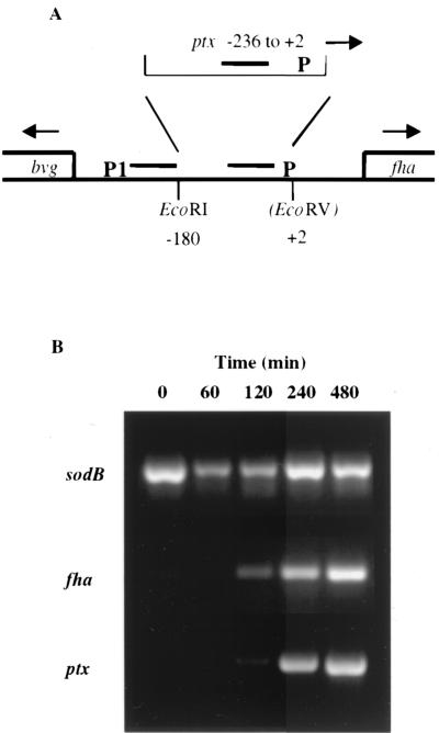
Although it is believed that the promoter regions of bvg-activated genes are responsible for the differential regulation of these genes, there is no direct in vivo evidence of this. We constructed a strain, KMC3, in which the upstream region from the early bvg-activated fha gene, including the promoter and BvgA-binding sequences (5), was replaced by the equivalent region from upstream of the late bvg-activated ptx gene (20) by allelic exchange (Fig. 1A). This placed fha under the control of the ptx promoter. Previous studies have determined that fha is expressed early following an inducing signal, while ptx is expressed late (17, 31). We used RT-PCR, as previously described (17), to analyze the activation kinetics of strain KMC3. Total RNA was prepared from cells at 0, 60, 120, 240, and 480 min. In strain KMC3, fha was expressed late, similar to ptx expression (Fig. 1B) and different from the early-induction kinetics of fha in wild-type strain Tohama I that we previously demonstrated (17). This result confirms the anticipated switch in promoter control compared to that for wild-type strain Tohama I, in which fha was expressed by 30 min (17). It also provides direct evidence that the promoter region is responsible for the differential regulation seen in vitro.
FIG. 1.
Promoter exchange activation kinetics. KMC3, a strain in which the promoter region of the early bvg-activated gene, fha, was replaced with the late bvg-activated ptx promoter region by allelic exchange, was used to determine if the promoter region is responsible for temporal transcriptional regulation. (A) Schematic diagram of the fha promoter replacement by the ptx promoter. Bars, BvgA-binding sequences; P, promoters of the respective genes. (B) RT-PCR analysis of KMC3 was used to detect transcripts of the bvg-independent standard gene (sodB) and fha and ptx at times 0, 60, 120, 240, and 480 min after induction of the Bvg system. RT-PCR products were run on ethidium bromide-stained agarose gels to determine the abundance of transcript at each time point. In wild-type strain Tohama I, fha transcription is present at 30 min after induction and maximal transcription is present at 60 min after induction (see Fig. 1 of reference 17).
Effect of fha promoter exchange on colonization of the mouse respiratory tract.
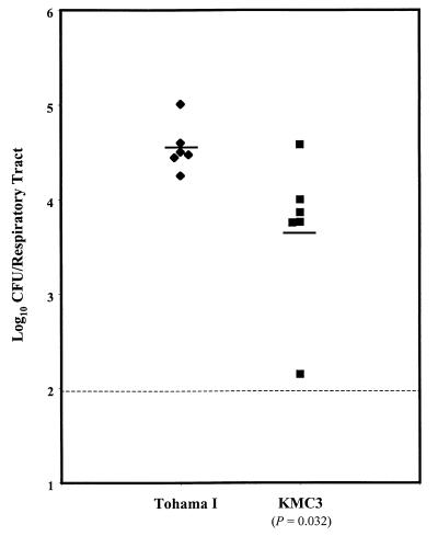
In an initial attempt to determine the effect of fha-to-ptx promoter exchange on the virulence of B. pertussis, we used an intranasal mouse model of infection and examined the ability of the mutant strain to colonize the respiratory tracts of mice. Mice were inoculated intranasally with approximately 5 × 10⁴ CFU of wild-type strain Tohama I or mutant strain KMC3 in 20 μl of PBS-CAA. After 7 days, the mice were sacrificed and the trachea and lungs were removed, homogenized, diluted, and plated to determine levels of colonization. Mice inoculated with late-expressing fha strain KMC3 had a statistically significant 86% reduction (approximately 1 log unit) (P = 0.032) in mean respiratory tract colonization compared to mice inoculated with wild-type strain Tohama I (Fig. 2).
FIG. 2.
Effect of mutation in KMC3 on colonization. Wild-type B. pertussis strain Tohama I and fha promoter mutant strain KMC3 (5 × 10⁴ CFU of each) were used to intranasally inoculate groups of six mice. There was a statistically significant reduction in the ability of the fha mutant strain to colonize the respiratory tracts of mice compared to that of wild-type strain Tohama I. The P value is shown. Bars, geometric means; dotted line, lower limit of detection.
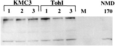
We performed Western immunoblotting of whole-cell lysates of strains Tohama I, KMC3, and NMD170 (an FHA− strain of B. pertussis) to determine the effect of our promoter alteration. Cultures were grown in triplicate and were diluted to an OD600 of 0.5. Twentyfold dilutions were analyzed by Western blotting. FHA was detected with an FHA-specific goat polyclonal antibody (obtained from Rino Rappuoli) (Fig. 3); densitometric analysis demonstrated that there was not a significant difference (P = 0.8) between levels of FHA produced by KMC3 and Tohama I. In addition, to determine that there was no effect of the presence of a duplicate copy of the ptx promoter on the production of PT in strain KMC3, we used a CHO cell clustering assay (14) and compared the levels of active PT secreted by KMC3 and Tohama I. Dilutions of supernatants (undiluted and 1:4, 1:16, and 1:64 dilutions) from triplicate cultures were assayed. There were no significant differences in clustering patterns induced by the supernatants of the wild-type and fha promoter mutant strains, indicating that there is no defect in PT production in KMC3. These data suggest that the altered expression of fha is responsible for the colonization defect of KMC3 in the intranasal mouse model of infection (Fig. 2).
FIG. 3.
Western blot analysis of FHA expression. Whole-cell lysates from three cultures each of KMC3 and Tohama I (TohI), as well as a whole-cell lysate from NMD170 (an FHA− strain of B. pertussis), were analyzed by Western immunoblotting. FHA was detected with an FHA-specific goat polyclonal antibody. The upper high-molecular-weight band, missing in NMD170, represents FHA, while the predominant lower band is presumably a cross-reacting protein. M, rainbow marker (Amersham).
Construction and analysis of activation kinetics of ptx promoter mutants.
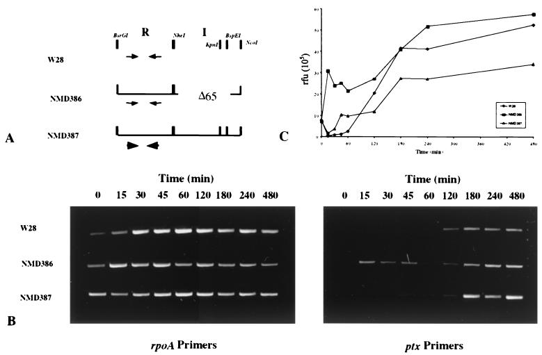
We have provided evidence that alteration of the kinetic pattern of fha expression from early to late is detrimental for optimal colonization and thus for full virulence. However, attempts to create the reciprocal strain, in which the late-activated ptx promoter is replaced by the early-activated fha promoter, resulted in a slowly growing strain that was not suitable for further study. As an alternative approach, we determined whether two strains with previously characterized ptx promoter alterations (20) were altered in their ptx activation kinetic patterns. These strains had retained promoter activities close to that of the wild type despite the ptx promoter modifications (20). NMD346 had a deletion of 65 bp in the intervening sequence, while NMD357 had a replacement of the ptx BvgA-binding heptameric inverted repeats with those of fha, but the native 10-bp sequence between the inverted repeats was maintained. These promoter alterations fused to the wild-type ptx open reading frame sequence (instead of lac) were introduced into the chromosome of B. pertussis strain W28 by allelic exchange to generate NMD386 and NMD387, respectively (Fig. 4A), the appropriate construction of which was confirmed by PCR.
FIG. 4.
RT-PCR analysis of ptx promoter mutant activation kinetics. (A) Schematic diagram of changes made to the promoter and bvg-activating regions of ptx. Changes were introduced to the chromosome of wild-type B. pertussis strain W28. NMD386 has a deletion of 65 bp in the intervening-sequence region (I), and NMD387 has a replacement of the ptx inverted repeats (arrows) with the inverted repeats of fha (arrowheads) in the repeat region (R). The inverted repeats are sites of primary BvgA binding at these promoters (20). (B) RT-PCR was used to detect transcripts of rpoA and ptx after induction of the Bvg system in wild-type B. pertussis strain W28 and the ptx promoter mutant strains, NMD386 and NMD387. The time course of induction is shown on ethidium bromide-stained agarose gels at 0, 15, 30, 45, 60, 120, 180, 240, and 480 min for the bvg-dependent ptx promoter and bvg-independent standard rpoA. Results from a typical experiment are shown. (C) Samples from the time course of induction were run on an agarose gel, stained with Vistra green, and quantitated using a FluorImager SI system from Molecular Dynamics. ⧫, W28; ■, NMD386; ▴, NMD387. The band intensities were normalized to the rpoA standard and plotted. rfu, relative fluorescence units.
In order to determine if the deletion of 65 bp within the intervening-sequence region (NMD386) and the replacement of the lower-affinity ptx BvgA-binding repeats with the higher-affinity fha repeats (NMD387) resulted in alterations in ptx activation kinetics, we again used the RT-PCR assay to detect ptx transcripts over the course of time after induction of the Bvg system. Total RNA was prepared from cells at times 0, 15, 30, 45, 60, 120, 180, 240, and 480 min. RT-PCR, with primers specific for prn, confirmed the appropriate intermediate expression of prn to verify correct modulation among the strains and confirmed that other bvg-dependent loci were not affected by the mutants (data not shown). In wild-type B. pertussis strain W28, ptx was not expressed until 2 h after induction of the Bvg system (Fig. 4B), results which are consistent with those described previously (17, 31). In B. pertussis strains NMD386 and NMD387, we saw reproducible expression of ptx at 15 and 45 min after induction, respectively (Fig. 4B). The same kinetic patterns were obtained when the band intensities were normalized to the rpoA standard and plotted (Fig. 4C). Therefore, the promoter alterations did indeed result in markedly earlier activation kinetics of ptx.
Effect of ptx promoter alterations on level of secreted PT.
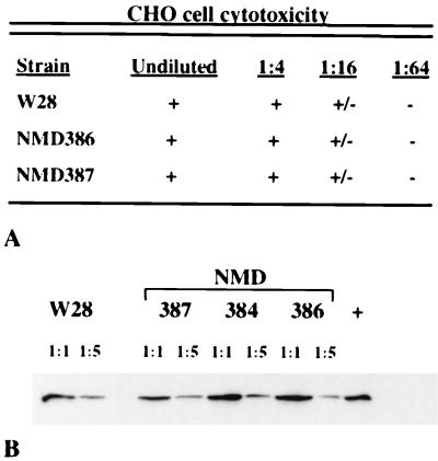
To compare the levels of active PT secreted by the strains with ptx promoter alterations with those secreted by wild-type B. pertussis W28, we used the CHO cell clustering assay described above. There were no significant differences in the overall patterns of clustering as a function of the supernatant PT concentrations of W28, NMD386, and NMD387 (Fig. 5A). This provides semiquantitative evidence that the cytotoxic activity and the levels of PT secreted by these ptx promoter mutant strains are the same as those for wild-type strain W28.
FIG. 5.
Analysis of PT expression: (A) CHO cell clustering activity of culture supernatants of strains W28, NMD386, and NMD387. (B) The levels of PT secreted by W28, NMD387, NMD384 (a strain similar to NMD386 with a 21-bp deletion in the intervening-sequence region), and NMD386 were compared by Western immunoblotting of TCA-precipitated supernatant proteins. The S1 subunit of PT was detected with monoclonal antibody X2X5. One set of supernatant proteins from cultures grown in triplicate are shown here. Densitometric analysis of bands from all cultures showed no significant difference in the level of secreted PT. +, purified PT control.
In addition, we compared the levels of PT secreted by NMD386 and NMD387 to that secreted by W28 by Western immunoblotting of TCA-precipitated supernatant proteins. Cultures were grown in triplicate, and TCA-precipitated supernatant proteins were analyzed by sodium dodecyl sulfate-polyacrylamide gel electrophoresis and transferred to nitrocellulose. The S1 subunit of PT was detected with monoclonal antibody X2X5 (courtesy of Drusilla Burns). The Western analysis of one set of the triplicate cultures is shown in Fig. 5B. NMD384, an additional B. pertussis mutant with a deletion of 21 bp in the intervening-sequence region, was not studied further due to its similarity to NMD386. Densitometric analysis demonstrated that, although there was culture-to-culture variation, there were no significant differences between the levels of PT secreted by NMD386 (P = 0.74) or NMD387 (P = 0.76) and those secreted by W28. Therefore, the ptx promoter alterations affect only the activation kinetics and not the steady-state production of PT.
Effect of ptx promoter alterations on colonization of the mouse respiratory tract.
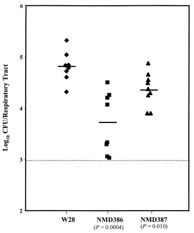
In order to determine the effect of the altered ptx activation kinetics on the virulence of B. pertussis, we used the intranasal mouse model of infection to examine the abilities of the mutant strains to colonize the respiratory tracts of mice. Eight or nine mice per group were inoculated with approximately 5 × 10⁴ CFU in 20 μl of PBS-CAA on day 0. On day 8, the mice were sacrificed and the trachea and lungs were removed, homogenized, diluted, and plated. Four to 5 days later colonies were counted and the CFU per respiratory tract were normalized to the initial inoculum level as determined by the viable counts of the inoculum (Fig. 6). Both mutant strains demonstrated a significant reduction in the ability to colonize the respiratory tracts of mice compared to wild-type strain W28. There was a 92% reduction (approximately 1 log unit) in the mean CFU per respiratory tract between W28 and NMD386 (P = 0.0004) and a 64% reduction (approximately 1/2 log unit) between W28 and NMD387 (P = 0.010) (Fig. 6). The extent of the defect in colonization correlated with the extent of alteration of promoter activation kinetics (NMD386 had the earliest ptx activation). These data indicate that the altered expression of ptx has an inhibitory effect on the ability of B. pertussis to colonize the respiratory tracts of mice. This suggests a role for differential regulation of bvg-activated genes in pathogenicity.
FIG. 6.
Colonization of mice by ptx promoter mutants. Wild-type B. pertussis strain W28 and ptx promoter mutant strains NMD386 (early in vitro ptx activation) and NMD387 (intermediate in vitro ptx activation) (5 × 10⁴ CFU of each) were intranasally inoculated into groups of eight or nine mice. P values for comparisons between the wild-type and mutant strains are shown. Bars, geometric means; dotted line, lower limit of detection.
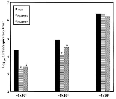
To determine the effect of the inoculation dose on the ability of the ptx promoter mutants to colonize the respiratory tracts of mice, we inoculated mice with either approximately 1 × 10⁴ or 5 × 10⁶ CFU in 20 μl of PBS-CAA and compared the results to those for the standard inoculum of 5 × 10⁴ CFU (Fig. 6). In other experiments, we found that a dose of 104 CFU is close to the minimum needed to consistently produce an infection by this protocol. At this lower dose, colonization by both NMD386 and NMD387 strains was significantly lower than that by W28 (P = 0.013 and 0.027, respectively) (Fig. 7). In fact, NMD387 showed a 1-log-unit decrease in the mean CFU per respiratory tract at the low dose compared to only a 1/2-log-unit decrease at the standard inoculum (Fig. 7). However, the virulence defect of the ptx promoter mutants appears to be overcome at the high-dose inoculum (Fig. 7). This indicates that the phenotype of the ptx promoter mutants is dose dependent.
FIG. 7.
Effect of inoculation dose on colonization by ptx promoter mutants. Groups of six to eight mice were inoculated with approximately 1 × 10⁴, 5 × 10⁴, and 5 × 10⁶ CFU of wild-type B. pertussis strain W28 and the ptx promoter mutant strains, NMD386 and NMD387. Mean values are plotted. Asterisk, statistically significant difference between the wild-type and mutant strains within a dose group.
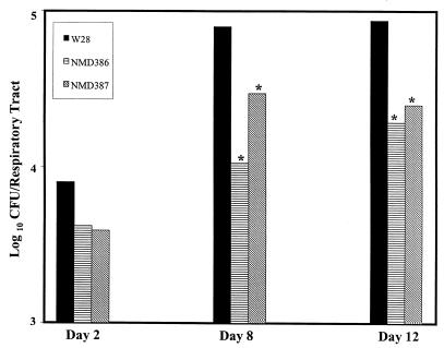
To determine if the reduction in colonization by the ptx promoter mutant is affected by the time of harvest of trachea and lungs during the course of infection, we examined the CFU per respiratory tract at 2, 8 (standard protocol), and 12 days after inoculation. There were no significant differences (P = 0.4) in the numbers of bacteria (approximately 25% of the inoculum) delivered to the respiratory tracts of mice inoculated with NMD386 (1.15 × 10⁵ CFU) versus W28 (1.27 × 10⁵ CFU) at 1 h after inoculation. Colonization at the 1-h time point was not determined for NMD387. At 2 days following inoculation, although there was not a statistically significant difference between the wild-type and mutant strains due to greater variation at lower infection levels, the pattern of reduced colonization was already apparent (Fig. 8). As described previously (Fig. 6), at 8 days following inoculation there was a statistically significant reduction in the ability of NMD386 and NMD387 to colonize the respiratory tracts of infected mice. At 12 days following infection, the virulence defect of the ptx promoter mutants, NMD386 (P = 0.019) and NMD387 (P = 0.012), was still apparent (Fig. 8). These data suggest that the pattern of reduced colonization by the ptx promoter mutants is manifested early and maintained for at least 12 days into the infection. The effect on persistence beyond day 12 was not examined.
FIG. 8.
Time course of colonization. Groups of six to eight mice were intranasally inoculated with 5 × 10⁴ CFU of wild-type B. pertussis strain W28 or the ptx promoter mutant strains, NMD386 and NMD387. After 2, 8, and 12 days the trachea and lungs of the mice were harvested and plated to determine CFU. Mean values are plotted. Asterisk, statistically significant difference between the wild-type and mutant strains within a time point.
Effect of addition of PT to B. pertussis inoculum on colonization.
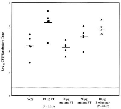
The above data indicate that the altered expression of ptx has a negative effect on the ability of B. pertussis to colonize the respiratory tracts of intranasally infected mice. A recent report indicated that when a human bronchial epithelial cell line was preincubated with increasing concentrations of purified PT, washed, and then incubated with B. pertussis strains, there was a dose-dependent reduction of adherence of the bacteria to the bronchial cells (38). These data provide evidence that PT can inhibit the adherence of B. pertussis to epithelial cells, which led us to the hypothesis that the virulence defect demonstrated by the ptx promoter mutants could be an effect of the altered expression of ptx resulting in inhibition of initial bacterial adherence to the respiratory tract by PT. To test this hypothesis, we determined whether the addition of purified PT to the inoculum of wild-type B. pertussis might mimic the virulence defect of the ptx promoter mutants, NMD386 and NMD387, by interfering with initial adherence. We therefore inoculated mice with 5 × 10⁴ CFU of B. pertussis W28 resuspended in buffer alone or with 1 or 10 μg of purified PT added. After 7 days, the mice were sacrificed and the trachea and lungs were removed, homogenized, diluted, and plated. Surprisingly, the addition of 1 and 10 μg of purified wild-type PT resulted in statistically significant 7-fold (not shown) and 10-fold (Fig. 9) increases, respectively, in the level of B. pertussis colonization of the respiratory tracts of the infected mice.
FIG. 9.
Effect on colonization of addition of purified PT to inoculum. Ten to 20 μg of purified holotoxin, mutant holotoxin, or the B oligomer of PT was added to 5 × 10⁴ CFU of wild-type B. pertussis strain W28 and intranasally inoculated into groups of five mice. Significant P values from comparisons to the no-toxin group (W28) are shown. Bars, geometric means; dotted line, lower limit of detection.
We hypothesized that this result could be due to the toxic activity of PT overwhelming the immune system and overcoming any inhibitory effect it may have on virulence. To test this, we analyzed the effect of coadministering, with the bacteria, inactive PT or the binding B oligomer, which lacks the ADP-ribosylating activity of PT but which could still bind and potentially inhibit the adherence of B. pertussis. We inoculated mice with 5 × 10⁴ CFU of W28 in buffer alone or with 10 μg of purified wild-type PT holotoxin, 10 or 20 μg of purified inactive PT holotoxin, or 10 μg of purified PT B oligomer added. After 7 days, the mice were sacrificed and the trachea and lungs were removed, homogenized, diluted, and plated. The CFU per respiratory tracts are shown in Fig. 9. The addition of either 10 μg of purified wild-type PT or 10 μg of purified B oligomer resulted in statistically significant increases in the level of B. pertussis colonization of the respiratory tracts of the infected mice (P = 0.013 and 0.016, respectively). The colonization seen when either 10 or 20 μg of mutant PT was added to the inoculum was not significantly different from that seen with no added toxin (Fig. 9). Although these data did not provide evidence to support an inhibitory role for the early presence of PT in the reduced ability of B. pertussis to colonize the respiratory tracts of mice, it does verify the role of PT as a virulence factor for B. pertussis.
Role of modulation in colonization.
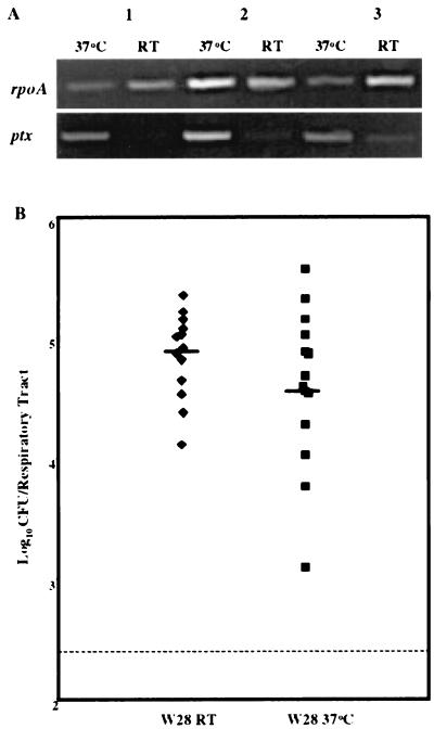
In order for the early expression of ptx to have an effect on the virulence of B. pertussis, we hypothesized that, at some point during the course of infection, down-regulation or modulation of ptx expression would be required. Using RT-PCR analysis, as described previously, we investigated whether the time of preparation of the bacterial inoculum (approximately 45 min to 1 h at room temperature in our standard protocol) was enough to down-regulate the expression of ptx. B. pertussis W28 cells were resuspended in PBS-CAA and maintained either at room temperature or at 37°C for 1 h, after which total RNA was isolated from both sets of cells for RT-PCR analysis with primers specific for bvg-independent gene rpoA and for ptx. The experiment was repeated three times, and the RT-PCR results are shown in Fig. 10A. The room temperature and 37°C ptx products were run on an agarose gel, stained with Vistra green, and normalized to the rpoA standard. An average 84% reduction in expression between the 37°C and room temperature ptx products was observed. This indicates that room temperature, a known modulator of the BvgAS system, is able to down-regulate expression of ptx within 1 h, the average time between removal of B. pertussis cells from the 37°C incubator for preparation of inocula and intranasal inoculation of mice.
FIG. 10.
Effect of preinoculation temperature on ptx expression and W28 colonization. (A) RT-PCR was used to detect transcripts of the bvg-independent rpoA gene and the bvg-dependent ptx gene after 1 h of incubation of resuspended W28 cells at either 37°C or room temperature (RT). Results of three independent experiments are shown on ethidium bromide-stained agarose gels. Normalization of the ptx products to the rpoA standard indicated an average 84% decrease in ptx expression between 37°C and room temperature incubations. (B) Wild-type B. pertussis strain W28 (5 × 10⁴) preincubated for 1 h at either room temperature or at 37°C was intranasally inoculated into groups of seven mice. Bars, geometric means; dotted line, lower limit of detection.
To examine the effect of this preinoculation down-regulation of ptx by our standard inoculation procedure, we inoculated two groups of mice with W28, one with an inoculum that was maintained at room temperature for 1 h prior to inoculation (standard protocol) and one with an inoculum that was maintained at 37°C for 1 h prior to inoculation. There was no statistically significant difference between the ability of the bacteria that were maintained at 37°C and that of the bacteria maintained at room temperature to colonize the respiratory tracts of mice (Fig. 10B). This suggests that, although modulation may occur during our standard procedure, it is not necessary for full virulence of B. pertussis W28 in mice.
DISCUSSION
In this study we have provided evidence that alterations to the promoter regions of bvg-activated genes can alter the kinetic patterns of expression of these genes. In addition, B. pertussis strains containing these promoter alterations demonstrate a reduction in the ability to colonize respiratory tracts in an intranasal mouse model of infection, i.e., a virulence defect. In particular, the altered regulation of the characteristically early-activated fha promoter and that of the characteristically late-activated ptx promoter both result in approximately a 1-log-unit reduction in the numbers of bacteria colonizing the respiratory tracts of mice. These data suggest a role for differential regulation of bvg-activated genes, and therefore for the BvgAS regulatory system, in the virulence of B. pertussis.
Changes as substantial as the replacement of the entire fha promoter region with the ptx promoter region, as well as more-subtle changes such as the deletion of 65 bp of the intervening-sequence region and the alteration of 4 bp in the heptameric inverted-repeat sequence in the ptx promoter region, resulted in altered kinetic patterns of expression of fha and ptx, respectively. This provides further evidence that the promoter regions of bvg-activated genes are responsible for the differential regulation seen in vitro. The early expression of ptx in NMD386, the strain with the deletion of the 65-bp intervening-sequence region, further supports the cooperative binding model of activation by BvgA at the ptx promoter put forth by Boucher and Stibitz (6). In this model, supported by biochemical and genetic data, phosphorylated BvgA binds first to a site which includes the heptameric inverted repeats. Subsequent cooperative binding of BvgA dimers along the intervening sequence between the initial binding site and the promoter allows BvgA to interact with RNA polymerase to promote transcription activation (6, 20). Although DNase I footprinting analysis will have to be performed to confirm this, we expect from the altered kinetic pattern that this mutant no longer requires the extensive cooperative binding, and thus the high concentration of BvgA, that is needed for activation at the wild-type ptx promoter and is therefore activated earlier (at lower BvgA concentrations) after an inducing signal. The intermediate kinetics of ptx in NMD387, in which the ptx heptameric inverted repeats are replaced by the fha inverted repeats, suggests that higher-affinity binding of BvgA to the primary binding sites of bvg-activated promoters also results in faster transcriptional activation. We believe that the altered activation kinetics of these strains supports the hypothesis that a combination of the affinity of BvgA for the primary binding site and the distance between this site and the core promoter sequences at bvg-activated promoters is primarily responsible for the differential regulation of bvg-activated genes. By using variations in the bvg-activating regions of genes in the Bvg regulon, B. pertussis appears to have optimized its virulence or colonization abilities through differential regulation of at least some of its virulence genes using the products of its two-component signal transduction system.
The above data suggest a mechanism for B. pertussis to be able to respond to subtle changes in the environment and control the expression of its positively regulated virulence factors accordingly. However, a role for differential regulation in the pathogenicity of Bordetella remains elusive. The Bvg+ phase is necessary and sufficient for colonization of animal respiratory tracts (11, 21), while the Bvg− phase appears not to have a role in virulence and may even be detrimental (2, 21, 23). These data suggest that there is no role for the Bvg− phase in vivo and that one function of the bvg locus in pathogenicity is repression of the bvg-repressed genes. However, there is some indication that the Bvgi phase of B. pertussis is expressed in vivo (21). We believed that if regulation by the BvgAS system is important for pathogenicity, changing the regulatory circuit between classes of bvg-activated promoters should affect virulence. We hypothesized that, by making changes to the promoter regions of bvg-activated genes, we could alter their patterns of regulation, manifested as a change in activation kinetics. Furthermore, we hypothesized that, if the differential regulation characterized in vitro has a role in the pathogenicity of B. pertussis, exchanging the regulatory control between classes of promoters should decrease the virulence of the organism.
Our analysis of B. pertussis mutants altered in their kinetic patterns of expression of fha and ptx indicates that the altered regulation of bvg-activated virulence factors reduces colonization of the mouse respiratory tract. In particular, ptx promoter mutants NMD386 and NMD387 demonstrate a statistically significant reduction in the ability to colonize the respiratory tracts of intranasally infected mice. Our data indicate that this colonization defect is dose dependent and that the pattern of reduced colonization is apparent throughout the course of a 12-day infection. Although the reduction of colonization is not statistically significant at the 2-day time point, the pattern of reduced colonization appears to be already established. Our data strongly suggest that the colonization defect of NMD386 and NMD387 is an effect of the altered regulation of ptx expression in these strains, as there are no significant differences in the steady-state levels of transcriptional activity or expression of PT between the ptx promoter mutant strains and wild-type strain W28. The colonization defect demonstrated by KMC3, the fha-to-ptx promoter replacement strain, also appears to be an effect of the altered regulation of fha expression.
We contend that, at some point prior to or during the course of infection, at least partial modulation of Bvg activity in B. pertussis would have to occur in order for the altered regulatory control in our mutants to have a deleterious effect. This could occur either in the respiratory tract of the host or prior to encounter with the host. It is possible that modulation occurs throughout the course of infection as the organism is exposed to different microenvironments in the host's respiratory tract. The immune response to the recently identified Bvgi phase factors provides evidence to support this (21). Our data indicate that 1 h at room temperature is sufficient to reduce ptx transcription by an average of 84%. However, our results also indicate that the maintenance of the wild-type B. pertussis inoculum at 37°C, thus inhibiting the ability of the Bvg system to modulate its virulence factors prior to inoculation, does not significantly affect the ability of the organism to colonize the respiratory tracts of intranasally infected mice. It remains possible that differential regulation is required during the course of infection within the host for optimal virulence of B. pertussis rather than prior to encounter with the host. Characterization of differential expression of bvg-activated genes throughout the course of infection would have to be examined to address this. Furthermore, we cannot rule out a role for differential regulation in the course of transmission between infected individuals during which B. pertussis may remain aerosolized for considerable periods of time (1). It is possible that an effect of the preinoculation modulation may be more dramatic at either an earlier or later harvest time point. Use of the aerosol challenge mouse model may also offer further insight into the role of differential regulation in transmission as it may provide a more relevant model of exposure to the organism and the early stages of infection. It is possible that the virulence defects of our regulatory mutants may be magnified using this model.
A recent study by van den Berg et al. (38) provided a possible explanation as to why the early presence of PT might have an inhibitory effect on the ability of B. pertussis to colonize the respiratory tracts of mice. This report indicated that, despite previous accounts that PT has a role as an adherence factor (36), PT does not augment B. pertussis adherence to cultured human cells and, in fact, can even inhibit B. pertussis adherence in vitro (38). However, we did not detect a dose-dependent inhibitory effect of added purified PT in our intranasal mouse model of infection to support this as a possible reason for the reduced ability of our ptx promoter mutants to colonize. It is possible that the addition of lower concentrations of the purified toxins or B oligomer may have demonstrated an inhibitory effect without enhancing bacterial virulence (presumably by overwhelming the immune system of the mice). However, the artificial addition of PT to the inoculum is probably quite different from B. pertussis production of PT and the local concentrations of bacteria and toxins present in a natural infection.
One can speculate that the complexity of an organism's regulatory mechanisms may correlate with the complexity of the bacterium's life cycle. It is possible that the adaptability afforded to B. pertussis by its complex regulatory components and phosphotransfer steps enables the bacterium to respond to a series of changing microenvironments throughout the course of infection. We demonstrate that B. pertussis utilizes differences in the promoters of its bvg-activated virulence factors to allow for differential expression of these genes using only the BvgAS system. Furthermore, we provide evidence that this differential regulation is required for optimal virulence. We believe that the temporal expression demonstrated in vitro likely reflects the sensitivity of a particular bvg-activated promoter to modulatory signals. This is consistent with the requirement for higher concentrations of transcriptional activator BvgA at the late-acting promoters (31). In further support of this idea, we have recently found that the intermediately activated prn promoter (17) is most transcriptionally active at intermediate levels of modulator MgSO4 (S. M. Kinnear and N. H. Carbonetti, unpublished data).
Two possible hypotheses may explain the role of differential regulation of bvg-activated genes during the course of infection. The first is that temporal regulation of virulence gene expression is important for colonization and the resulting infection. Incorporating our data, a possible model to explain the role of temporal regulation of bvg-activated virulence factors is as follows. Following a coughing paroxysm of a pertussis patient, the aerosolized organisms are exposed to the lower temperature of the environment and the BvgAS system is thereby modulated or down-regulated. As the aerosol droplet reaches the nasal passages of the next host, the least-sensitive fha promoter is either still on or is immediately activated and the intermediately sensitive prn promoter is transcribed at its highest level to facilitate the ability of FHA to colonize the respiratory tract. It has been proposed that pertactin (PRN) acts as an accessory adherence factor, perhaps in conjunction with FHA. PRN may be important as a scaffolding or support component of an adhesin complex (27, 28, 29). Our data indicate that, if there is some modulation of the BvgAS system in the time between aerosolization of the organism and transmission to the next host, prn expression would be at its peak. At the same time, the most sensitive ptx promoter is turned off so as not to have an inhibitory effect on initial adherence.
An alternative hypothesis is that differential regulation of bvg-activated virulence gene expression occurs in response to spatial rather than temporal cues. This hypothesis suggests that in vivo microenvironments differ in their modulatory signals. Some microenvironments may present cues that are perceived as semimodulating and thus would induce expression of adhesins (such as FHA and PRN) but repress toxin expression. Other microenvironments may be nonmodulating and thus would induce expression of both adhesins and toxins. From previous data (21, 23), it seems unlikely that there are fully modulating in vivo microenvironments that would inhibit the expression of the bvg-activated factors and induce the expression of the bvg-repressed factors. According to this hypothesis of spatial cues, virulence genes would be differentially regulated depending on the location of the bacteria within the host respiratory tract to result in optimal infection. The two hypotheses are not necessarily mutually exclusive, since the location of bacteria within a host may be dependent to some extent on the time after the first encounter with the host.
An additional, more trivial possibility is that the differences in colonization levels between the wild-type and mutant strains are due to differences in the steady-state expression levels of FHA and PT rather than differences in regulatory control. However densitometric analysis of Western blots and cytotoxicity assays for PT activity indicated that there were no significant differences in the levels of FHA and PT expressed between the strains in vitro (Fig. 3 and 5, respectively). While it may seem surprising that merely altering the regulatory pattern of expression of FHA should have a measurable effect on virulence when it has been demonstrated that FHA− mutants were not affected in virulence in a mouse model (39), the steady-state lack of a virulence factor is different than an alteration of regulatory patterns, as in our mutants. The permanent lack of FHA may be compensated for by an alternative adherence factor in the animal model, whereas altered regulation of FHA may result in inappropriate temporal or spatial expression and interference with optimal bacterium-host interactions for infection. Although we cannot rule out a possible difference in expression levels in vivo, we believe this to be an unlikely cause of the virulence defect in our mutant strains and therefore favor either the temporal or spatial hypotheses presented. The ability of the BvgAS system to be sensitive to small changes in environmental signals via the complexity of its phosphorelay together with differences in the promoter regions of genes in its regulon may allow B. pertussis to respond to the intricacy of its life cycle through the efficiency of one regulatory system.
ACKNOWLEDGMENTS
We thank Kyle McKenna for the construction of KMC3; Monica Castro and Uli McNamara for technical assistance; Abdu Azad and Suzana Radulovic for mice; Abdu Azad, Drusilla Burns, and Rino Rappuoli for reagents; Alla Romashko and the University of Maryland Animal Facility for assistance with mouse experiments; and Colin O'Connell for help with figures. We also thank Steve Wasserman for advice on statistics and Jim Kaper and Jim Nataro for critical reading of the manuscript. We are also grateful to both anonymous reviewers, whose painstakingly thorough critiques of the manuscript helped to improve it significantly.
This work was supported by NIH grants AI32946 and AI38979.
REFERENCES
- 1.Aintablian N, Walpita P, Sawyer M H. Detection of Bordetella pertussis and respiratory syncytial virus in air samples from hospital rooms. Infect Control Hosp Epidemiol. 1998;19:918–923. doi: 10.1086/647764. [DOI] [PubMed] [Google Scholar]
- 2.Akerley B J, Cotter P A, Miller J F. Ectopic expression of the flagellar regulon alters development of the Bordetella-host interaction. Cell. 1995;80:611–620. doi: 10.1016/0092-8674(95)90515-4. [DOI] [PubMed] [Google Scholar]
- 3.Arico B, Miller J F, Roy C R, Stibitz S, Monack D, Falkow S, Gross R, Rappuoli R. Sequences required for expression of Bordetella pertussis virulence factors share homology with prokaryotic signal transduction proteins. Proc Natl Acad Sci USA. 1989;86:6671–6675. doi: 10.1073/pnas.86.17.6671. [DOI] [PMC free article] [PubMed] [Google Scholar]
- 4.Bartoloni A, Pizza M, Bigio M, Nucci D, Ashworth L A, Irons L I, Robinson A, Burns D, Manclark C, Sato H, Rappuoli R. Binding of a protective epitope of pertussis toxin by in vitro refolding of recombinant fragments. Bio/Technology. 1988;6:709–712. [Google Scholar]
- 5.Boucher P E, Murakami K, Ishihama A, Stibitz S. Nature of DNA binding and RNA polymerase interaction of the Bordetella pertussis BvgA transcriptional activator at the fha promoter. J Bacteriol. 1997;179:1755–1763. doi: 10.1128/jb.179.5.1755-1763.1997. [DOI] [PMC free article] [PubMed] [Google Scholar]
- 6.Boucher P E, Stibitz S. Synergistic binding of RNA polymerase and BvgA phosphate to the pertussis toxin promoter of Bordetella pertussis. J Bacteriol. 1995;177:6486–6491. doi: 10.1128/jb.177.22.6486-6491.1995. [DOI] [PMC free article] [PubMed] [Google Scholar]
- 7.Boucher P E, Menozzi F D, Locht C. The modular architecture of bacterial response regulators: insights into the activation mechanism of the BvgA transactivator of Bordetella pertussis. J Mol Biol. 1994;241:363–377. doi: 10.1006/jmbi.1994.1513. [DOI] [PubMed] [Google Scholar]
- 8.Carbonetti N H, Irish T J, Chen C H, O'Connell C B, Hadley G A, McNamara U, Tuskan R G, Lewis G K. Intracellular delivery of a cytolytic T-lymphocyte epitope peptide by pertussis toxin to major histocompatibility complex class I without involvement of the cytosolic class I antigen processing pathway. Infect Immun. 1999;67:602–607. doi: 10.1128/iai.67.2.602-607.1999. [DOI] [PMC free article] [PubMed] [Google Scholar]
- 9.Carbonetti N H, Fuchs T M, Patamawenu A A, Irish T J, Deppisch H, Gross R. Effect of mutations causing overexpression of RNA polymerase α subunit on regulation of virulence factors in Bordetella pertussis. J Bacteriol. 1994;176:7267–7273. doi: 10.1128/jb.176.23.7267-7273.1994. [DOI] [PMC free article] [PubMed] [Google Scholar]
- 10.Cotter P A, Miller J F. A mutation in the Bordetella bronchiseptica bvgS gene results in reduced virulence and increased resistance to starvation, and identifies a new class of Bvg-regulated antigens. Mol Microbiol. 1997;24:671–685. doi: 10.1046/j.1365-2958.1997.3821741.x. [DOI] [PubMed] [Google Scholar]
- 11.Cotter P A, Miller J F. BvgAS-mediated signal transduction: analysis of phase-locked regulatory mutants of Bordetella bronchiseptica in a rabbit model. Infect Immun. 1994;62:3381–3390. doi: 10.1128/iai.62.8.3381-3390.1994. [DOI] [PMC free article] [PubMed] [Google Scholar]
- 12.DeShazer D, Bannan J D, Moran M J, Friedman R L. Characterization of the gene encoding superoxide dismutase of Bordetella pertussis and construction of a SOD-deficient mutant. Gene. 1994;142:85–89. doi: 10.1016/0378-1119(94)90359-x. [DOI] [PubMed] [Google Scholar]
- 13.Gross R, Rappuoli R. Positive regulation of pertussis toxin expression. Proc Natl Acad Sci USA. 1988;85:3913–3917. doi: 10.1073/pnas.85.11.3913. [DOI] [PMC free article] [PubMed] [Google Scholar]
- 14.Hewlett E L, Sauer K T, Meyers G A, Cowell J L, Guerrant R L. Induction of a novel morphological response in Chinese hamster ovary cells by pertussis toxin. Infect Immun. 1983;40:1198–1230. doi: 10.1128/iai.40.3.1198-1203.1983. [DOI] [PMC free article] [PubMed] [Google Scholar]
- 15.Kasuga T, Nakase Y, Ukishima K, Takatsu K. Studies on Haemophilus pertussis. Relation between the phase of bacilli and the progress of the whooping cough. Kitasato Arch Exp Med. 1954;27:57–62. [PubMed] [Google Scholar]
- 16.Kimura A, Mountzouros K T, Relman D A, Falkow S, Cowell J L. Bordetella pertussis filamentous hemagglutinin: evaluation as a protective antigen and colonization factor in a mouse respiratory infection model. Infect Immun. 1990;58:7–16. doi: 10.1128/iai.58.1.7-16.1990. [DOI] [PMC free article] [PubMed] [Google Scholar]
- 17.Kinnear S M, Boucher P E, Stibitz S, Carbonetti N H. Analysis of BvgA activation of the pertactin gene promoter in Bordetella pertussis. J Bacteriol. 1999;181:5234–5241. doi: 10.1128/jb.181.17.5234-5241.1999. [DOI] [PMC free article] [PubMed] [Google Scholar]
- 18.Knapp S, Mekalanos J. Two trans-acting regulatory genes (vir and mod) control antigenic modulation in Bordetella pertussis. J Bacteriol. 1988;170:5059–5066. doi: 10.1128/jb.170.11.5059-5066.1988. [DOI] [PMC free article] [PubMed] [Google Scholar]
- 19.Leininger E, Roberts M, Kenimer J G, Charles I G, Fairweather N, Novotny P, Brennan M J. Pertactin, an Arg-Gly-Asp-containing Bordetella pertussis surface protein that promotes adherence to mammalian cells. Proc Natl Acad Sci USA. 1991;88:345–349. doi: 10.1073/pnas.88.2.345. [DOI] [PMC free article] [PubMed] [Google Scholar]
- 20.Marques R R, Carbonetti N H. Genetic analysis of pertussis toxin promoter activation in Bordetella pertussis. Mol Microbiol. 1997;24:1215–1224. doi: 10.1046/j.1365-2958.1997.4371792.x. [DOI] [PubMed] [Google Scholar]
- 21.Martinez de Tejeda G, Cotter P A, Heininger U, Camilli A, Akerley B J, Mekalanos J J, Miller J F. Neither the Bvg− phase nor the vrg6 locus of Bordetella pertussis is required for respiratory infection in mice. Infect Immun. 1998;66:2762–2768. doi: 10.1128/iai.66.6.2762-2768.1998. [DOI] [PMC free article] [PubMed] [Google Scholar]
- 22.Masure H R. Modulation of adenylate cyclase toxin production as Bordetella pertussis enters human macrophages. Proc Natl Acad Sci USA. 1992;89:6521–6525. doi: 10.1073/pnas.89.14.6521. [DOI] [PMC free article] [PubMed] [Google Scholar]
- 23.Merkel T J, Stibitz S, Keith J M, Leef M, Shahin R. Contribution of regulation by the bvg locus to respiratory infection of mice by Bordetella pertussis. Infect Immun. 1998;66:4367–4373. doi: 10.1128/iai.66.9.4367-4373.1998. [DOI] [PMC free article] [PubMed] [Google Scholar]
- 24.Merkel T J, Stibitz S. Identification of a locus required for the regulation of bvg-repressed genes in Bordetella pertussis. J Bacteriol. 1995;177:2727–2736. doi: 10.1128/jb.177.10.2727-2736.1995. [DOI] [PMC free article] [PubMed] [Google Scholar]
- 25.Pizza M, Covacci A, Bartoloni A, Perugini M, Nencioni L, DeMagistris M T, Villa L, Nucci D, Manetti R, Bugnoli M, Giovannoni F, Olivieri R, Barbieri J T, Sato H, Rappuoli R. Mutants of pertussis toxin suitable for vaccine development. Science. 1989;246:497–500. doi: 10.1126/science.2683073. [DOI] [PubMed] [Google Scholar]
- 26.Rappuoli R, Arico B, Scarlato V. Thermoregulation and reversible differentiation in Bordetella: a model for pathogenic bacteria. Mol Microbiol. 1992;6:2209–2211. doi: 10.1111/j.1365-2958.1992.tb01395.x. [DOI] [PubMed] [Google Scholar]
- 27.Relman D A, Falkow S. A molecular perspective of microbial pathogenicity. In: Mandell G L, Douglas R E, Bennett E, editors. Principles and practice of infectious diseases. 4th ed. New York, N.Y: Churchill Livingstone; 1994. pp. 19–29. [Google Scholar]
- 28.Relman D A. Bordetella pertussis: determinants of virulence. In: Moss J, Iglewski B, Vaughn M, Tu A T, editors. Bacterial toxins and virulence factors in disease. M. Dekker, New York, N.Y. 367–405. 1995. pp. 367–405. [Google Scholar]
- 29.Roberts M, Fairweather N F, Leininger E, Pickard D, Hewlett E L, Robinson A, Hayward C, Dougan G, Charles I G. Construction and characterization of Bordetella pertussis mutants lacking the vir-regulated P.69 outer membrane protein. Mol Microbiol. 1991;5:1393–1404. doi: 10.1111/j.1365-2958.1991.tb00786.x. [DOI] [PubMed] [Google Scholar]
- 30.Robinson A L, Ashworth A E, Baskerville A, Irons L I. Proceedings of the 4th International Symposium on Pertussis. Dev Biol Stand. 1985;61:165–172. [PubMed] [Google Scholar]
- 31.Scarlato V, Prugnola A, Arico B, Rappuoli R. Positive transcriptional feedback at the bvg locus controls expression of virulence factors in Bordetella pertussis. Proc Natl Acad Sci USA. 1990;87:6753–6757. doi: 10.1073/pnas.87.17.6753. [DOI] [PMC free article] [PubMed] [Google Scholar]
- 32.Simon R, Priefer U, Pühler A. A broad host range mobilization system for in vivo genetic engineering: transposon mutagenesis in gram negative bacteria. Bio/Technology. 1983;1:784–791. [Google Scholar]
- 33.Stainer D W, Scholte M J. A simple chemically defined medium for the production of phase I Bordetella pertussis. J Gen Microbiol. 1970;63:211–220. doi: 10.1099/00221287-63-2-211. [DOI] [PubMed] [Google Scholar]
- 34.Steffen P, Goyard S, Ullmann A. Phosphorylated BvgA is sufficient for transcriptional activation of virulence-regulated genes in Bordetella pertussis. EMBO J. 1996;15:102–109. [PMC free article] [PubMed] [Google Scholar]
- 35.Stibitz S, Yang M-S. Subcellular location and immunological detection of proteins encoded by the vir locus of Bordetella pertussis. J Bacteriol. 1991;173:4288–4296. doi: 10.1128/jb.173.14.4288-4296.1991. [DOI] [PMC free article] [PubMed] [Google Scholar]
- 36.Tuomanen E, Weiss A. Characterization of two adhesins of Bordetella pertussis for human ciliated respiratory-epithelial cells. J Infect Dis. 1985;152:118–125. doi: 10.1093/infdis/152.1.118. [DOI] [PubMed] [Google Scholar]
- 37.Uhl M A, Miller J F. Integration of multiple domains in a two-component sensor protein: the Bordetella pertussis BvgAS phosphorelay. EMBO J. 1996;15:1028–1036. [PMC free article] [PubMed] [Google Scholar]
- 38.van den Berg B M, Beekhuzuizen H, Willems R J L, Mooi F R, van Furth R. Role of Bordetella pertussis virulence factors in adherence to epithelial cell lines derived from the human respiratory tracts. Infect Immun. 1999;67:1056–1062. doi: 10.1128/iai.67.3.1056-1062.1999. [DOI] [PMC free article] [PubMed] [Google Scholar]
- 39.Weiss A A, Goodwin M S. Lethal infection by Bordetella pertussis mutants in the infant mouse model. Infect Immun. 1989;57:3757–3764. doi: 10.1128/iai.57.12.3757-3764.1989. [DOI] [PMC free article] [PubMed] [Google Scholar]


