Endoscopic ultrasound (EUS)-guided gallbladder drainage (GBD) is an effective rescue therapy for biliary drainage after failed endoscopic retrograde cholangiopancreatography (ERCP) for unresectable malignant distal bile duct obstruction 1 . The use of the electrocautery-enhanced lumen-apposing metal stent (EC-LAMS) has been demonstrated to be safe and highly effective for EUS-GBD, and facilitates a single-step procedure 2 .
An 83-year-old man was admitted to our hospital with malignant distal biliary obstruction. Repeated attempts at ERCP were unsuccessful. Percutaneous transhepatic gallbladder drainage (PTGD) was performed following a complication of biliary peritonitis. The patient was deemed a poor surgical candidate due to advanced age and cardiorespiratory comorbidities. EUS-guided biliary drainage (EUS-BD) was not feasible considering the limited diameter of the bile duct after PTGD drainage. Therefore, EUS-GBD was performed using EC-LAMS ( Video 1 ).
Video 1 Endoscopic salvage treatment of contralateral gallbladder perforation caused by the electrocautery-enhanced lumen-apposing metal stent for endoscopic ultrasound-guided gallbladder drainage.
Under EUS guidance, the EC-LAMS (Hot Axios, 15 mm × 10 mm in diameter; Boston Scientific, Marlborough, Massachusetts, USA) was used to puncture the neck of the gallbladder via the transduodenal route. However, the endosonographic view was obscured due to coagulation artefacts, and the contralateral gallbladder neck was also accidentally penetrated by the electrocautery tip ( Fig. 1 ), resulting in the perforation of the gallbladder, bile leakage, and extraluminal bleeding.
Fig. 1.
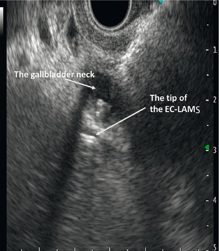
The contralateral gallbladder neck was accidentally penetrated by the electrocautery tip of the lumen-apposing metal stent (EC-LAMS).
The EC-LAMS was pulled back into the gallbladder immediately and deployed in the correct position. A dilation balloon was used to dilate the EC-LAMS up to 11.5 mm ( Fig. 2 ), which allowed a therapeutic gastroscope (GIF-Q260J; Olympus, Tokyo, Japan) to pass through the stent into the gallbladder. Endoscopic inspection revealed blood clots in the gallbladder ( Fig. 3 ), obscuring the endoscopic view. An endoscopic snare was used to remove the blood clots, exposing the perforation of the gallbladder wall ( Fig. 4 ). Endoscopic closure of the perforation was performed using through-the-scope clips ( Fig. 5 ).
Fig. 2.
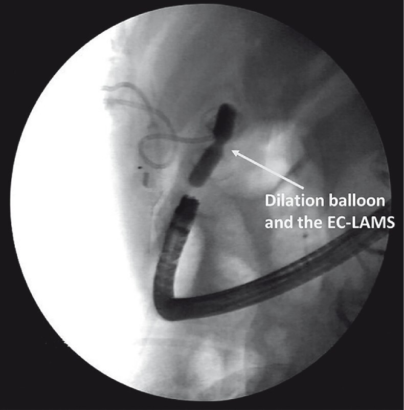
A dilation balloon was used to dilate the electrocautery-enhanced lumen-apposing metal stent (EC-LAMS) up to 11.5 mm, which allowed a therapeutic gastroscope to pass through the stent into the gallbladder.
Fig. 3.
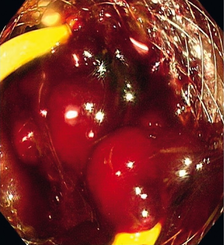
Endoscopic view of blood clots in the gallbladder.
Fig. 4.
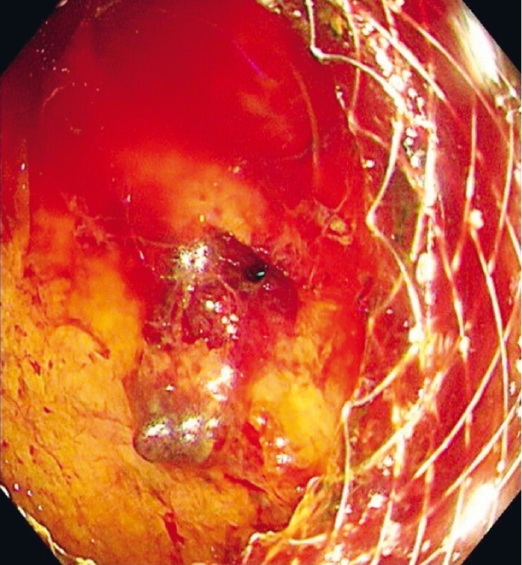
An endoscopic snare was used to remove the blood clots, exposing the perforation of the gallbladder wall.
Fig. 5.
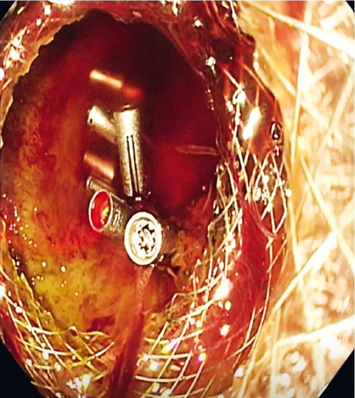
Endoscopic closure of the perforation was performed using through-the-scope clips.
After the procedure, the patient developed abdominal bleeding and peritonitis, and received percutaneous peritoneal drainage, blood transfusions (4 units, suspension of red blood cells), and antibiotics. Percutaneous peritoneal drainage decreased gradually, and abdominal pain was relieved after conservative treatment without laparotomy. The patient was discharged after removing the PTGD and abdominal drainage tube 7 days later.
Adverse events following EUS-GBD include bleeding, stent migration, capnoperitoneum, and stent occlusion, with the reporting rates between 8 % and 18 % 3 . The current case illustrates a rare adverse event of EUS-GBD. Perforation of the contralateral gallbladder wall was caused by the electrocautery tip of the EC-LAMS during EUS-GBD. Coagulation artefacts may obscure the endosonographic view even if pure cutting current is used. Therefore, care should be taken to identify the electrocautery tip under EUS guidance during the procedure. EC-LAMS used in EUS-GBD allowed endoscopic gallbladder inspection and closure of the gallbladder perforation through the stent. Successful endoscopic salvage treatment obviated the need for emergency surgery.
Endoscopy_UCTN_Code_CPL_1AL_2AD
Footnotes
Competing Interest The authors declare that they have no conflict of interest.
Endoscopy E-Videos : https://eref.thieme.de/e-videos .
Endoscopy E-Videos is an open access online section, reporting on interesting cases and new techniques in gastroenterological endoscopy. All papers include a high quality video and all contributions are freely accessible online. Processing charges apply (currently EUR 375), discounts and wavers acc. to HINARI are available. This section has its own submission website at https://mc.manuscriptcentral.com/e-videos
References
- 1.van der Merwe S W, van Wanrooij R LJ, Bronswijk M et al. Therapeutic endoscopic ultrasound: European Society of Gastrointestinal Endoscopy (ESGE) Guideline. Endoscopy. 2022;54:185–205. doi: 10.1055/a-1717-1391. [DOI] [PubMed] [Google Scholar]
- 2.Krishnamoorthi R, Dasari C S, Thoguluva Chandrasekar V et al. Effectiveness and safety of EUS-guided choledochoduodenostomy using lumen-apposing metal stents (LAMS): a systematic review and meta-analysis. Surg Endosc. 2020;34:2866–2877. doi: 10.1007/s00464-020-07484-w. [DOI] [PubMed] [Google Scholar]
- 3.van Wanrooij R LJ, Bronswijk M, Kunda R et al. Therapeutic endoscopic ultrasound: European Society of Gastrointestinal Endoscopy (ESGE) Technical Review. Endoscopy. 2022;54:310–332. doi: 10.1055/a-1738-6780. [DOI] [PubMed] [Google Scholar]


