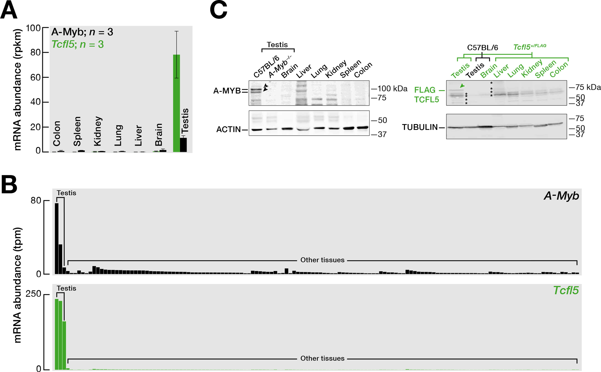
Fig. 1. TCFL5 is specifically expressed in primary spermatocytes of testis.

(A) Steady-state mRNA abundance of A-Myb and Tcfl5 measured by RNA sequencing of various mouse tissues (Merkin et al., 2012). The bar represents the mean mRNA abundance of A-Myb and Tcfl5 from three independent trials. Whiskers show standard error.
(B) mRNA abundance of A-Myb and Tcfl5 measured by RNA sequencing of various tissues. Data is from the mouse ENCODE project.
(C) Abundance of A-MYB and FLAG–TCFL5 proteins in various mouse tissues. FLAG-TCFL5 protein was detected in various tissues from Tcfl5+/FLAG mouse. ACTIN serves as a loading control. Each lane contained 50 μg testis protein. See Fig. S1A for uncropped western blot images. Asterisks represent background from the secondary antibody: See Fig. S1B.
