Abstract
Background
Childhood chronic haematogenous osteomyelitis (CCHOM) is a severe condition in paediatric patients. The optimal timing of debridement and the subsequent method of bone reconstruction in CCHOM patients remain controversial. The purpose of this study was to assess the treatment efficacy of Masquelet technique with early debridement and internal fixation in CCHOM of long bones.
Methods
Between January 2016 and January 2021, a total of 21 patients (18 males, 3 females) with CCHOM of long bone were included. The mean age was 10.4 years (range, 2–18 years). All cases were treated by a two-stage surgical protocol of Masquelet technique. In the first stage, aggressive debridement, sequestrectomy, and inducing membrane by bone cement spacer were performed after definite diagnosis. In the second stage, cement spacer was removed, and autologous and allogeneic bone was grafted. Internal fixation was used for the first and/or second stage depending on stability requirements. The patients’ clinical and imaging results were retrospectively analysed.
Results
The mean follow-up was 31.7 months (range, 21–61 months). None of the patients experienced recurrence of infection. Radiographic bone union time was 4.3 months (range, 2.5–11 months). Five cases underwent re-operation due to complications such as bone resorption or refracture. By the last follow-up visit, bones had healed and all of the patients had resumed daily living and sports activities.
Conclusion
The Masquelet technique with early debridement and internal fixation is a viable surgical method for the management of large long bone defects of CCHOM patients.
Keywords: Chronic osteomyelitis, Paediatric, Internal fixation, Masquelet technique
Background
Although childhood chronic haematogenous osteomyelitis (CCHOM) has become less prevalent in developed nations, it remains a major source of morbidity and a ponderous burden on public health care in developing countries [1]. In southwest China, haematogenous osteomyelitis accounts for 17.9% of all cases of chronic osteomyelitis [2]. CCHOM is frequently accompanied by clinical manifestations such as pain, chronic sinus tracts, bone exposure, and pathological fractures. Physical manifestations of the disease can lead to secondary problems for these paediatric patients, such as psychological and economic problems [3].
CCHOM mainly involves metaphyses of long bones in an early stage and occurs at a single site in majority of cases [3]. It may lead to extensive necrosis of bone and formation of sequestra. Surgery is the most important means of treating CCHOM. However, there are many controversies regarding the optimal surgical method. Sequestrectomy, external fixator, bone graft or Ilizarov technique, and systemic antibiotic treatment are standard treatment methods [4–13]. Although many patients are cured, these treatment methods are often accompanied by complaints such as poor patient tolerance, high complication rates, and long-term recurrence rates (Table 1). The Masquelet technique, also known as the induced membrane technique, is a two-stage approach used in the reconstruction of bone defects. It involves filling the defect with polymethyl methacrylate (PMMA) cement spacer to induce a membrane in the first stage, and removing the spacer and grafting cancellous bone in the second stage [14]. It has widely been used in the treatment of chronic osteomyelitis [15, 16]. The technique has many advantages such as a high rate of infection eradiation, a relatively short time of bone union, and few complications, even for critical size bone defects [17]. Combined with internal fixation, it can provide better comfort for the patients and does not increase the risk of infection recurrence [18]. The clinical effect has been reported in adults [18]. However, in children, the outcomes of this technique are still uncertain. The aim of this study was to assess the efficacy of the Masquelet technique with early debridement and internal fixation for the treatment of CCHOM of long bones.
Table 1.
Literature review of childhood chronic haematogenous osteomyelitis (CCHOM) of long bones
| Work | Cases (n) | Mean age (years) | Gender (M/F) | Location (n or %) | Surgical methods used | Follow-up (mean) | Results | Complications at the last follow-up |
|---|---|---|---|---|---|---|---|---|
| Daoud et al. [4] | 34 | 7.7 | 17/17 |
Tibia: 24 Femur: 8 Humerus: 2 |
Sequestrectomy, Debridement, Immobilisation Corticocancellous iliac grafts |
37 months |
Limb length discrepancy, Fracture, Axial deformity, Non-union |
|
| Liu et al. [5] | 11 | 14 | 7/4 | Humerus: 11 |
Osteotomy, Callus distraction by monolateral external fixator |
106 months |
Excellent: 7 Good: 3 Poor:1 |
Pin track infection, Local inflammation, Flexion contracture of elbow, Pin loosening, Inferior subluxation of the glenohumeral joint |
| Hoang et al. [6] | 6 | 9.8 | 6/0 | Tibia: 6 |
Debridement, Free gracilis muscle flap, |
3 years | Satisfactory: 6 | No complication |
| Kucukkaya et al. [7] | 7 | 7.2 | 5/2 | Tibia: 7 |
Sequestrectomy, Debridement, Ilizarov technique |
4.6 years | Excellent: 7 | No complication |
| Stevenson et al. [8] | 145 | 10 |
Tibia: 46% Femur: 26% Humerus: 10% |
Drilling and curettage of abscess, Sequestrectomy, Stabilisation, Reconstruction of bone defect |
At least 3 years | |||
| Beckles et al. [9] | 167 | 8 | 102/65 |
Tibia: 79 Femur: 47 Humerus: 18 |
Sequestrectomy, Drilling Curettage, Incision and drainage of abscess, Bone grafting, Bone transport |
At least one year |
Infection recurrence, Below-knee amputation |
|
| Ellur et al. [10] | 34 | 4 | 22/9 |
Tibia: 17 Femur: 12 Humerus: 1 Metacarpal: 1 |
Debridement, Antibiotic-impregnated calcium sulphate beads |
42 months |
No infection recurrence No reoperation No systemic adverse reaction |
Length discrepancy (< 1 cm) |
| El-Rosasy et al. [11] | 14 | 9.2 years | 9/5 | Tibia: 14 |
Ilizarov techniques, Fibular osteotomy |
36.9 months |
Satisfactory 12 Unsatisfactory 2 |
Refracture, Limb shortening |
| Wirbel et al. [12] | 16 (out of 11 patients) |
Debridement, Sequestrectomy External fixator Vacuum-assisted closure (VAC) system, Coverage of the soft tissue, Fibula or rib transplantation |
24 months |
Functional restrictions, Length discrepancy |
||||
| Lauschke et al. [13] | 30 (out of 24 patients) | Tibia: 30 |
Drainage of abscess, Cortical fenestration, Sequestrectomy, A composite transposition of the ipsilateral fibula |
2 years |
Restriction of motion, Limb shortening |
|||
| Wang et al. [27] | Tibia |
Debridement, Masquelet technique |
25 months |
Bone union, No infection recurrence |
Small bone diameter, Limitation of knee activity |
Methods
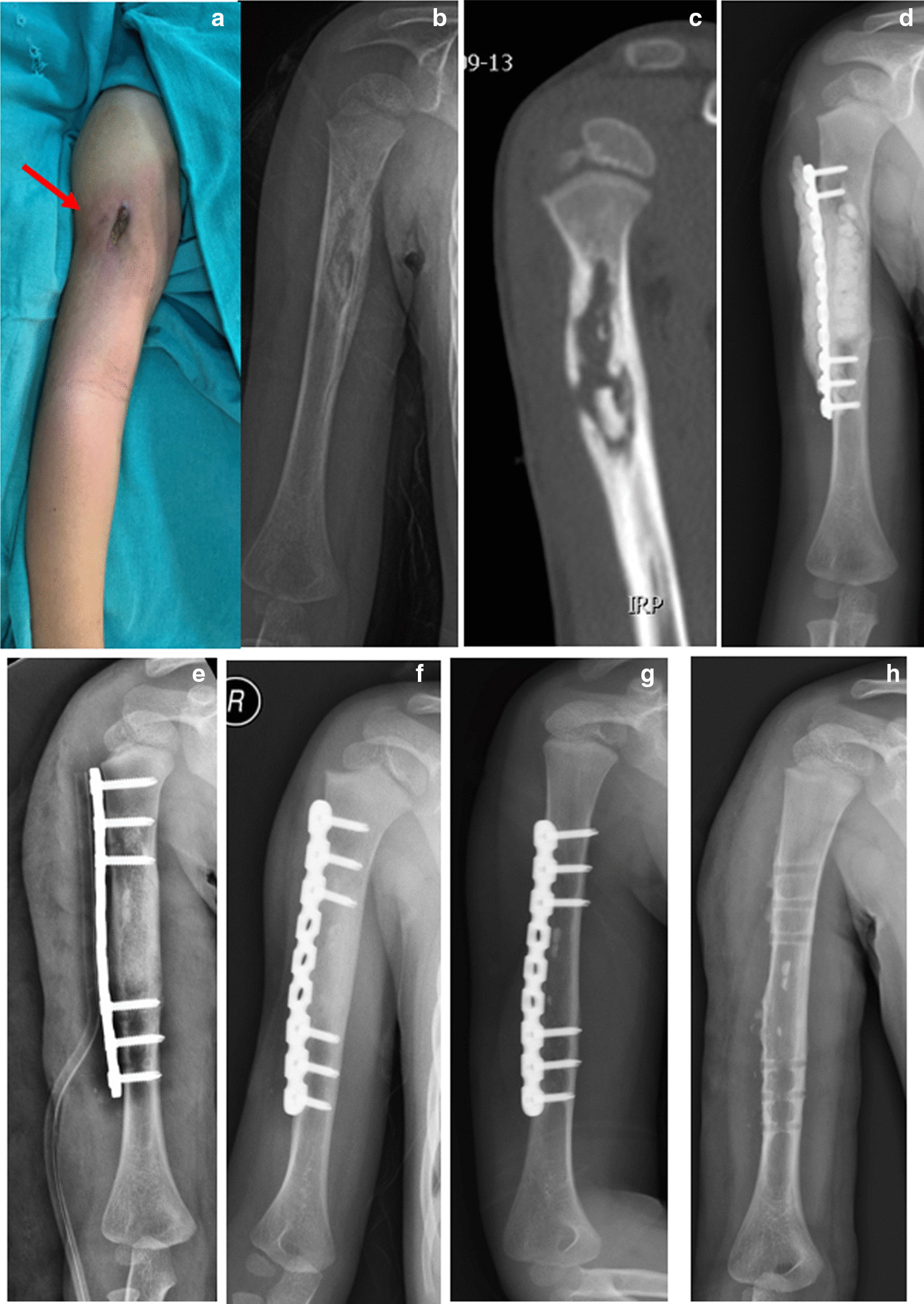
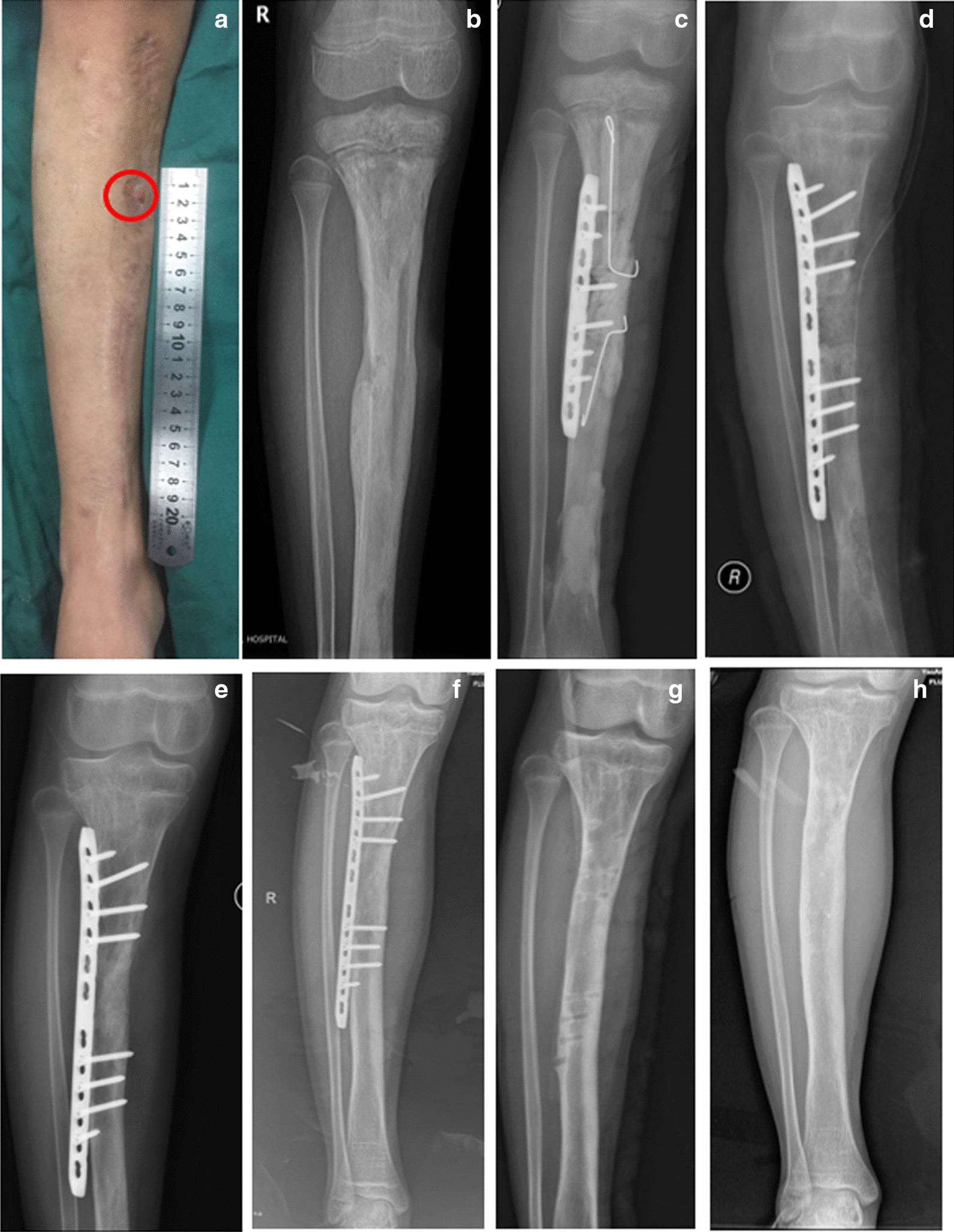
After the approval from the institutional review board, the procedures were performed from January 2016 to January 2021 at the Department of Orthopaedic Surgery, 920 Hospital of Joint Logistic Support Force of PLA, China. The inclusion criteria were as follows: (1) chronic osteomyelitis of long bones, which was confirmed by clinical features and imaging (plain radiographs, CT, and MRI); (2) lack of history of fracture or surgery before the onset of bone infection at the affected site; (3) type III or IV Cierny–Mader anatomic type; (4) treatment with the Masquelet technique; (5) bone defect was stabilised by internal fixation in the first or the second stages; (6) age less than 18 years; (7) A or B-host Cierny–Mader physiologic class. Patients with insufficient follow-up information were excluded. Table 2 summarises the patients’ data (Figs. 1 and 2).
Table 2.
Patients’ demographic and clinical data at the last follow-up
| Patient number | Sex | Age (years) | Location | Cierny–Mader type | Fixation | Follow-up (months) | Radiological bone union (months) | Full weight-bearing (months) | Outcomes | |
|---|---|---|---|---|---|---|---|---|---|---|
| 1st | 2nd | |||||||||
| 1 | F | 13 | Tibia | III | None | IP | 25 | 3 | 3 | Good |
| 2 | M | 17 | Tibia | IV | IP | IP + Nail | 36 | 7 | 8 | Bone resorption (local) |
| 3 | M | 3 | Ulna | IV | IP | IP | 37 | 11 | 12 | Bone resorption (segmental) |
| 4 | M | 18 | Radius | IV | IP | IP | 26 | 4 | 5 | Good |
| 5 | M | 3 | Humerus | III | IP | IP | 46 | 3 | 4 | Good |
| 6 | M | 14 | Femur | III | IP | IP | 33 | 4 | 6 | Good |
| 7 | F | 8 | Femur | III | IP | IP | 27 | 3 | 4 | Good |
| 8 | M | 6 | Femur | III | IP | IP | 24 | 3 | 4 | Good |
| 9 | M | 15 | Tibia | IV | IP | Nail | 36 | 6 | 9 | Good |
| 10 | M | 13 | Femur | III | IP | IP | 22 | 5 | 6 | Good |
| 11 | M | 2 | Tibia | III | None | IP | 22 | 2 | 3 | Good |
| 12 | M | 11 | Tibia | IV | IP | IP | 25 | 3 | 4 | Good |
| 13 | M | 12 | Tibia | IV | IP | IP | 21 | 4 | 5 | Refracture |
| 14 | F | 10 | Tibia | III | IP | IP | 29 | 2.5 | 3 | Good |
| 15 | F | 7 | Femur | III | None | IP | 27 | 3 | 3 | Good |
| 16 | M | 9 | Tibia | IV | IP | IP | 24 | 3 | 4 | Good |
| 17 | M | 11 | Tibia | IV | IP | IP | 44 | 9 | 12 | Broken plate |
| 18 | M | 9 | Tibia | III | None | IP | 44 | 3 | 6 | Varus deformity of the ankle joint |
| 19 | M | 10 | Tibia | IV | IP | IP | 61 | 3 | 4 | Good |
| 20 | M | 16 | Fibula | IV | IP | IP | 30 | 3 | 4 | Good |
| 21 | M | 11 | Tibia | IV | IP | IP | 26 | 5 | 5 | Good |
F female; M male, IP internal plate, nail intramedullary nail
Fig. 1.

Case 5: a 3-year-old boy who has recently presented with recurrent swelling and drainage from his right upper arm. At the age of 2, he underwent surgery due to swelling, redness, and pain with no preceding trauma. Afterward, a sinus tract formed at the incision (red arrow). a Pre-operative presentation of an affected limb. b Anteroposterior (AP) radiograph shows chronic osteomyelitis of the right humerus. c CT shows a sequestrum. d In the first stage of surgery, debridement and locking plate were performed and antibiotic-loaded bone cement was used to fill the bone defect area. e AP radiograph after bone graft in the second stage of surgery. f AP radiograph shows bone defect healed at 3 months after bone grafting. g AP radiograph shows bone defect remodelled at 26 months after bone grafting. h AP radiograph shows that the internal fixation was removed at 26 months after bone grafting
Fig. 2.

Case 19: a 10-year-old boy with recurrent pain and drainage from his right tibia. Three months before, he underwent surgery twice due to swelling, redness, and pain with no preceding trauma. However, the drainage from sinus tract (red circle) and pain did not disappear after the operations. a Pre-operative presentation of the right lower leg. b Anteroposterior (AP) radiograph shows chronic osteomyelitis of the right tibia. c In the first-stage surgery, debridement and internal fixation were performed, and antibiotic-loaded bone cement was used to fill the bone defect and medullary cavity. d AP radiograph after bone graft in the second stage of surgery. e AP radiograph shows bone defect healed at 3 months after bone grafting. f AP radiograph shows bone defect remodelled at 23 months after bone grafting. g AP radiograph shows that internal fixation was removed at 26 months after bone grafting. h AP radiograph shows the right tibia at 47 months after bone grafting
Surgical technique
All of the patients were treated with the Masquelet technique by a two-stage procedure.
The first stage began with aggressive debridement of all infected, necrotic tissues. Sinus was resected; sequestrum was removed; and medullary cavity was rimmed. The extent of debridement was determined by pre-operative radiographs, CT, and bone scintigraphy. Debridement was carried out until punctate bleeding (Paprika sign) was seen at the bony and soft tissue margins. After abundant lavage, a narrow locking plate was used for the stabilisation of the bone defect. In principle, when the bone defect was segmental or for the local bone defect whose width exceeded one-third of the circumference of the bone cortex, internal fixation was used. Fluoroscopy was used to confirm that the screw was not placed in the epiphyseal plate and that the alignment and length of the affected bone were appropriate. Then, we placed antibiotic-loaded (Vancomycin, Lilly, 2–4 g per 40 g cement) PMMA bone cement (PALACOS®R+G, HERAEUS, Germany) into the bone defect and then coated the plate and both bone ends of the defect with the cement. Two drainage tubes were used for 7–14 days. The deep tissue was harvested for bacterial culture and pathological examination. Weight bearing was forbidden on the affected limb.
All of the patients received intravenous broad-spectrum antibiotics for two weeks post-operatively. According to the results of bacterial culture, they were replaced with sensitive antibiotics, which were then changed to oral antibiotics for 4 weeks. White blood cell (WBC) count, erythrocyte sedimentation rate (ESR), and C-reactive protein (CRP) level were examined every two weeks. The next stage of surgery was performed after 6–8 weeks. The prerequisite was that WBC count, ESR, and CRP level were normal for 2–3 times. During the second stage of procedure, the membrane was incised sharply longitudinally to reach the bone cement, and the antibiotic-loaded bone cement and the locking plate were removed carefully, avoiding damage to the induced membrane. After rinsing thoroughly, a new similar locking plate was used for defect fixation with simultaneous osteosynthesis performed as the first stage. The bone was decorticated proximally and distally until bone bleeding was obtained. Then, the bone defect was filled with autogenous cancellous bone within the induced membrane. The graft was morselised into very small grafts measuring 1–2 mm3. If autologous cancellous bone from the iliac crests was insufficient, allogeneic bone (BIO-GENE, Beijing Cojoing, China) was added. The allogeneic bone was placed in the middle of the defect, and the autologous bone was placed at the surrounding area near the induction membrane. Before using, the allograft was soaked in the blood harvested from the donor site for at least 30 min.
Sensitive antibiotics were continued for 2 weeks after the second stage, and weight bearing of the affected limb was restricted unless allowed by the surgeons.
Follow-up
All of the patients had regular clinical and radiological follow-up every month after surgery until the bone healing. Thereafter, the patients were followed up every three months. Radiological bone union was defined as at least three continuous cortices visible on anteroposterior (AP) and lateral radiographs of the bone defect. The follow-up radiographs were independently assessed by two authors in an unblinded fashion. Disagreements between the two authors were judged by another author.
During each follow-up visit, the patients underwent a clinical evaluation and laboratory analyses. WBC, CRP, and ESR, as well as other clinical features (such as discharge, redness and swelling, warmth, and pain) were assessed to exclude the recurrence of infection. Furthermore, range of motion (ROM) of joint in the affected limb and growth disturbances were recorded at the final follow-up to determine the treatment effect. Complications were assessed by the surgeons involved in the treatment of the patients. At the last follow-up, if the patient had no symptoms of recurrent infections such as local redness, swelling, pain, and sinus in the affected limb, the radiologic imaging showed good bone healing without deformity, and there was no other complication; the treatment outcome could be considered “good”.
Results
The study included a total of 21 consecutive children (18 males, 3 females) with CCHOM of long bones. The mean patients’ age at the time of the first hospitalisation was 10.4 (range, 2–18) years. The femur was involved in five cases, the tibia in 12 cases, the humerus in one case, the radius in one case, the fibula in one case, and the ulna in one case. Cierny–Mader Type III and Type IV were observed in 10 cases and 11 cases, respectively. All patients had A-host Cierny–Mader physiologic class. Thirteen cases had sinus tracts, and 12 cases had a history of surgery in other hospitals.
Two patients required repeated operation due to recurrent infection during the interval between the first and the second stage, and infection was controlled after re-debridement. Nine patients had positive bacterial cultures. The causative pathogen was Staphylococcus aureus in six cases, Enterobacter cloacae in two cases, and Staphylococcus epidermidis in one case.
The volume of bone defect was estimated based on CT measurement before the second-stage surgery. The average volume of bone defect was 36.4 cm3 (9–78 cm3). Four patients were implanted with autologous bone, and 17 patients were implanted with autologous plus allograft in different proportions (16–50%), of which five patients were implanted with allograft more than 25% of the total volume. The mean follow-up period after the second stage of surgery was 31.7 months (range, 21–61 months). Table 2 summarises the management procedures and results. The mean radiographic bone union occurred in 4.3 months (range, 2–11 months), and full weight bearing was noted at 5.5 months (range, 3–12 months).
Two cases underwent bone re-grafting due to partial or segmental bone resorption at the defect. The proportion of allograft bone was 38% and 33% in these two cases. In one case, internal fixation was broken due to an accidental fall, and surgery was required to replace it with a new plate, which resulted in bone union delay of 9 months. One case with distal tibia osteomyelitis had a varus deformity (about 17°) of the ankle joint at 16 months after grafting, but did not have pain, lameness, or other symptoms. In one case, a refracture occurred at the bone defect due to a fall one month after removal of the internal fixation. Reduction, fixation, and grafting were performed again, and the fracture healed six months after surgery.
By the last follow-up visit, all of the patients achieved bone union and were pain-free; their inflammatory markers remained within the normal range; no infection recurrence was noted; and they resumed daily living and sports activities.
Discussion
We showed that the early debridement and internal fixation with the Masquelet technique could effectively control infection, relieve patients’ symptoms, achieve bone healing, and facilitate early functional exercise in patients with CCHOM.
The Beit-CURE (BC) classification is the first classification specific for CCHOM [1, 8–10]. It considers the presence of both sequestrum and involucrum. Some studies have confirmed that the BC classification can guide surgical strategy and help predict length of inpatient treatment and number and type of procedures required [8–10]. However, in our hospital, most of the patients had undergone one or more surgeries in local hospitals before they were admitted. According to the BC classification, they should be categorised as unclassifiable, and therapy recommendations are not conceivable. Cierny–Mader’s classification is based on the extent of infection (medullary, superficial, localised, diffuse) and host status (healthy, compromised immune system, failed immune system) [19, 20]. This is the most widely used classification for chronic osteomyelitis. It represents the pathological progression of osteomyelitis and is useful in planning treatment strategy [13]. Although it is mainly suitable for adult patients, it can be applied to the paediatric population as well; namely, in this study, we adopted different strategies of debridement and stabilisation according to this classification.
The timing of debridement of CCHOM is controversial. Some authors believe that the intact involucrum develops, which can provide good stability, before sequestrectomy is required to reduce the risk of complication such as pathological fractures, deformities, and segmental bone defects [5, 11, 13]. Other authors, however, advocate for the early debridement to control infection, create a better environment for the periosteum to respond, and minimise damage to the surrounding soft tissues [1, 5]. In our cases, due to the application of internal fixation, the stability of the affected limb could be maintained; thus, debridement could be performed after diagnosis is confirmed, regardless of whether involucrum has formed or not. This undoubtedly shortened the treatment period and accelerated the recovery of the children.
The Masquelet’s technique is regarded as the “gold standard” for treating various types of long bone defects in adults and children [21, 22]. Auregan et al. [23] found that paediatric patients treated with the Masquelet technique had a 58% success rate, which increased to 87% when iterative surgery was considered. Canavese et al. [24] and Rousset et al. [25] reported five and eight children with chronic osteomyelitis treated by the Masquelet technique, respectively, and achieved satisfactory treatment results. Shen et al. treated 26 children with chronic osteomyelitis with the Masquelet technique, and the bone defects were healed in 4.0–5.0 months after the operation. Wang et al. [26] achieved good clinical efficacy in treating chronic osteomyelitis in both adult and paediatric patients. These reports confirm that the Masquelet technique is an effective treatment for chronic osteomyelitis in children, but it is mainly used for post-traumatic osteomyelitis, while there are no specific studies on CCHOM.
The application of internal fixation is typically contraindicated in the treatment of chronic osteomyelitis. However, some scholars have used antibiotic-loaded bone cement–coated plate as a temporary fixation after debridement, which can kill planktonic bacteria and inhibit the formation of biofilms [16, 18]. In the second stage, the application of internal fixation for bone reconstruction can reduce the burden of care, avoid pin tract infections, and allow early functional exercise. In this study, for the Cierny–Mader type III patients, according to the protocol by Kinik et al. [27], more than 30% of cortical bone removal for debridement necessitates prophylactic fixation to prevent iatrogenic fracture risk. For the Cierny–Mader type IV, we used locking plates for internal fixation in the first and second stages. This did not increase the chance of infection recurrence, but it reduced the difficulty of postoperative care and improved the comfort of the child. In the second stage, significant solidification of the bone graft was seen 2–3 months after the second stage of surgery, so that full weight bearing could be gradually achieved. Children could return to society early and participate in classroom learning and activities, which is beneficial to the physical and mental development, especially in younger patients.
In the second stage of the Masquelet's technique, the need for a large amount of autologous cancellous bone graft to reconstruct bone defects is a limiting factor, especially in very young children. The application of some alternative materials has been tried in clinical practice. Fitoussi et al. [28] used autologous cancellous bone particles combined with autologous fibula scaffold to fill the bone defect in the second stage for eight children with post-operative bone defect larger than 15 cm due to primary malignant tumour. All of the bone defects healed within 5.6 (range, 4–8) months. Gouron et al. [29] performed bone reconstruction in 14 children with trauma, tumour resection, or tibial congenital pseudarthrosis. They added allograft bone, biphasic calcium phosphate (BCP), or tibial bone strut to increase the graft volume. Bone union was achieved in 9.5 (range, 2–25) months. Canavese et al. [24] and Rousset et al. [25] used β-tricalcium phosphate (BTP) as a bone graft substitute. Their results showed that BTP was even more effective in osteogenesis than bone graft. Shen et al. [30] used bone marrow concentrator–modified allograft or bone marrow aspirate–mixed allograft to improve the osteogenic ability of allograft. In this study, allograft bone was added as an alternative graft material to increase the graft volume. Prior to use, in accordance with the method reported by Gouron [22, 28], the allograft was immersed in blood from the donor site. Two patients had resorption, and we believe the higher proportion of allogeneic bone was the main reason. Therefore, it is recommended that the proportion of allograft bone does not exceed one-third to avoid hindering consolidation according to an empirical advice of Masquelet [21].
There were several limitations to our study: (1) small sample size; (2) different skeletal sites involved; (3) no comparison with other treatment methods. Thus, additional studies are mandatory.
Conclusion
CCHOM is a relatively complex disease, and once the diagnosis is confirmed, early debridement can effectively prevent further bacterial damage to the bone. Depending on the infection, the choice of an appropriate surgical method with aggressive debridement, the use of antibiotic-loaded bone cement to induce membrane formation, and then reconstruction by bone grafting in the second stage can achieve satisfactory results in patients with CCHOM. Early debridement can shorten the course of treatment; internal fixation can provide a stable osteogenic environment; and induced membrane in the Masquelet technique can rapidly promote the graft corticalisation. These measures are conducive to early joint functional exercise and reduce joint functional damage.
Acknowledgements
We thank LetPub (www.letpub.com) for linguistic assistance and pre-submission expert review.
Abbreviations
- CCHOM
Childhood chronic haematogenous osteomyelitis
- WBC
White blood cell
- CRP
C-reactive protein
- ESR
Erythrocyte sedimentation rate
- PMMA
Polymethyl methacrylate
- ROM
Range of motion
- BCP
Biphasic calcium phosphate
- BTP
β-tricalcium phosphate
Author contributions
JS and YX contributed to the study conception and design. JS, LQ, HZ, XY, MS, and XZ contributed to the material preparation and data collection. Statistical analyses were managed by JS, TZ, and XC. The first draft of the manuscript was written by JS and YX. All authors commented on previous versions of the manuscript. All authors read and approved the final manuscript.
Funding
This study was funded by the Clinical Orthopaedic Trauma Medical Centre Program of Yunnan Province, China (ZX20191001), and the Applied Basic Research Joint Project of Yunnan Science and Technology Department and Kunming Medical University (202101AY070001-294).
Availability of data and materials
The data presented in this study are available in the article or supplementary material.
Declarations
Ethics approval and consent to participate
This study was approved by the institutional review board with approval No. 091-01 from the 920 Hospital of the Joint Logistics Support Force of the PLA. Informed consent was obtained from all individual participants included in the study.
Consent for publication
Not applicable.
Competing interests
The authors declare that they have no competing interests.
Footnotes
Publisher's Note
Springer Nature remains neutral with regard to jurisdictional claims in published maps and institutional affiliations.
Contributor Information
Jian Shi, Email: doctorshijian920@sina.com.
Yongqing Xu, Email: xuyongqingkm@163.net.
References
- 1.Jones HW, Beckles VL, Akinola B, Stevenson AJ, Harrison WJ. Chronic haematogenous osteomyelitis in children: an unsolved problem. J Bone Jt Surg Br. 2011;93:1005–1010. doi: 10.1302/0301-620X.93B8.25951. [DOI] [PubMed] [Google Scholar]
- 2.Wang X, Yu S, Sun D, Fu J, Wang S, Huang K, et al. Current data on extremities chronic osteomyelitis in southwest China: epidemiology, microbiology and therapeutic consequences. Sci Rep. 2017;7:16251. doi: 10.1038/s41598-017-16337-x. [DOI] [PMC free article] [PubMed] [Google Scholar]
- 3.Lew DP, Waldvogel FA. Osteomyelitis. Lancet. 2004;364:369–379. doi: 10.1016/S0140-6736(04)16727-5. [DOI] [PubMed] [Google Scholar]
- 4.Daoud A, Saighi-Bouaouina A. Treatment of sequestra, pseudarthroses, and defects in the long bones of children who have chronic hematogenous osteomyelitis. J Bone Jt Surg Am. 1989;71:1448–1468. doi: 10.2106/00004623-198971100-00003. [DOI] [PubMed] [Google Scholar]
- 5.Liu T, Zhang X, Li Z, Zeng W, Peng D, Sun C. Callus distraction for humeral nonunion with bone loss and limb shortening caused by chronic osteomyelitis. J Bone Jt Surg Br. 2008;90:795–800. doi: 10.1302/0301-620X.90B6.20392. [DOI] [PubMed] [Google Scholar]
- 6.Hoang NT, Staudenmaier R, Feucht A, Hoehnke C. Effectiveness of free gracilis muscle flaps in the treatment of chronic osteomyelitis with purulent fistulas at the distal third of the tibia in children. J Pediatr Orthop. 2009;29:305–311. doi: 10.1097/BPO.0b013e31819903e1. [DOI] [PubMed] [Google Scholar]
- 7.Kucukkaya M, Kabukcuoglu Y, Tezer M, Kuzgun U. Management of childhood chronic tibial osteomyelitis with the Ilizarov method. J Pediatr Orthop. 2002;22:632–637. doi: 10.1097/01241398-200209000-00012. [DOI] [PubMed] [Google Scholar]
- 8.Stevenson AJ, Jones HW, Chokotho LC, Beckles VL, Harrison WJ. The beit CURE classification of childhood chronic haematogenous osteomyelitis–a guide to treatment. J Orthop Surg Res. 2015;10:144. doi: 10.1186/s13018-015-0282-9. [DOI] [PMC free article] [PubMed] [Google Scholar]
- 9.Beckles VL, Jones HW, Harrison WJ. Chronic haematogenous osteomyelitis in children: a retrospective review of 167 patients in Malawi. J Bone Jt Surg Br. 2010;92:1138–1143. doi: 10.1302/0301-620X.92B8.23413. [DOI] [PubMed] [Google Scholar]
- 10.Ellur V, Kumar G, Sampath JS. Treatment of chronic hematogenous osteomyelitis in children with antibiotic impregnated calcium sulphate. J Pediatr Orthop. 2021;41:127–131. doi: 10.1097/BPO.0000000000001723. [DOI] [PubMed] [Google Scholar]
- 11.El-Rosasy MA. Ilizarov treatment for pseudarthrosis of the tibia due to haematogenous osteomyelitis. J Pediatr Orthop B. 2013;22:200–206. doi: 10.1097/BPB.0b013e328360268b. [DOI] [PubMed] [Google Scholar]
- 12.Wirbel R, Hermans K. Surgical treatment of chronic osteomyelitis in children admitted from developing countries. Afr J Paediatr Surg. 2014;11:297–303. doi: 10.4103/0189-6725.143133. [DOI] [PubMed] [Google Scholar]
- 13.Lauschke FH, Frey CT. Hematogenous osteomyelitis in infants and children in the northwestern region of Namibia. Management and two-year results. J Bone Jt Surg Am. 1994;76:502–510. doi: 10.2106/00004623-199404000-00004. [DOI] [PubMed] [Google Scholar]
- 14.Yu X, Wu H, Li J, Xie Z. Antibiotic cement-coated locking plate as a temporary internal fixator for femoral osteomyelitis defects. Int Orthop. 2017;41:1851–1857. doi: 10.1007/s00264-016-3258-4. [DOI] [PubMed] [Google Scholar]
- 15.Masquelet A, Kanakaris NK, Obert L, Stafford P, Giannoudis PV. Bone repair using the Masquelet technique. J Bone Jt Surg Am. 2019;101:1024–1036. doi: 10.2106/JBJS.18.00842. [DOI] [PubMed] [Google Scholar]
- 16.Wu H, Shen J, Yu X, Fu J, Yu S, Sun D, et al. Two stage management of Cierny-Mader type IV chronic osteomyelitis of the long bones. Injury. 2017;48:511–518. doi: 10.1016/j.injury.2017.01.007. [DOI] [PubMed] [Google Scholar]
- 17.Masquelet AC, Kishi T, Benko PE. Very long-term results of post-traumatic bone defect reconstruction by the induced membrane technique. Orthop Traumatol Surg Res. 2019;105:159–166. doi: 10.1016/j.otsr.2018.11.012. [DOI] [PubMed] [Google Scholar]
- 18.Luo F, Wang X, Wang S, Fu J, Xie Z. Induced membrane technique combined with two-stage internal fixation for the treatment of tibial osteomyelitis defects. Injury. 2017;48:1623–1627. doi: 10.1016/j.injury.2017.04.052. [DOI] [PubMed] [Google Scholar]
- 19.Cierny G, 3rd. Mader JT, Penninck JJ. A clinical staging system for adult osteomyelitis. Clin Orthop Relat Res. 2003;414:7–24. doi: 10.1097/01.blo.0000088564.81746.62. [DOI] [PubMed] [Google Scholar]
- 20.Cierny G., 3rd Surgical treatment of osteomyelitis. Plast Reconstr Surg. 2011;127(Suppl 1):190s–204s. doi: 10.1097/PRS.0b013e3182025070. [DOI] [PubMed] [Google Scholar]
- 21.Masquelet AC. The induced membrane technique. Orthop Traumatol Surg Res. 2020;106:785–787. doi: 10.1016/j.otsr.2020.06.001. [DOI] [PubMed] [Google Scholar]
- 22.Gouron R. Surgical technique and indications of the induced membrane procedure in children. Orthop Traumatol Surg Res. 2016;102:S133–S139. doi: 10.1016/j.otsr.2015.06.027. [DOI] [PubMed] [Google Scholar]
- 23.Aurégan JC, Bégué T, Rigoulot G, Glorion C, Pannier S. Success rate and risk factors of failure of the induced membrane technique in children: a systematic review. Injury. 2016;47(Suppl 6):S62–S67. doi: 10.1016/S0020-1383(16)30841-5. [DOI] [PubMed] [Google Scholar]
- 24.Canavese F, Corradin M, Khan A, Mansour M, Rousset M, Samba A. Successful treatment of chronic osteomyelitis in children with debridement, antibiotic-laden cement spacer and bone graft substitute. Eur J Orthop Surg Traumatol. 2017;27:221–228. doi: 10.1007/s00590-016-1859-7. [DOI] [PubMed] [Google Scholar]
- 25.Rousset M, Walle M, Cambou L, Mansour M, Samba A, Pereira B, et al. Chronic infection and infected non-union of the long bones in paediatric patients: preliminary results of bone versus beta-tricalcium phosphate grafting after induced membrane formation. Int Orthop. 2018;42:385–393. doi: 10.1007/s00264-017-3693-x. [DOI] [PubMed] [Google Scholar]
- 26.Wang X, Wang Z, Fu J, Huang K, Xie Z. Induced membrane technique for the treatment of chronic hematogenous tibia osteomyelitis. BMC Musculoskelet Disord. 2017;18:33. doi: 10.1186/s12891-017-1395-6. [DOI] [PMC free article] [PubMed] [Google Scholar]
- 27.Kinik H, Karaduman M. Cierny-Mader Type III chronic osteomyelitis: the results of patients treated with debridement, irrigation, vancomycin beads and systemic antibiotics. Int Orthop. 2008;32:551–558. doi: 10.1007/s00264-007-0342-9. [DOI] [PMC free article] [PubMed] [Google Scholar]
- 28.Fitoussi F, Ilharreborde B. Is the induced-membrane technique successful for limb reconstruction after resecting large bone tumors in children? Clin Orthop Relat Res. 2015;473:2067–2075. doi: 10.1007/s11999-015-4164-6. [DOI] [PMC free article] [PubMed] [Google Scholar]
- 29.Gouron R, Deroussen F, Plancq MC, Collet LM. Bone defect reconstruction in children using the induced membrane technique: a series of 14 cases. Orthop Traumatol Surg Res. 2013;99:837–843. doi: 10.1016/j.otsr.2013.05.005. [DOI] [PubMed] [Google Scholar]
- 30.Shen J, Sun D, Yu S, Fu J, Wang X, Wang S, et al. Radiological and clinical outcomes using induced membrane technique combined with bone marrow concentrate in the treatment of chronic osteomyelitis of immature patients. Bone Jt Res. 2021;10:31–40. doi: 10.1302/2046-3758.101.BJR-2020-0229.R1. [DOI] [PMC free article] [PubMed] [Google Scholar]
Associated Data
This section collects any data citations, data availability statements, or supplementary materials included in this article.
Data Availability Statement
The data presented in this study are available in the article or supplementary material.


