Abstract
Objectives
Macrophage subsets, activated by T cells, are increasingly recognised to play a central role in rheumatoid arthritis (RA) pathogenesis. Janus kinase (JAK) inhibitors have proven beneficial clinical effects in RA. In this study, we investigated the effect of JAK inhibitors on the generation of cytokine-activated T (Tck) cells and the production of cytokines and chemokines induced by Tck cell/macrophage interactions.
Methods
CD14+ monocytes and CD4+ T cells were purified from peripheral blood mononuclear cells from buffy coats of healthy donors. As representative JAK inhibitors, tofacitinib or ruxolitinib were added during Tck cell differentiation. Previously validated protocols were used to generate macrophages and Tck cells from monocytes and CD4+ T cells, respectively. Cytokine and chemokine including TNF, IL-6, IL-15, IL-RA, IL-10, MIP1α, MIP1β and IP10 were measured by ELISA.
Results
JAK inhibitors prevented cytokine-induced maturation of Tck cells and decreased the production of proinflammatory cytokines TNF, IL-6, IL-15, IL-1RA and the chemokines IL-10, MIP1α, MIP1β, IP10 by Tck cell-activated macrophages in vitro (p<0.05).
Conclusions
Our findings show that JAK inhibition disrupts T cell-induced macrophage activation and reduces downstream proinflammatory cytokine and chemokine responses, suggesting that suppressing the T cell-macrophage interaction contributes to the therapeutic effect of JAK inhibitors.
Keywords: antirheumatic agents; arthritis, rheumatoid; chemokines; inflammation; T-lymphocyte subsets
WHAT IS ALREADY KNOWN ON THIS TOPIC
Janus kinase (JAK) inhibitors modulate the activity of proinflammatory cytokines implicated in rheumatoid arthritis (RA progression.
WHAT THIS STUDY ADDS
JAK inhibition disrupts cytokine-activated T cell-induced macrophage activation, an in vitro model for RA synovial T-cell-mediated stimulation of monocytes/macrophages, and reduces proinflammatory cytokine and chemokine responses.
Suppression of the T cell: macrophage interaction may contribute to the therapeutic efficacy of JAK inhibitors.
HOW THIS STUDY MIGHT AFFECT RESEARCH, PRACTICE OR POLICY
Although this study may not directly change policy or practice, it highlights that JAK inhibition may affect multiple steps involved in mediating inflammation by inhibiting cytokine and chemokine production and disrupting the function of innate and acquired immune cells.
Introduction
The hallmarks of synovitis in rheumatoid arthritis (RA) include the infiltration of inflammatory cells, predominantly blood-derived lymphoid and myeloid cells.1 Infiltrating T-cells and monocytes/macrophages reside collaboratively within the inflamed RA synovium; hence, the established heterogeneity of inflammatory synovial macrophages2 can drive synovitis via cytokine production by activated T-cells.3 Conversely, T-cells activate macrophages, leading to a proinflammatory cytokine forward feedback loop.4
Perpetuation of inflammation by monocytes/macrophages stimulated by cytokine-activated T (Tck) cells may be modelled in vitro by the macrophage: T-cell coculture assay.5 6 Signalling pathways elicited by Tck cells in responding monocytes were identical to those induced by T-cells isolated from RA synovial tissue.6 7 Thus, Tck cells have been proposed as a model for RA synovial T-cell-mediated stimulation of monocytes/macrophages via cell-cell contact-dependent mechanisms.5 7
Janus kinases (JAKs) are tyrosine kinases that recruit and activate signal transducers and activators of transcription, which drive proinflammatory cellular responses. There are four JAKs in humans, JAK1, JAK2, JAK3 and tyrosine kinase 2 (TYK2)8 that subserve the biology of a range of cytokines critical in the pathogenesis of RA.9 The JAK inhibitor tofacitinib interacts with multiple JAKs, although preferentially inhibiting JAK1-dependent and JAK3-dependent cytokines.10 At the pathophysiological level, tofacitinib and now other JAK inhibitors, modulate the activity of proinflammatory cytokines implicated in RA progression.10 11 Though the biological roles of JAK inhibitors in lymphocytes are well known, their function in the context of macrophage: T-cell interactions has not been defined. Here, we investigated the effect of tofacitinib, ruxolitinib and Tyrphostin AG-490 on the generation of Tck cells and assessed whether JAK inhibition in Tck cells impacts cytokine and chemokine production by macrophages in the vitro coculture system.
Materials and methods
Isolation of peripheral blood mononuclear cells from buffy coats
Peripheral blood mononuclear cells (PBMC) were isolated from buffy coats using Histopaque 1077 (Sigma) density centrifugation. The buffy coats were obtained from healthy volunteers from the Scottish National Blood Transfusion Service UK (SNBTS). All subjects provided informed consent with the appropriate ethical approvals in place.
Reagents
Tofacitinib, ruxolitinib and Tyrphostin AG-490 were obtained from LC-Labs and dissolved in dimethyl sulfoxide (DMSO) (Sigma). Serial dilutions were employed to ensure that DMSO concentration did not exceed 0.001% in cell culture. Recombinant TNF, IL-6, IL-2 and MCSF (Peprotech); IFN-γ, GM-CSF and IL-4 (R&D Systems); Lipopolysaccharide (Salmonella typhimurium; Sigma).
Purification of CD14+ monocytes and differentiation into macrophages
CD14+ monocytes were purified by positive magnetic separation using CD14 immunomagnetic microbeads (Miltenyi Biotec). Purities of >98% were routinely confirmed by flow cytometry (data not shown). CD14+ monocytes were cultured in 24 well-plates with MCSF (50 ng/mL) for 3 days. Cells were detached using a non-enzymatic dissociation solution (Sigma), washed in Dulbecco’s phosphate-buffered saline and then replated in 96 well plates at a density of 5×10⁵ cells/mL with MCSF (50 ng/mL) for the additional 3 days before harvesting and used in subsequent experiments.
Purification and differentiation of CD4+ Tck cells and the effect of JAK inhibition on Tck cell maturation
CD4+ T-cells were isolated from PBMC using CD4 microbeads (Miltenyi Biotec). To generate Tck cells, CD4+ T-cells were cultured at 1×10⁶ cells/mL with IL-2 (25 ng/mL), IL-6 (100 ng/mL) and TNF (25 ng/mL) (Peprotech) for 6 days.12 In separate experiments, the JAK inhibitor tofacitinib was added to the culture medium during the 6-day maturation period to assess the effect of tofacitinib on Tck cell maturation. The cells were washed and cultured with mature macrophages at a 4:1 Tck: macrophage ratio. The supernatants were collected and assessed for TNF using ELISA.
Macrophage activation by Tck cells in coculture and the effect of JAK inhibition
Fixed numbers of monocyte-derived macrophages (5.0×10⁵ cells) were cocultured with autologous Tck cells, achieving macrophage: Tck ratios of 1:8, 1:4, 1:2 and 1:1. Macrophages and Tck cells were also cultured alone. A 1:4 macrophage/Tck cell ratio was optimal and was used to assess the effect of JAK inhibitors. DMSO (0.001%) was used as a control. The cocultured cells were incubated for 24 hours, and supernatants were collected for analysis using ELISA or Luminex. Trans-well inserts (0.4 µm pore size; Corning) were used to assess cell-cell contact independent mechanisms.
Measurement of cytokines in culture supernatants
IL-6, IL-15, IL-1RA, IL-10, MIP1α, MIP1β, IP10 and MIG were assessed using Luminex (Invitrogen), and TNF was determined using ELISA (Invitrogen); all following the manufacturer’s protocols.
Statistical analysis
Results were analysed using GraphPad Prism V.9 Software, (San Diego, California, USA). Statistical differences between groups were analysed using the Wilcoxon rank test, Student’s t-test or one-way analysis of variance with Bonferroni post hoc corrections as appropriate.
Results
Tck induce contact-dependent and concentration-dependent TNF production by human macrophages
To delineate the effect of JAK inhibition on macrophages, we revisited the generation of Tck cells and their ability to activate monocyte-derived macrophages. In coculture, we established a 1:4 macrophage/Tck cell ratio as the optimal ratio of macrophages to Tck cells. In agreement with previous reports,12 13 Tck cells activated macrophages contact-dependently, secreting TNF (online supplemental figure 1A, B).
rmdopen-2022-002671supp001.pdf (424.2KB, pdf)
JAK inhibitors prevent cytokine-induced maturation of Tck cells and inhibit the production of IL-6, IL-15 and IL-1RA by TcK-activated macrophages
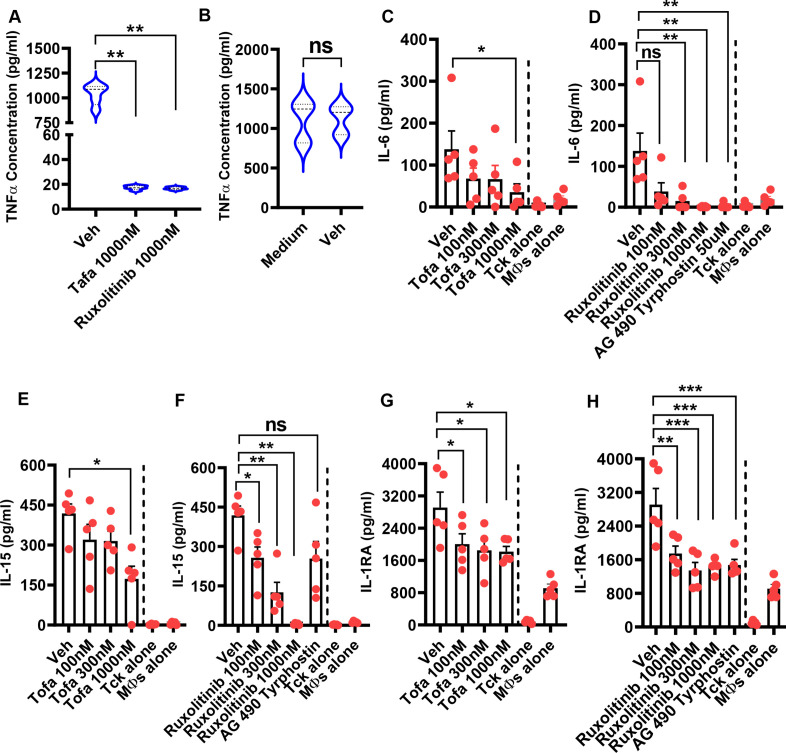
The effect of tofacitinib or ruxolitinib was evaluated on Tck cell maturation. JAK inhibitors diminished the capacity to induce TNF production by macrophages in coculture, suggesting that Tck cells failed to mature sufficiently to be able to activate macrophages (figure 1A). The reduced TNF production was not due to cell death caused by the toxicity of DMSO (figure 1B). Tofacitinib was selected as it is in clinical use, while ruxolitinib and AG-490 tyrphostin extended the generability of the findings. We chose a range of concentrations (online supplemental figure 2A, B) corresponding to approximate blood levels reflected by peak and trough dosage of tofacitinib by mouth.14 We also explored the effect of JAK inhibitors on IL-6, IL-15 and IL-1RA synthesis, relevant cytokines in inflammatory processes in RA. Tofacitinib, ruxolitinib and AG-490 tyrphostin reduced IL-6, IL-15 and IL-1RA (figure 1C–H).
Figure 1.
Janus kinase (JAK) inhibitors prevent cytokine-induced maturation of cytokine-activated T (Tck) cells and inhibit the production of IL-6, IL-15 and IL-1RA by activated macrophages. CD14+ monocytes (5×10⁵ cells/mL) were differentiated into macrophages by culturing them in the presence of macrophage-colony stimulating factor (MCSF; 50 ng/mL) for 6 days in a complete medium in a 96-well plate. Purified CD4+ T cells (1×10⁶ cells/mL) were stimulated with a cocktail of IL-2, IL-6 and TNF in the absence or presence of 0.001% DMSO (used as Vehicle), tofacitinib (1000 nM) or ruxolitinib (1000 nM). The cells were incubated for 6 days to generate Tck. The Tck populations were washed and then cocultured with monocyte-derived macrophages at a concentration of 4:1 Tck-macrophage ratio. The supernatants were harvested, and the level of TNF was measured by ELISA. (A) The comparison of TNF production between DMSO and Tofacitinib (Tofa)-treated or ruxolitinib-treated Tck. (B) The comparison of TNF production between medium and DMSO-treated Tck. In separate experiments, cytokine-generated Tck were cocultured with monocyte-derived macrophages (MФs) at 4:1 ratio in the absence or presence of 0.001% DMSO (Veh) or tofacitinib (100 nM, 300 nM and 1000 nM) or ruxolitinib (100 nM, 300 nM, 1000 nM, 50 µM). Culture supernatants were harvested, and the levels of (C, D) IL-6, (E, F) IL-15 and (G, H) IL-1RA were measured using Luminex. Data represent a mean of triplicate cultures±SE of the mean of at least five experiments. Statistically significant differences are indicated (*p< 0.05; **p<0.01; ***p<0.001). DMSO, dimethyl sulfoxide; ns, not significant; Veh, vehicle.
JAK inhibitors reduce MIP1α, MIP1β, MIG and IP10 production by TcK-activated macrophages
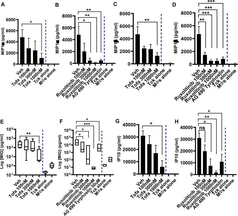
We then evaluated the effect of JAK inhibitors on chemokines, including macrophage inflammatory protein MIP1α and MIP1β, monokine induced by interferon-γ (MIG)⁄CXCL9) and interferon-γ-inducible protein-10 (IP10)/CXCL10. These chemokines induce the synthesis and release of other proinflammatory cytokines that play pathological roles. At rest, macrophages or Tck cells did not produce MIP1α, MIP1β, MIG and IP10 (data not shown). Activation of macrophages with Tck cells induced increased production of MIP1α, MIP1β and MIG. The output of IP10 was generated by the treatment of macrophages with LPS. Treatment with tofacitinib, ruxolitinib or AG 490 tyrphostin reduced the release of MIP1α, MIP1β, MIG and IP10 (figure 2A–H).
Figure 2.
Janus kinase (JAK) inhibitors reduce MIP1α, MIP1β, MIG and IP10 production by activated macrophages. Cytokine-generated cytokine-activated T (Tck) cells were washed and then cocultured with monocyte-derived macrophages (MФs) at a 4:1 ratio. The cells were treated with 0.001% dimethyl sulfoxide (DMSO) (Veh) or tofacitinib (100 nM, 300 nM and 1000 nM) or ruxolitinib (100 nM, 300 nM, 1000 nM, 50 µM). In some experiments, MФs were also cultured alone. Culture supernatants were harvested, and the levels of (A, B) MIP1α; (C, D) MIP1β; (E, F) MIG; and (G, H) IP10 were measured using Luminex. Data represent a mean of triplicate cultures±SE of the mean of at least five experiments. Statistically significant differences are indicated (*p<0.05; **p<0.01; ***p <0.001; ns=not significant). Veh, vehicle.
Discussion
We show that tofacitinib and ruxolitinib, two structurally unrelated small molecule inhibitors, inhibit the production of TNF, IL-6, IL-15, IL-1RA and chemokines MIP1α, MIP1β, MIG and IP10 by macrophages interacting with cytokine activated memory T-cells. This was due to tofacitinib and ruxolitinib preventing cytokine-induced maturation of Tck cells. Thus, tofacitinib and ruxolitinib impact the production of proinflammatory cytokines and chemokines crucial in the pathogenesis of immune-mediated inflammatory diseases. Although the effects of JAK inhibition on macrophages and T-cells have been studied,15–18 no study has thus far demonstrated the impact of JAK inhibition on Tck cell-activated macrophages.
Our observations concur with studies showing that JAK inhibitors mediate the suppression of TNF and IL-6 release, in keeping with the suggestion that JAK inhibitors work, in part, by suppressing the TNF-IFNβ-JAK-STAT1 autocrine loop.17 Tofacitinib, ruxolitinib and AG490 Tyrphostin also led to the inhibition of Tck cell maturation. Likely, the inhibition of IL-6 and IL-15 by tofacitinib and ruxolitinib contributed to the failure to generate mature Tck cells since these cytokines are crucial in forming mature Tck cells, as previously suggested.12 13 Indeed, we previously demonstrated that IL-15 (which signals via JAK1 and JAK3) is sufficient to form Tck cells and alters the balance between effector and memory T cells.13 These observations suggest that JAK inhibition may effectively disrupt cell contact-mediated inflammation at the synovial site and prevent a priori development of Tck cells.
Since Tck cells in our setting were not fixed, it is likely that a small proportion of the cytokines came from Tck cells and not all from macrophages. However, using fixed Tck cells, a previous study demonstrated that Tck cell-activated macrophages induced TNF19; therefore, it is reasonable to suggest that the TNF synthesis seen in our study is of macrophage origin.
The limitation of this study is that the investigation did not include RA patient samples, which would provide direct clinical relevance. However, the data suggest that JAK inhibition may effectively treat established inflammation by disrupting the crosstalk between macrophages and T-cells (online supplemental figure 3). Overall, JAK inhibition may affect multiple steps in mediating inflammation by targeting cytokine and chemokine production and affecting the function of innate and acquired immune cells, contributing to the beneficial effect of JAK inhibitors in inflammatory diseases like RA.
Footnotes
Contributors: MHN analysed the data and wrote the manuscript. JSN performed the assays. MF-G, DSG, SS, DP, CSG and IBM supervised the study and edited the manuscript. All authors approved the final version of the manuscript.
Funding: Funding was provided by vs Arthritis, UK and the Scottish Clinical Pharmacology and Pathology Programme (SCP3), Medical Research Council (Ref: G1000419).
Competing interests: DP has received consultancy and sponsorship from Abbvie and Eli Lilly to attend scientific meetings. SS has received institutional research grants from Amgen (previously Celgene), Boehringer Ingelheim, Bristol Myers Squibb, Eli Lilly, GlaxoSmithKline (GSK), Janssen, and UCB; and honoraria/speaker fees from AbbVie, Amgen, Eli Lilly, GSK, Janssen, and UCB. IBM has received consultancy and research support from Bristol Myers Squibb, Pfizer, Abbvie, Eli Lilly and Gilead, all of whom manufacture inhibitors of JAKs.
Provenance and peer review: Not commissioned; externally peer reviewed.
Supplemental material: This content has been supplied by the author(s). It has not been vetted by BMJ Publishing Group Limited (BMJ) and may not have been peer-reviewed. Any opinions or recommendations discussed are solely those of the author(s) and are not endorsed by BMJ. BMJ disclaims all liability and responsibility arising from any reliance placed on the content. Where the content includes any translated material, BMJ does not warrant the accuracy and reliability of the translations (including but not limited to local regulations, clinical guidelines, terminology, drug names and drug dosages), and is not responsible for any error and/or omissions arising from translation and adaptation or otherwise.
Ethics statements
Patient consent for publication
Not applicable.
Ethics approval
This study complied with the World Medical Association Declaration of Helsinki and was approved by the Glasgow East ethics committee. Participants gave informed consent to participate in the study before taking part.
References
- 1.Croft AP, Campos J, Jansen K, et al. Distinct fibroblast subsets drive inflammation and damage in arthritis. Nature 2019;570:246–51. 10.1038/s41586-019-1263-7 [DOI] [PMC free article] [PubMed] [Google Scholar]
- 2.Alivernini S, MacDonald L, Elmesmari A, et al. Distinct synovial tissue macrophage subsets regulate inflammation and remission in rheumatoid arthritis. Nat Med 2020;26:1295–306. 10.1038/s41591-020-0939-8 [DOI] [PubMed] [Google Scholar]
- 3.Van Raemdonck K, Umar S, Palasiewicz K, et al. CCL21/CCR7 signaling in macrophages promotes joint inflammation and TH17-mediated osteoclast formation in rheumatoid arthritis. Cell Mol Life Sci 2020;77:1387–99. 10.1007/s00018-019-03235-w [DOI] [PMC free article] [PubMed] [Google Scholar]
- 4.Udalova IA, Mantovani A, Feldmann M. Macrophage heterogeneity in the context of rheumatoid arthritis. Nat Rev Rheumatol 2016;12:472–85. 10.1038/nrrheum.2016.91 [DOI] [PubMed] [Google Scholar]
- 5.McInnes IB, Leung BP, Liew FY. Cell-Cell interactions in synovitis. interactions between T lymphocytes and synovial cells. Arthritis Res 2000;2:374–8. 10.1186/ar115 [DOI] [PMC free article] [PubMed] [Google Scholar]
- 6.Brennan F, Foey A. Cytokine regulation in RA synovial tissue: role of T cell/macrophage contact-dependent interactions. Arthritis Res 2002;4 Suppl 3:S177–82. 10.1186/ar556 [DOI] [PMC free article] [PubMed] [Google Scholar]
- 7.Brennan FM, McInnes IB. Evidence that cytokines play a role in rheumatoid arthritis. J Clin Invest 2008;118:3537–45. 10.1172/JCI36389 [DOI] [PMC free article] [PubMed] [Google Scholar]
- 8.Schwartz DM, Kanno Y, Villarino A, et al. Jak inhibition as a therapeutic strategy for immune and inflammatory diseases. Nat Rev Drug Discov 2017;16:843–62. 10.1038/nrd.2017.201 [DOI] [PubMed] [Google Scholar]
- 9.Gao W, McGarry T, Orr C, et al. Tofacitinib regulates synovial inflammation in psoriatic arthritis, inhibiting STAT activation and induction of negative feedback inhibitors. Ann Rheum Dis 2016;75:311–5. 10.1136/annrheumdis-2014-207201 [DOI] [PMC free article] [PubMed] [Google Scholar]
- 10.Fragoulis GE, McInnes IB, Siebert S. JAK-inhibitors. new players in the field of immune-mediated diseases, beyond rheumatoid arthritis. Rheumatology 2019;58:i43–54. 3. 10.1093/rheumatology/key276 [DOI] [PMC free article] [PubMed] [Google Scholar]
- 11.McInnes IB, Byers NL, Higgs RE, et al. Comparison of baricitinib, upadacitinib, and tofacitinib mediated regulation of cytokine signaling in human leukocyte subpopulations. Arthritis Res Ther 2019;21:183. 10.1186/s13075-019-1964-1 [DOI] [PMC free article] [PubMed] [Google Scholar]
- 12.Brennan FM, Hayes AL, Ciesielski CJ, et al. Evidence that rheumatoid arthritis synovial T cells are similar to cytokine-activated T cells: involvement of phosphatidylinositol 3-kinase and nuclear factor kappaB pathways in tumor necrosis factor alpha production in rheumatoid arthritis. Arthritis Rheum 2002;46:31–41. [DOI] [PubMed] [Google Scholar]
- 13.McInnes IB, Leung BP, Sturrock RD, et al. Interleukin-15 mediates T cell-dependent regulation of tumor necrosis factor-alpha production in rheumatoid arthritis. Nat Med 1997;3:189–95. 10.1038/nm0297-189 [DOI] [PubMed] [Google Scholar]
- 14.Cohen S, Zwillich SH, Chow V, et al. Co-administration of the JAK inhibitor CP-690,550 and methotrexate is well tolerated in patients with rheumatoid arthritis without need for dose adjustment. Br J Clin Pharmacol 2010;69:143–51. 10.1111/j.1365-2125.2009.03570.x [DOI] [PMC free article] [PubMed] [Google Scholar]
- 15.Ghoreschi K, Jesson MI, Li X, et al. Modulation of innate and adaptive immune responses by tofacitinib (CP-690,550). J Immunol 2011;186:4234–43. 10.4049/jimmunol.1003668 [DOI] [PMC free article] [PubMed] [Google Scholar]
- 16.Kubo S, Yamaoka K, Kondo M, et al. The JAK inhibitor, tofacitinib, reduces the T cell stimulatory capacity of human monocyte-derived dendritic cells. Ann Rheum Dis 2014;73:2192–8. 10.1136/annrheumdis-2013-203756 [DOI] [PubMed] [Google Scholar]
- 17.Yarilina A, Xu K, Chan C, et al. Regulation of inflammatory responses in tumor necrosis factor-activated and rheumatoid arthritis synovial macrophages by JAK inhibitors. Arthritis Rheum 2012;64:3856–66. 10.1002/art.37691 [DOI] [PMC free article] [PubMed] [Google Scholar]
- 18.Maeshima K, Yamaoka K, Kubo S, et al. The JAK inhibitor tofacitinib regulates synovitis through inhibition of interferon-γ and interleukin-17 production by human CD4+ T cells. Arthritis Rheum 2012;64:1790–8. 10.1002/art.34329 [DOI] [PubMed] [Google Scholar]
- 19.Sebbag M, Parry SL, Brennan FM, et al. Cytokine stimulation of T lymphocytes regulates their capacity to induce monocyte production of tumor necrosis factor-alpha, but not interleukin-10: possible relevance to pathophysiology of rheumatoid arthritis. Eur J Immunol 1997;27:624–32. 10.1002/eji.1830270308 [DOI] [PubMed] [Google Scholar]
Associated Data
This section collects any data citations, data availability statements, or supplementary materials included in this article.
Supplementary Materials
rmdopen-2022-002671supp001.pdf (424.2KB, pdf)


