Abstract
We report here that sera of children and adults infected with Schistosoma mansoni, S. haematobium, or S. japonicum contain antibodies against GalNAcβ1-4(Fucα1-2Fucα1-3)GlcNAc (LDN-DF) and to a lesser extent to Galβ1-4(Fucα1-3)GlcNAc (Lewisx) and GalNAcβ1-4GlcNAc (LDN). Surface plasmon resonance (SPR) spectroscopy was used to monitor the presence of serum antibodies to neoglycoconjugates containing these carbohydrate epitopes and to define the immunoglobulin M (IgM) and IgG subclass distribution of the antibodies. The serum levels of antibodies to LDN-DF are high related to LDN and Lewisx for all examined groups of Schistosoma-infected individuals. A higher antibody response to the LDN-DF epitope was found in sera of infected children than in sera of infected adults regardless of the schistosome species. With respect to the subclasses, we found surprisingly that individuals infected with S. japonicum have predominantly IgG antibodies, while individuals infected with S. mansoni mainly show an IgM response; high levels of both isotypes were measured in sera of individuals infected with S. haematobium. These data provide new insights in the human humoral immune response to schistosome-derived glycans.
In schistosomiasis, a major tropical parasitic disease caused mainly by Schistosoma mansoni, S. haematobium, and S. japonicum, a strong humoral immune response against several schistosomal glycoconjugates in infected animals or humans has been found (10, 20). Vaccination studies of mice have shown that the humoral immune response contributes to protection and that protective sera contain anticarbohydrate antibodies (5, 22). The structures of a number of potentially immunogenic schistosome glycans have recently been identified (reviewed in references 3, 4, and 24). Among these structures are glycans containing the Galβ1-4(Fucα1-3)GlcNAc (Lewisx), the GalNAcβ1-4GlcNAc (LacdiNAc, LDN), or the GalNAcβ1-4(Fucα1-3)GlcNAc (LDN-F) antigen. Other glycans that have been structurally defined are the circulating cathodic antigen (CCA) and circulating anodic antigen (2, 23) and several multifucosylated structures present on O-glycans of the cercarial glycocalyx and on egg glycoproteins and egg glycosphingolipids (12–14).
Antibodies from sera of animals or humans infected with Schistosoma have been shown to recognize several of these glycan epitopes. A strong and early humoral immune response has been found against CCA (6), a poly-Lewisx-containing excretory glycoconjugate antigen originating from the schistosome gut (23). Cytolytic immunoglobulin M (IgM) and IgG antibodies directed against Lewisx-containing structures have been demonstrated in infected humans and primates (17, 25). Patients infected with S. mansoni elicit antibodies against CCA which also show binding to synthetic trimeric Lewisx but with lower affinity (1). Nyame et al.(19) reported that mice infected with S. mansoni generate IgM and IgG antibody responses to the LDN epitope.
Recently, we constructed neoglycoproteins containing the glycan structures Lewisx, LDN, LDN-F, and GalNAcβ1-4(Fucα1-2Fucα1-3)GlcNAc (LDN-DF). From a large panel of monoclonal antibodies (MAbs) derived from Schistosoma- infected mice, several MAbs recognizing the aforementioned glycan epitopes were obtained. This indicates that in mice, an immune response against antigens containing these epitopes is elicited during infection (28).
In this study, we determined the antibody responses against Lewisx, LDN, and LDN-DF in different groups of patients infected with S. mansoni, S. japonicum, or S. haematobium, using surface plasmon resonance (SPR) analysis. These data provide new insights in the human humoral immune response to schistosomal glycans and the diagnostic potential of the constructed neoglycoproteins.
MATERIALS AND METHODS
Sera.
Sera from S. mansoni- and S. haematobium-infected individuals were obtained from the WHO/TDR Reference Serum Bank for African Schistosomiasis. These sera were collected in areas in Kenya where Schistosomiasis is endemic. Sera from S. japonicum-infected subjects were obtained from the Philippines (29). All sera were selected on the basis of positive stool egg counts in case of S. mansoni and S. japonicum or positive urine egg counts in the case of S. haematobium. Negative controls consisted of 20 sera from Dutch blood donors with no history of schistosomiasis. The characteristics of the study groups are given in Table 1.
TABLE 1.
Characteristics of schistosomiasis patient and control groups
| Group | n | Minimum–maximum (median) age | Minimum–maximum (median) S. mansoni or S. japonicum eggs/g of feces or S. haematobium eggs/10 ml of urine |
|---|---|---|---|
| S. haematobium | |||
| Children | 12 | 10–18 (13.5) | 2–624 (36) |
| Adults | 8 | 19–53 (22) | 0.3–17 (7.5) |
| S. mansoni | |||
| Children | 10 | 8–17 (12) | 7–3,287 (180) |
| Adults | 12 | 19–53 (32.5) | 7–1,680 (100) |
| S. japonicum | |||
| Children | 12 | 8–18 (12.5) | 23–9,913 (161) |
| Adults | 12 | 25–48 (31) | 23–4,600 (86.5) |
| Control | 20 | 19–52 (34) |
SPR spectroscopy.
SPR analysis was carried out using a BIAcore 3000 instrument with a computer interface for system control, data acquisition, and data analysis (Biacore AB, Uppsala, Sweden). Sensor chip CM5, surfactant P-20 (Tween 20), and an amine coupling kit were also obtained from Biacore AB.
Lewisx, LDN, and LDN-DF were enzymatically synthesized and coupled to bovine serum albumin (BSA) as described elsewhere (28). The amounts of (molecules) oligosaccharides per molecule of BSA were 14, 12, and 11 for Lewisx, LDN, and LDN-DF, respectively. The neoglycoproteins were immobilized at a flow rate of 5 μl/min in 10 mM sodium acetate (pH 4.0) onto a carboxylmethylated dextran CM5 sensor chip by covalent amine coupling according to the instructions of the manufacturer until an increase of approximately 4,000 response units (RU) was observed. All analyses were performed at a flow rate of 5 μl/min at 25°C using HEPES-buffered saline as an eluent. Sera were diluted 1:40 in running buffer with 0.5% P-20. Injection times of sera were 2 min followed by 2 min of buffer injection to allow dissociation. The isotypes of the antibodies were determined subsequently by 2-min injection pulses with goat anti-human IgM (GaHuIgM) and goat anti-human IgG (GaHuIgG), each followed by 2 min of dissociation time. Both anticlass antibodies were diluted 1:100 in running buffer with 0.5% P-20. Regeneration was performed using a 2-min pulse of 100 mM HCl.
Data analysis.
The data were analyzed using the BIA evaluation software (version 3.0). To correct for refractive index change and nonspecific binding, the BSA control surface was used as a blank. The negative control group was used for cutoff definition (average and 3 standard deviations).
Spearman's rank correlation coefficients were computed to check associations between the various BSA-corrected responses. The correlation of total serum antibody responses with IgG and IgM and the sum of IgG and IgM responses (total) were calculated. Statistical analysis was performed using the SPSS for Windows statistical package (SPSS Inc., Chicago, Ill.).
RESULTS
In this study, sera of individuals infected either with S. japonicum, S. mansoni, or S. haematobium were analyzed by SPR for binding of IgG and IgM antibodies to the immobilized LDN, Lewisx, and LDN-DF epitopes. Sera of uninfected individuals were used as a control. For each sample, the total antibody responses as well as the specific IgG and IgM antibody responses were determined in a single run. Similar IgG and IgM levels were measured independent of the order of administration of the GaHuIgG and GaHuIgM antibodies. The sensor chips have been regenerated at least 500 times with excellent reproducibility of the measurements.
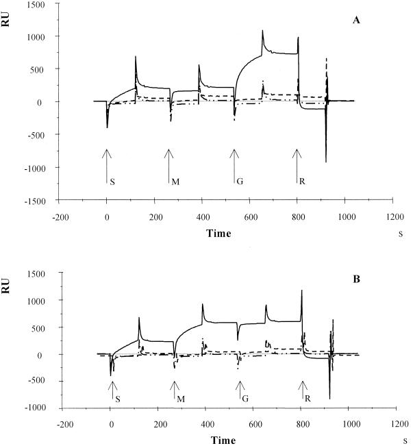
A typical example of a sensorgram illustrating binding of antibodies to different neoglycoproteins and subsequent isotype determination is shown in Fig. 1. The antiglycan antibody levels (IgG and IgM) are summarized in Table 2. In all groups of patients, the median of the antibody responses to LDN as well as to Lewisx was found to be lower than the median of responses to LDN-DF. Only adult individuals infected with S. haematobium showed elevated antibody responses for the LDN epitope.
FIG. 1.
Sensorgram illustrating binding of serum antibodies interacting with LDN (–––), Lewisx (—- · ·), and LDN-DF (——) for individuals infected with S. japonicum (A) or S. mansoni (B). The times of start of injection of serum (S), followed by GaHuIgM (M), GaHuIgG (G), and 100 mM HCl (R), are indicated by arrows.
TABLE 2.
Ranges of IgM and IgG antibody levels for all groups of schistosomiasis patients to the LDN-DF, LDN, and Lewisx epitopes
| Response in: | Minimum–maximum (median) RUa
|
|||||
|---|---|---|---|---|---|---|
| IgG
|
IgM
|
|||||
| LDN-DF | LDN | Lewisx | LDN-DF | LDN | Lewisx | |
| Children | ||||||
| S. haematobium | 24–571 (122.5) | 25–179 (39.5) | 5–35 (13) | 43–335 (165) | 10–120 (46) | 13–35 (18) |
| S. mansoni | 3–1351 (35) | 7–176 (25.5) | 2–30 (6) | 45–348 (103.5) | 19–74 (32) | 11–28 (12) |
| S. japonicum | 33–382 (157) | 12–186 (28.5) | 4–11 (6.5) | 15–222 (53.5) | 8–125 (38.5) | 5–37 (12) |
| Adults | ||||||
| S. haematobium | 42–254 (97.5) | 14–155 (62) | 0–18 (15.5) | 31–528 (122) | 33–169 (86.5) | −3–99 (11) |
| S. mansoni | 16–125 (24.5) | 14–185 (27) | 5–27 (7) | 5–165 (52.5) | 3–50 (25) | 6–19 (10.5) |
| S. japonicum | 36–515 (75) | 17–79 (43) | 1–38 (7) | 12–62 (25) | −8–54 (18.5) | 3–17 (6) |
| Control | 9–28 (12) | 7–77 (11) | −17–19 (3) | 10–24 (15) | −5–39 (11.5) | −26–35 (5.5) |
After subtraction of the response of the BSA surface.
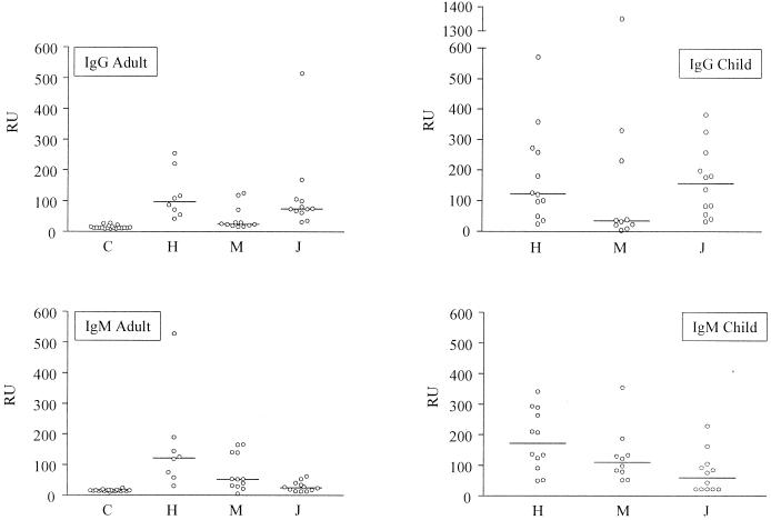
The anti-LDN-DF antibody responses (IgG and IgM) were analyzed in more detail (Fig. 2). In general, all groups of children gave higher antibody responses towards the LDN-DF epitope than the adult groups. Infection with S. japonicum seems to induce mainly an IgG antibody response against the LDN-DF epitope, while patients infected with S. mansoni mainly have an IgM response. In sera of individuals infected with S. haematobium, high levels of both IgM and IgG were found.
FIG. 2.
Levels of IgG and IgM antibodies binding to the LDN-DF epitope for individuals infected with S. mansoni (M), S. japonicum (J), or S. haematobium (H) and uninfected individuals (C). Thin lines indicate medians.
The specificity for schistosomiasis of the IgG and IgM responses to the different glycan epitopes was determined. Sera of 20 uninfected individuals were used as a negative control. For each isotype, the percentage of positive sera was calculated (Table 3). Almost all infected individuals displayed a clear positive immune response to the LDN-DF epitope, whereas a lower and more variable positive response was shown to the LDN or the Lewisx epitope. Individuals infected with S. haematobium had both IgG and IgM antibodies against the LDN-DF epitope (children, 92 and 100% respectively; adults, 100 and 100%, respectively). Sera of individuals infected with S. japonicum contained predominantly IgG antibodies against the LDN-DF epitope (children, 92% for IgG and 58% for IgM; adults, 92% for IgG and 50% for IgM), while individuals infected with S. mansoni showed mainly IgM antibodies (children, 50% for IgG and 100% for IgM; adults, 25% for IgG and 83% for IgM). To check if the most important isotypes reacting with the LDN-DF epitope had been identified, Spearman's rank correlations were calculated between the different RU levels measured for total serum antibody levels and IgG and/or IgM separately. It was shown that associations between total serum antibody responses and either IgM or IgG were in most cases highly significant. Associations between the sum of the two isotype responses and the total serum antibody responses were in all cases highly significant (Table 4). This implies that by measuring IgM and IgG antibody classes, the most abundant isotypes were identified. In the cases of Lewisx and LDN, which display low RU, no associations were found (data not shown).
TABLE 3.
Adults and children infected with S. mansoni, S. japonicum or S. haematobium testing positive for the LDN-DF, LDN, and Lewisx epitope
| Group | % Positivea
|
|||||
|---|---|---|---|---|---|---|
| LDN-DF
|
LDN
|
Lewisx
|
||||
| IgG | IgM | IgG | IgM | IgG | IgM | |
| Children | ||||||
| S. haematobium | 92 | 100 | 17 | 66 | 42 | 100 |
| S. mansoni | 50 | 100 | 30 | 20 | 20 | 50 |
| S. japonicum | 92 | 58 | 17 | 33 | 0 | 50 |
| Adults | ||||||
| S. haematobium | 100 | 100 | 38 | 63 | 63 | 50 |
| S. mansoni | 25 | 83 | 25 | 8 | 8 | 63 |
| S. japonicum | 92 | 50 | 25 | 17 | 17 | 8 |
| Control | 0 | 0 | 0 | 5 | 10 | 5 |
Cutoff levels were based on the negative controls average and 3 standard deviations.
TABLE 4.
Correlations between total serum antibody responses and the sum of IgG and IgM or IgG and IgM separately against the LDN-DF epitope measured in adults and children infected with S. mansoni, S. japonicum, or S. haematobium
| Group | LDN-DF
|
||
|---|---|---|---|
| IgM + IgG | IgG | IgM | |
| Children | |||
| S. haematobium | 0.986a | 0.979a | 0.937a |
| S. mansoni | 0.936a | 0.505 | 0.474 |
| S. japonicum | 0.977a | 0.935a | 0.861a |
| Adults | |||
| S. haematobium | 0.802b | 0.095 | 0.738a |
| S. mansoni | 0.979a | 0.732a | 0.867a |
| S. japonicum | 0.904a | 0.739a | −0.221 |
Correlation is significant at the 0.05 level (two-tailed test).
Correlation is significant at 0.01 level (two-tailed test).
DISCUSSION
An increasing number of studies indicate that carbohydrates on glycoproteins, glycolipids, and glycosaminoglycans synthesized by schistosomes are targets of humoral immunity and may play a role in modulating host immune responses. To achieve more insight in the host's immune response to schistosomes, we used neoglycoconjugates in combination with SPR technology to analyze sera of infected individuals for their antibody reactivities with specific glycans. The advantage of using SPR technology over enzyme-linked immunosorbent assay (ELISA) is that only small amounts of the neoglycoconjugates, which are usually hard to synthesize and are available only in low quantities, are needed. In addition, the sensor chips can be regenerated many times. The SPR analysis is fully automatic, and in one run different subtypes of immunoglobulins can be determined without the use of labels.
The LDN, Lewisx, and LDN-DF epitopes studied here have been shown by structural analysis to occur in schistosomes (12, 13). In several studies, mouse anti-carbohydrate MAbs that recognize these epitopes have been identified (7, 18, 19, 28). These MAbs were isolated from Schistosoma-infected or -immunized mice. This reactivity confirms that the respective carbohydrate epitopes are indeed present on schistosomes and indicates that at least in the mouse, they are presented to the immune system. Furthermore, immunofluorescent assay studies showed that the glycans mentioned above are expressed by the schistosomes in various life cycle stages (28).
In this study, we demonstrate immunoreactivity against the LDN-DF epitope and to a lesser extent to the LDN and Lewisx epitopes in sera of different groups of patients, each infected with one of the three major species of Schistosoma. Interestingly, it was found that anti-LDN-DF antibodies from individuals infected with S. japonicum were predominantly of the IgG isotype and those from S. mansoni-infected individuals were of the IgM isotype, whereas both isotypes were observed in S. haematobium infected individuals. We have no clear explanation for this finding.
Sera of animals or humans infected with Schistosoma contained Lewisx antibodies (15, 17, 23), and recently Nyame et al. found LDN-reactive antibodies in mice infected with S. mansoni (19). In this study, high levels of LDN-DF-binding antibodies were observed in Schistosoma-infected individuals, which indicates that LDN-DF-containing glycoconjugates are strongly immunogenic. Antibody reactivities against LDN and Lewisx were found to be much lower than those against LDN-DF, possibly because those epitopes are not unique for Schistosoma but are also expressed on a number of human glycoproteins. For example, the LDN sequence has been found on several vertebrate glycoproteins (26). Lewisx epitopes have been found on a number of human tissues, cells, glycoproteins, and glycolipids, e.g., on α1-acid glycoprotein, granulocytes and respiratory mucins (8, 27). Still, anti-LDN and anti-Lewisx antibodies were detected in sera of most patients, possibly due to the persistent presentation of the glycan antigen to the immune system. An alternative explanation may be that these carbohydrate structures presented by the schistosomes to the immune system as a repetitive glycan give rise to a more immunogenic and specific epitope, as illustrated by the strong and early antibody response against CCA, a poly-Lewisx-containing structure (6).
The anti-poly-Lewisx antibodies from infected humans are potent in mediating the complement dependent cytolysis in vitro of human granulocytes, thus implying an autoimmune phenomenon (25). Lewisx glycans may alter cellular immunity in infected hosts, by facilitating a shift from Th1 and Th2 response (21, 30). Recently, it was demonstrated that mice sensitized with Lewisx-containing glycoconjugates displayed an increased cellular immune response toward soluble egg antigen-coated beads implanted in the liver, resulting in the formation of large periparticular granulomas (11). The effect of LDN or LDN-DF on the cellular immune response is not known. In view of the highly immunogenic character of LDN-DF, it may be postulated that LDN-DF containing glycoconjugates also play an important role in the pathology of schistosomiasis.
In a previous study, we showed that MAb 114-5B1-A recognizes the LDN-DF epitope (28). Using this antibody in a capture ELISA, circulating soluble egg antigens were detected in serum pools of mice heavily infected with S. mansoni. This assay was shown to be useful for the quantitative determination of egg antigens in urine samples of S. haematobium-infected individuals (16). This illustrates that in the infected host, antigens with the LDN-DF epitope are continuously released by the eggs in relatively large amounts. The anti-LDN-DF antibodies measured in the present study may be directed against cercarial and worm as well as egg antigens, since immunofluorescent assay studies have shown that each of the different stages stain to some extent with MAb 114-5B1-A (28). However, in view of the relatively abundant release of LDN-DF-containing egg antigens in relation to LDN-DF-containing worm antigens, it is likely that the antibody responses measured here are mainly directed against these egg antigens.
In view of the number of sera having a positive antibody response against the LDN-DF epitope, this glycan epitope might be useful in the development of a specific diagnostic assay for Schistosoma infections. A high sensitivity was observed in particular for patients infected with S. haematobium, which makes the LDN-DF interesting as a possible diagnostic tool in follow-up and reinfection studies for individuals infected with this species.
In conclusion, it should be mentioned that SPR is a valuable technique for monitoring antiglycan antibody levels in serum. Using this technique, we have shown that humans infected with Schistosoma display specific antibody responses against the LDN, the Lewisx, and in particular the LDN-DF epitope.
ACKNOWLEDGMENTS
We are grateful to L. van Lieshout for helpful discussion. We are also grateful to T. M. Falcao Ferreira for providing the control sera and D. Kornelis for critical reading of the manuscript. We acknowledge the WHO/TDR Reference Serum Bank for African Schistosomiasis for providing the sera.
This study was supported by The Netherlands Foundation for Chemical Research and Life Science Foundation, with financial aid from The Netherlands Organization for Scientific Research.
REFERENCES
- 1.Appelmelk B J, Simmons-Smit I M, Verboom T, Negrini R, van Dam G J, Moran A P, Vandenbroucke-Grauls C M J E. Serological response to Helicobacter pylori lipopolysaccharide. In: Moran A P, O'Morain C A, editors. Pathogenesis and host response in Helicobacter pylori infections. Englewood, N.J: Normed Verlag; 1997. pp. 43–52. [Google Scholar]
- 2.Bergwerff A A, van Dam G J, Rotmans J P, Deelder A M, Kamerling J P, Vliegenthart J F. The immunologically reactive part of immunopurified circulating anodic antigen from Schistosoma mansoni is a threonine-linked polysaccharide consisting of → 6)-(β-d-GlcpA-(1 → 3))-β-d-GalpNAc-(1 → repeating units. J Biol Chem. 1994;269:31510–31517. [PubMed] [Google Scholar]
- 3.Cummings R D, Nyame A K. Glycobiology of schistosomiasis. FASEB J. 1996;10:838–848. doi: 10.1096/fasebj.10.8.8666160. [DOI] [PubMed] [Google Scholar]
- 4.Cummings R D, Nyame A K. Schistosome glycoconjugates. Biochim Biophys Actal. 1999;1455:363–374. doi: 10.1016/s0925-4439(99)00063-0. [DOI] [PubMed] [Google Scholar]
- 5.Dalton J P, Strand M, Mangold B L, Dean D A. Identification of Schistosoma mansoni glycoproteins recognized by protective antibodies from mice immunized with irradiated cercariae. J Immunol. 1986;136:4689–4694. [PubMed] [Google Scholar]
- 6.Deelder A M, De Jonge N, Boerman O C, Fillié Y E, Hilberath G W, Rotmans J P, Gerritse M J, Schut D W O A. Sensitive determination of circulating anodic antigen in Schistosoma mansoni infected individuals by an enzyme-linked immunosorbent assay using monoclonal antibodies. Am J Trop Med Hyg. 1989;40:939–943. doi: 10.4269/ajtmh.1989.40.268. [DOI] [PubMed] [Google Scholar]
- 7.Deelder A M, van Dam G J, Kornelis D, Fillié Y E, van Zeyl R J M. Schistosoma: analysis of monoclonal antibodies reactive with the circulating antigens CCA and CAA. Parasitology. 1996;112:21–35. doi: 10.1017/s0031182000065045. [DOI] [PubMed] [Google Scholar]
- 8.De Graaf T W, van der Stelt M E, Angebergen M G, van Dijk W. Inflammation-induced expression of sialyl Lewis x-containing glycan structures on alphal-acid glycoprotein (orosomucoid) in human sera. J Exp Med. 1993;177:657–666. doi: 10.1084/jem.177.3.657. [DOI] [PMC free article] [PubMed] [Google Scholar]
- 9.Dell A, Morris H R, Easton R L, Panico M, Patankar M, Oehniger S, Koistinen R, Koistinen H, Seppala M, Clark G F. Structural analysis of the oligosaccharides derived from glycodelin, a human glycoprotein with potent immunosuppressive and contraceptive activities. J Biol Chem. 1995;270:24116–24126. doi: 10.1074/jbc.270.41.24116. [DOI] [PubMed] [Google Scholar]
- 10.Dunne D W. Schistosome carbohydrates. Parasitol Today. 1990;6:45–48. doi: 10.1016/0169-4758(90)90068-f. [DOI] [PubMed] [Google Scholar]
- 11.Jacobs W, Deelder A M, Van Marck E. Schistosomal granuloma modulation. II. Specific immunogenic carbohydrates can modulate schistosome-egg-antigen-induced hepatic granuloma formation. Parasitol Res. 1999;85:14–18. doi: 10.1007/s004360050500. [DOI] [PubMed] [Google Scholar]
- 12.Khoo K-H, Sarda S, Xu X, Caulfield J P, McNeil M R, Homans S W, Morris H R, Dell A. A unique multifucosylated -3GalNAcβ1-4GlcNAcβ1-3Galα1- motif constitutes the repeating unit of the complex O-glycans derived from the cercarial glycocalyx of Schistosoma mansoni. J Biol Chem. 1995;270:17114–17123. doi: 10.1074/jbc.270.29.17114. [DOI] [PubMed] [Google Scholar]
- 13.Khoo K-H, Chatterjee D, Caulfield J P, Morris H R, Dell A. Structural characterization of glycosphingolipids from the eggs of Schistosoma mansoni and Schistosoma japonicum. Glycobiology. 1997;7:653–661. doi: 10.1093/glycob/7.5.653. [DOI] [PubMed] [Google Scholar]
- 14.Khoo K-H, Chatterjee D, Caulfield J P, Morris H R, Dell A. Structural mapping of the glycans from the egg glycoproteins of Schistosoma mansoni and Schistosoma japonicum—identification of novel core structures and terminal sequences. Glycobiology. 1997;7:663–677. doi: 10.1093/glycob/7.5.663. [DOI] [PubMed] [Google Scholar]
- 15.Ko A I, Drager U C, Harn D A. A Schistosoma mansoni epitope recognized by a protective monoclonal antibody is identical to the stage-specific embryonic antigen 1. Proc Natl Acad Sci USA. 1990;87:4159–4163. doi: 10.1073/pnas.87.11.4159. [DOI] [PMC free article] [PubMed] [Google Scholar]
- 16.Nibbeling H M, Kahama A I, Van Zeyl R M, Deelder A M. Use of monoclonal antibodies prepared against Schistosoma mansoni hatching fluid antigens for demonstration of circulating egg antigens in urine. Am J Trop Med Hyg. 1998;58:543–550. doi: 10.4269/ajtmh.1998.58.543. [DOI] [PubMed] [Google Scholar]
- 17.Nyame A K, Pilcher J B, Tsang V C W, Cummings R D. Schistosoma mansoni infection in humans and primates induces cytolytic antibodies to surface Lex determinants on myeloid cells. Exp Parasitol. 1996;82:191–200. doi: 10.1006/expr.1996.0024. [DOI] [PubMed] [Google Scholar]
- 18.Nyame A K, Debose-Boyd R, Long T D, Tsang V C W, Cummings R D. Expression of Lex antigen in Schistosoma japonicum and S. haematobium and immune responses to Lex in infected animals: lack of Lex expression in other trematodes and nematodes. Glycobiology. 1998;8:615–624. doi: 10.1093/glycob/8.6.615. [DOI] [PubMed] [Google Scholar]
- 19.Nyame A K, Leppänen A M, Debose-Boyd R, Cummings R D. Mice infected with Schistosoma mansoni generate antibodies to LDN (GalNAcβ1-4GlcNAc) determinants. Glycobiology. 1999;9:1029–1035. doi: 10.1093/glycob/9.10.1029. [DOI] [PubMed] [Google Scholar]
- 20.Omer Ali P, Smithers S R, Bickle Q, Phillips S M, Harn D, Simpson A J G. Analysis of the anti-Schistosoma mansoni surface antibody response during murine infection and its potential contribution to protective immunity. J Immunol. 1988;140:258–264. [PubMed] [Google Scholar]
- 21.Palanivel V, Posey C, Horauf A M, Solbach W, Piessens W F, Harn D A. B-cell outgrowth and ligand-specific production of IL-10 correlate with Th2 dominance in certain parasitic diseases. Exp Parasitol. 1996;84:168–177. doi: 10.1006/expr.1996.0102. [DOI] [PubMed] [Google Scholar]
- 22.Richter D, Incani R N, Harn D A. Lacto-N-fucopantaose III (Lewisx), a target of the antibody response in mice vaccinated with irradiated cercariae of Schistosoma mansoni. Infect Immun. 1996;64:1826–1831. doi: 10.1128/iai.64.5.1826-1831.1996. [DOI] [PMC free article] [PubMed] [Google Scholar]
- 23.van Dam G J, Bergwerff A A, Thomas-Oates J E, Rotmans J P, Kamerling J P, Vliegenthart J F G, Deelder A M. The immunological reactive O-linked polysaccharide chains derived from circulating cathodic antigen isolated from the human blood fluke Schistosoma mansoni have Lewisx as repeating unit. Eur J Biochem. 1994;225:467–482. doi: 10.1111/j.1432-1033.1994.00467.x. [DOI] [PubMed] [Google Scholar]
- 24.van Dam G J, Deelder A M. Glycoproteins of parasites. In: Montreuil J, Vliegenthart J F G, Schachter H, editors. Glycoproteins and disease. Amsterdam, The Netherlands: Elsevier; 1996. pp. 159–182. [Google Scholar]
- 25.van Dam G J, Claas F H J, Yazdanbakhsh M, Kruize Y C M, van Keulen A C I, Ferreira S T M F, Rotmans J P, Deelder A M. Schistosoma mansoni excretory circulating cathodic antigen shares Lewis-x epitopes with a human granulocyte surface antigen and evokes host antibodies mediating complement-dependent lysis of granulocytes. Blood. 1996;88:4246–4251. [PubMed] [Google Scholar]
- 26.van den Eijnden D H, Bakker H, Neeleman A P, van den Nieuwenhof I M, van Die I. Novel pathways in complex-type oligosaccharide synthesis: new vistas opened by studies in invertebrates. Biochem Soc Trans. 1997;25:887–893. doi: 10.1042/bst0250887. [DOI] [PubMed] [Google Scholar]
- 27.van Kuik J A, de Waard P, Vliegenthart J F, Klein A, Carnoy C, Lamblin G, Roussel P. Isolation and structural characterization of novel neutral oligosaccharide-alditols from respiratory-mucus glycoproteins of a patient suffering from bronchiectasis. 2. Structure of twelve hepta-to-nonasaccharides, six of which possess the GlcNAc beta(1-3)[Gal beta(1-4)GlcNAc beta(1-6)]Gal beta(1-3)GalNAc-ol common structural element. Eur J Biochem. 1991;198:169–182. doi: 10.1111/j.1432-1033.1991.tb15998.x. [DOI] [PubMed] [Google Scholar]
- 28.van Remoortere A, Hokke C H, van Dam G J, van Die I, Deelder A M, van den Eijnden D H. Various stages of Schistosoma express Lewisx, LacdiNAc, GalNAcβ1-4(Fucα1-3)GlcNAc and GalNAcβ1-4(Fucα1-2Fucα1-3)GlcNAc carbohydrate epitopes: detection with monoclonal antibodies that are characterized by enzymatically synthesized neoglycoproteins. Glycobiology. 2000;10:601–609. doi: 10.1093/glycob/10.6.601. [DOI] [PubMed] [Google Scholar]
- 29.van ‘t Wout A B, De Jonge N, Tiu W U, Garcia E E, Mitchell G F, Deelder A M. Schistosome circulating anodic antigen in serum of individuals infected with Schistosoma japonicum from the Philippines before and after chemotherapy with praziquantel. Trans R Soc Trop Med Hyg. 1992;86:410–413. doi: 10.1016/0035-9203(92)90243-6. [DOI] [PubMed] [Google Scholar]
- 30.Velupillai P, Harn D A. Oligosaccharide-specific induction of interleukin 10 production by B220+ cells from schistosome-infected mice: a mechanism for regulation of CD4+ T-cell subsets. Proc Natl Acad Sci USA. 1994;91:18–22. doi: 10.1073/pnas.91.1.18. [DOI] [PMC free article] [PubMed] [Google Scholar]


