Abstract
Background: Decreased peak oxygen consumption during exercise (peak Vo2) is a well-established prognostic marker for mortality in ambulatory heart failure. After heart transplantation, the utility of peak Vo2 as a marker of post-transplant survival is not well established. Methods and Results: We performed a retrospective analysis of adult heart transplant recipients at the Hospital of the University of Pennsylvania who underwent cardiopulmonary exercise testing within a year of transplant between the years 2000 to 2011. Using time-to-event models, we analyzed the hazard of mortality over nearly two decades of follow-up as a function of post-transplant percent predicted peak Vo2 (%Vo2). A total of 235 patients met inclusion criteria. The median post-transplant %Vo2 was 49% (IQR 42 to 60). Each standard deviation (±14%) increase in %Vo2 was associated with a 32% decrease in mortality in adjusted models (HR 0.68, 95% CI 0.53 to 0.87, p = 0.002). A %Vo2 below 29%, 64% and 88% predicted less than 80% survival at 5, 10, and 15 years, respectively. Conclusions: Post-transplant peak Vo2 is a highly significant prognostic marker for long-term post-transplant survival. It remains to be seen whether decreased peak Vo2 post-transplant is modifiable as a target to improve post-transplant longevity.
Keywords: transplant, exercise, survival, prognosis
1. Introduction
Diminished exercise capacity is a cardinal feature of advanced heart failure, and a strong association between decreased peak oxygen consumption (Vo2) and increased mortality has been recognized in ambulatory heart failure patients for decades [1,2]. Given its association with mortality, peak Vo2 is commonly used to estimate prognosis, and current practice guidelines recommend its use as a threshold for advanced heart failure therapies [3]. Mechanistically, however, the relationship between Vo2 and mortality in heart failure with reduced ejection fraction (HFrEF) is confounded by multiple convergent pathophysiologic processes resulting in the diminished consumption of oxygen. While it is plausible that decreased peak Vo2 in HFrEF reflects, at least in part, inadequate tissue oxygen delivery during exercise due to a reduced cardiac output [4], a dissociation between peak Vo2 and hemodynamic dysfunction has long been observed [5].
Hypothetically, if cardiac output were the main limitation barring further increases in peak Vo2 then peak Vo2 should increase dramatically after heart transplantation, and Vo2 would no longer distinguish risk for mortality. Yet, across the population of heart-transplant recipients, post-transplant Vo2 is highly heterogeneous [6,7]. Peak Vo2 only improves in a small number of individuals, and those with the lowest pre-transplant peak Vo2 often have the lowest post-transplant Vo2 [8]. The reasons for this and the usefulness of Vo2 as a marker of short- or long-term survival are not well understood. Significant extracardiac parameters may mediate the link between mortality and peak Vo2, and advanced heart failure therapies that treat cardiac failure may not directly influence these parameters, including mechanical circulatory support and heart transplantation.
Because multiple comorbidities factor into the peak Vo2, there may be significant information captured in this metric post-transplant, even after transplant. This information could be clinically useful, improving risk-stratification and shedding new light on causal mechanisms for patient decline and transplant outcomes, with the potential for intervention. In this study, we sought to (1) quantify the association between post-transplant peak Vo2 and mortality and (2) identify potential pathophysiologic processes that may explain this association.
2. Materials and Methods
The first and last authors (T.C.H. and E.Y.B.) had full access to all the data in the study and take responsibility for its integrity and the data analysis. Requests to access deidentified data from qualified researchers trained in human subject confidentiality protocols should be sent to the corresponding author. Unidentified Stata code for this analysis is available upon request.
2.1. Design and Participants
We performed a retrospective analysis of all adult heart transplant recipients who were transplanted from March 2000 to November 2011 at the Hospital of the University of Pennsylvania, excluding multiorgan recipients and patients undergoing cardiac re-transplantation. Survival status was observed through October 2019 using data merged from the Penn Transplant Institute. This era was selected due to a clinical protocol at the time in which all patients underwent a post-transplant cardiopulmonary exercise test (CPET) within one year of transplant if clinically able. Patients who underwent CPET outside of their first post-transplant year were excluded, as were four patients with missing peak Vo2. Baseline pre- and post-transplant clinical data were recorded from the electronic medical record, including demographics, pre- and post-transplant comorbidities up to the time of CPET, laboratory values at the time of CPET, post-transplant echocardiographic data up to one year post-CPET, the incidence of acute cellular rejection grade 2R or 3R or antibody mediated rejection prior to CPET, and the CPET results themselves. This study was approved by the University of Pennsylvania Institutional Review Board with a waiver of informed consent.
2.2. Cardiopulmonary Exercise Metrics
Data from CPET were acquired retrospectively from the electronic medical record. All patients performed one symptom-limited CPET within the first post-transplant year according to our standard clinical practice, using a treadmill with a low- or moderate-intensity ramped protocol until exhaustion, the onset of limiting symptoms, or the development of a contraindication to continued exercise. Breath-by-breath expired gas analysis was used to obtain continuous estimates of Vo2 and carbon dioxide production (Vco2) at peak exercise. Studies were interpreted by an advanced heart failure cardiologist.
Absolute peak Vo2 was determined as the highest 10-s averaged samples obtained during exercise. We estimated the % of predicted peak Vo2 using the Fitness Registry and the Importance of Exercise National Database (FRIEND) equation based on age, sex, and weight [9]. Peak Vo2 is conventionally standardizing for total body mass; however, because Vo2 is influenced primarily by muscle mass and can be reduced out of proportion to heart failure severity in obese subjects [10], we examined values of Vo2 indexed separately for total body mass in kilograms (peak Vo2, body mass) and estimated lean body mass (peak Vo2, lean). Lean body mass was estimated from weight, height, age, and ethnicity using a formula validated against dual-energy X-ray absorptiometry (i.e., DXA scan) in a sample of 14,000 US adults [11]. Heart rate, systolic blood pressure, and diastolic blood pressure were recorded at rest and at peak exercise. O2 pulse was calculated as the ratio of absolute peak Vo2 to peak heart rate.
The respiratory exchange ratio (RER) was determined from the ratio of Vco2 to Vo2 at the time of exhaustion. Maximal Volitional exercise was determined using a peak RER ≥ 1.0, based on guideline recommendation and application in recent large studies of exercise in HFrEF [12,13,14,15]. Only studies demonstrating evidence of maximal effort were used in analyses. The first post-transplant transthoracic echocardiogram obtained up to one year after CPET was used to compare peak Vo2 to the left ventricular ejection fraction.
2.3. Statistical Analysis
Continuous data are expressed as mean ± standard deviation (SD), while categorical data are expressed as frequency and proportions. We compared baseline characteristics between patients who had peak Vo2 values above and below the sample median using the two-sample t-test for continuous variables (irrespective of normality due to large sample size) and the Chi-squared test or Fisher’s exact test as appropriate for categorical variables. All time-to-event analyses considered the time to death or re-transplantation from the time of transplant, referred to hereafter as simply “mortality” given the low incidence of re-transplantation. Differences in mortality over time in relation to absolute peak Vo2, %Vo2, peak Vo2, body mass, peak Vo2, lean and O2 pulse were assessed in univariate and multivariable models as described below.
Three Cox proportional hazard models were constructed for each Vo2 parameter to quantify its crude and adjusted association with mortality. Model 1 was unadjusted. Model 2 was adjusted for demographics including age, race/ethnicity, sex, and the number of days from transplant to CPET. Finally, model 3 was adjusted for model 2 variables plus pre-transplant pulmonary vascular resistance (PVR), type 2 diabetes, ischemic cardiomyopathy, peripheral vascular disease, chronic obstructive pulmonary disease and post-transplant hemoglobin (measured at the time of CPET) [16]. All independent variables were normalized as a Z-score to their underlying SD to allow direct comparison of relative effect size. The proportional hazard assumption for each model was tested with Schoenfeld residuals [17]. A two-sided Type I error rate of 0.05 was used, without adjustment for multiple comparisons. The rate of missing data for each covariate did not exceed 15%. Missing values for covariates were imputed via multiple imputation with chained equations.
Next, we modeled survival at 1, 5, 10 and 15 years post-transplant as a function of post-transplant %Vo2 using flexible parametric survival models [18]. The %Vo2 cutoffs for predicted survival below 80% are presented at 5, 10, and 15 years post-transplant. One-year survival was also modeled, but a cutoff for 80% survival at 1-year did not exist within the measured range of %Vo2. All analyses were performed in Stata version 16 (College Station, TX, USA: StataCorp LLC).
3. Results
3.1. Characteristics of the Sample Population
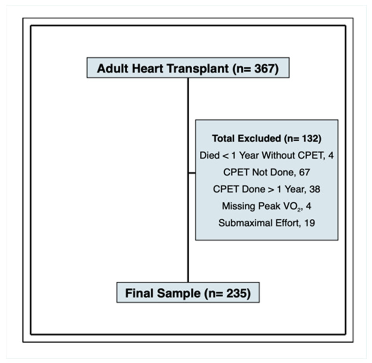
A total of 367 patients underwent single-organ, first-time heart transplantation at the University of Pennsylvania during the study period (Figure 1). Of these, we obtained peak Vo2 measurements on 235 patients. Reasons for missing peak Vo2 measurements were: 4 died prior to one year without undergoing CPET, 38 performed CPET after the 1-year cutoff, 4 performed CPET but were missing peak Vo2, 67 were unable to perform CPET due to prolonged index hospitalizations or severe deconditioning, and 19 performed CPET with submaximal exercise. The RER for the patients excluded for submaximal exercise ranged from 0.8 to 1.0, while the RER for patients performing maximal exercise ranged from 1.0 to 1.44.
Figure 1.
Study Inclusion and Exclusion. The final sample consists of 235 patients who had peak Vo2 measured within the first post-transplant year. Acronyms: CPET, cardiopulmonary exercise test; RER, respiratory exchange ratio.
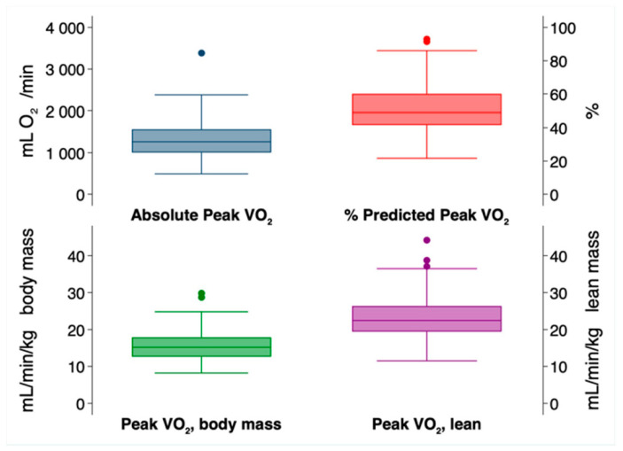
During follow-up of the 235 patients in the cohort, there were 93 deaths (40%) and 4 retransplants (1.7%). The range of follow-up was 115 days to 18.9 years (mean 10.5 years, SD ± 4.4 years). Figure 2 shows the distributions of post-transplant absolute peak Vo2, %Vo2, and Vo2 indexed to body mass or lean mass. The median %Vo2 was 49% (IQR 42 to 60%).
Figure 2.
Distribution of Post-Transplant Peak Vo2 Indices. Box plots displaying the median, 1.5× interquartile range, and outliers for each Vo2 metric.
Several baseline parameters were associated with a %Vo2 below median (Table 1). This included younger age (50 vs. 56 years, p < 0.001), male sex (87 vs. 77%, p = 0.038), lower peak heart rate (116 vs. 125 bpm, p < 0.001), lower weight (82 vs. 88 kg, p = 0.013), and lower BMI (26 vs. 29 kg/m2, p < 0.001). Some of these relationships may seem paradoxical (i.e., younger age), but this is because younger patients tended to have lower peak Vo2 relative to their age-predicted values than older patients, in spite of having greater absolute peak Vo2 measurements. O2 pulse, the ratio of absolute Vo2 to peak heart rate, was also lower in individuals with %Vo2 below median (10 vs. 12 mL/beat, p < 0.001), suggesting impairments in the arteriovenous O2 content or stroke volume at peak exercise. Distributions of pre-transplant comorbidities and ethnicity were similar. No differences in post-transplant LVEF were observed between the groups (64% vs. 66%, p = 0.34), and there was no association between acute cellular rejection and %Vo2 (35% vs. 35%, p = 0.97). No patients who performed maximal exercise CPET had experienced antibody mediated rejection prior to CPET.
Table 1.
Baseline Characteristics of Patients Above and Below Median % Predicted Vo2.
| % Predicted Peak Vo2, <Median of 48.6% n = 118 | % Predicted Peak Vo2, ≥Median of 48.6% n = 117 | p-Value | |
|---|---|---|---|
| Baseline Characteristics | |||
| Age at CPET, years | 50 ± 13 | 56 ± (9) | <0.001 |
| Sex, % male | 103 (87%) | 90 (77%) | 0.038 |
| Ethnicity | 0.066 | ||
| Non-Hispanic White | 91 (77%) | 95 (81%) | |
| Non-Hispanic Black | 20 (17%) | 22 (19%) | |
| Hispanic | 4 (3%) | 0 (0%) | |
| Other | 3 (3%) | 0 (0%) | |
| Diabetic Pre-Transplant | 52 (44%) | 43 (37%) | 0.25 |
| Ischemic Etiology Pre-Transplant | 51 (43%) | 47 (40%) | 0.64 |
| PVD Pre-Transplant | 8 (8%) | 3 (3%) | 0.12 |
| COPD Pre-Transplant | 9 (9%) | 8 (8%) | 0.82 |
| PVR Pre-Transplant, dynes * s * cm−5 | 146 ± 114 | 128.5 ± 72 | 0.19 |
| Serum Creatinine (mg/dL) | 1.6 ± 0.9 | 1.5 ± 1.0 | 0.39 |
| Daily Prednisone Dose, mg | 3.5 ± 3.5 | 3.1 ± 3.0 | 0.40 |
| Cyclosporine Use | 18 (15.3%) | 16 (13.8%) | 0.75 |
| CPET Parameters | |||
| Days from Transplant to CPET | 88 ± 70 | 103 ± 79 | 0.12 |
| Absolute Peak Vo2, mL O2/min | 1095 ± 283 | 1528 ± 412 | <0.001 |
| % Predicted Peak Vo2 | 40 ± 6 | 62 ± 10 | <0.001 |
| O2 pulse, mL/beat | 10 ± 3 | 12 ± 3 | <0.001 |
| Total Exercise Time, min | 8 ± 3 | 9 ± 3 | 0.11 |
| Peak Heart Rate, bpm | 116 ± 17 | 125 ± 17 | <0.001 |
| Peak SBP, mmHg | 149 ± 22 | 153 ± 22 | 0.18 |
| Peak DBP, mmHg | 81 ± 12 | 82 ± 11 | 0.71 |
| Peak Respiratory Exchange Ratio | 1.1 ± 0.1 | 1.1 ± 0.1 | 0.71 |
| Hemoglobin at CPET, g/dL | 13 ± 2 | 13 ± 2 | 0.23 |
| Height, cm | 176 ± 10 | 175 ± 10 | 0.35 |
| Body Weight, kg | 82 ± 19 | 88 ± 17 | 0.013 |
| BMI at CPET, kg/m2 | 26 ± 5 | 29 ± 5 | <0.001 |
| Beta Blocker Use at CPET | 25 (21%) | 20 (17%) | 0.46 |
| Acute Cellular Rejection, % | 35 (29.7%) | 35 (29.9%) | 0.97 |
| Post-Transplant LVEF, % | 64 ± 12 | 66 ± 7 | 0.34 |
Values are mean ± standard deviation or number (proportion). Acronyms: BMI, body mass index; COPD, chronic obstructive pulmonary disease; CPET, cardiopulmonary exercise test; DBP, diastolic blood pressure; SBP, systolic blood pressure; PVD, peripheral vascular disease; PVR, pulmonary vascular resistance; Vco2, carbon dioxide output; Vo2, oxygen consumption.
3.2. Association of Vo2 Metrics with Post-Transplant Mortality
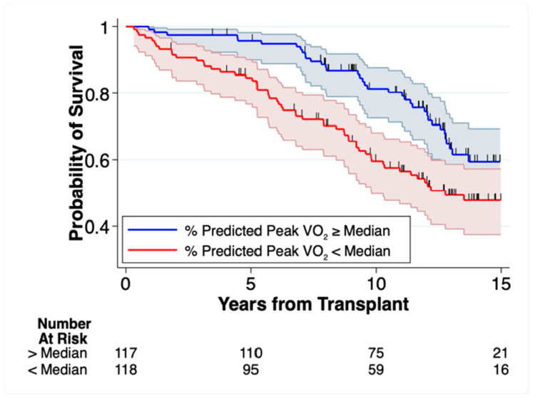
All metrics of post-transplant peak Vo2 were significantly associated with mortality in unadjusted and multivariable adjusted models, with a large effect size (Table 2). Estimates for the effect of different peak Vo2 metrics on mortality were only marginally different. In univariate models, each SD increase in %Vo2 (14%) was associated with a 34% decrease in mortality (HR 0.66, 95% CI 0.53 to 0.84, p = 0.001), equivalent to a 2.4% decrease in mortality per each %Vo2. After adjusting for demographics, comorbidities, and the time from transplant to CPET (i.e., Model 3)—each SD increase in %Vo2 was associated with a 32% decrease in mortality (HR 0.68, 95 CI 0.53 to 0.87, p < 0.002), equating to a 2.3% decrease in mortality per %Vo2. We analyzed the association between peak Vo2 and mortality using alternative weight indices, including unindexed estimates (absolute peak Vo2) and peak Vo2 indexed to total body mass (Vo2, body mass) or estimated lean mass (peak Vo2, lean). All of these had estimates comparable to that of the %Vo2 (Table 2). Lastly, we evaluated whether the observed association between %Vo2 and mortality was conditional on heart rate by looking at the association of O2 pulse with mortality. The O2 pulse, in which the absolute peak Vo2 is indexed to the peak heart rate, demonstrated an equally strong association with mortality as other peak Vo2 indices (Table 2). In unadjusted models, each SD increase in the O2 pulse (3.0 mL O2/beat) was associated with a 19% decrease in the hazard of mortality (HR 0.81, 95% CI 0.65 to 0.99, p = 0.043). After multivariable adjustment, the HR for O2 pulse was 0.66 (95% CI 0.51 to 0.85, p = 0.002). Figure 3 displays the unadjusted Kaplan–Meier survival probability over time stratified by the median %Vo2 of 49%.
Table 2.
Hazard Associated with each Standard Deviation Change in Peak Vo2.
| Model 1 Univariate | Model 2 Demographics | Model 3 Clinical Covariates | ||||
|---|---|---|---|---|---|---|
| HR § (95% CI) | p-Value | HR (95% CI) | p-Value | HR (95% CI) | p-Value | |
| Peak Oxygen Consumption Absolute Peak Vo2 (SD ± 414 mL O2/min) | 0.75 (0.60, 0.93) | 0.010 | 0.60 (0.46, 0.78) | 0.000 | 0.62 (0.47, 0.81) | 0.001 |
| % Predicted Peak Vo2 (SD ± 14%) | 0.66 (0.53, 0.84) | 0.001 | 0.63 (0.50, 0.81) | 0.000 | 0.68 (0.53, 0.87) | 0.002 |
| Peak Vo2, body mass (SD ± 4 mL O2/min/kg body mass) | 0.69 (0.55, 0.87) | 0.002 | 0.66 (0.52, 0.83) | 0.000 | 0.72 (0.56, 0.91) | 0.007 |
| Peak Vo2, lean (SD ± 6 mL/min/kg lean mass) | 0.62 (0.49, 0.78) | 0.000 | 0.63 (0.50, 0.79) | 0.000 | 0.68 (0.53, 0.86) | 0.002 |
| O2 Pulse (SD ± 3 mL O2/beat) | 0.81 (0.65, 0.99) | 0.043 | 0.66 (0.52, 0.85) | 0.001 | 0.66 (0.51, 0.85) | 0.002 |
Higher Peak Vo2 is associated with a decreased hazard of death or retransplantation. Model 1—unadjusted Cox proportional hazard model. Model 2—adjusted for age, race/ethnicity, sex, and days from transplant to CPET. Model 3—adjusted for Model 2 variables plus pulmonary vascular resistance, type 2 diabetes, ischemic cardiomyopathy, peripheral-vascular disease, chronic obstructive pulmonary disease, and hemoglobin. Hazard ratios (HR) are for a one standard deviation (SD) change in each metric. Acronyms: Vo2, oxygen consumption.
Figure 3.
Kaplan–Meier Survival Estimates Stratified by Median % Predicted Peak Vo2. Patients with post-transplant % Predicted Peak Vo2 (%Vo2) below median had significantly lower long-term survival. The median %Vo2 was 49%. Vertical dash marks indicate censored events. Shaded areas represent the 95% Confidence Intervals.
3.3. Predicting Post-Transplant Survival
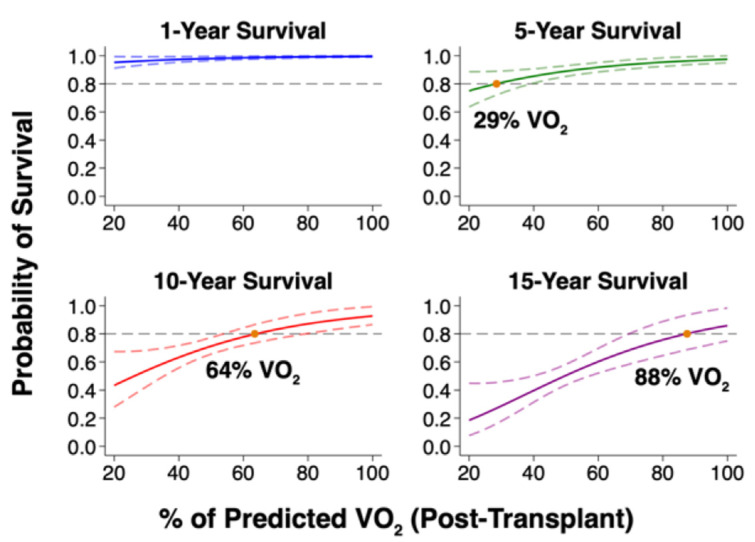
We derived four models predicting survival at 1, 5, 10 and 15 years post-transplant as a function of post-transplant %Vo2 (Figure 4). From each model, we identified a %Vo2 cutoff below which the predicted survival to each timepoint was less than 80%. At 5 years post-transplant, 80% survival (95% CI 72 to 89%) was predicted by a %Vo2 below 29%. At 10 years, the 80% survival threshold (95% CI 74 to 87%) was predicted by a %Vo2 below 64%. Finally, at 15 years, 80% survival (95% CI 69 to 94%) was predicted by a %Vo2 below 88%. Only 5 patients in our sample died within the first year (2%), so no cutoff predicting 1-year survival less than 80% could be estimated.
Figure 4.
Predicted Survival Free from Re-transplantation based on Post-Transplant % Predicted Peak Vo2. Post-Transplant % Predicted Peak Vo2 predicts long-term survival. Plots based on unadjusted flexible parametric survival models, displaying mean survival (solid lines) along with upper and lower 95% confidence intervals (dashed lines). Acronyms: Vo2, oxygen consumption.
4. Discussion
An association between pre-transplant peak Vo2 and mortality in ambulatory heart failure was first established over two decades ago [1], and numerous studies have corroborated this association across multiple populations [2,19,20]. In this retrospective cohort analysis, we evaluated whether lower peak Vo2 in the early post-transplant period is associated with long-term post-transplant mortality. Our major findings were that: (1) diminished post-transplant peak Vo2 was strongly associated with mortality over nearly two decades of follow-up and (2) this association was independent of parameters known to directly influence Vo2, such as hemoglobin, sex, heart rate, body mass, and the number of days from transplant to CPET.
The etiology of diminished peak Vo2 is multifactorial, inVOlving cardiac and extracardiac parameters. Only some of these will respond directly to improved cardiac function, while some deficits are likely to remain, explaining why several studies have shown little improvement in peak Vo2 after heart transplant or mechanical circulatory support [6,8]. In our analysis, we attempted to differentiate whether the association between diminished peak Vo2 and mortality post-transplant was inherent to peak Vo2 or instead confounded by several common comorbidities. In the fully adjusted model, each SD (14%) increase in %Vo2 remained associated with a 32% decrease in the hazard of mortality (HR 0.68, 95 CI 0.53 to 0.87, p = 0.002). This effect size is in accordance with another single-center study out of Norway that analyzed transplants from the decade prior [21], confirming the generalizability of this association across time and institution. Moreover, we showed that this effect was independent of several additional extracardiac factors such as COPD, diabetes, peripheral vascular disease, and pulmonary vascular resistance.
These latter comorbidities, though not severe enough to preclude transplant in the first place, could still significantly confound the relationship between Vo2 and mortality. Instead, their adjustment had negligible effect. Similarly, the association between Vo2 and mortality was independent of post-transplant factors that can significantly impact the peak Vo2 or mortality, including body mass, LVEF, hemoglobin, creatinine, a history of acute cellular or antibody mediated rejection, the number of days from transplant to CPET, and immunosuppressive regimen. Additionally, the O2 pulse—which indexes the peak Vo2 to peak heart rate—was strongly associated with mortality in all multivariable models. This confirms that the association was not conditional on heart rate response, which can be diminished post-transplant secondary to sympathetic denervation. Based on the strong association between peak Vo2 and survival, higher %Vo2 predicted a greater probability of survival each year post-transplant. Using 80% survival probability as a target, this threshold was predicted at 5, 10 and 15 years by increasing %Vo2 cutoffs of 29%, 64%, and 88%, respectively. If externally validated, this metric could provide a useful prognostic screening tool for the early post-transplant period.
Diminished peak Vo2 that is unrelated to cardiac dysfunction or major comorbidities may indicate impaired skeletal muscle function, consistent with the “muscle hypothesis” in chronic heart failure [22]. Even in a healthy population, diminished exercise capacity is associated with increased mortality [23]; even more so in a heart failure population with deranged skeletal muscle oxidative capacity. Inadequate skeletal muscle oxidative capacity during exercise appears as a common element in multiple heart failure phenotypes, including heart failure with either reduced or preserved ejection fraction [24]. In chronic heart failure, this may be related to reduced O2 diffusion or deficiencies in mitochondrial density and efficiency [25], and these deficiencies post-transplant may be a holdover from the pre-transplant period. This is corroborated by the observation that lower pre-transplant peak Vo2 predicts lower post-transplant peak Vo2 [8], although skeletal muscle oxidative capacity could also worsen post-transplant secondary to chronic exposure to steroids and calcineurin inhibitors or prolonged hospitalizations [25]. Furthermore, it may be possible to detect reduced skeletal muscle oxidative capacity via a decline in the arteriovenous oxygen difference, implying decreased extraction and utilization of oxygen. Thus, it is notable that mortality in this study was strongly associated with decreased O2 pulse, which in addition to correlating with stroke volume also provides an indirect measure of the arteriovenous oxygen difference [26].
5. Limitations of Our Study
This study is retrospective in nature, which increases the possibility for misclassification bias using variables measured for clinical intent. In particular, peak Vo2 were determined via a 10 s average rather than 30 s, which may overestimate peak Vo2 (although this was measured in similar fashion for all subjects). As a single center study with a predominantly white population, generalizability to other sites may be reduced. In this study, most patients did not have pre-transplant CPET to analyze the prognostic importance of the change in parameters from pre- to post-transplant—this is a topic of future interest. Selection bias likely exists in the ability to perform a CPET, per se, thus estimates of association with mortality in the CPET population likely underestimate mortality in the overall heart transplant population that includes patients who were unable to perform CPET. Some covariate data were missing in this study, but no variables had greater than 15% missingness, and we were able to minimize bias through multiple imputation of missing datapoints.
6. Conclusions
Post-transplant peak Vo2 during maximal exercise is strongly associated with long-term post-transplant survival. The independent association between peak Vo2 metrics and mortality was independent of several important confounders and factors that contribute directly to Vo2. We observed an association between O2 pulse and mortality, suggesting that diminished post-transplant skeletal muscle oxidative capacity is a link between decreased peak Vo2 and survival. High-intensity exercise conditioning after transplant can safely augment peak Vo2 [27], but future studies are needed to evaluate whether improved peak Vo2 would lead to improved survival. If so, more aggressive cardiopulmonary rehabilitation post-transplant would be warranted, with peak Vo2 as an objective target.
Abbreviations
| CPET | cardiopulmonary exercise test |
| HFrEF | heart failure with reduced ejection fraction |
| Peak Vo2 | peak oxygen consumption |
| Peak Vo2, body mass | peak oxygen consumption indexed to total body mass |
| Peak Vo2, lean | peak oxygen consumption indexed to estimated lean body mass |
| RER | respiratory exchange ratio |
| %Vo2 | percent of predicted peak oxygen consumption |
| VT | ventilatory threshold |
Author Contributions
Conceptualization, T.C.H. and E.Y.B.; Formal analysis, T.C.H.; Data curation, T.C.H., Y.Z., R.S.Z., M.V.G. and M.M.; Writing—original draft, T.C.H.; Writing—review & editing, Y.Z., R.S.Z., M.V.G., M.M., R.C.M., J.A.M., M.S.T., J.W.W., P.A., M.A.A., L.R.G., R.S.Z., P.Z. and E.Y.B.; Supervision, E.Y.B. All authors have read and agreed to the published version of the manuscript.
Institutional Review Board Statement
The study was conducted in accordance with the Declaration of Helsinki, and approved by the Institutional Review Board of the University of Pennsylvania.
Informed Consent Statement
Ethical review and approval were waived for this study. This is a retrospective study of preexisting clinical data and a signed informed consent document is not feasible, as many patients are no longer be available to obtain authorization for use or disclosure of protected health information (PHI). Furthermore, a consent form will increase the risk of potential harm from a breach of confidentiality by becoming a “link” stored to identify the subject.
Data Availability Statement
The data that support the findings are available on request from the first author, T.C.H.
Conflicts of Interest
The authors declare no conflict of interest. All investigators are affiliated with the University of Pennsylvania, which has received clinical research support from Abbott and Medtronic. M.A.A. is on the advisory board for FineHeart and has a consulting relationship with Abbott. PZ has consulted for Vyaire. E.Y.B. has received additional research support from Impulse Dynamics to the University of Pennsylvania.
Funding Statement
TCH was supported by the National Institute of Health (NIH)/National Heart, Lung, and Blood Institute (NHLBI) T32 Training Grant HL-007891.
Footnotes
Disclaimer/Publisher’s Note: The statements, opinions and data contained in all publications are solely those of the individual author(s) and contributor(s) and not of MDPI and/or the editor(s). MDPI and/or the editor(s) disclaim responsibility for any injury to people or property resulting from any ideas, methods, instructions or products referred to in the content.
References
- 1.Mancini D.M., Eisen H., Kussmaul W., Mull R., Edmonds L.H., Wilson J.R. Value of peak exercise oxygen consumption for optimal timing of cardiac transplantation in ambulatory patients with heart failure. Circulation. 1991;83:778–786. doi: 10.1161/01.CIR.83.3.778. [DOI] [PubMed] [Google Scholar]
- 2.Elmariah S., Goldberg L.R., Allen M.T., Kao A. Effects of Gender on Peak Oxygen Consumption and the Timing of Cardiac Transplantation. J. Am. Coll. Cardiol. 2006;47:2237–2242. doi: 10.1016/j.jacc.2005.11.089. [DOI] [PubMed] [Google Scholar]
- 3.Metra M., Ponikowski P., Dickstein K., McMurray J.J.V., Gavazzi A., Bergh C.-H., Fraser A.G., Jaarsma T., Pitsis A., Mohacsi P., et al. Heart Failure Association of the European Society of Cardiology. Advanced chronic heart failure: A position statement from the Study Group on Advanced Heart Failure of the Heart Failure Association of the European Society of Cardiology. Eur. J. Heart Fail. 2007;9:684–694. doi: 10.1016/j.ejheart.2007.04.003. [DOI] [PubMed] [Google Scholar]
- 4.Weber K.T., Kinasewitz G.T., Janicki J.S., Fishman A.P. Oxygen utilization and ventilation during exercise in patients with chronic cardiac failure. Circulation. 1982;65:1213–1223. doi: 10.1161/01.CIR.65.6.1213. [DOI] [PubMed] [Google Scholar]
- 5.Wilson J.R., Rayos G., Yeoh T.K., Gothard P. Dissociation between peak exercise oxygen consumption and hemodynamic dysfunction in potential heart transplant candidates. J. Am. Coll. Cardiol. 1995;26:429–435. doi: 10.1016/0735-1097(95)80018-C. [DOI] [PubMed] [Google Scholar]
- 6.Borrelli E., Pogliaghi S., Molinello A., Diciolla F., Maccherini M., Grassi B. Serial Assessment of Peak Vo2 and Vo2 Kinetics Early after Heart Transplantation. Med. Sci. Sports Exerc. 2003;35:1798–1804. doi: 10.1249/01.MSS.0000093610.71730.02. [DOI] [PubMed] [Google Scholar]
- 7.Carter R., Al-Rawas O.A., Stevenson A., Mcdonagh T., Stevenson R.D. Exercise responses following heart transplantation: 5 year follow-up. Scott. Med. J. 2006;51:6–14. doi: 10.1258/RSMSMJ.51.3.6. [DOI] [PubMed] [Google Scholar]
- 8.Williams T.J., Mckenna M.J. Exercise Limitation Following Transplantation. Compr. Physiol. 2012;2:1937–1979. doi: 10.1002/cphy.c110021. [DOI] [PubMed] [Google Scholar]
- 9.Myers J., Kaminsky L.A., Lima R., Christle J.W., Ashley E., Arena R. A Reference Equation for Normal Standards for Vo2 Max: Analysis from the Fitness Registry and the Importance of Exercise National Database (FRIEND Registry) Prog. Cardiovasc. Dis. 2017;60:21–29. doi: 10.1016/j.pcad.2017.03.002. [DOI] [PubMed] [Google Scholar]
- 10.Malhotra R., Bakken K., D’Elia E., Lewis G.D. Cardiopulmonary Exercise Testing in Heart Failure. J. Am. Coll. Cardiol. HF. 2016;4:607–616. doi: 10.1016/j.jchf.2016.03.022. [DOI] [PubMed] [Google Scholar]
- 11.Lee D.H., Keum N., Hu F.B., Orav E.J., Rimm E.B., Sun Q., Willett W.C., Giovannucci E.L. Development and validation of anthropometric prediction equations for lean body mass, fat mass and percent fat in adults using the National Health and Nutrition Examination Survey (NHANES) 1999–2006. Br. J. Nutr. 2017;118:858–866. doi: 10.1017/S0007114517002665. [DOI] [PubMed] [Google Scholar]
- 12.Fleg J.L., Pina I.L., Balady G.J., Chaitman B.R., Flether B., Lavie C., Limacher M.C., Stein R.A., Williams M., Bazzarre T. Assessment of functional capacity in clinical and research applications: An advisory from the committee on exerise, rehabilitation, and prevention, council on clinical cardiology, American Heart Association. Circ. Lippincott Williams Wilkins. 2000;102:1591–1597. doi: 10.1161/01.CIR.102.13.1591. [DOI] [PubMed] [Google Scholar]
- 13.Mezzani A., Corrà U., Bosimini E., Giordano A., Giannuzzi P. Contribution of peak respiratory exchange ratio to peak Vo2 prognostic reliability in patients with chronic heart failure and severely reduced exercise capacity. Am. Heart J. 2003;145:1102–1107. doi: 10.1016/S0002-8703(03)00100-5. [DOI] [PubMed] [Google Scholar]
- 14.O’Connor C.M., Whellan D.J., Lee K.L., Keteyian S.J., Cooper L.S., Ellis S.J., Leifer E.S., Kraus W.E., Kitzman D.W., Blumenthal J.A., et al. Efficacy and safety of exercise training in patients with chronic heart failure HF-ACTION randomized controlled trial. J. Am. Med. Assoc. 2009;301:1439–1450. doi: 10.1001/jama.2009.454. [DOI] [PMC free article] [PubMed] [Google Scholar]
- 15.Whellan D.J., O’Connor C.M., Lee K.L., Keteyian S.J., Cooper L.S., Ellis S.J., Leifer E.S., Kraus W.E., Kitzman D.W., Blumenthal J.A., et al. Heart Failure and a Controlled Trial Investigating Outcomes of Exercise TraiNing (HF-ACTION): Design and rationale. Am. Heart J. 2007;153:201–211. doi: 10.1016/j.ahj.2006.11.007. [DOI] [PubMed] [Google Scholar]
- 16.Cox D.R., Oakes D. Analysis of Survival Data. Analysis of Survival Data. CRC Press; Boca Raton, FL, USA: 2018. pp. 1–201. [Google Scholar]
- 17.Schoenfeld D. Partial Residuals for The Proportional Hazards Regression Model. Biometrika. 1982;69:239. doi: 10.1093/biomet/69.1.239. [DOI] [Google Scholar]
- 18.Royston P., Parmar M.K.B. Flexible parametric proportional-hazards and proportional-odds models for censored survival data, with application to prognostic modelling and estimation of treatment effects. Stat. Med. 2002;21:2175–2197. doi: 10.1002/sim.1203. [DOI] [PubMed] [Google Scholar]
- 19.Elmariah S., Goldberg L.R., Allen M.T., Kao A. The Effects of Race on Peak Oxygen Consumption and Survival in Patients with Systolic Dysfunction. J. Card. Fail. 2010;16:332–339. doi: 10.1016/j.cardfail.2009.12.010. [DOI] [PubMed] [Google Scholar]
- 20.Menachem J.N., Reza N., Mazurek J.A., Burstein D., Birati E.Y., Fox A., Kim Y.Y., Molina M., Partington S.L., Tanna M., et al. Cardiopulmonary Exercise Testing—A Valuable Tool, Not Gatekeeper When Referring Patients with Adult Congenital Heart Disease for Transplant Evaluation. World J. Pediatr. Congenit. Heart Surg. 2019;10:286–291. doi: 10.1177/2150135118825263. [DOI] [PMC free article] [PubMed] [Google Scholar]
- 21.Yardley M., Havik O.E., Grov I., Relbo A., Gullestad L., Nytrøen K. Peak oxygen uptake and self-reported physical health are strong predictors of long-term survival after heart transplantation. Clin. Transplant. 2016;30:161–169. doi: 10.1111/ctr.12672. [DOI] [PubMed] [Google Scholar]
- 22.Coats A.J.S. The “Muscle Hypothesis” of chronic heart failure. J. Mol. Cell. Cardiol. 1996;28:2255–2262. doi: 10.1006/jmcc.1996.0218. [DOI] [PubMed] [Google Scholar]
- 23.Myers J., Prakash M., Froelicher V., Do D., Partington S., Atwood J.E. Exercise capacity and mortality among men referred for exercise testing. N. Engl. J. Med. 2002;346:793–801. doi: 10.1056/NEJMoa011858. [DOI] [PubMed] [Google Scholar]
- 24.Weiss K., Schär M., Panjrath G.S., Zhang Y., Sharma K., Bottomley P.A., Golozar A., Steinberg A., Gerstenblith G., Russell S.D., et al. Fatigability, Exercise Intolerance, and Abnormal Skeletal Muscle Energetics in Heart Failure. Circ. Heart Fail. 2017;10:e004129. doi: 10.1161/CIRCHEARTFAILURE.117.004129. [DOI] [PMC free article] [PubMed] [Google Scholar]
- 25.Houstis N.E., Eisman A.S., Pappagianopoulos P.P., Wooster L., Bailey C.S., Wagner P.D., Lewis G.D. Exercise intolerance in heart failure with preserved ejection fraction: Diagnosing and ranking Its causes using personalized O2 pathway analysis. Circulation. 2018;137:148–161. doi: 10.1161/CIRCULATIONAHA.117.029058. [DOI] [PMC free article] [PubMed] [Google Scholar]
- 26.Gmada N., Al-Hadabi B., Haj Sassi R., Abdel Samia B., Bouhlel E. Relationship between oxygen pulse and arteriovenous oxygen difference in healthy subjects: Effect of exercise intensity. Sci. Sports. 2019;34:e297–e306. doi: 10.1016/j.scispo.2019.04.005. [DOI] [Google Scholar]
- 27.Nytrøen K., Rolid K., Andreassen A.K., Yardley M., Gude E., Dahle D.O., Bjørkelund E., Relbo Authen A., Grov I., Philip Wigh J., et al. Effect of High-Intensity Interval Training in De Novo Heart Transplant Recipients in Scandinavia: One-Year Follow-Up of the HITTS Randomized, Controlled Study. Circulation. 2019;139:2198–2211. doi: 10.1161/CIRCULATIONAHA.118.036747. [DOI] [PubMed] [Google Scholar]
Associated Data
This section collects any data citations, data availability statements, or supplementary materials included in this article.
Data Availability Statement
The data that support the findings are available on request from the first author, T.C.H.


