Abstract
The introduction into Brucella suis 1330 of a plasmid allowing the heterologous expression of a hybrid cytolysin containing listeriolysin from Listeria monocytogenes, and its export via the Escherichia coli hemolysin secretion pathway, resulted in secretion of active listeriolysin monitored by erythrocyte lysis. In contrast to observations with the nonhemolytic control strain, the phagosomes of infected human monocytes containing the hemolytic B. suis were partially disrupted, and this strain failed to multiply in human macrophage-like cells. These results added strong evidence supporting the proposal that the phagosome of the macrophage was the predominant niche of brucellae in their mammalian hosts.
The facultative intracellular bacterium Brucella suis, which is one of the causative agents of brucellosis in humans and animals (4), resists intracellular killing by macrophages and replicates within the phagosome (9). In contrast, the other intracellular pathogen we refer to in this study, Listeria monocytogenes, rapidly enters the host cell cytoplasm. Membrane disruption caused by the action of listeriolysin (15) is responsible for this escape from the phagosome. Listeriae then replicate freely within the cytoplasm, and actin polymerization leads to intracellular movements (15).
We were interested in the impact of phagosomal membrane lysis on intracellular multiplication of B. suis in the phagocyte. The question of whether B. suis could survive or even replicate in the absence of an intact phagosomal membrane and switch to an efficient cytoplasmic lifestyle was of interest for the general understanding of Brucella virulence mechanisms, especially as it has been recently reported that the early acidification of B. suis-containing phagosomes is crucial for the intracellular replication of the bacteria in macrophages (14). To answer this question, the use of listeriolysin appeared promising to us, as the optimum of its biological activity is reached at acidic pH.
In a heterologous system, listeriolysin was first used with Bacillus subtilis, allowing its release into the cytoplasm after phagocytosis (1). More recently, the construction of a Salmonella enterica serovar Dublin aroA strain secreting an active hybrid cytolysin made up of listeriolysin and the C-terminal secretion signal of Escherichia coli hemolysin, resulting in release of the hemolytic serovar Dublin strain into the cytoplasm of the host cell, was described (5). The plasmid used, named pILH-1, also contained the genes hlyR, -C, -B, and -D for regulation of hemolysin expression and for transport (5). The same construct was also used for the development of antigen delivery systems (6).
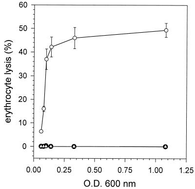
To allow expression of the hybrid cytolysin in B. suis, pILH-1 was cut by PvuI and SalI, eliminating a 3-kb fragment containing the origin of replication and the ampicillin resistance gene. The remaining fragment of 10 kb was ligated to pBBRIMCS (12) digested by PvuI and SalI, resulting in the intermediate construct pBBRI-HLY, capable of replication in brucellae, and conferring resistance to chloramphenicol following transformation by electroporation (11). A kanamycin resistance cassette isolated from pUC4K (Pharmacia Biotech, Orsay, France) was then ligated to Asp718 linkers and inserted into pBBR1-HLY linearized by Asp718, yielding the 15.9-kb plasmid pBBR1-HLYK, which allows selection for kanamycin and chloramphenicol. Standard protocols (17) were used for all DNA manipulations. In E. coli DH5α with pBBR1-HLYK, secretion of the hybrid cytolysin was confirmed by sodium dodecyl sulfate-polyacrylamide gel electrophoresis and Western blotting using antilisteriolysin antiserum according to the previously described protocols (5), and the cytolysin showed lytic activity on erythrocytes (data not shown). To evidence hemolytic activity of B. suis(pBBR1-HLYK), an erythrocyte lysis assay was performed as follows. Stationary-phase cultures of B. suis(pBBR1-HLYK) and of the control strain B. suis(pMR10-CM) were diluted 50-fold in tryptic soy (TS) broth containing 1% washed human erythocytes and incubated with shaking at 37°C for 24 h. Plasmid pMR10-CM was derived from pMR10 (obtained from R. Wright, Stanford University, Palo Alto, Calif.) and contained a chloramphenicol resistance gene inserted into the SmaI site of the polylinker, hence allowing selective pressure with both kanamycin and chloramphenicol, as for pBBR1-HLYK. At 4, 6, 8, 10, and 24 h postinoculation, 1 ml of each culture was centrifuged, and the concentration of free hemoglobin was measured spectrophotometrically as the optical density at 543 nm (OD543) (A. Schrettenbrunner, personal communication). A supernatant of TS medium containing erythrocytes and incubated under the same conditions was used as a blank. The OD values obtained with erythrocytes (1% final concentration) in phosphate-buffered saline and in H2O were considered to represent 0 and 100% lysis, respectively. Bacterial growth of the cultures was monitored by measuring the OD600, and growth rates were identical for both strains (not shown). Over a period of 24 h, approximately 50% of the erythrocytes lysed in the culture containing B. suis(pBBR1-HLYK), as opposed to the complete absence of lysis with the control strain (FIG. 1). The rate of lysis was strongest in the early logarithmic growth phase at an OD600 of 0.1 to 0.25. To confirm that the lytic activity observed with this strain was due to secretion of active cytolysin, a competition experiment was performed in the presence of polyclonal antilisteriolysin rabbit antiserum. This antiserum was added to the culture together with the erythrocytes, and lysis was measured as described above. The presence of antilisteriolysin antibodies in the culture of B. suis(pBBR1-HLYK) completely inhibited erythrocyte lysis, yielding a curve identical to the one obtained with the control strain (FIG. 1). A nonimmune serum had no effect on lysis (data not shown). We failed, however, to detect hemolytic activity in culture supernatants of B. suis at various growth phases harvested prior to performance of the hemolysin assay, and we also failed to visualize the cytolysin by Western blotting in culture supernatants and total cell lysates from B. suis with pBBR1-HLYK taken at various time points during growth. This could be due either (i) to lower efficiency of expression of the E. coli hly genes in B. suis or (ii) to instability or rapid degradation of the secreted cytolysin in the supernatant. Nevertheless, a weak zone of hemolytic activity was visible on blood agar plates in the immediate vicinity of bacterial colonies (data not shown).
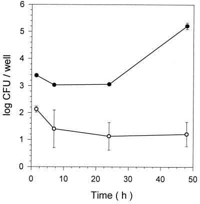
The cytolytic B. suis strain containing pBBR1-HLYK was then assayed for its capacity of intracellular replication compared to that of the wild-type strain with plasmid pMR10-CM. Human THP-1 macrophage-like cells were cultured for 3 days in RPMI 1640–10% fetal calf serum at 37°C with 5% CO2 in the presence of 1,25-dihydroxyvitamin D3 (10−7 M). Adherent cells were scraped off the flask, seeded at 500,000 cells per well in 24-well plates, and incubated in RPMI 1640–10% fetal calf serum for another 24 h. Bacterial strains were grown in TS broth in the presence of antibiotics. Infection at a multiplicity of infection of 20, washing, reincubation of the infected cells in the presence of gentamicin, kanamycin, and chloramphenicol, and lysis with Triton X-100 were performed as described previously (3). Enumeration of viable intracellular brucellae was done by plating on TS agar and incubation for 3 days. We expected that the secretion of the cytolysin would damage the phagosomal membrane surrounding B. suis inside the macrophage, thereby affecting the intracellular environment of the bacteria. In human THP-1 macrophages, the strain secreting the hybrid cytolysin was unable to replicate intracellularly but survived at a low level of infection for at least 48 h, in contrast to the wild-type strain, which multiplied approximately 100-fold during the same period (FIG. 2). In the absence of selective pressure, pBBR1-HLYK was rapidly lost from the mutant strain, and antibiotic-sensitive bacteria were recovered that multiplied normally within the macrophage. From these experiments we concluded that the secretion of the cytolysin by B. suis modified its intracellular niche so that intracellular multiplication was no longer possible.
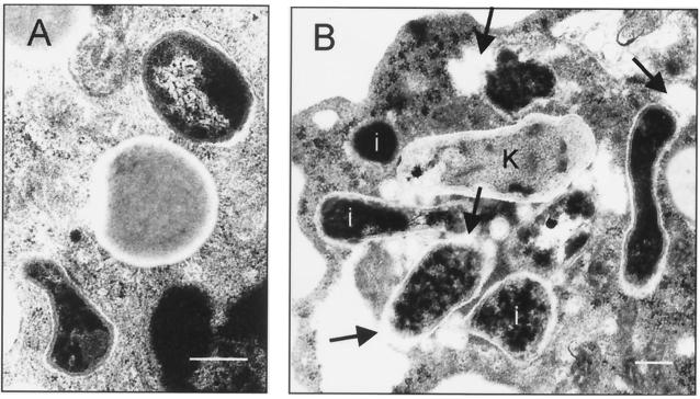
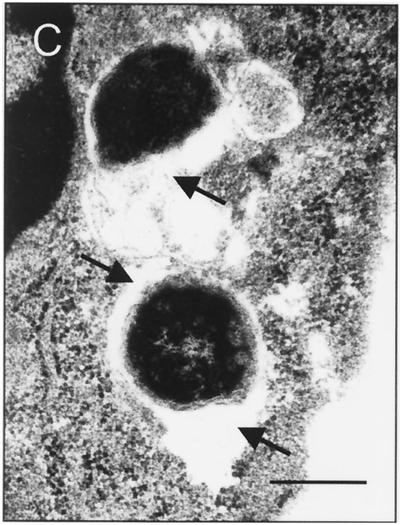
To address the question of a possible modification in the local environment of intracellular B. suis(pBBR1-HLYK), transmission electron microscopy was performed on infected human monocytes at 1 and 9 h postinfection. As a control, the wild-type strain, B. suis 1330, was used in parallel. Freshly isolated peripheral blood monocytes were infected at a multiplicity of infection of 500 for 20 min, washed, and chased in RPMI 1640–10% fetal calf serum for 60 min or 9 h. The incubation was stopped by adding an excess amount of cold Ito's fixative (7) to the tubes. The samples were then processed for electron microscopy by following standard protocols (7). Ultrathin sections were stained with a mixture of uranyl acetate and lead citrate. Sections were viewed with a Zeiss type 906 transmission electron microscope. At 1 h after infection, phagosomes containing the cytolysin-producing strain were indistinguishable from those enclosing the wild-type B. suis strain (not shown). At 9 h, however, approximately 50% of the phagosomes with B. suis(pBBR1-HLYK) showed partially disrupted membranes resulting in leakage of phagosomal content (FIG. 3B and C), a phenomenon never observed with the wild-type strain (FIG. 3A). The actual number of leaky phagosomes may be larger, since the disruption of the membrane could be outside the plane of section. Membrane lysis was less efficient than with L. monocytogenes, which produces a phosphatidylinositol-specific phospholipase C in addition to listeriolysin (2) and readily escapes into the cytoplasm. Earlier work on listeriolysin secretion by intracellular Mycobacterium bovis BCG describes formation of pores in the phagosome but also the inability of the bacteria to egress into the cytoplasm, which was attributed to a possible suboptimal endosomal pH for maximal biological activity of listeriolysin (10).
In summary, our results showed that the expression of a previously described hybrid cytolysin (5) by the intracellular pathogen B. suis resulted in damage of the phagosomal membrane and that both events correlated with the loss of the ability of B. suis to replicate within the macrophage. As it is known that B. suis requires an acidic phagosome for replication in the macrophage (14), this lack of multiplication could be explained by an early neutralization due to listeriolysin-induced membrane lysis. Collapse of the pH gradient between the phagosomal compartment and the cytosol would be a direct consequence of this lysis. It is interesting that the expression of an essential virulence factor of the gram-positive pathogen L. monocytogenes in the gram-negative pathogen B. suis completely abolishes the capacity of intramacrophagic replication of the latter, hence demonstrating that a specific intracellular strategy for a given pathogen cannot be altered. B. suis is not adapted to replication in a cytoplasmic environment, and the identification of macrophage-induced proteins (11, 13, 16) most likely reflects this adaptation of brucellae to their specific intracellular niche, the phagosome.
A recent study reported that functional gene transfer from intracellular E. coli K-12 to mammalian cells was enhanced when listeriolysin was released following intraphagosomal killing and lysis of the bacteria (8). In the future, direct release of brucellae or their antigens into the cytoplasm of macrophages may therefore be a promising approach for class I antigen presentation analysis and for the development of new types of vaccine strains.
FIG. 1.
Erythrocyte lysis in B. suis cultures containing washed human erythrocytes, as a function of bacterial growth (OD600). Strains used were B. suis with the control plasmid pMR10-CM (●) or with plasmid pBBR1-HLYK and in the absence (○) or the presence (▵) of antilisteriolysin antiserum. Experiments were performed three times in triplicate. Error bars represent standard deviations.
FIG. 2.
Intracellular replication of the B. suis control strain with plasmid pMR10-CM (●) and of the strain with plasmid pBBR1-HLYK (○) in differentiated human macrophage-like THP-1 cells. Experiments were performed three times in triplicate. Error bars represent standard deviations.
FIG. 3.
Transmission electron microscopic images of Brucella-containing phagosomes in human monocytes infected with live B. suis 1330 (A) or B. suis(pBBR1-HLYK) (B and C). Phagosomes with the wild-type strain were always intact (A). Images of infection with B. suis(pBBR1-HLYK) show intact and damaged phagosomal membranes (B) and a cross section of two damaged phagosomes (C). Arrows point out areas of membrane degradation and leaking of the phagosomal content. Three apparently intact phagosomes are indicated (i). K, killed bacterium. Bars, 0.5 μm.
Acknowledgments
We thank J. Kreft for methodological advice and his generous gift of antiserum to listeriolysin, and we thank W. Goebel and F. Porte for helpful discussion. J. Teyssier is acknowledged for technical assistance.
This work was supported by grant BIO-CT98-0089 from the European Union.
REFERENCES
- 1.Bielecki J, Youngman P, Connelly P, Portnoy D A. Bacillus subtilis expressing a haemolysin gene from Listeria monocytogenes can grow in mammalian cells. Nature (London) 1990;345:175–176. doi: 10.1038/345175a0. [DOI] [PubMed] [Google Scholar]
- 2.Camilli A, Tilney L G, Portnoy D A. Dual roles of plcA in Listeria monocytogenes pathogenesis. Mol Microbiol. 1993;8:143–157. doi: 10.1111/j.1365-2958.1993.tb01211.x. [DOI] [PMC free article] [PubMed] [Google Scholar]
- 3.Caron E, Liautard J P, Köhler S. Differentiated U937 cells exhibit increased bactericidal activity upon LPS activation and discriminate between virulent and avirulent Listeria and Brucella species. J Leukoc Biol. 1994;56:174–181. doi: 10.1002/jlb.56.2.174. [DOI] [PubMed] [Google Scholar]
- 4.Corbel M J. Brucella. In: Parker M T, Collier L H, editors. Principles of bacteriology, virology, and immunity. 2. E. London, United Kingdom: Arnold; 1990. pp. 339–353. [Google Scholar]
- 5.Gentschev I, Sokolovic Z, Mollenkopf H-J, Hess J, Kaufmann S H E, Kuhn M, Krohne G F, Goebel W. Salmonella strain secreting active listeriolysin changes its intracellular localization. Infect Immun. 1995;63:4202–4205. doi: 10.1128/iai.63.10.4202-4205.1995. [DOI] [PMC free article] [PubMed] [Google Scholar]
- 6.Gentschev I, Mollenkopf H, Sokolovic Z, Hess J, Kaufmann S H E, Goebel W. Development of antigen-delivery systems based on Escherichia coli hemolysin secretion pathway. Gene. 1996;179:133–140. doi: 10.1016/s0378-1119(96)00424-6. [DOI] [PubMed] [Google Scholar]
- 7.Glauert A. Fixation, dehydration and embedding of biological specimens. Vol. 3 1975. , part I. Coordinating ed., A. Glauert. North-Holland Publishing Co. Amsterdam, The Netherlands. [Google Scholar]
- 8.Grillot-Courvalin C, Goussard S, Huetz F, Ojcius D M, Courvalin P. Functional gene transfer from intracellular bacteria to mammalian cells. Nat Biotechnol. 1998;16:862–866. doi: 10.1038/nbt0998-862. [DOI] [PubMed] [Google Scholar]
- 9.Harmon B G, Adams L G, Frey M. Survival of rough and smooth strains of Brucella abortus in bovine mammary gland macrophages. Am J Vet Res. 1988;49:1092–1097. [PubMed] [Google Scholar]
- 10.Hess J, Miko D, Catic A, Lehmensiek V, Russell D G, Kaufmann S H E. Mycobacterium bovis bacille Calmette-Guérin strains secreting listeriolysin of Listeria monocytogenes. Proc Natl Acad Sci USA. 1998;95:5299–5304. doi: 10.1073/pnas.95.9.5299. [DOI] [PMC free article] [PubMed] [Google Scholar]
- 11.Köhler S, Teyssier J, Cloeckaert A, Rouot B, Liautard J P. Participation of the molecular chaperone DnaK in intracellular growth of Brucella suis within U937-derived phagocytes. Mol Microbiol. 1996;20:701–712. doi: 10.1111/j.1365-2958.1996.tb02510.x. [DOI] [PubMed] [Google Scholar]
- 12.Kovach M E, Phillips R W, Elzer P H, Roop II R M, Peterson K M. pBBR1MCS: a broad-host-range cloning vector. BioTechniques. 1994;16:800–802. [PubMed] [Google Scholar]
- 13.Lin J, Ficht T A. Protein synthesis in Brucella abortus induced during macrophage infection. Infect Immun. 1995;63:1409–1414. doi: 10.1128/iai.63.4.1409-1414.1995. [DOI] [PMC free article] [PubMed] [Google Scholar]
- 14.Porte F, Liautard J-P, Köhler S. Early acidification of phagosomes containing Brucella suis is essential for intracellular survival in murine macrophages. Infect Immun. 1999;67:4041–4047. doi: 10.1128/iai.67.8.4041-4047.1999. [DOI] [PMC free article] [PubMed] [Google Scholar]
- 15.Portnoy D A, Chakraborty T, Goebel W, Cossart P. Molecular determinants of Listeria monocytogenes pathogenesis. Infect Immun. 1992;60:1263–1267. doi: 10.1128/iai.60.4.1263-1267.1992. [DOI] [PMC free article] [PubMed] [Google Scholar]
- 16.Rafie-Kolpin M, Essenberg R C, Wyckoff J H., III Identification and comparison of macrophage-induced proteins and proteins induced under various stress conditions in Brucella abortus. Infect Immun. 1996;64:5274–5283. doi: 10.1128/iai.64.12.5274-5283.1996. [DOI] [PMC free article] [PubMed] [Google Scholar]
- 17.Sambrook J, Fritsch E F, Maniatis T. Molecular cloning: a laboratory manual. 2nd ed. Cold Spring Harbor, N.Y: Cold Spring Harbor Laboratory; 1989. [Google Scholar]


