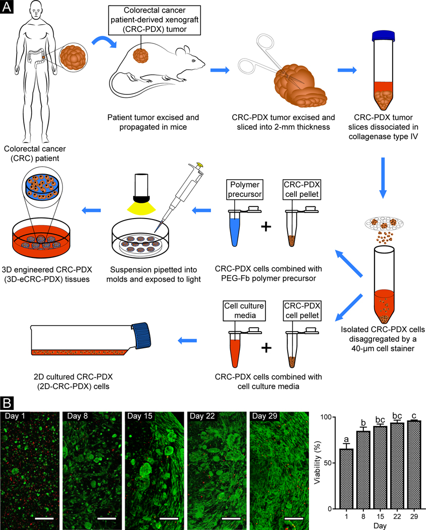
Figure 1. Following encapsulation in PEG-Fb, CRC-PDX tumor cells were viable during long-term culture.
(A) All PDX cells, including both human cancer cells and mouse stromal cells, were isolated from CRC-PDX tumors and used to create 3D-eCRC-PDX tissues and cultured as 2D-CRC-PDX cells. (B) Representative confocal images of live (green) and dead (red) cells within 3D-eCRC-PDX tissues (scale bar = 200 μm). Day 1 cell viability was equivalent to that of non-encapsulated cells (Suppl. Figure 2). Cell elongation and increasing cell colony size were observed from Day 1 to Day 29 post-encapsulation. As a result of cell growth, the percentage of viable cells within the 3D-eCRC-PDX tissues increased significantly during in vitro culture. Bar = mean ± SD. Means that do not share a letter are significantly different (p ≤ 0.05; n = 3 tissues).

