



Abstract
Coronavirus has brought about three massive outbreaks in the past two decades. Each step of its life cycle invariably depends on the interactions among virus and host molecules. The interaction between virus RNA and host protein (IVRHP) is unique compared to other virus–host molecular interactions and represents not only an attempt by viruses to promote their translation/replication, but also the host's endeavor to combat viral pathogenicity. In other words, there is an urgent need to develop a database for providing such IVRHP data. In this study, a new database was therefore constructed to describe the interactions between coronavirus RNAs and host proteins (CovInter). This database is unique in (a) unambiguously characterizing the interactions between virus RNA and host protein, (b) comprehensively providing experimentally validated biological function for hundreds of host proteins key in viral infection and (c) systematically quantifying the differential expression patterns (before and after infection) of these key proteins. Given the devastating and persistent threat of coronaviruses, CovInter is highly expected to fill the gap in the whole process of the ‘molecular arms race’ between viruses and their hosts, which will then aid in the discovery of new antiviral therapies. It's now free and publicly accessible at: https://idrblab.org/covinter/
Graphical Abstract
Graphical Abstract.
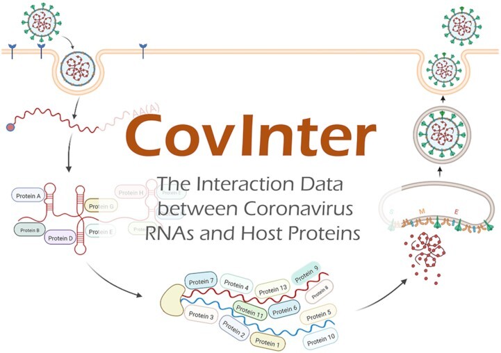
CovInter is unique in (a) unambiguously characterizing the interactions between virus RNA and host protein, (b) comprehensively providing experimentally validated biological function for hundreds of host proteins key in viral infection and (c) systematically quantifying the differential expression patterns (before and after infection) of these key proteins.
INTRODUCTION
The coronaviruses have brought about three massive outbreaks over the past two decades: severe acute respiratory syndrome (SARS), middle eastern respiratory syndrome (MERS) and corona virus disease 2019 (COVID-19) (1–4). The general life cycle of coronavirus (illustrated in Figure 1) is composed of multiple steps, such as fusion, proteolysis, translation, replication, packaging, and release (5–7), each step of which invariably depends on the interactions among the molecules of viruses and hosts (8–12). Particularly, the virus–host protein–protein interactions (VHPPIs) are reported to be essential for virus entry, replication, and dysregulation of host's innate immune response (13–16); the virus–host RNA–RNA interactions (VHRRIs) are identified as the critical building blocks of the pathways for coronaviruses’ transcription/replication (17–19); and the interactions between virus protein and host RNA (IVPHRs) are found essential for not only the transcription of virus proteins but also the packaging during the coronavirus infection (20–23).
Figure 1.

A schematic representation of the life cycle of coronavirus, which consisted of multiple steps (such as fusion, proteolysis, translation, replication, packaging & release). The interactions between virus RNAs and host proteins (IVRHPs, highlighted using blue bold font) are unique in representing not only the virus’ attempts to accelerate their translation & replication, but also the host's endeavor to combat virus pathogenicity.
Compared with the interactions above (VHPPIs, VHRRIs and VPHRIs), the interactions between virus RNA and host protein (IVRHPs, highlighted using blue & bold font in Figure 1) are unique in representing not only the virus’ attempts to accelerate their translation and replication (24–26), but also the host's endeavor to combat virus pathogenicity (27–29). With the recent technological breakthrough in detecting such invaluable interactions (30–33), the discovery of new IVRHP has attracted broad attention from the research community and a huge number of IVRHP data have thus been accumulated (34–38). Particularly, some of these newly discovered IVRHPs are reported to promote the translation/replication of virus (pro-virus (39,40)), while some others are found to suppress the infection (anti-virus (41–43)). Both types of IVRHP data are valuable for filling the missing blanks in the entire process of the ‘molecular arms race’ between virus and host (44–47), and identifying new therapeutic targets to facilitate drug discovery/repurposing (48–53). Therefore, it is essential to have a coronavirus-related knowledge base to provide such valuable IVRHP data that facilitate (pro-virus) or inhibit (anti-virus) virus infection.
Until now, a variety of molecular interaction-based databases that discusses virus infections have been constructed (54–59). The majority of them focus on describing VHPPI data (like COVINET (54), HVIDB (55), VirHostNet (56) and VirusMentha (57)); some others specialize in demonstrating the VHRRI data (such as ViRBase (58)); and the remaining one aims at offering IVPHR data (such as IntACT (59)). However, as an integral & unique part of the comprehensive interacting network in viruses’ life cycle, the IVRHPs have not been covered by any existing databases, which urgently asks for the development of a new database to provide the interaction data between virus RNAs and host proteins (IVRHPs).
In this study, a new knowledge base titled ‘interaction data between coronavirus RNAs and host proteins (CovInter)’ was thus constructed. First, a systematic literature review was conducted by keyword searching in PubMed, which resulted in a total of 10 180 IVRHP data between 310 virus RNAs and 1281 host proteins. Second, the functions of host proteins interacting with virus RNA were then systematically collected by the literature review, and a total of 808 host proteins were identified to describe their pro/anti-virus functions. Particularly, there were 364 pro-virus proteins and 444 anti-virus ones. Third, the differential expression patterns of these host proteins were further analyzed based on the data collected from the GEO database (60). Finally, a total of 316 infectious signaling pathways (the definition of this type of pathway was explicitly described below) that those host proteins involved in were extracted.
All in all, CovInter database was introduced to (a) explicitly describe the interaction data between virus RNAs and host proteins, (b) systematically provide the experimentally verified function for hundreds of host proteins key in virus infection and (c) quantitatively demonstrate the differential expression patterns (before and after infection) of these key proteins. Considering the devastating and long-lasting threat of coronavirus, the data shown in CovInter are expected to help to fill the missing blanks in the entire process of ‘molecular arms race’ between virus and host, which will therefore facilitate the identification of new therapeutic targets for drug discovery/repurposing.
FACTUAL CONTENT AND DATA RETRIEVAL
Collection of interaction data between virus RNA and host protein (IVRHPs)
A comprehensive literature review on the interactions between coronavirus RNA and host protein was first conducted by PubMed searching using the keywords of ‘coronavirus RNA interaction’, ‘SARS-CoV-2 RNA protein interaction’, ‘COVID RNA protein interaction’, ‘SARS-CoV RNA protein interactions’, ‘MERS RNA protein interactions’, ‘coronavirus RNA binding’, and so on. As a result, the IVRHP data of seven types of coronavirus were collected, which included severe acute respiratory syndrome coronavirus (SARS), severe acute respiratory syndrome coronavirus 2 (SARS-CoV-2), middle eastern respiratory syndrome coronavirus (MERS-CoV) together with other four human coronaviruses (HCoV-OC43, HCoV-NL63, HCoV-HKU1, HCoV-229E), and a total of 10 180 IVRHPs between 310 virus RNAs and 1281 host proteins were identified.
Moreover, the additional referencing data for host proteins, virus RNAs, and their corresponding interactions were systematically collected and provided in CovInter database. For a host protein, a variety of referencing data were given, which included protein name, protein family, gene name, EC number (if available), subcellular location, sequence, UniProt ID (61), gene ID (62), Ensembl ID (63), HGNC ID (64) and biological function. The 2D and 3D structures of these proteins from PDB (65), SWISS-MODEL (66) and AlphaFold (67) were also collected. For a coronavirus RNA, its referencing data included virus name, taxonomy ID (68), strains name, strains family, GISAID accession ID (69), strains mutation site, and its structure predicted using RNAfold. For an IVRHP, a variety of experimental data were collected and described, which contained virus infection time, infection cells, cell-originated tissue, detection method, interaction type, interaction binding type and so on. All these interactions were validated using diverse living systems, which included 30 cell lines from 14 tissues and various model organisms. As shown in Figure 2 (a typical CovInter page for virus RNA), a full list of host proteins that interacted with this virus RNA together with the detailed experimental information was provided and explicitly described.
Figure 2.

A typical CovInter page for virus RNA describing a comprehensive list of host proteins that interacted with this RNA. The detailed experimental information was provided and explicitly discussed, which included the virus infection time, infection cell, cell-originated tissue, detection method, interaction types, interaction binding type and so on. All the interactions were validated using diverse living systems including 30 cell lines from 14 tissues and various model organisms. Detailed information of the interacting proteins can be found by clicking the dark blue button.
Illustration of the level of conservation among different IVRHP interactions
Conservation of IVRHPs among various virus strains
Because of the rapid sequence variations in coronavirus RNA, significant gain/loss of interaction (especially IVRHPs) has been frequently reported, which is highly expected to lead to substantial changes in the rate of both virus transmission and case fatality (46,70,71). Thus, it is key to have an in-depth understanding of the level of the conservation among the IVRHPs of various virus variants/strains. In CovInter, a circular plot (comprehensively describing all IVRHPs for a particular virus RNA) was therefore constructed and then provided to demonstrate their level of conservation among virus strains. As provided in Figure 3, for a particular RNA region from the France/IDF-220–95 stain of SARS-CoV-2, all interactions between this RNA (red circle) and different host proteins (green circles) were first visualized by linking them using red lines. Then, other RNAs (from other strains of SARS-CoV-2) which interacted with the same host protein as that of the studied RNA (RNA region from SARS-CoV-2 France/IDF-220-95 stain), were also highlighted using orange circles, and their corresponding interactions with host protein were visualized using orange lines. The diameter of green circle indicated the number of virus’ RNAs that could interact with the host protein. The larger the diameter of a protein is, the more virus RNAs this protein interacts with. In other words, the diameter of a green circle denoted the level of conservation among the corresponding IVRHPs of various virus strains.
Figure 3.

A typical circular plot in online CovInter comprehensively describing all IVRHPs for a particular RNA region from the France/IDF-220–95 stain of SARS-CoV-2 (indicated by a red circle). All interactions between this RNA and different host proteins (green circles) were visualized by linking them using the red line. Other RNAs (from other SARS-CoV-2 strains) which interacted with the same host proteins as that of the studied RNA region above, were highlighted by orange circles, and their corresponding interactions with host proteins were visualized using orange lines. The diameter of a green circle indicated the number of virus’ RNAs interacting with the corresponding protein. The larger the diameter of a protein is, the more virus RNAs this protein interacts with. Specifically, the diameter of a green circle denoted the level of conservation among the corresponding IVRHPs of different virus variants/strains. The circular plot is drawn using the Pychart 1.91 package in Python 3.8 environment, which can be readily viewed online and freely downloaded from the CovInter website.
Taking SRSF1 protein as an example (described in Figure 3), it interacted with the RNAs of six different SARS-CoV-2 strains (not only the France/IDF-220-95 one, but also the remaining five strains: Argentina/INEI104133, Zimbabwe/CERI-KRISP, Namibia/N17380, IPBCAMS-YL01 and Wuhan-Hu-1). This suggested that the IVRHP between SARS-CoV-2′s RNA region and host SRSF1 protein was greatly conserved despite the viral evolution (25,26,34,37,72). Moreover, some other host proteins were found to selectively interact with specific virus strains. Taking the CCAR2 protein as another example, it only interacted with the RNA region from France/IDF-220–95 stain of SARS-CoV-2 (34). That is to say, the corresponding interactions between virus RNA region and host CCAR2 protein were deprived during the virus evolution from the France strain to others. All in all, the CovInter is unique in showing the level of conservation among the IVRHPs of different virus strains, which can contribute to our understandings of virus infection process and the identification and selection of new drug targets and therapies.
Conservation of IVRHPs among different virus types
It is also very important to understand the level of conservation of IVRHPs among different types of coronaviruses, since such interactions usually indicate the fundamental mechanisms underlying virus’ survival and transmission (35,73). In CovInter, a hierarchical plot (that illustrated all IVRHPs for specific host protein) was therefore drawn to show their level of conservations among virus types. As shown in Figure 4, for a host protein named ‘RPS3′, all interactions between virus RNAs (as shown in the middle column) and this protein (on the left side) were first shown by linking them using purple lines. Then, the strains of the same type of coronaviruses were shown in the same color on the right side. As illustrated, the IVRHPs between the virus 5′-UTR RNAs and host RPS3 were largely conserved among different SARS-CoV-2 strains, but this type of IVRHP has not been reported in other types of coronaviruses yet. Different from the 5′-UTR RNA, the 3′-UTR RNA was found to be conserved among different coronavirus types (which were highlighted using different colors). All in all, CovInter is unique in illustrating the level of conservation among the IVRHPs of various types of coronaviruses (such as SARS-CoV-2, SARS-CoV and MERS-CoV).
Figure 4.

A typical hierarchical plot in CovInter illustrating all IVRHPs for specific host protein named ‘RPS3’ (on the far-left side). All interactions between virus RNAs (as provided in the middle column) and this protein were described by linking them using purple line. The strains of the same type of coronaviruses were illustrated in the same color on the right side (e.g. all SARS-CoV-2 strains were shown using blue lines one right side). As illustrated, the IVRHPs between virus 5′-UTR RNA and host RPS3 protein were substantially conserved among different SARS-CoV-2 strains, but this type of IVRHP has not been found in other types of coronaviruses. Different from the 5′-UTR RNA, the 3′-UTR RNA was reported to be conserved among different coronavirus types (highlighted using different colors). The hierarchical plot above is drawn using the Pychart 1.91 package in the Python 3.8 environment, which can be readily viewed online and freely downloaded from the CovInter website.
Explicit description on the interacting protein from multiple perspectives
Current studies on the discovery or the analysis of new IVRHPs have provided great implications on the development of novel antiviral strategies (74–77), and it is also necessary to have detailed descriptions on interacting proteins from multiple perspectives (78–82). Thus, the additional data of these host proteins were provided, such as the function of the proteins in coronavirus infection, available molecular regulators (especially drugs) of the proteins, differential expression pattern of the proteins before and after infection, and infectious pathways that the proteins involved in.
Function and regulators of the host interacting proteins
The function of the host interacting proteins in coronavirus infection was identified by searching the keywords combination in PubMed, such as ‘coronaviruses + host protein + loss of function’, ‘SARS-CoV-2 + host factor + CRISPR knockout screens’, ‘SARS-CoV + genome wide screen’ and ‘MERS-CoV + siRNA screens’. As a result, a total of 808 host proteins were discovered as pro-viral (facilitating viral infection) and anti-viral (hampering infectious progression) proteins. Particularly, there were 364 pro-viral proteins (such as HMGB1 promoting the fusion of SARS-CoV-2 (83)) and 444 anti-viral ones (such as DDX1 inhibiting the RNA amplification of multiple types of coronavirus (84)). The detailed experiments for validating the protein function were also described, such as infection time & cells, cell-originated tissue, detection method and so on.
Furthermore, the available molecular regulators (especially drugs) of the host interacting proteins were also collected by literature review using the keywords combinations of ‘coronavirus + host protein + drug’, ‘SARS-CoV-2 + drug repurposing’, ‘SARS-CoV-2 + drug discovery’, ‘MERS-CoV + antiviral drugs’ and so on. As a result, a total of 391 drugs that targeted 110 host proteins were collected and the links for these drugs to several popular databases such as DrugBank (85), PubChem (86) and TTD (87) were also provided. In the CovInter webpage describing both functions and regulators of host proteins (shown in Figure 5), various data of the corresponding protein was systematically described, and all data can be freely downloaded from the website.
Figure 5.

A typical CovInter webpage showing the biological function and molecular regulation data of the host interacting proteins. A total of 808 host proteins were offered in CovInter as pro-viral (facilitating viral infection) and anti-viral (hampering infectious progression). The detailed experiments for validating the protein function were described here, such as infection time/cells, cell-originated tissue, detection method and so on. Moreover, the available molecular regulators (especially drugs) of the host protein were collected, which resulted in a total of 391 drugs targeting 110 host proteins. All data can be freely downloaded from the CovInter website.
Expression and pathways of the host interacting proteins
Virus infection elicits the differential expression of certain protein and regulates infection-related pathways (88–91). The discovery of proteins and biological pathways that are changed in virus infection can substantially facilitate the explanation of virus’ pathology (12,19), discovery of drug targets (92–94), and advance of precision medicine (95,96). Therefore, the differential expression patterns of the host interacting proteins were analyzed and provided in CovInter database using the following procedures. First, three benchmarks generated by Illumina NovaSeq 6000 and Illumina NextSeq 500 were collected from GEO (60), including GSE152641, GSE162835, and GSE175779. GSE152641 is composed of 24 and 62 blood samples before and after SARS-COV-2 infections; GSE162835 consists of nasopharyngeal swab samples of 37, 10 and 3 patients with mild, moderate, and severe symptom, respectively; GSE175779 contains the bronchial epithelial cell samples from 4 healthy people and 4 SARS-COV-2 patients at different time points (0, 24, 48, 72 and 96 h). Then, the differential expression patterns of host proteins were collected from the original publications of these benchmarks, and illustrated in the CovInter using the Seaborn 0.11.2 package in Python 3.8 environment (as shown in Figure 6).
Figure 6.

Differential expression patterns of host interacting proteins illustrated in CovInter. The differential expression pattern data were collected using the following process. First, three benchmarks were collected from GEO (GSE152641, GSE162835 and GSE175779). GSE152641 is composed of 24 and 62 blood samples before and after SARS-COV-2 infections; GSE162835 consists of the nasopharyngeal swab samples of 37, 10 and 3 patients with mild, moderate, and severe symptom, respectively; GSE175779 contains the bronchial epithelial cell samples from 4 healthy people and 4 SARS-COV-2 patients at different time points (0, 24, 48, 72 and 96 h). Second, the differential expression pattern of host proteins was collected from the original studies of these benchmarks and illustrated in CovInter using the Seaborn 0.11.2 package in Python. Green: protein expression in healthy individuals; red: protein expression in the infected patients. Mild: protein expression in patients with mild symptom; Moderate: protein expression in patients with moderate symptom; Severe: protein expression in patients with severe symptom.
Moreover, to achieve an in-depth insight into the host response pathway activated by host protein during coronavirus infection, a total of 316 infection-associated pathways that these host proteins involved in were identified. Particularly, the pathway information of each host protein was first extracted from KEGG database, and these extracted pathways were checked one-by-one on their relation to virus infection. Then, to determine whether a pathway is related to the infection, two critical procedures were conducted, which included the adoption of KEGG subclasses (such as ‘viral infectious disease’ and ‘information processing in viruses’) and the literature review in PubMed by searching some keyword combinations (such as ‘Pathway Name + infection’ and ‘Pathway Name + infectious disease’). All in all, these efforts helped us to identify any pathways reported to be closely related to virus infection, which were therefore considered as ‘infectious signaling pathways’ in our database. Some of the typical infectious pathways included COVID-19 infectious pathway, chemokine signaling pathway, MAPK and JAK-STAT signaling pathways etc. (97). All infectious pathway maps can be readily viewed online and freely downloaded directly from CovInter website. As reported, interactions between virus RNAs and host proteins could be regulated by protein phosphorylation. Analysis of the phosphorylation of host protein after SARS-CoV-2 infection was therefore considered in CovInter based on two reputable phosphorylated proteomic studies (54,98). The dynamic phosphorylation of proteins at different sites over time (2, 4, 8, 12, 24, and 36 h after SARS-CoV-2 infection) was drawn using the Python package Matplotlib 0.11.2, and a total of 4047 types of phosphorylation occurring in 631 host proteins at 6 time-points were therefore collected and provided.
CONCLUSION AND PERSPECTIVES
Herein, CovInter was developed to unambiguously characterize, for the first time, the interaction data between virus RNA and host protein, comprehensively provide the experimentally verified functions for hundreds of host proteins key in coronavirus infection, and systematically quantify the differential expression patterns of these key proteins before and after infection. CovInter has been smoothly running for months and tested by different research labs, and its data can now be fully accessed without any login requirement by all users at: https://idrblab.org/covinter/.
DATA AVAILABILITY
To view the contributors of each individual sequence that downloaded from GISAID database in this study (provided in Supplementary Table), visit via www.gsaid.org/ EPI_SET_220823ya to get the detailed information such as accession number, virus name, collection date, originating lab and submitting lab and the list of authors.
Supplementary Material
ACKNOWLEDGEMENTS
We gratefully acknowledge all data contributors, i.e. the authors and their originating laboratories responsible for obtaining the specimens, and their submitting laboratories for generating the genetic sequence and metadata and sharing via the GISAID Initiative, on which this research is based.
Contributor Information
Kuerbannisha Amahong, College of Pharmaceutical Sciences, The Second Affiliated Hospital, Zhejiang University School of Medicine, Zhejiang University, Hangzhou 310058, China; Innovation Institute for Artificial Intelligence in Medicine of Zhejiang University, Alibaba-Zhejiang University Joint Research Center of Future Digital Healthcare, Hangzhou 330110, China.
Wei Zhang, College of Pharmaceutical Sciences, The Second Affiliated Hospital, Zhejiang University School of Medicine, Zhejiang University, Hangzhou 310058, China; Innovation Institute for Artificial Intelligence in Medicine of Zhejiang University, Alibaba-Zhejiang University Joint Research Center of Future Digital Healthcare, Hangzhou 330110, China.
Ying Zhou, State Key Laboratory for Diagnosis and Treatment of Infectious Disease, Collaborative Innovation Center for Diagnosis and Treatment of Infectious Diseases, Zhejiang Provincial Key Laboratory for Drug Clinical Research and Evaluation, The First Affiliated Hospital, Zhejiang University, Hangzhou 310000, China.
Song Zhang, College of Pharmaceutical Sciences, The Second Affiliated Hospital, Zhejiang University School of Medicine, Zhejiang University, Hangzhou 310058, China.
Jiayi Yin, College of Pharmaceutical Sciences, The Second Affiliated Hospital, Zhejiang University School of Medicine, Zhejiang University, Hangzhou 310058, China; Innovation Institute for Artificial Intelligence in Medicine of Zhejiang University, Alibaba-Zhejiang University Joint Research Center of Future Digital Healthcare, Hangzhou 330110, China.
Fengcheng Li, College of Pharmaceutical Sciences, The Second Affiliated Hospital, Zhejiang University School of Medicine, Zhejiang University, Hangzhou 310058, China; Innovation Institute for Artificial Intelligence in Medicine of Zhejiang University, Alibaba-Zhejiang University Joint Research Center of Future Digital Healthcare, Hangzhou 330110, China.
Hongquan Xu, Key Laboratory of Elemene Class Anti-cancer Chinese Medicines, School of Pharmacy, Hangzhou Normal University, Hangzhou 311121, China.
Tianci Yan, Key Laboratory of Elemene Class Anti-cancer Chinese Medicines, School of Pharmacy, Hangzhou Normal University, Hangzhou 311121, China.
Zixuan Yue, Key Laboratory of Elemene Class Anti-cancer Chinese Medicines, School of Pharmacy, Hangzhou Normal University, Hangzhou 311121, China.
Yuhong Liu, Key Laboratory of Elemene Class Anti-cancer Chinese Medicines, School of Pharmacy, Hangzhou Normal University, Hangzhou 311121, China.
Tingjun Hou, College of Pharmaceutical Sciences, The Second Affiliated Hospital, Zhejiang University School of Medicine, Zhejiang University, Hangzhou 310058, China.
Yunqing Qiu, State Key Laboratory for Diagnosis and Treatment of Infectious Disease, Collaborative Innovation Center for Diagnosis and Treatment of Infectious Diseases, Zhejiang Provincial Key Laboratory for Drug Clinical Research and Evaluation, The First Affiliated Hospital, Zhejiang University, Hangzhou 310000, China.
Lin Tao, Key Laboratory of Elemene Class Anti-cancer Chinese Medicines, School of Pharmacy, Hangzhou Normal University, Hangzhou 311121, China.
Lianyi Han, Greater Bay Area Institute of Precision Medicine (Guangzhou), School of Life Sciences, Fudan University, Shanghai 315211, China.
Feng Zhu, College of Pharmaceutical Sciences, The Second Affiliated Hospital, Zhejiang University School of Medicine, Zhejiang University, Hangzhou 310058, China; Innovation Institute for Artificial Intelligence in Medicine of Zhejiang University, Alibaba-Zhejiang University Joint Research Center of Future Digital Healthcare, Hangzhou 330110, China.
SUPPLEMENTARY DATA
Supplementary Data are available at NAR Online.
FUNDING
Natural Science Foundation of Zhejiang Province [LR21H300001]; National Natural Science Foundation of China [U1909208, 81872798]; Leading Talent of ‘Ten Thousand Plan’ of the National High-Level Talents Special Support Plan of China; Fundamental Research Fund of Central University [2018QNA7023]; Key R&D Program of Zhejiang Province [2020C03010]; Chinese ‘Double Top-Class’ Universities [181201*194232101]; Westlake Laboratory (Westlake Laboratory of Life Science & Biomedicine); Alibaba-Zhejiang University Joint Research Center of Future Digital Healthcare; Alibaba Cloud; Information Tech Center of Zhejiang University. Funding for open access charge: National Natural Science Foundation of China [81872798].
Conflict of interest statement. None declared.
REFERENCES
- 1. Zhou P., Yang X.L., Wang X.G., Hu B., Zhang L., Zhang W., Si H.R., Zhu Y., Li B., Huang C.L.et al.. A pneumonia outbreak associated with a new coronavirus of probable bat origin. Nature. 2020; 579:270–273. [DOI] [PMC free article] [PubMed] [Google Scholar]
- 2. Corona S. The species severe acute respiratory syndrome-related coronavirus: classifying 2019-nCoV and naming it SARS-CoV-2. Nat Microbiol. 2020; 5:536–544. [DOI] [PMC free article] [PubMed] [Google Scholar]
- 3. Oudshoorn D., Rijs K., Limpens R., Groen K., Koster A.J., Snijder E.J., Kikkert M., Barcena M.. Expression and cleavage of middle east respiratory syndrome coronavirus nsp3-4 polyprotein induce the formation of double-membrane vesicles that mimic those associated with coronaviral RNA replication. Mbio. 2017; 8:e01658-17. [DOI] [PMC free article] [PubMed] [Google Scholar]
- 4. Rodriguez-Morales A.J., Bonilla-Aldana D.K., Balbin-Ramon G.J., Rabaan A.A., Sah R., Paniz-Mondolfi A., Pagliano P., Esposito S.. History is repeating itself: probable zoonotic spillover as the cause of the 2019 novel coronavirus epidemic. Infez. Med. 2020; 28:3–5. [PubMed] [Google Scholar]
- 5. V’Kovski P., Kratzel A., Steiner S., Stalder H., Thiel V.. Coronavirus biology and replication: implications for SARS-CoV-2. Nat. Rev. Microbiol. 2021; 19:155–170. [DOI] [PMC free article] [PubMed] [Google Scholar]
- 6. Kupferschmidt K., Cohen J.. Race to find COVID-19 treatments accelerates. Science. 2020; 367:1412–1413. [DOI] [PubMed] [Google Scholar]
- 7. Pickett B.E., Sadat E.L., Zhang Y., Noronha J.M., Squires R.B., Hunt V., Liu M., Kumar S., Zaremba S., Gu Z.et al.. ViPR: an open bioinformatics database and analysis resource for virology research. Nucleic Acids Res. 2012; 40:D593–D598. [DOI] [PMC free article] [PubMed] [Google Scholar]
- 8. Hoffmann M., Kleine-Weber H., Schroeder S., Krüger N., Herrler T., Erichsen S., Schiergens T.S., Herrler G., Wu N.H., Nitsche A.et al.. SARS-CoV-2 cell entry depends on ACE2 and TMPRSS2 and is blocked by a clinically proven protease inhibitor. Cell. 2020; 181:271–280. [DOI] [PMC free article] [PubMed] [Google Scholar]
- 9. Jackson C.B., Farzan M., Chen B., Choe H.. Mechanisms of SARS-CoV-2 entry into cells. Nat. Rev. Mol. Cell Biol. 2022; 23:3–20. [DOI] [PMC free article] [PubMed] [Google Scholar]
- 10. Wong H.H., Kumar P., Tay F.P., Moreau D., Liu D.X., Bard F.. Genome-wide screen reveals valosin-containing protein requirement for coronavirus exit from endosomes. J. Virol. 2015; 89:11116–111128. [DOI] [PMC free article] [PubMed] [Google Scholar]
- 11. Tan Y., Schneider T., Leong M., Aravind L., Zhang D.. Novel immunoglobulin domain proteins provide insights into evolution and pathogenesis of SARS-CoV-2-related viruses. Mbio. 2020; 11:e00760-20. [DOI] [PMC free article] [PubMed] [Google Scholar]
- 12. Zhang S., Amahong K., Sun X., Lian X., Liu J., Sun H., Lou Y., Zhu F., Qiu Y.. The miRNA: a small but powerful RNA for COVID-19. Brief. Bioinform. 2021; 22:1137–1149. [DOI] [PMC free article] [PubMed] [Google Scholar]
- 13. Kumar R., Afsar M., Khandelwal N., Chander Y., Riyesh T., Dedar R.K., Gulati B.R., Pal Y., Barua S., Tripathi B.N.et al.. Emetine suppresses SARS-CoV-2 replication by inhibiting interaction of viral mRNA with eIF4E. Antiviral Res. 2021; 189:105056. [DOI] [PMC free article] [PubMed] [Google Scholar]
- 14. Suryawanshi R.K., Koganti R., Agelidis A., Patil C.D., Shukla D.. Dysregulation of cell signaling by SARS-CoV-2. Trends Microbiol. 2021; 29:224–237. [DOI] [PMC free article] [PubMed] [Google Scholar]
- 15. Khorsand B., Savadi A., Naghibzadeh M.. SARS-CoV-2-human protein–protein interaction network. Inform Med Unlocked. 2020; 20:100413. [DOI] [PMC free article] [PubMed] [Google Scholar]
- 16. Nelemans T., Kikkert M.. Viral innate immune evasion and the pathogenesis of emerging RNA virus infections. Viruses. 2019; 11:961. [DOI] [PMC free article] [PubMed] [Google Scholar]
- 17. Ziv O., Price J., Shalamova L., Kamenova T., Goodfellow I., Weber F., Miska E.A.. The short- and long-range RNA–RNA interactome of SARS-CoV-2. Mol. Cell. 2020; 80:1067–1077. [DOI] [PMC free article] [PubMed] [Google Scholar]
- 18. Yang S.L., DeFalco L., Anderson D.E., Zhang Y., Aw J.G.A., Lim S.Y., Lim X.N., Tan K.Y., Zhang T., Chawla T.et al.. Comprehensive mapping of SARS-CoV-2 interactions in vivo reveals functional virus–host interactions. Nat. Commun. 2021; 12:5113. [DOI] [PMC free article] [PubMed] [Google Scholar]
- 19. Zhang S., Amahong K., Zhang C., Li F., Gao J., Qiu Y., Zhu F.. RNA–RNA interactions between SARS-CoV-2 and host benefit viral development and evolution during COVID-19 infection. Brief Bioinform. 2022; 23:bbab397. [DOI] [PMC free article] [PubMed] [Google Scholar]
- 20. Schubert K., Karousis E.D., Jomaa A., Scaiola A., Echeverria B., Gurzeler L.A., Leibundgut M., Thiel V., Mühlemann O., Ban N.. SARS-CoV-2 nsp1 binds the ribosomal mRNA channel to inhibit translation. Nat. Struct. Mol. Biol. 2020; 27:959–966. [DOI] [PubMed] [Google Scholar]
- 21. Carlson C.R., Asfaha J.B., Ghent C.M., Howard C.J., Hartooni N., Safari M., Frankel A.D., Morgan D.O.. Phosphoregulation of phase separation by the SARS-CoV-2 n protein suggests a biophysical basis for its dual functions. Mol. Cell. 2020; 80:1092–1103. [DOI] [PMC free article] [PubMed] [Google Scholar]
- 22. Iserman C., Roden C.A., Boerneke M.A., Sealfon R.S.G., McLaughlin G.A., Jungreis I., Fritch E.J., Hou Y.J., Ekena J., Weidmann C.A.et al.. Genomic RNA elements drive phase separation of the SARS-CoV-2 nucleocapsid. Mol. Cell. 2020; 80:1078–1091. [DOI] [PMC free article] [PubMed] [Google Scholar]
- 23. Martianez-Vendrell X., Kikkert M.. Proteomics approaches for the identification of protease substrates during virus infection. Adv. Virus Res. 2021; 109:135–161. [DOI] [PubMed] [Google Scholar]
- 24. White K.M., Rosales R., Yildiz S., Kehrer T., Miorin L., Moreno E., Jangra S., Uccellini M.B., Rathnasinghe R., Coughlan L.et al.. Plitidepsin has potent preclinical efficacy against SARS-CoV-2 by targeting the host protein eEF1A. Science. 2021; 371:926–931. [DOI] [PMC free article] [PubMed] [Google Scholar]
- 25. Flynn R.A., Belk J.A., Qi Y., Yasumoto Y., Wei J., Alfajaro M.M., Shi Q., Mumbach M.R., Limaye A., DeWeirdt P.C.et al.. Discovery and functional interrogation of SARS-CoV-2 RNA-host protein interactions. Cell. 2021; 184:2394–2411. [DOI] [PMC free article] [PubMed] [Google Scholar]
- 26. Zhang S., Huang W., Ren L., Ju X., Gong M., Rao J., Sun L., Li P., Ding Q., Wang J.et al.. Comparison of viral RNA-host protein interactomes across pathogenic RNA viruses informs rapid antiviral drug discovery for SARS-CoV-2. Cell Res. 2022; 32:9–23. [DOI] [PMC free article] [PubMed] [Google Scholar]
- 27. Lee S., Lee Y.S., Choi Y., Son A., Park Y., Lee K.M., Kim J., Kim J.S., Kim V.N.. The SARS-CoV-2 RNA interactome. Mol. Cell. 2021; 81:2838–2850. [DOI] [PMC free article] [PubMed] [Google Scholar]
- 28. Puschnik A.S., Majzoub K., Ooi Y.S., Carette J.E.. A CRISPR toolbox to study virus–host interactions. Nat. Rev. Microbiol. 2017; 15:351–364. [DOI] [PMC free article] [PubMed] [Google Scholar]
- 29. Scutigliani E.M., Kikkert M.. Interaction of the innate immune system with positive-strand RNA virus replication organelles. Cytokine Growth Factor Rev. 2017; 37:17–27. [DOI] [PMC free article] [PubMed] [Google Scholar]
- 30. Ramanathan M., Porter D.F., Khavari P.A.. Methods to study RNA-protein interactions. Nat. Methods. 2019; 16:225–234. [DOI] [PMC free article] [PubMed] [Google Scholar]
- 31. Chu C., Chang H.Y.. ChIRP-MS: RNA-directed proteomic discovery. Methods Mol. Biol. 2018; 1861:37–45. [DOI] [PubMed] [Google Scholar]
- 32. McHugh C.A., Chen C.K., Chow A., Surka C.F., Tran C., McDonel P., Pandya-Jones A., Blanco M., Burghard C., Moradian A.et al.. The xist lncRNA interacts directly with SHARP to silence transcription through HDAC3. Nature. 2015; 521:232–236. [DOI] [PMC free article] [PubMed] [Google Scholar]
- 33. Shen W.X., Zeng X., Zhu F., Wang Y.L., Qin C., Tan Y., Jiang Y.Y., Chen Y.Z.. Out-of-the-box deep learning prediction of pharmaceutical properties by broadly learned knowledge-based molecular representations. Nat Mach Intell. 2021; 3:334–343. [Google Scholar]
- 34. Labeau A., Fery-Simonian L., Lefevre-Utile A., Pourcelot M., Bonnet-Madin L., Soumelis V., Lotteau V., Vidalain P.O., Amara A., Meertens L.. Characterization and functional interrogation of the SARS-CoV-2 RNA interactome. Cell Rep. 2022; 39:110744. [DOI] [PMC free article] [PubMed] [Google Scholar]
- 35. Verma R., Saha S., Kumar S., Mani S., Maiti T.K., Surjit M.. RNA-Protein interaction analysis of SARS-CoV-2 5′ and 3′ untranslated regions reveals a role of lysosome-associated membrane protein-2A during viral infection. Msystems. 2021; 6:e0064321. [DOI] [PMC free article] [PubMed] [Google Scholar]
- 36. Schmidt N., Lareau C.A., Keshishian H., Ganskih S., Schneider C., Hennig T., Melanson R., Werner S., Wei Y., Zimmer M.et al.. The SARS-CoV-2 RNA-protein interactome in infected human cells. Nat. Microbiol. 2021; 6:339–353. [DOI] [PMC free article] [PubMed] [Google Scholar]
- 37. Kamel W., Noerenberg M., Cerikan B., Chen H., Jarvelin A.I., Kammoun M., Lee J.Y., Shuai N., Garcia-Moreno M., Andrejeva A.et al.. Global analysis of protein-RNA interactions in SARS-CoV-2-infected cells reveals key regulators of infection. Mol. Cell. 2021; 81:2851–2867. [DOI] [PMC free article] [PubMed] [Google Scholar]
- 38. Kerr C.H., Skinnider M.A., Andrews D.D.T., Madero A.M., Chan Q.W.T., Stacey R.G., Stoynov N., Jan E., Foster L.J.. Dynamic rewiring of the human interactome by interferon signaling. Genome Biol. 2020; 21:140. [DOI] [PMC free article] [PubMed] [Google Scholar]
- 39. Zhu Y., Feng F., Hu G., Wang Y., Yu Y., Zhu Y., Xu W., Cai X., Sun Z., Han W.et al.. A genome-wide CRISPR screen identifies host factors that regulate SARS-CoV-2 entry. Nat. Commun. 2021; 12:961. [DOI] [PMC free article] [PubMed] [Google Scholar]
- 40. Israeli M., Finkel Y., Yahalom-Ronen Y., Paran N., Chitlaru T., Israeli O., Cohen-Gihon I., Aftalion M., Falach R., Rotem S.et al.. Genome-wide CRISPR screens identify GATA6 as a proviral host factor for SARS-CoV-2 via modulation of ACE2. Nat. Commun. 2022; 13:2237. [DOI] [PMC free article] [PubMed] [Google Scholar]
- 41. Daniloski Z., Jordan T.X., Wessels H.H., Hoagland D.A., Kasela S., Legut M., Maniatis S., Mimitou E.P., Lu L., Geller E.et al.. Identification of required host factors for SARS-CoV-2 infection in human cells. Cell. 2021; 184:92–105. [DOI] [PMC free article] [PubMed] [Google Scholar]
- 42. Ciccosanti F., Di Rienzo M., Romagnoli A., Colavita F., Refolo G., Castilletti C., Agrati C., Brai A., Manetti F., Botta L.et al.. Proteomic analysis identifies the RNA helicase DDX3X as a host target against SARS-CoV-2 infection. Antiviral Res. 2021; 190:105064. [DOI] [PMC free article] [PubMed] [Google Scholar]
- 43. Hoffmann H.H., Sanchez-Rivera F.J., Schneider W.M., Luna J.M., Soto-Feliciano Y.M., Ashbrook A.W., Le Pen J., Leal A.A., Ricardo-Lax I., Michailidis E.et al.. Functional interrogation of a SARS-CoV-2 host protein interactome identifies unique and shared coronavirus host factors. Cell Host Microbe. 2021; 29:267–280. [DOI] [PMC free article] [PubMed] [Google Scholar]
- 44. Wada M., Lokugamage K.G., Nakagawa K., Narayanan K., Makino S.. Interplay between coronavirus, a cytoplasmic RNA virus, and nonsense-mediated mRNA decay pathway. Proc. Natl. Acad. Sci. U.S.A. 2018; 115:E10157–E10166. [DOI] [PMC free article] [PubMed] [Google Scholar]
- 45. Di Giorgio S., Martignano F., Torcia M.G., Mattiuz G., Conticello S.G.. Evidence for host-dependent RNA editing in the transcriptome of SARS-CoV-2. Sci. Adv. 2020; 6:eabb5813. [DOI] [PMC free article] [PubMed] [Google Scholar]
- 46. Banerjee A.K., Blanco M.R., Bruce E.A., Honson D.D., Chen L.M., Chow A., Bhat P., Ollikainen N., Quinodoz S.A., Loney C.et al.. SARS-CoV-2 disrupts splicing, translation, and protein trafficking to suppress host defenses. Cell. 2020; 183:1325–1339. [DOI] [PMC free article] [PubMed] [Google Scholar]
- 47. Guerrero-Arguero I., Tellez-Freitas C.M., Weber K.S., Berges B.K., Robison R.A., Pickett B.E.. Alphaviruses: host pathogenesis, immune response, and vaccine and treatment updates. J. Gen. Virol. 2021; 102:jgv.0.001644. [DOI] [PubMed] [Google Scholar]
- 48. Gordon D.E., Jang G.M., Bouhaddou M., Xu J., Obernier K., White K.M., O’Meara M.J., Rezelj V.V., Guo J.Z., Swaney D.L.et al.. A SARS-CoV-2 protein interaction map reveals targets for drug repurposing. Nature. 2020; 583:459–468. [DOI] [PMC free article] [PubMed] [Google Scholar]
- 49. Sa Ribero M., Jouvenet N., Dreux M., Nisole S.. Interplay between SARS-CoV-2 and the type i interferon response. PLoS Pathog. 2020; 16:e1008737. [DOI] [PMC free article] [PubMed] [Google Scholar]
- 50. Van Nostrand E.L., Pratt G.A., Shishkin A.A., Gelboin-Burkhart C., Fang M.Y., Sundararaman B., Blue S.M., Nguyen T.B., Surka C., Elkins K.et al.. Robust transcriptome-wide discovery of RNA-binding protein binding sites with enhanced CLIP (eCLIP). Nat. Methods. 2016; 13:508–514. [DOI] [PMC free article] [PubMed] [Google Scholar]
- 51. Wang X., Li F., Qiu W., Xu B., Li Y., Lian X., Yu H., Zhang Z., Wang J., Li Z.et al.. SYNBIP: synthetic binding proteins for research, diagnosis and therapy. Nucleic Acids Res. 2022; 50:D560–D570. [DOI] [PMC free article] [PubMed] [Google Scholar]
- 52. Tang J., Fu J., Wang Y., Li B., Li Y., Yang Q., Cui X., Hong J., Li X., Chen Y.et al.. ANPELA: analysis and performance assessment of the label-free quantification workflow for metaproteomic studies. Brief Bioinform. 2020; 21:621–636. [DOI] [PMC free article] [PubMed] [Google Scholar]
- 53. Li F., Yin J., Lu M., Yang Q., Zeng Z., Zhang B., Li Z., Qiu Y., Dai H., Chen Y.et al.. ConSIG: consistent discovery of molecular signature from OMIC data. Brief Bioinform. 2022; 23:bbac253. [DOI] [PubMed] [Google Scholar]
- 54. Stukalov A., Girault V., Grass V., Karayel O., Bergant V., Urban C., Haas D.A., Huang Y., Oubraham L., Wang A.et al.. Multilevel proteomics reveals host perturbations by SARS-CoV-2 and SARS-CoV. Nature. 2021; 594:246–252. [DOI] [PubMed] [Google Scholar]
- 55. Yang X., Lian X., Fu C., Wuchty S., Yang S., Zhang Z.. HVIDB: a comprehensive database for human-virus protein–protein interactions. Brief Bioinform. 2021; 22:832–844. [DOI] [PubMed] [Google Scholar]
- 56. Guirimand T., Delmotte S., Navratil V.. VirHostNet 2.0: surfing on the web of virus/host molecular interactions data. Nucleic Acids Res. 2015; 43:D583–D587. [DOI] [PMC free article] [PubMed] [Google Scholar]
- 57. Calderone A., Licata L., Cesareni G.. VirusMentha: a new resource for virus–host protein interactions. Nucleic Acids Res. 2015; 43:D588–D592. [DOI] [PMC free article] [PubMed] [Google Scholar]
- 58. Cheng J., Lin Y., Xu L., Chen K., Li Q., Xu K., Ning L., Kang J., Cui T., Huang Y.et al.. ViRBase v3.0: a virus and host ncRNA-associated interaction repository with increased coverage and annotation. Nucleic Acids Res. 2022; 50:D928–D933. [DOI] [PMC free article] [PubMed] [Google Scholar]
- 59. Del Toro N., Shrivastava A., Ragueneau E., Meldal B., Combe C., Barrera E., Perfetto L., How K., Ratan P., Shirodkar G.et al.. The intact database: efficient access to fine-grained molecular interaction data. Nucleic Acids Res. 2022; 50:D648–D653. [DOI] [PMC free article] [PubMed] [Google Scholar]
- 60. Clough E., Barrett T.. The gene expression omnibus database. Methods Mol. Biol. 2016; 1418:93–110. [DOI] [PMC free article] [PubMed] [Google Scholar]
- 61. UniProt, C. UniProt: a worldwide hub of protein knowledge. Nucleic Acids Res. 2019; 47:D506–D515. [DOI] [PMC free article] [PubMed] [Google Scholar]
- 62. Brown G.R., Hem V., Katz K.S., Ovetsky M., Wallin C., Ermolaeva O., Tolstoy I., Tatusova T., Pruitt K.D., Maglott D.R.et al.. Gene: a gene-centered information resource at NCBI. Nucleic Acids Res. 2015; 43:D36–D42. [DOI] [PMC free article] [PubMed] [Google Scholar]
- 63. Howe K.L., Achuthan P., Allen J., Allen J., Alvarez-Jarreta J., Amode M.R., Armean I.M., Azov A.G., Bennett R., Bhai J.et al.. Ensembl 2021. Nucleic Acids Res. 2021; 49:D884–D891. [DOI] [PMC free article] [PubMed] [Google Scholar]
- 64. Bruford E.A., Braschi B., Denny P., Jones T.E.M., Seal R.L., Tweedie S.. Guidelines for human gene nomenclature. Nat. Genet. 2020; 52:754–758. [DOI] [PMC free article] [PubMed] [Google Scholar]
- 65. Burley S.K., Berman H.M., Bhikadiya C., Bi C., Chen L., Di Costanzo L., Christie C., Dalenberg K., Duarte J.M., Dutta S.et al.. RCSB protein data bank: biological macromolecular structures enabling research and education in fundamental biology, biomedicine, biotechnology and energy. Nucleic Acids Res. 2019; 47:D464–D474. [DOI] [PMC free article] [PubMed] [Google Scholar]
- 66. Waterhouse A., Bertoni M., Bienert S., Studer G., Tauriello G., Gumienny R., Heer F.T., de Beer T.A.P., Rempfer C., Bordoli L.et al.. SWISS-MODEL: homology modelling of protein structures and complexes. Nucleic Acids Res. 2018; 46:W296–W303. [DOI] [PMC free article] [PubMed] [Google Scholar]
- 67. Varadi M., Anyango S., Deshpande M., Nair S., Natassia C., Yordanova G., Yuan D., Stroe O., Wood G., Laydon A.et al.. AlphaFold protein structure database: massively expanding the structural coverage of protein-sequence space with high-accuracy models. Nucleic Acids Res. 2022; 50:D439–D444. [DOI] [PMC free article] [PubMed] [Google Scholar]
- 68. Federhen S. The NCBI taxonomy database. Nucleic Acids Res. 2012; 40:D136–D143. [DOI] [PMC free article] [PubMed] [Google Scholar]
- 69. Khare S., Gurry C., Freitas L., Schultz M.B., Bach G., Diallo A., Akite N., Ho J., Lee R.T., Yeo W.et al.. GISAID’s role in pandemic response. China CDC Wkly. 2021; 3:1049–1051. [DOI] [PMC free article] [PubMed] [Google Scholar]
- 70. Galan C., Sola I., Nogales A., Thomas B., Akoulitchev A., Enjuanes L., Almazan F.. Host cell proteins interacting with the 3' end of TGEV coronavirus genome influence virus replication. Virology. 2009; 391:304–314. [DOI] [PMC free article] [PubMed] [Google Scholar]
- 71. Mukherjee M., Goswami S.. Global cataloguing of variations in untranslated regions of viral genome and prediction of key host RNA binding protein-microRNA interactions modulating genome stability in SARS-CoV-2. PLoS One. 2020; 15:e0237559. [DOI] [PMC free article] [PubMed] [Google Scholar]
- 72. Ferrarini M.G., Lal A., Rebollo R., Gruber A.J., Guarracino A., Gonzalez I.M., Floyd T., de Oliveira D.S., Shanklin J., Beausoleil E.et al.. Genome-wide bioinformatic analyses predict key host and viral factors in SARS-CoV-2 pathogenesis. Commun Biol. 2021; 4:590. [DOI] [PMC free article] [PubMed] [Google Scholar]
- 73. Sola I., Mateos-Gomez P.A., Almazan F., Zuniga S., Enjuanes L.. RNA–RNA and RNA-protein interactions in coronavirus replication and transcription. RNA Biol. 2011; 8:237–248. [DOI] [PMC free article] [PubMed] [Google Scholar]
- 74. Li Y.H., Li X.X., Hong J.J., Wang Y.X., Fu J.B., Yang H., Yu C.Y., Li F.C., Hu J., Xue W.W.et al.. Clinical trials, progression-speed differentiating features and swiftness rule of the innovative targets of first-in-class drugs. Brief. Bioinform. 2020; 21:649–662. [DOI] [PMC free article] [PubMed] [Google Scholar]
- 75. Fu T., Li F., Zhang Y., Yin J., Qiu W., Li X., Liu X., Xin W., Wang C., Yu L.et al.. VARIDT 2.0: structural variability of drug transporter. Nucleic Acids Res. 2022; 50:D1417–D1431. [DOI] [PMC free article] [PubMed] [Google Scholar]
- 76. Tang J., Mou M., Wang Y., Luo Y., Zhu F.. MetaFS: performance assessment of biomarker discovery in metaproteomics. Brief. Bioinform. 2021; 22:bbaa105. [DOI] [PubMed] [Google Scholar]
- 77. Yang Q., Li B., Tang J., Cui X., Wang Y., Li X., Hu J., Chen Y., Xue W., Lou Y.et al.. Consistent gene signature of schizophrenia identified by a novel feature selection strategy from comprehensive sets of transcriptomic data. Brief Bioinform. 2020; 21:1058–1068. [DOI] [PubMed] [Google Scholar]
- 78. Gray M., Guerrero-Arguero I., Solis-Leal A., Robison R.A., Berges B.K., Pickett B.E.. Chikungunya virus time course infection of human macrophages reveals intracellular signaling pathways relevant to repurposed therapeutics. PeerJ. 2022; 10:e13090. [DOI] [PMC free article] [PubMed] [Google Scholar]
- 79. Bailey-Elkin B.A., Knaap R.C.M., Kikkert M., Mark B.L.. Structure and function of viral deubiquitinating enzymes. J. Mol. Biol. 2017; 429:3441–3470. [DOI] [PMC free article] [PubMed] [Google Scholar]
- 80. Wu Q., Zheng X., Leung K.S., Wong M.H., Tsui S.K., Cheng L.. meGPS: a multi-omics signature for hepatocellular carcinoma detection integrating methylome and transcriptome data. Bioinformatics. 2022; 38:3513–3522. [DOI] [PubMed] [Google Scholar]
- 81. Zhang S., Sun X., Mou M., Amahong K., Sun H., Zhang W., Shi S., Li Z., Gao J., Zhu F.. REGLIV: molecular regulation data of diverse living systems facilitating current multiomics research. Comput. Biol. Med. 2022; 148:105825. [DOI] [PubMed] [Google Scholar]
- 82. Li B., Tang J., Yang Q., Li S., Cui X., Li Y., Chen Y., Xue W., Li X., Zhu F.. NOREVA: normalization and evaluation of MS-based metabolomics data. Nucleic Acids Res. 2017; 45:W162–W170. [DOI] [PMC free article] [PubMed] [Google Scholar]
- 83. Wei J., Alfajaro M.M., DeWeirdt P.C., Hanna R.E., Lu-Culligan W.J., Cai W.L., Strine M.S., Zhang S.M., Graziano V.R., Schmitz C.O.et al.. Genome-wide CRISPR screens reveal host factors critical for SARS-CoV-2 infection. Cell. 2021; 184:76–91. [DOI] [PMC free article] [PubMed] [Google Scholar]
- 84. Schneider W.M., Luna J.M., Hoffmann H.H., Sánchez-Rivera F.J., Leal A.A., Ashbrook A.W., Le Pen J., Ricardo-Lax I., Michailidis E., Peace A.et al.. Genome-scale identification of SARS-CoV-2 and pan-coronavirus host factor networks. Cell. 2021; 184:120–132. [DOI] [PMC free article] [PubMed] [Google Scholar]
- 85. Wishart D.S., Feunang Y.D., Guo A.C., Lo E.J., Marcu A., Grant J.R., Sajed T., Johnson D., Li C., Sayeeda Z.et al.. DrugBank 5.0: a major update to the drugbank database for 2018. Nucleic Acids Res. 2018; 46:D1074–D1082. [DOI] [PMC free article] [PubMed] [Google Scholar]
- 86. Kim S., Chen J., Cheng T., Gindulyte A., He J., He S., Li Q., Shoemaker B.A., Thiessen P.A., Yu B.et al.. PubChem in 2021: new data content and improved web interfaces. Nucleic Acids Res. 2021; 49:D1388–D1395. [DOI] [PMC free article] [PubMed] [Google Scholar]
- 87. Zhu F., Shi Z., Qin C., Tao L., Liu X., Xu F., Zhang L., Song Y., Liu X., Zhang J.et al.. Therapeutic target database update 2012: a resource for facilitating target-oriented drug discovery. Nucleic Acids Res. 2012; 40:D1128–D1136. [DOI] [PMC free article] [PubMed] [Google Scholar]
- 88. Lin H., Satizabal C., Xie Z., Yang Q., Huan T., Joehanes R., Wen C., Munson P.J., Beiser A., Levy D.et al.. Whole blood gene expression and white matter hyperintensities. Mol Neurodegener. 2017; 12:67. [DOI] [PMC free article] [PubMed] [Google Scholar]
- 89. Wang R., Zheng X., Wang J., Wan S., Song F., Wong M.H., Leung K.S., Cheng L.. Improving bulk RNA-seq classification by transferring gene signature from single cells in acute myeloid leukemia. Brief Bioinform. 2022; 23:bbac002. [DOI] [PubMed] [Google Scholar]
- 90. Martinez Viedma M.D.P., Pickett B.E.. Characterizing the different effects of zika virus infection in placenta and microglia cells. Viruses. 2018; 10:649. [DOI] [PMC free article] [PubMed] [Google Scholar]
- 91. Yin J., Sun W., Li F., Hong J., Li X., Zhou Y., Lu Y., Liu M., Zhang X., Chen N.et al.. VARIDT 1.0: variability of drug transporter database. Nucleic Acids Res. 2020; 48:D1042–D1050. [DOI] [PMC free article] [PubMed] [Google Scholar]
- 92. Liu X., Xu Y., Wang R., Liu S., Wang J., Luo Y., Leung K.S., Cheng L.. A network-based algorithm for the identification of moonlighting noncoding RNAs and its application in sepsis. Brief Bioinform. 2021; 22:581–588. [DOI] [PubMed] [Google Scholar]
- 93. Tan Y., Wang C., Schneider T., Li H., de Souza R.F., Tang X., Swisher Grimm K.D., Hsieh T.F., Wang X., Li X.et al.. Comparative phylogenomic analysis reveals evolutionary genomic changes and novel toxin families in endophytic liberibacter pathogens. Microbiol. Spectr. 2021; 9:e0050921. [DOI] [PMC free article] [PubMed] [Google Scholar]
- 94. Li F., Zhou Y., Zhang Y., Yin J., Qiu Y., Gao J., Zhu F.. POSREG: proteomic signature discovered by simultaneously optimizing its reproducibility and generalizability. Brief Bioinform. 2022; 23:bbac040. [DOI] [PubMed] [Google Scholar]
- 95. Fu J., Zhang Y., Wang Y., Zhang H., Liu J., Tang J., Yang Q., Sun H., Qiu W., Ma Y.et al.. Optimization of metabolomic data processing using NOREVA. Nat. Protoc. 2022; 17:129–151. [DOI] [PubMed] [Google Scholar]
- 96. Yang Q., Wang Y., Zhang Y., Li F., Xia W., Zhou Y., Qiu Y., Li H., Zhu F.. NOREVA: enhanced normalization and evaluation of time-course and multi-class metabolomic data. Nucleic Acids Res. 2020; 48:W436–W448. [DOI] [PMC free article] [PubMed] [Google Scholar]
- 97. Kanehisa M., Furumichi M., Tanabe M., Sato Y., Morishima K.. KEGG: new perspectives on genomes, pathways, diseases and drugs. Nucleic Acids Res. 2017; 45:D353–D361. [DOI] [PMC free article] [PubMed] [Google Scholar]
- 98. Bouhaddou M., Memon D., Meyer B., White K.M., Rezelj V.V., Correa Marrero M., Polacco B.J., Melnyk J.E., Ulferts S., Kaake R.M.et al.. The global phosphorylation landscape of SARS-CoV-2 infection. Cell. 2020; 182:685–712. [DOI] [PMC free article] [PubMed] [Google Scholar]
Associated Data
This section collects any data citations, data availability statements, or supplementary materials included in this article.
Supplementary Materials
Data Availability Statement
To view the contributors of each individual sequence that downloaded from GISAID database in this study (provided in Supplementary Table), visit via www.gsaid.org/ EPI_SET_220823ya to get the detailed information such as accession number, virus name, collection date, originating lab and submitting lab and the list of authors.