ABSTRACT
This study aimed to validate the prognostic value of Immunoscore (IS) in stage II colorectal cancer (CRC), and explore the roles of IS and circulating tumor DNA (ctDNA) in the adjuvant treatment for early-stage CRC. Resected tumor samples from stage II CRC patients were collected from the Sun Yat-sen University Cancer Center. The densities of CD3+ and CD8+ lymphocytes were quantified and converted to IS and classified into Low, Intermediate (Int), and High groups according to predefined cutoffs. A total of 113 patients were included in the study. Patients with IS-High, Int, and Low were 43 (38%), 62 (55%), and 8 (7%), respectively. Patients with IS-High had an excellent clinical outcome, with none recurring during a median follow-up of 3 years, including 15 (35%) clinical high-risk patients. The 3-year disease-free survival (DFS) was 100% for IS-High, 76% for IS-Int, and 47% for IS-Low (P < .001). In the multivariate Cox analysis, IS was the only significant parameter associated with DFS. IS-Int and IS-Low patients with adjuvant chemotherapy had improved DFS compared to those who did not receive adjuvant chemotherapy (HR = 0.3; 95% CI 0.1–0.92; P = .026). Among the 49 patients with postoperative ctDNA data, IS-High patients had the lowest ctDNA positivity rate, suggesting that they were most eligible for chemotherapy-free treatment. IS had a strong prognostic value in Chinese patients with stage II CRC and demonstrates its clinical utility. IS and ctDNA will jointly optimize the adjuvant treatment strategies for early-stage CRC.
KEYWORDS: Immunoscore, colorectal cancer, prognostic, predictive, adjuvant chemotherapy, ctDNA
Introduction
Colorectal cancer (CRC) is the third most common cancer worldwide, with over 1.9 million new cases and 935,000 deaths in 20201. Stage II colon cancer (CC) is an early-stage tumor that has not spread to lymph nodes or distant organs, with a cure rate of about 80% by surgery alone.2 Although the benefit of adjuvant chemotherapy in stage III CC patients is well established, it remains controversial in stage II patients. Clinical practice guidelines recommend risk classification based on microsatellite instability and clinical risk factors to guide the adjuvant chemotherapy decisions.3–6 Patients with poor prognostic features (clinical high-risk patients) can be considered for adjuvant chemotherapy. However, the definition of high-risk is inadequate, as many patients with high-risk features do not have a recurrence.7,8 Also, there is a lack of data on the predictive value of the features for the chemotherapy benefit or the correlation between the features and the selection of chemotherapy. More importantly, even in high-risk patients, fluorouracil-based adjuvant chemotherapy improved survival by less than 5%,9,10 resulting in many patients being exposed to the toxicity of unnecessary chemotherapy. Thus, there is an urgent need to find the optimal prognostic and predictive markers to guide adjuvant treatment decisions for stage II CC patients.
With the growing recognition of the importance of the tumor immune microenvironment, multiple types of tumor-infiltrating immune cells have been reported to correlate with cancer prognosis, including CRC.11,12 However, in addition to Immunoscore (IS), a scoring system assessing the densities of CD3 and CD8 positive lymphocytes at the invasive margin and the tumor core, few immune cell-based prognostic markers have comparable standardized methodologies and extensive clinical data.13 In an international study of IS in stage I–III CC, IS showed a superior prognostic value to all the existing tumor parameters, including the AJCC-TNM classification-system.14 Among the 1434 patients with stage II CC in the SITC study, IS could identify patients with different risks of tumor recurrence. From the same cohort, among patients who were not treated with adjuvant therapy, those with high-risk features and high IS had a similar recurrence risk to those with low clinical risk, suggesting that IS may identify good prognostic patients with high-risk features who may avoid adjuvant therapy.15
However, the prognostic value of the consensus IS in stage II CC has only been demonstrated in the SITC cohort,14 and external validation in independent cohorts is lacking. Also, the evidence for the association between IS and the chemotherapy benefit in stage II CC is insufficient. In addition, the IS scoring-system was based on data from more than 2500 CC patients in the international SITC study, of whom 15% were Asian and only 1.3% (36 cases) were Chinese.14 It is therefore unknown whether the IS thresholds are equally applicable to an independent population. It was previously reported that a high density of CD3+, CD8+ and effector-memory T-cells were associated with decreased venous emboli, lymphatic invasion or perineural invasion (collectively VELIPI).16 Since patients with VELIPI+ are more likely to have detectable ctDNA, we hypothesized a possible relationship between Immunoscore a ctDNA detection. Here, we validated the IS prognostic value in a Chinese stage II CRC population and explored the correlation between IS and the benefit of adjuvant chemotherapy. Besides, in a subset of the cohort, we investigated the association between IS and another potential prognostic marker circulating tumor DNA (ctDNA) and envisaged the future direction of adjuvant treatment-decisions for stage II CC based on these two biomarkers.
Materials and methods
Study design and patients
We collected tumor samples in 137 stage II CRC patients at Sun Yat-sen University Cancer Center (SYSUCC) from January 2017 to September 2019 to verify the prognostic performance of IS in the Chinese population. Patients receiving neoadjuvant therapy were excluded and those receiving adjuvant chemotherapy or not were included. Adjuvant chemotherapy regimens in this study included single-agent capecitabine or capecitabine plus oxaliplatin (CAPOX). Clinical high-risk disease was defined as pathological T4 (pT4) tumor or proficient mismatch-repair (pMMR) tumor with at least one of the following risk factors: poorly differentiated/undifferentiated histology, VELIPI (vascular, lymphatic, or perineural invasion), bowel obstruction, <12 lymph nodes (LN) examined, localized perforation, or close, indeterminate, or positive margins. MMR status was determined by immunohistochemistry for the four MMR proteins MLH1, MSH2, MSH6, and PMS2. If any MMR protein was negatively stained, referred to as MMR deficiency (dMMR). The primary endpoint was to evaluate the prognostic value of IS for disease-free survival (DFS), defined as the time from surgery to the first observed disease recurrence or death due to any cause. This study was approved by the Ethics Committee of SYSUCC. All patients provided written informed consent, and the study was conducted in accordance with the Declaration of Helsinki.
Immunoscore assay
The Immunoscore assay was conducted as previously reported.14 Briefly, tumor blocks containing the core of the tumor and invasive margin were selected for histological review. Sections of 4 μm were processed for standardized CD3 and CD8 Immunohistochemical staining. The densities of the two markers in both regions of the whole slide were determined by dedicated Immunoscore software and then converted into percentiles by comparison with the Immunoscore database. Finally, the mean of the four percentiles was calculated and translated into an Immunoscore. According to predefined cutoffs, Immunoscore was usually analyzed in three groups (0%-25%, low; >25%-70%, intermediate; >70%-100%, high) or in two groups (0%-25%, low; >25%-100%, high). Immunohistochemical analysis and full scan of staining images were performed in Genecast laboratory (Wuxi, China), and digital pathological analysis of Immunoscore was carried out in Veracyte laboratory.
Circulating tumor DNA analysis
In this cohort, 49 patients were enrolled in a prospective observational study investigating the utility of ctDNA in predicting tumor recurrence risk.17 The peripheral blood leukocytes, primary tumor tissue and plasma samples were collected and sequenced by the Geneseeq PrimeTM 425-gene panel. Tumor-specific somatic variants of each patient were used for ctDNA tracking, and more than 5% of the total tracking variants detected in plasma samples were considered ctDNA positive. The postoperative plasma samples were collected at day 3–7, 6 months after surgery, and then every 3 months until month 24. For more details please refer to the previous study.17 Patients with the detection of ctDNA at any postoperative time-point were included in the positive ctDNA group.
Statistical analysis
Patient characteristics were summarized as frequencies and percentages. Differences between groups were evaluated by Fisher’s exact test. DFS was estimated using the Kaplan-Meier method and compared by the log-rank test. Hazard ratios (HRs) for factors associated with DFS were estimated by univariate and multivariate COX regression analysis (survival, R package). Between-group differences in DFS were analyzed using the restricted mean survival time (RMST), a survival analysis that calculates the area under the survival curve to a specific time point and is independent of the proportional-hazards assumption (survRM2, R package). Subgroup analysis for the association between chemotherapy and DFS was carried out and summarized with the forest plot. All statistical tests were two-sided, and P < .05 was considered statistical significance.
Results
Patient characteristics and Immunoscore distribution
A total of 113 patients with IS results were included in the analysis (Table 1). The median age of the patients was 56 years (Interquartile range [IQR] 48–64), 47 of them (42%) were female. Among the patients, 106 (94%) had colon cancer, and the remaining 7 (6%) had high rectal cancer eligible for IS testing. 49 (43%) of them were clinical high-risk, with pMMR tumors and at least one clinical risk factor. The median follow-up was 37.3 months (IQR 32.1–40.7).
Table 1.
Patient population characteristics according to Immunoscore (3 groups).
| Total | IS Hi | IS Int | IS Lo | P | |
|---|---|---|---|---|---|
| Characteristics | n = 113 | n = 43 | n = 62 | n = 8 | |
| Age (years) | 0.015 | ||||
| <65 | 90 (80%) | 37 (86%) | 50 (81%) | 3 (38%) | |
| ≥65 | 23 (20%) | 6 (14%) | 12 (19%) | 5 (63%) | |
| Gender | 0.913 | ||||
| Female | 47 (42%) | 17 (40%) | 27 (44%) | 3 (38%) | |
| Male | 66 (58%) | 26 (60%) | 35 (56%) | 5 (63%) | |
| Cancer type | 0.822 | ||||
| Colon | 106 (94%) | 41 (95%) | 57 (92%) | 8 (100%) | |
| Rectal | 7 (6%) | 2 (5%) | 5 (8%) | 0 (0%) | |
| T stage | 0.410 | ||||
| T3 | 103 (91%) | 41 (95%) | 55 (89%) | 7 (88%) | |
| T4 | 10 (9%) | 2 (5%) | 7 (11%) | 1 (13%) | |
| Tumor site† | 0.146 | ||||
| Left | 55 (49%) | 16 (37%) | 34 (55%) | 5 (63%) | |
| Right | 58 (51%) | 27 (63%) | 28 (45%) | 3 (38%) | |
| Differentiation | 0.755 | ||||
| Mod/Well | 83 (73%) | 32 (74%) | 46 (74%) | 5 (63%) | |
| Poor | 30 (27%) | 11 (26%) | 16 (26%) | 3 (38%) | |
| Mucinous | 0.044 | ||||
| No | 107 (95%) | 42 (98%) | 59 (95%) | 6 (75%) | |
| Yes | 6 (5%) | 1 (2%) | 3 (5%) | 2 (25%) | |
| Examined LN | 1.000 | ||||
| <12 | 11 (10%) | 4 (9%) | 6 (10%) | 1 (13%) | |
| ≥12 | 102 (90%) | 39 (91%) | 56 (90%) | 7 (88%) | |
| VELIPI | 0.168 | ||||
| No | 85 (75%) | 35 (81%) | 46 (74%) | 4 (50%) | |
| Yes | 28 (25%) | 8 (19%) | 16 (26%) | 4 (50%) | |
| MMR status | 0.456 | ||||
| dMMR | 13 (12%) | 7 (16%) | 6 (10%) | 0 (0%) | |
| pMMR | 100 (88%) | 36 (84%) | 56 (90%) | 8 (100%) | |
| Clinical risk | 0.344 | ||||
| High risk | 49 (43%) | 15 (35%) | 30 (48%) | 4 (50%) | |
| Low risk | 64 (57%) | 28 (65%) | 32 (52%) | 4 (50%) | |
| Preoperative CEA | |||||
| <5 ng/ml | 77 (68%) | 33 (77%) | 39 (63%) | 5 (63%) | 0.434 |
| ≥5 ng/ml | 35 (31%) | 10 (23%) | 22 (35%) | 3 (38%) | |
| Missing | 1 (1%) | 0 (0%) | 1 (2%) | 0 (0%) | |
| Adjuvant chemo | 0.103 | ||||
| Yes | 46 (41%) | 15 (35%) | 27 (44%) | 4 (50%) | |
| No | 60 (53%) | 27 (63%) | 31 (50%) | 2 (25%) | |
| Missing | 7 (6%) | 1 (2%) | 4 (6%) | 2 (25%) |
†Left-sided cancer was defined as tumors arising from the splenic flexure, descending, sigmoid, rectosigmoid colon, or the rectum; right-sided cancer was defined as tumors arising from the cecum, ascending, hepatic flexure, or transverse colon.
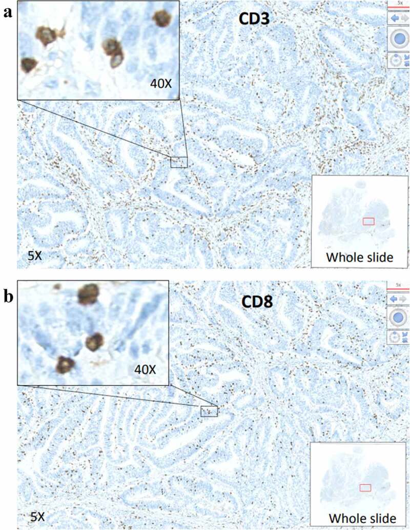
IS-High, Intermediate (Int), and Low were observed in 43 (38%), 62 (55%) and 8 (7%) patients, respectively. According to the three IS groups, no significant differences in baseline features were found, except for a higher percentage of patients with mucinous adenocarcinoma or over 65 years of age in the IS-Low group than in the IS-High or Int groups. Notably, the IS distribution was quite different from that previously reported in the SITC study (Figure S1). Among the 1434 stage II CC patients in the SITC cohort, IS-High, Int, and Low accounted for 26%, 47%, and 27%, respectively.14 There was a striking difference in the proportion of IS-Low which accounted for 27% in the SITC cohort compared to 7% in our SYSUCC cohort (P < .001). Then, we further examined IS data from 151 patients with stage II CC who underwent IS diagnostic testing in the Genecast Medical Laboratory (Wuxi, China). Results revealed that the proportions of IS-Low, Int, and High were 9%, 64% and 27%, respectively, similar to those of the SYSUCC cohort but significantly different from those of the SITC cohort. The quality of the staining for CD3 and CD8 were excellent, with no background and a very good signal intensity that could easily quantified by the Immunoscore software. Representative images of the software are illustrated (Figure 1). Therefore, we considered that the IS distribution in the Chinese population might be distinct from that in Western countries. IS was usually classified into 3-groups (High, Int, Low) or 2-groups (High+Int, Low).14,18,19 In this study, we also tested the modified IS 2-groups of High and Int+Low owing to the small size of IS-Low in the SYSUCC cohort. No significant differences in baseline features were observed according to the modified IS 2-groups (IS-High and IS-Int+Low) (Table S1).
Figure 1.

Representative images of CD3 and CD8 staining. Whole tumor slides of patients with stage II CRC from SYSUCC cancer center were stained for CD3 (A) and for CD8 (B). Whole slide images, 5X magnification and 40X magnification are illustrated.
Patients with IS-High have a superior clinical outcome for DFS
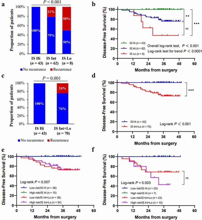
We first validated the prognostic performance of IS in the SYSUCC CRC cohort. Consistent with previous studies, higher IS was associated with a lower risk of recurrence. The recurrence rates were 0%, 21% and 50% in patients with IS-High, Int, and Low, respectively (P < .001) (Figure 2a). Most importantly, the clinical outcomes of DFS for patients identified in the three IS groups were remarkably different, with a 3-year DFS of 47% (95% CI 0.22–1.0) for IS-Low, 76% (95% CI 0.65–0.89) for IS-Int, and 100% for IS-High (Figure 2b). Thus, patients with IS-High had a significantly better outcome than patients with IS-Int or than patients with IS-Low Figure 2b). The prognosis of the 43 IS-High patients were favorable and none of them experienced recurrence during a median follow-up of 3 years. Furthermore, of the patients with IS-High, 15 (35%) were clinically high-risk, 2 (5%) had pT4 tumors, and 36 (84%) were pMMR patients, indicating that IS can effectively identify patients with a good prognosis from the predefined high-risk population.
Figure 2.

Immunoscore (IS) and clinical outcomes of patients with stage II CRC. Overall recurrence rates according to the IS 3-groups (a) and the modified IS 2-groups (c). Kaplan-Meier curves for disease-free survival (DFS) according to IS 3-groups (b) and the modified IS 2-groups (d). Kaplan-Meier curves for DFS in patients with different clinical risk and IS levels (e). Kaplan-Meier curves for DFS in untreated patients with different clinical risk and IS levels (f). Hi, high; Int, intermediate; Lo, low; (*) indicates significant log-rank P-value, *P < .05, **P < .01, ***P < .001; ns, non-significant.
Significant and similar results were also found with the IS 2-groups, both classical and modified. According to the modified IS 2-groups, patients with IS-Int and IS-Low had a recurrence rate of 25% (Figure 2c) and a 3-year DFS rate of 73% (95% CI 0.62–0.85) (Figure 2d). A significant survival difference was also observed between the classical IS 2-groups (IS-High+Int vs Low) (Figure S2). As clinical risk based on histopathological features was used to stratify patients and guide adjuvant treatment decisions, we compared it with the modified 2-group IS, which showed a superior prognostic value in this cohort (Figure 2e). Notably, patients with an excellent prognosis for IS-High accounted for almost one-third of the clinical high-risk patients. This was in line with the result for the subgroup of 60 patients without adjuvant chemotherapy (figure 2f). In this no-treatment group, 7 (41%) of clinical high-risk patients were IS-High and had no recurrences, indicating that patients with IS-High may be spared from chemotherapy.
Univariate Cox analysis showed that age and IS were associated with DFS in all patients (Table 2). According to the Kaplan-Meier analysis, IS (High and Int+Low) could stratify the DFS of patients in different age groups (Figure S3). Also, RMST analysis revealed a significant gain of 8.1 and 14.5 months for the IS-Int and IS-High groups, respectively, compared to the IS-Low group. Moreover, the RMST analysis showed a significant difference of 10.7 months between patients with IS-High+Int and IS-Low (classsical IS-2 groups) and of 7.8 months between patients with IS-High and IS-Int+Low (P < .0001) (Table S2). The 3-group IS as a continuous variable and the classical 2-group IS could be assessed in multivariate analysis. IS was demonstrated to be an independent prognostic factor by the adjusted multivariate analysis of gender, tumor site, T stage, examined LN, VELIPI, and MMR-status in all patients or patients without adjuvant chemotherapy (Table 3). A similar result was also shown for the classical 2-group IS (Table S3). Therefore, IS was the strongest and the most significant parameter in the multivariate analysis of the SYSUCC cohort.
Table 2.
Univariate Cox analysis for disease-free survival in all available patients.
| Characteristics | n | Events | HR (95%CI) | Wald P | Log-rank P | |
|---|---|---|---|---|---|---|
| Age (years) | <65 | 90 | 10 | reference | ||
| ≥65 | 23 | 7 | 2.99 (1.14–7.86) | 0.026 | 0.02 | |
| Gender | Male | 66 | 11 | reference | ||
| Female | 47 | 6 | 0.78 (0.29–2.12) | 0.632 | 0.631 | |
| Cancer type | Colon | 106 | 17 | reference | ||
| Rectal | 7 | 0 | 0.04 (0–211.67) | 0.472 | 0.272 | |
| Tumor site† | Right | 58 | 9 | reference | ||
| Left | 55 | 8 | 0.93 (0.36–2.41) | 0.876 | 0.876 | |
| T Stage | T3 | 103 | 15 | reference | ||
| T4 | 10 | 2 | 1.5 (0.34–6.55) | 0.592 | 0.589 | |
| Differentiation | Mod/Well | 83 | 14 | reference | ||
| Poor | 30 | 3 | 0.61 (0.18–2.13) | 0.438 | 0.434 | |
| Mucinous | No | 107 | 16 | reference | ||
| Yes | 6 | 1 | 1 (0.13–7.53) | 0.999 | 0.999 | |
| Examined LN | <12 | 11 | 2 | reference | ||
| ≥12 | 102 | 15 | 0.76 (0.17–3.35) | 0.72 | 0.719 | |
| VELIPI | No | 85 | 12 | reference | ||
| Yes | 28 | 5 | 1.4 (0.49–3.96) | 0.532 | 0.53 | |
| Clinical risk | Low | 64 | 9 | reference | ||
| High | 49 | 8 | 1.16 (0.45–3.02) | 0.755 | 0.755 | |
| MMR status | pMMR | 100 | 16 | reference | ||
| dMMR | 13 | 1 | 0.47 (0.06–3.58) | 0.47 | 0.459 | |
| Preoperative CEA | <5 ng/ml | 77 | 10 | reference | ||
| ≥5 ng/ml | 35 | 6 | 1.44 (0.52–3.96) | 0.481 | 0.479 | |
| Modified IS 2-groups | Int+Lo | 70 | 17 | reference | ||
| Hi | 43 | 0 | 0.02 (0–1.12) | 0.057 | 0.001 | |
| Classical IS 2-groups | Lo | 8 | 4 | reference | ||
| Hi+Int | 105 | 13 | 0.21 (0.07–0.64) | 0.002 | 0.006 | |
| IS 3-groups (numeric) | - | - | 0.2 (0.09–0.44) | <0.0001 | ||
†Left-sided cancer was defined as tumors arising from the splenic flexure, descending, sigmoid, rectosigmoid colon, or the rectum; right-sided cancer was defined as tumors arising from the cecum, ascending, hepatic flexure, or transverse colon.
Table 3.
Multivariate DFS analysis in all patients and patients without ACT.
| Variables | All patients (n = 113) |
Patients without ACT (n = 60) |
||
|---|---|---|---|---|
| Adjusted HR (95% CI) | Wald P | Adjusted HR (95% CI) | Wald P | |
| IS 3-groups (numeric) | 0.19 (0.08–0.43) | <0.0001 | 0.09 (0.02–0.33) | <0.0001 |
| Gender (Female vs Male) | 0.69 (0.24–1.93) | 0.475 | 1.23 (0.25–6.1) | 0.798 |
| Tumor site† (Left vs Right) | 0.5 (0.17–1.47) | 0.209 | 1.32 (0.31–5.65) | 0.705 |
| T stage (T4 vs T3) | 1.37 (0.28–6.72) | 0.694 | - | - |
| Examined LN (≥12 vs <12) | 0.76 (0.15–3.85) | 0.742 | 0.54 (0.08–3.74) | 0.533 |
| VELIPI (Yes vs No) | 0.91 (0.28–2.96) | 0.879 | 2.45 (0.35–17.43) | 0.37 |
| MMR status (dMMR vs pMMR) | 0.58 (0.07–4.78) | 0.613 | 0.74 (0.09–6.46) | 0.789 |
†Left-sided cancer was defined as tumors arising from the splenic flexure, descending, sigmoid, rectosigmoid colon, or the rectum; right-sided cancer was defined as tumors arising from the cecum, ascending, hepatic flexure, or transverse colon.
Patients with IS-Int and IS-Low significantly benefit from adjuvant chemotherapy
In previous studies, the predictive value of IS for adjuvant chemotherapy benefit was only reported in patients with stage III CC.18,19 Here, we explored whether IS could predict the benefit of adjuvant chemotherapy for stage II CRC. In the present study, a total of 106 patients had data on adjuvant therapy, where 60 (56.6%) did not receive chemotherapy. Of the 46 patients who received chemotherapy, approximately three-quarters were treated with capecitabine, and others were treated with CAPOX. Adjuvant chemotherapy was primarily used for clinical high-risk patients, including those with pMMR and positive VELIPI, and all patients with pT4 tumors (Table S4).
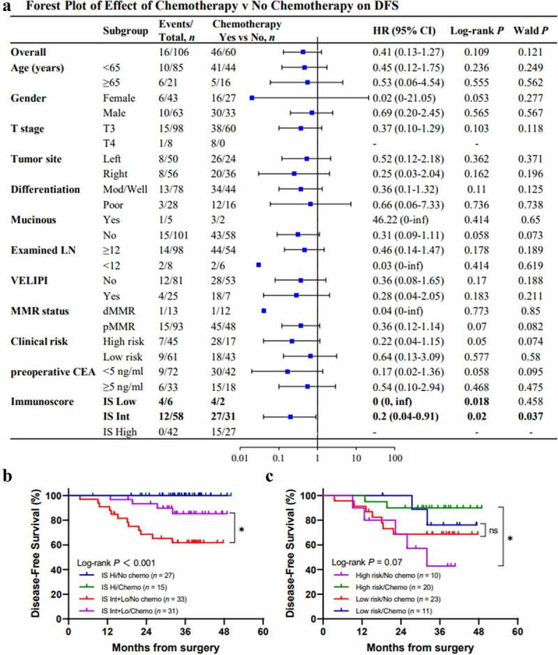
We comprehensively investigated the DFS in different subgroups with or without chemotherapy. Although chemotherapy may be beneficial in almost all subgroups, a significant benefit association was only observed in the IS-Int and IS-Low groups (Figure 3a). In the IS-High group, no patient experienced relapse or death regardless of whether they received adjuvant chemotherapy. In the IS-Int and IS-Low group, patients who received chemotherapy had a better DFS than those who did not (HR = 0.3; 95% CI 0.1–0.92; P = .026), with the 3-year DFS of 85% (95% CI 0.73–1.0) and 62% (95% CI 0.47–0.82), respectively (Figure 3b), indicating that IS could predict chemotherapy benefit. In addition, in the IS-Int and IS-Low group, we found that clinical high-risk patients were more likely to benefit from adjuvant chemotherapy than clinical low-risk patients. For patients with IS-Int and IS-Low and clinical high-risk, chemotherapy could markedly improve the DFS (HR = 0.16; 95% CI 0.03–0.81; P = .01), which it was not statistically significant in clinical low-risk patients (Figure 3c).
Figure 3.

Immunoscore (IS) and the benefit from adjuvant chemotherapy. Forest plot representing the predictive value of response to chemotherapy (disease-free survival [DFS]) in different groups according to clinical parameters and IS levels (a). Kaplan-Meier curves for DFS in patients with or without chemotherapy in different IS groups (b). Kaplan-Meier curves for DFS in patients with IS-Int+Low stratified by clinical risk and chemotherapy (c). (*) indicates significant log-rank P-value, *P < .05; ns, non-significant.
These results suggest that IS has a predictive value for chemotherapy benefit in stage II CRC. Patients with IS-Int and IS-Low had improved DFS when receiving adjuvant chemotherapy, especially those with clinical high-risk factors simultaneously. Strikingly, all patients (100%) with IS-High had no recurrence with or without chemotherapy.
A combination of IS and postoperative ctDNA might optimize the adjuvant therapy strategy for stage II CC
In addition to IS, postoperative ctDNA has been recently reported as a strong prognostic marker for stage I–III CRC. The relationship between postoperative ctDNA and IS will be a very interesting topic since both are promising prognostic markers. ctDNA represents minimal residual disease (MRD) from cancerous cells and tumors, while IS reflects the local immune status of the tumor.
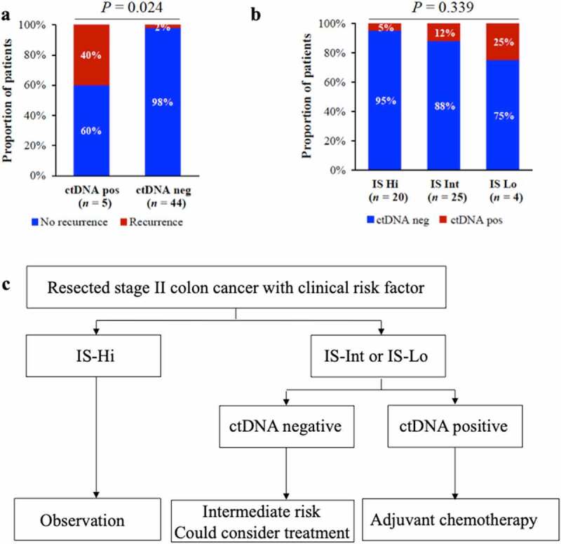
Postoperative ctDNA results could be analyzed in 49 patients. Among these patients, 3 had recurrences and 2 died of colon cancer within 3 years (Table S5). Nevertheless, the 5 patients with positive ctDNA had a higher recurrence rate than those with negative ctDNA (40% vs 2%, P = .024) (Figure 4a), confirming the poor prognosis of ctDNA-positive patients. We explored the association between IS and ctDNA-status. Numerically, the lower the IS, the higher the postoperative ctDNA positivity rate (ctDNA positivity rates in the IS-High, IS-Int, and IS-Low groups: 5%, 12%, 25%; P = .339) (Figure 4b). The trend was not statistically significant, however, most likely due to the small sample size. But it was reasonable as patients with higher IS were demonstrated to have a lower risk of recurrence. It should be noted that the only patient with IS-High and positive ctDNA did not receive adjuvant chemotherapy and did not relapse during nearly two years of follow-up.
Figure 4.

Postoperative ctDNA status and Immunoscore (IS) in the adjuvant setting. Overall recurrence rates according to postoperative ctDNA status (a). Positive ctDNA rates in different IS groups (b). A scheme proposed to guide adjuvant treatment for patients with clinically high-risk stage II colon cancer based on postoperative ctDNA and IS (c). pos, positive; neg, negative; Hi, high; Int, intermediate; Lo, low.
We hypothesized that ctDNA and IS were promising biomarkers that could potentially address the issue of over- or under-treatment. According to previous studies, ctDNA had a high positive predictive value in detecting recurrence in patients with colorectal cancer, colon cancer, and rectal cancer (Table S6).17,20–24 However, ctDNA assay at a single time point before the adjuvant treatment-decision could identify only a small number of patients with MRD (less than 10% of patients with stage II CC)24 and had a sensitivity of less than 50% in predicting disease recurrence.17,20–24 Therefore, it is not safe to de-escalate or avoid adjuvant chemotherapy in patients with stage II CC based on postoperative negative ctDNA alone. In contrast, IS could play a better role in identifying patients with a good prognosis who might avoid chemotherapy. Unlike ctDNA-negative patients, patients with IS-High typically accounted for about 30% of stage II CC patients and were the definite subset with the best prognosis. Importantly, this study found no recurrence of 100% of IS-High, both in the ctDNA positive and ctDNA negative populations. In short, the postoperative ctDNA assay had an advantage in identifying patients at a high risk of recurrence, whereas IS was the opposite. Therefore, we proposed a decision algorithm for adjuvant treatment of stage II CC based on ctDNA and IS (Figure 4c): i) patients with IS-High had the lowest risk of recurrence and could be spared from chemotherapy; ii) patients with postoperative negative ctDNA and IS-Int or IS-Low had an intermediate risk of recurrence and could be considered for treatment; iii) patients with postoperative positive ctDNA and IS-Int or IS-Low had the highest risk of recurrence and were highly recommended to receive chemotherapy.
Discussion
The present study validated the prognostic value of IS for the first time in a Chinese population and demonstrated the predictive value of IS for the chemotherapy benefit in patients with stage II CRC. We also forecasted the future application pattern of IS and ctDNA in guiding adjuvant therapy for early-stage CC.
The distribution of IS in our study is different from that in several studies abroad. Although the number of cases in this work was small, there may be differences in the IS distribution by ethnicity. Earlier research has reported the prognostic performance of IS in CC within the Asian population (mostly Japanese patients) of the SITC cohort.25 The IS distribution of stage II CC in the Asian subgroup was markedly different from that of the overall SITC cohort and our cohort, with the percentage of IS-Low as high as 41%. Moreover, according to the data tracked by the Genecast lab on Chinese stage II CC patients (less than 200 patients as of June this year) commercially tested with IS, the proportion of patients with IS-Low remained below 15%. Thus, more data are needed to prove the differences in the distribution of IS among various populations.
The IS 3-groups including IS-High, IS-Int and IS-Low are more applicable for risk classification of stage II CC in Chinese population and decision-making for adjuvant treatment in IS-Int and IS-Low. Compared with clinical parameters, IS can identify patients with a good prognosis, and help them to be spared from chemotherapy to solve the problem of overtreatment in practice. However, owing to the absence of randomized controlled studies of IS in the adjuvant setting, chemotherapy-free treatment can only be considered for a small number of patients who are safe enough, i.e., IS-High patients. A previous study suggested that 69.5% of clinical high-risk patients could be classified as IS-High+Int and might avoid adjuvant treatment as their risk of recurrence is similar to that of the clinical low-risk patients.15 But the proportion of IS-High+Int is too high, and it could be 92% in our Chinese cohort, thus it would be unsafe to change treatment for such a large proportion of patients.
Our study demonstrated that patients with IS-Int and IS-Low could benefit from chemotherapy, which seems different with the results of stage III CC where only patients with IS-High+Int could benefit from adjuvant chemotherapy.18,19 But we believe these results are non-conflicting and reasonable. First, the tumor stage was different: patients with stage II CC and IS-High had a favorable prognosis, hence little room for survival improvement with adjuvant chemotherapy; patients with stage III CC and IS-High did not have such good prognosis to avoid chemotherapy, which further improved survival in these patients, possibly due to the dependence of chemotherapy efficacy on immune status.26, 27 Second, in our cohort, patients with IS-Int accounted for the most and greatly contributed to the significant chemotherapy benefit observed in the IS-Int and IS-Low groups. As for whether patients with IS-Low could benefit from adjuvant chemotherapy, it might vary at different tumor stages. Although there was no statistical significance, a trend toward chemotherapy benefit was observed in patients with stage III CC and IS-Low.18
Taken together, the association between the chemotherapy benefit and IS deserves further mechanistic investigation.
In recent years, both ctDNA and IS are prognostic markers that have been proven to be superior to all other clinical factors in early-stage CRC and are expected to guide adjuvant therapy for patients in the near future.27,28 However, the correlation between them and the application direction has not been reported. It was previously reported that higher Immunoscore correlate with a cytotoxic and Th1 type response (including increased IFNG, IRF1, TBX21 expression),29–31 and that increased effector memory T-cells correlated with the absence of VELIPI.16 In this study, although the ctDNA cohort was small, we observed a reasonable trend of the association, namely, patients with higher IS would have lower positive rates of postoperative ctDNA. More data are needed to illustrate whether patients with IS-High have ctDNA-MRD detected before adjuvant therapy after radical surgery. Nevertheless, it was suggested that IS is more effective than ctDNA assay in selecting patients who can safely avoid adjuvant chemotherapy. Only when the sensitivity level of ctDNA-MRD testing exceeds 95% can it be used for treatment de-escalation33. According to published data, negative ctDNA still missed some patients who would relapse, and clinical risk-features were needed to increase the negative predictive value.22 Based on the features of IS and ctDNA, we proposed a simple decision algorithm for adjuvant therapy in Figure 4, expecting to facilitate individualized treatment for patients with stage II CC. We believe that with the advancement of detection technology and the accumulation of research evidence, these markers will be applied in a more precise way to clinical decision-making.32 Thus, Immunoscore is an important immunoprofiling biomarker and a predictive tool for cancer treatment.26,29,32–36
Our study adds to the evidence for the prognostic and predictive value of IS in Chinese patients with stage II CRC. Patients with IS-High had the best prognosis, independent of chemotherapy and ctDNA status, while patients with IS-Low had the worse prognosis. Furthermore, patients with IS-High did not need adjuvant chemotherapy, whereas patients with IS-Int and especially IS-Low with the poorest DFS could benefit from adjuvant chemotherapy. IS will help identify patients with excellent prognostic, for whom chemotherapy may be safely avoided. This study demonstrates the clinical utility of IS in the adjuvant setting for early-stage CRC.
Supplementary Material
Acknowledgments
The authors are very grateful to all the participants and their families.
Funding Statement
This research received no external funding.
Highlights
Immunoscore (IS) is a strong prognostic biomarker for Chinese stage II colorectal cancer patients.
Patients with high IS had a 100% DFS rate during a median follow-up of 3 years
Patients with intermediate to low IS could significantly benefit from adjuvant chemotherapy demonstrating IS clinical utility
IS and postoperative ctDNA will jointly optimize the adjuvant treatment strategy for stage II colon cancer.
List of abbreviations
- IS
Immunoscore
- CRC
Colorectal cancer
- CC
Colon cancer
- Int
Intermediate
- DFS
Disease-free survival
- ctDNA
Circulating tumor DNA
- SITC
Society for Immunotherapy of Cancer
- SYSUCC
Sun Yat-sen University Cancer Center
- CAPOX
Capecitabine plus oxaliplatin
- CEA
carcinoembryonic antigen
- chemo
chemotherapy
- Hi
high
- Int
intermediate
- pMMR
Proficient mismatch-repair
- VELIPI
Vascular, lymphatic, or perineural invasion
- dMMR
Mismatch-repair deficiency
- Mod/Well
moderate or well
- RMST
Restricted mean survival time
- IQR
Interquartile range
- HR
Hazard ratio
- MRD
Minimal residual disease
- ACT
adjuvant chemotherapy
Disclosure statement
JQ is employed by Genecast Biotechnology Co., Ltd. AC and JG are employed by Veracyte company. JG has a patent for Immunoscore and is a co-founder of HalioDx, now Veracyte. Immunoscore® is a registered trademark from the National Institute of Health and Medical Research (INSERM) licensed to HalioDx. All remaining authors have declared no conflicts of interest.
Data availability statement
The data presented in this study are available on request from the corresponding author (http://www.ici.upmc.fr/contact.shtml).
Institutional Review Board Statement
The study was conducted according to the guidelines of the Declaration of Helsinki, and approved by the Ethics Committee of Sun Yat-sen University Cancer Center (approval number: B2020-178-01).
Informed Consent Statement
Informed consent was obtained from all subjects involved in the study.
Supplementary material
Supplemental data for this article can be accessed online at https://doi.org/10.1080/2162402X.2022.2161167
References
- 1.Sung H, Ferlay J, Siegel RL, Laversanne M, Soerjomataram I, Jemal A, Bray F.. Global Cancer Statistics 2020: GLOBOCAN estimates of incidence and mortality worldwide for 36 cancers in 185 countries. CA Cancer J Clin. 2021;71:209–11. PMID: 33538338. 10.3322/caac.21660. [DOI] [PubMed] [Google Scholar]
- 2.Rebuzzi SE, Pesola G, Martelli V, Sobrero AF. Adjuvant Chemotherapy for Stage II Colon Cancer. Cancers (Basel). 2020;12 PMID: 32927771. doi: 10.3390/cancers12092584. [DOI] [PMC free article] [PubMed] [Google Scholar]
- 3.Argilés G, Tabernero J, Labianca R, Hochhauser D, Salazar R, Iveson T, Laurent-Puig P, Quirke P, Yoshino T, Taieb J, et al. Localised colon cancer: ESMO clinical practice guidelines for diagnosis, treatment and follow-up. Ann Oncol. 2020;31:1291–1305. PMID: 32702383. doi: 10.1016/j.annonc.2020.06.022. [DOI] [PubMed] [Google Scholar]
- 4.Costas-Chavarri A, Nandakumar G, Temin S, Lopes G, Cervantes A, Cruz Correa M, Engineer R, Hamashima C, Ho GF, Huitzil FD, et al. Treatment of patients with early-stage colorectal cancer: ASCO resource-stratified guideline. J Glob Oncol. 2019;5:1–19. PMID: 30802158. doi: 10.1200/JGO.18.00214. [DOI] [PMC free article] [PubMed] [Google Scholar]
- 5.Yoshino T, Argilés G, Oki E, Martinelli E, Taniguchi H, Arnold D, Mishima S, Li Y, Smruti BK, Ahn JB, et al. Pan-Asian adapted ESMO Clinical Practice Guidelines for the diagnosis treatment and follow-up of patients with localised colon cancer. Ann Oncol. 2021;32:1496–1510. PMID: 34411693. doi: 10.1016/j.annonc.2021.08.1752. [DOI] [PubMed] [Google Scholar]
- 6.NCCN clinical practice guidelines in oncology: colon cancer. Available online: 2022. [accessed 2022]. Version 1; https://wwwnccn.org/professionals/physician_gls/pdf/colon_blocks.pdf
- 7.Babcock BD, Aljehani MA, Jabo B, Choi AH, Morgan JW, Selleck MJ, Luca F, Raskin E, Reeves ME, Garberoglio CA, et al. High-Risk Stage II colon cancer: not all risks are created equal. Ann Surg Oncol. 2018;25:1980–1985. PMID: 29675762. doi: 10.1245/s10434-018-6484-8. [DOI] [PubMed] [Google Scholar]
- 8.Benson AB 3rd, Hamilton SR. Path toward prognostication and prediction: an evolving matrix. J Clin Oncol. 2011;29:4599–4601. PMID: 22067398. doi: 10.1200/JCO.2011.37.8646. [DOI] [PubMed] [Google Scholar]
- 9.O’Connor ES, Greenblatt DY, LoConte NK, Gangnon RE, Liou JI, Heise CP, Smith MA. Adjuvant chemotherapy for stage II colon cancer with poor prognostic features. J Clin Oncol. 2011;29:3381–3388. PMID: 21788561. doi: 10.1200/JCO.2010.34.3426. [DOI] [PMC free article] [PubMed] [Google Scholar]
- 10.Quasar Collaborative G, Gray R, Barnwell J, McConkey C, Hills RK, Williams NS, Kerr DJ. Adjuvant chemotherapy versus observation in patients with colorectal cancer: a randomised study. Lancet. 2007;370:2020–2029. PMID: 18083404. doi: 10.1016/S0140-6736(07)61866-2. [DOI] [PubMed] [Google Scholar]
- 11.Bruni D, Angell HK, Galon J. The immune contexture and Immunoscore in cancer prognosis and therapeutic efficacy. Nat Rev Cancer. 2020. PMID: 32753728 10.1038/s41568-020-0285-7 [DOI] [PubMed] [Google Scholar]
- 12.Fridman WH, Pages F, Sautes-Fridman C, Galon J. The immune contexture in human tumours: impact on clinical outcome. Nat Rev Cancer. 2012;12:298–306. PMID: 22419253. [DOI] [PubMed] [Google Scholar]
- 13.Angell HK, Bruni D, Barrett JC, Herbst R, Galon J. The immunoscore: colon cancer and beyond. Clin Cancer Res. 2020;26:332–339. PMID: 31413009. doi: 10.1158/1078-0432.CCR-18-1851. [DOI] [PubMed] [Google Scholar]
- 14.Pages F, Mlecnik B, Marliot F, Bindea G, Ou FS, Bifulco C, Lugli A, Zlobec I, Rau TT, Berger MD, et al. International validation of the consensus Immunoscore for the classification of colon cancer: a prognostic and accuracy study. Lancet. 2018;391:2128–2139. PMID: 29754777. doi: 10.1016/S0140-6736(18)30789-X. [DOI] [PubMed] [Google Scholar]
- 15.Galon J, Hermitte F, Mlecnik B, Marliot F, Bifulco CB, Lugli A, Nagtegaal ID, Hartmann A, Van den Eynde M, Roehrl MHA, et al. Immunoscore clinical utility to identify good prognostic colon cancer stage II patients with high-risk clinicopathological features for whom adjuvant treatment may be avoided. J Clin Oncol. 2019;37. PMID: WOS:000489107600493. doi: 10.1200/JCO.2019.37.4_suppl.487. [DOI] [Google Scholar]
- 16.Pages F, Berger A, Camus M, Sanchez-Cabo F, Costes A, Molidor R, Mlecnik B, Kirilovsky A, Nilsson M, Damotte D, et al. Effector memory T cells, early metastasis, and survival in colorectal cancer. PMID: 16371631. N Engl J Med. 2005;353:2654–2666. [DOI] [PubMed] [Google Scholar]
- 17.Chen G, Peng J, Xiao Q, Wu HX, Wu X, Wang F, Li L, Ding P, Zhao Q, Li Y, et al. Postoperative circulating tumor DNA as markers of recurrence risk in stages II to III colorectal cancer. J Hematol Oncol. 2021;14:80. PMID: 34001194. doi: 10.1186/s13045-021-01089-z. [DOI] [PMC free article] [PubMed] [Google Scholar]
- 18.Mlecnik B, Bifulco C, Marliot F, Bindea G, Lee JJ, Lugli A, Zlobec I, Rau TT, Berger MD, Nagtegaal ID, et al. Multicenter international SITC study of the consensus Immunoscore for the prediction of survival and response to chemotherapy in Stage III colon cancer. J Clin Oncol. 2020;38(31):3638–3651. PMID: 32897827. doi: 10.1200/JCO.19.03205. [DOI] [PMC free article] [PubMed] [Google Scholar]
- 19.Pagès F, André T, Taieb J, Vernerey D, Henriques J, Borg C, Marliot F, Ben Jannet R, Louvet C, Mineur L, et al. Prognostic and predictive value of the Immunoscore in stage III colon cancer patients treated with oxaliplatin in the prospective IDEA France PRODIGE-GERCOR cohort study. Ann Oncol. 2020;31:921–929. PMID: 32294529. doi: 10.1016/j.annonc.2020.03.310. [DOI] [PubMed] [Google Scholar]
- 20.Henriksen TV, Tarazona N, Frydendahl A, Reinert T, Gimeno-Valiente F, Carbonell-Asins JA, Sharma S, Renner D, Hafez D, Roda D, et al. Circulating tumor DNA in stage III colorectal cancer, beyond minimal residual disease detection, toward assessment of adjuvant therapy efficacy and clinical behavior of recurrences. Clin Cancer Res. 2022;28:507–517. PMID: 34625408. doi: 10.1158/1078-0432.CCR-21-2404. [DOI] [PMC free article] [PubMed] [Google Scholar]
- 21.Reinert T, Henriksen TV, Christensen E, Sharma S, Salari R, Sethi H, Knudsen M, Nordentoft I, Wu H-T, Tin AS, et al. Analysis of plasma cell-free DNA by ultradeep sequencing in patients with stages I to III colorectal cancer. JAMA oncol. 2019;5:1124–1131. PMID: 31070691. doi: 10.1001/jamaoncol.2019.0528. [DOI] [PMC free article] [PubMed] [Google Scholar]
- 22.Tie J, Cohen JD, Lahouel K, Lo SN, Wang Y, Kosmider S, Wong R, Shapiro J, Lee M, Harris S, et al. Circulating tumor DNA analysis guiding adjuvant therapy in stage II colon cancer. N Engl J Med. 2022;386:2261–2272. PMID: 35657320. doi: 10.1056/NEJMoa2200075. [DOI] [PMC free article] [PubMed] [Google Scholar]
- 23.Tie J, Cohen JD, Wang Y, Christie M, Simons K, Lee M, Wong R, Kosmider S, Ananda S, McKendrick J, et al. Circulating tumor DNA analyses as markers of recurrence risk and benefit of adjuvant therapy for stage III colon cancer. JAMA oncol. 2019;5:1710–1717. PMID: 31621801. doi: 10.1001/jamaoncol.2019.3616. [DOI] [PMC free article] [PubMed] [Google Scholar]
- 24.Tie J, Wang Y, Tomasetti C, Li L, Springer S, Kinde I, Silliman N, Tacey M, Wong HL, Christie M, et al. Circulating tumor DNA analysis detects minimal residual disease and predicts recurrence in patients with stage II colon cancer. Sci Transl Med. 2016;8:346ra392. PMID: 27384348. doi: 10.1126/scitranslmed.aaf6219. [DOI] [PMC free article] [PubMed] [Google Scholar]
- 25.Mlecnik B, Torigoe T, Bindea G, Popivanova B, Xu M, Fujita T, Hazama S, Suzuki N, Nagano H, Okuno K, et al. Clinical Performance of the Consensus Immunoscore in Colon Cancer in the Asian Population from the Multicenter International SITC Study. Cancers (Basel). 2022;14(18): 4346. PMID: 36139506. doi: 10.3390/cancers14184346. [DOI] [PMC free article] [PubMed] [Google Scholar]
- 26.Vacchelli E, Galluzzi L, Fridman WH, Galon J, Sautes-Fridman C, Tartour E, Kroemer G. Trial watch: chemotherapy with immunogenic cell death inducers. Oncoimmunology. 2012;1:179–188. PMID: 22720239. doi: 10.4161/onci.1.2.19026. [DOI] [PMC free article] [PubMed] [Google Scholar]
- 27.Naidoo M, Gibbs P, Tie J. ctDNA and adjuvant therapy for colorectal cancer: time to re-invent our treatment paradigm. Cancers (Basel). 2021;13 PMID: 33477814. doi: 10.3390/cancers13020346. [DOI] [PMC free article] [PubMed] [Google Scholar]
- 28.Taieb J, Karoui M, Basile D. How I treat stage II colon cancer patients. ESMO Open. 2021;6:100184. PMID: 34237612. doi: 10.1016/j.esmoop.2021.100184. [DOI] [PMC free article] [PubMed] [Google Scholar]
- 29.Bindea G, Mlecnik B, Angell HK, Galon J. The immune landscape of human tumors: implications for cancer immunotherapy. Oncoimmunology. 2014;3:e27456. PMID: 24800163. doi: 10.4161/onci.27456. [DOI] [PMC free article] [PubMed] [Google Scholar]
- 30.Galon J, Costes A, Sanchez-Cabo F, Kirilovsky A, Mlecnik B, Lagorce-Pages C, Tosolini M, Camus M, Berger A, Wind P, et al. Type, density, and location of immune cells within human colorectal tumors predict clinical outcome. PMID: 17008531. Science. 2006;313:1960–1964. [DOI] [PubMed] [Google Scholar]
- 31.Galon J, Sudarshan C, Ito S, Finbloom D, O’Shea JJ. IL-12 induces IFN regulating factor-1 (IRF-1) gene expression in human NK and T cells. J Immunol. 1999;162:7256–7262. PMID: 10358173. [PubMed] [Google Scholar]
- 32.Antoniotti C, Rossini D, Pietrantonio F, Catteau A, Salvatore L, Lonardi S, Boquet I, Tamberi S, Marmorino F, Moretto R, et al. Upfront FOLFOXIRI plus bevacizumab with or without atezolizumab in the treatment of patients with metastatic colorectal cancer (AtezoTRIBE): a multicentre, open-label, randomised, controlled, phase 2 trial. Lancet Oncol. 2022;23:876–887. PMID: 35636444. doi: 10.1016/s1470-2045(22)00274-1. [DOI] [PubMed] [Google Scholar]
- 33.Vacchelli E, Senovilla L, Eggermont A, Fridman WH, Galon J, Zitvogel L, Kroemer G, Galluzzi L. Trial watch: chemotherapy with immunogenic cell death inducers. Oncoimmunology. 2013;2:e23510. PMID: 23687621. doi: 10.4161/onci.23510. [DOI] [PMC free article] [PubMed] [Google Scholar]
- 34.Dasari A, Morris VK, Allegra CJ, Atreya C, Benson AB 3rd, Boland P, Chung K, Copur MS, Corcoran RB, Deming DA, et al. ctDNA applications and integration in colorectal cancer: an NCI colon and rectal-anal task forces whitepaper. Nat Rev Clin Oncol. 2020;17:757–770. PMID: 32632268. doi: 10.1038/s41571-020-0392-0. [DOI] [PMC free article] [PubMed] [Google Scholar]
- 35.Ascierto PA, Capone M, Urba WJ, Bifulco CB, Botti G, Lugli A, Marincola FM, Ciliberto G, Galon J, Fox BA. The additional facet of immunoscore: immunoprofiling as a possible predictive tool for cancer treatment. J Transl Med. 2013;11:54. PMID: 23452415. doi: 10.1186/1479-5876-11-54. [DOI] [PMC free article] [PubMed] [Google Scholar]
- 36.Bindea G, Mlecnik B, Fridman WH, Galon J. The prognostic impact of anti-cancer immune response: a novel classification of cancer patients. Semin Immunopathol. 2011;33:335–340. PMID: 21461991. doi: 10.1007/s00281-011-0264-x. [DOI] [PMC free article] [PubMed] [Google Scholar]
Associated Data
This section collects any data citations, data availability statements, or supplementary materials included in this article.
Supplementary Materials
Data Availability Statement
The data presented in this study are available on request from the corresponding author (http://www.ici.upmc.fr/contact.shtml).


