Abstract
Microsphere-assisted microscopy utilizing a microsphere in immediate proximity of the specimen boosts the imaging resolution mainly as a result of an increase in the effective numerical aperture of the system.
Subject terms: Interference microscopy, Imaging and sensing
Light microscopes are one of the most widely used tools for sample inspection in life and material sciences. Their achievable spatial resolution, however, is fundamentally limited to ~λ/(2 NA) due to the diffraction of light waves, in which λ is the wavelength of light and NA=n Sinθ is the numerical aperture of the system where θ is the maximum collection half-angle of light by the lens. Improving microscopy resolution beyond this limit or even enhancing the resolution of a given system to reach this limit without significant modifications would be of significant interest for various applications.
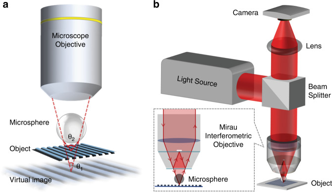
It has been shown in the past decade that micron-scale dielectric spheres and cylinders can be used as supplementary lenses to improve microscopy resolution, a technique termed “microsphere-assisted microscopy” (MAM)1. MAM is a simple, yet efficient approach in which a microsphere (MS) is placed in the immediate vicinity of a specimen, as schematically shown in Fig. 1a. Focusing the objective lens on the virtual image formed underneath the specimen by the MS enables microscopy with higher resolution and magnification compared to using the objective lens without the MS (θ2 > θ1 in Fig. 1a). MAM can be performed with low-index (n~1.5) MSs2 as well as high-index (n > 1.9) MSs3 placed in a background medium with a lower refractive index than that of the microsphere4. The latter approach would allow fabrication of novel resolution-improving optical devices5. MAM is a versatile technique and can be incorporated with different microscopes, such as fluorescent6,7, confocal8, and two-photon9 setups. MAM can also be extended to interference and digital holographic microscopies for 3D imaging with enhanced resolution. This can be simply achieved in an interferometric arrangement, by placing the MS within the working distance of either a conventional objective lens10–15 or a dedicated interferometric objective lens (Fig. 1b)16,17.
Fig. 1. Microsphere-assisted microscopy.
Schematic of MAM: (a) integration with a conventional microscope objective, and (b) extension to 3D microscopy in combination with an interferometric objective lens
MAM has well matched up with interferometric-based approaches, such that both have benefited from each other’s features. Although interferometric arrangements have significantly higher axial resolution compared to conventional microscopy, their lateral resolution would still be restricted by the diffraction limit. However, MAM combined with interferometric setups has shown promising results in producing 3D label-free images with enhanced lateral resolution. Additionally, MAM has shown its potential to be easily integrated with other resolution-enhancing schemes, such as structured and oblique illumination, to achieve even further resolution enhancements in 3D microscopy systems18,19. Microsphere-assisted interference microscopy provides an inexpensive and non-destructive imaging technique for 3D surface metrology17,20 and microsphere-assisted digital holographic microscopy (DHM) brings forward a cost-effective and easy-to-implement high-quality quantitative phase imaging approach in transmission and reflection modes21. The advantages of MAM over other resolution enhancement techniques for DHM are further featured;22,23 in MAM, due to the symmetric structure of the MS, the resolution enhancement is achieved simultaneously in all spatial directions, which is extremely important in real-time imaging of specimens with rapid dynamics12,24. On the other hand, MAM benefits from DHM’s advantageous features. Adding a spherical lens to a conventional microscope introduces non-negligible spherical aberrations1. It also brings about a curved deformation in the measured phase in interference microscopy, which in turn further sacrifices the effective field-of-view (FoV)13. However, in DHM, thanks to the phase compensation possibility, the effect of any possible contaminations and aberrations in the optical train on the final data can be removed. To this end, phase information of the reference hologram (taken for no-sample state in the setup) is subtracted from the object phase during the numerical reconstruction process25. Another challenging issue in MAM that can be less important in microsphere-assisted DHM is precise positioning of the MS and in some cases preserving the relative sample-to-microsphere distance26 during the imaging. However, in DHM, it is consistently possible to be numerically focused on the image plane post-recording of the digital hologram to achieve the best possible imaging quality27. Nevertheless, similar to all kinds of MAM, microsphere-assisted interferometric-based approaches also suffer from the limited FoV, which still needs to be addressed in a sustainable manner.
The open questions and challenges in MAM have been listed in a recent review1; among them were the exact mechanism for resolution enhancement and a robust measurement of the achieved resolution. The enhancement of the numerical aperture was listed as the main contributing factor in resolution enhancement in MAM. Other factors included evanescent wave collection, photonic nanojet effects, resonant effects, and substrate and specimen-specific effects.
In an article published in this issue28, the investigation team performed numerical simulation using finite element method (FEM) to investigate the resolution enhancement mechanism in a microsphere-enhanced interferometry setup29,30. Although previous simulation works31,32 have been done, their model involves rigorous treatment of light scattering at the specimen’s surface and modeling the high NA objective lenses used for illumination and imaging. They considered full 3D conical Kӧhler illumination as well as conical imaging of the scattered waves by the MS. The resolution enhancement and magnification were studied with respect to the NA of the objective lens. Their case study was done for a 5-μm-diameter sphere with refractive index of 1.5 placed on a silicon sinusoidal phase grating with 25 nm peak-to-valley amplitude and 13.2 μm period. A 100× (0.9 NA) objective lens and monochromatic light at a non-resonant (440 nm) and resonant (480.76 nm) wavelength was considered; more simulation detail is provided in their previous work33. Their results confirmed that the enhancement of the NA is the main factor in resolution enhancement in MAM. No significant difference between the resonant and non-resonant wavelengths was observed.
The proposed method can be extended to conventional and confocal microscopies to help better understand and shed more light on MAM. Their approach can also be used to investigate parameters influencing MAM and find the optimum MS in terms of the refractive index, size, and index of the background medium34 for a given application.
Acknowledgements
We would like to dedicate this work to the memory of Kian Pirfalak (2013–2022), a student from Izeh, Iran, who was passionate to become an engineer.
References
- 1.Darafsheh A. Microsphere-assisted microscopy. J. Appl. Phys. 2022;131:031102. doi: 10.1063/5.0068263. [DOI] [Google Scholar]
- 2.Wang ZB, et al. Optical virtual imaging at 50 nm lateral resolution with a white-light nanoscope. Nat. Commun. 2011;2:218. doi: 10.1038/ncomms1211. [DOI] [PubMed] [Google Scholar]
- 3.Darafsheh A, et al. Optical super-resolution by high-index liquid-immersed microspheres. Appl. Phys. Lett. 2012;101:141128. doi: 10.1063/1.4757600. [DOI] [Google Scholar]
- 4.Darafsheh, A. Optical super-resolution and periodical focusing effects by dielectric microspheres. PhD thesis, University of North Carolina at Charlotte, Charlotte (2013).
- 5.Darafsheh A. Photonic nanojets and their applications. J. Phys. Photonics. 2021;3:022001. doi: 10.1088/2515-7647/abdb05. [DOI] [Google Scholar]
- 6.Yang H, et al. Super-resolution biological microscopy using virtual imaging by a microsphere nanoscope. Small. 2014;10:1712–1718. doi: 10.1002/smll.201302942. [DOI] [PubMed] [Google Scholar]
- 7.Darafsheh A, et al. Optical super-resolution imaging by high-index microspheres embedded in elastomers. Opt. Lett. 2015;40:5–8. doi: 10.1364/OL.40.000005. [DOI] [PubMed] [Google Scholar]
- 8.Darafsheh A, et al. Advantages of microsphere-assisted super-resolution imaging technique over solid immersion lens and confocal microscopies. Appl. Phys. Lett. 2014;104:061117. doi: 10.1063/1.4864760. [DOI] [Google Scholar]
- 9.Forouhesh Tehrani K, et al. Resolution enhancement of 2-photon microscopy using high-refractive index microspheres. Proc. SPIE. 2018;10498:1049833. [Google Scholar]
- 10.O’Connor T, Anand A, Javidi B. Field-portable microsphere-assisted high resolution digital holographic microscopy in compact and 3D-printed Mach-Zehnder Interferometer. OSA Contin. 2020;3:1013–1020. doi: 10.1364/OSAC.389832. [DOI] [Google Scholar]
- 11.Wang WC, et al. Microsphere-assisted Fabry–Perot interferometry: proof of concept. Appl. Opt. 2022;61:5442–5448. doi: 10.1364/AO.455341. [DOI] [PubMed] [Google Scholar]
- 12.Wang YX, et al. Resolution enhancement phase-contrast imaging by microsphere digital holography. Opt. Commun. 2016;366:81–87. doi: 10.1016/j.optcom.2015.12.031. [DOI] [Google Scholar]
- 13.Perrin S, et al. Microsphere-assisted phase-shifting profilometry. Appl. Opt. 2017;56:7249–7255. doi: 10.1364/AO.56.007249. [DOI] [PubMed] [Google Scholar]
- 14.Abbasian V, Rasouli S, Moradi AR. Microsphere-assisted self-referencing digital holographic microscopy in transmission mode. J. Opt. 2019;21:045301. doi: 10.1088/2040-8986/ab0815. [DOI] [Google Scholar]
- 15.Wang FF, et al. Three-dimensional super-resolution morphology by near-field assisted white-light interferometry. Sci. Rep. 2016;6:24703. doi: 10.1038/srep24703. [DOI] [PMC free article] [PubMed] [Google Scholar]
- 16.Aakhte M, et al. Microsphere-assisted super-resolved Mirau digital holographic microscopy for cell identification. Appl. Opt. 2017;56:D8–D13. doi: 10.1364/AO.56.0000D8. [DOI] [PubMed] [Google Scholar]
- 17.Kassamakov I, et al. 3D super-resolution optical profiling using microsphere enhanced mirau interferometry. Sci. Rep. 2017;7:3683. doi: 10.1038/s41598-017-03830-6. [DOI] [PMC free article] [PubMed] [Google Scholar]
- 18.Xie ZY, et al. 3D super-resolution reconstruction using microsphere-assisted structured illumination microscopy. IEEE Photonics Technol. Lett. 2019;31:1783–1786. doi: 10.1109/LPT.2019.2946793. [DOI] [Google Scholar]
- 19.Abbasian V, et al. Super-resolved microsphere-assisted Mirau digital holography by oblique illumination. J. Opt. 2018;20:065301. doi: 10.1088/2040-8986/aac22f. [DOI] [Google Scholar]
- 20.Hüser L, Lehmann P. Microsphere-assisted interferometry with high numerical apertures for 3D topography measurements. Appl. Opt. 2020;59:1695–1702. doi: 10.1364/AO.379222. [DOI] [PubMed] [Google Scholar]
- 21.Abbasian V, et al. Digital holographic microscopy for 3D surface characterization of polymeric nanocomposites. Ultramicroscopy. 2018;185:72–80. doi: 10.1016/j.ultramic.2017.11.013. [DOI] [PubMed] [Google Scholar]
- 22.Micó V, et al. Resolution enhancement in quantitative phase microscopy. Adv. Opt. Photonics. 2019;11:135–214. doi: 10.1364/AOP.11.000135. [DOI] [Google Scholar]
- 23.Gao P, Yuan CJ. Resolution enhancement of digital holographic microscopy via synthetic aperture: a review. Light Adv. Manuf. 2022;3:105–120. doi: 10.37188/lam.2022.006. [DOI] [Google Scholar]
- 24.Chen LW, et al. Microsphere enhanced optical imaging and patterning: from physics to applications. Appl. Phys. Rev. 2019;6:021304. doi: 10.1063/1.5082215. [DOI] [Google Scholar]
- 25.Anand A, Chhaniwal VK, Javidi B. Real-time digital holographic microscopy for phase contrast 3D imaging of dynamic phenomena. J. Disp. Technol. 2010;6:500–505. doi: 10.1109/JDT.2010.2052020. [DOI] [Google Scholar]
- 26.Darafsheh A. Comment on ‘Super-resolution microscopy by movable thin-films with embedded microspheres: resolution analysis’ [Ann. Phys. (Berlin) 527, 513 (2015)] Ann. Phys. 2016;528:898–900. doi: 10.1002/andp.201500359. [DOI] [Google Scholar]
- 27.Kim, M. K. Digital Holographic Microscopy: Principles, Techniques, and Applications (Springer, 2011).
- 28.Pahl T, et al. FEM-based modeling of microsphere-enhanced interferometry. Light Adv. Manuf. 2022;3:49. [Google Scholar]
- 29.Hüser L, Lehmann P. Microsphere-assisted interference microscopy for resolution enhancement. tm. - Technisches Mess. 2021;88:311–318. doi: 10.1515/teme-2020-0101. [DOI] [Google Scholar]
- 30.Hüser L, et al. Microsphere assistance in interference microscopy with high numerical aperture objective lenses. J. Optical Microsyst. 2022;2:044501. doi: 10.1117/1.JOM.2.4.044501. [DOI] [Google Scholar]
- 31.Sundaram VM, Wen SB. Analysis of deep sub-micron resolution in microsphere based imaging. Appl. Phys. Lett. 2014;105:204102. doi: 10.1063/1.4902247. [DOI] [Google Scholar]
- 32.Hoang TX, et al. Focusing and imaging in microsphere-based microscopy. Opt. Express. 2015;23:12337–12353. doi: 10.1364/OE.23.012337. [DOI] [PubMed] [Google Scholar]
- 33.Pahl T, et al. 3D modeling of coherence scanning interferometry on 2D surfaces using FEM. Opt. Express. 2020;28:39807–39826. doi: 10.1364/OE.411167. [DOI] [PubMed] [Google Scholar]
- 34.Darafsheh A. Influence of the background medium on imaging performance of microsphere-assisted super-resolution microscopy. Opt. Lett. 2017;42:735–738. doi: 10.1364/OL.42.000735. [DOI] [PubMed] [Google Scholar]


