Abstract
Eosinophils participate in allergic inflammation and may have roles in the body's defense against helminthic infestation. Even under noninflammatory conditions, eosinophils are present in the mucosa of the large intestine, where large numbers of gram-negative bacteria reside. Therefore, roles for eosinophils in host defenses against bacterial invasion are possible. In a system for bacterial viable counts, the bactericidal activity of eosinophils and the contribution of different cellular antibacterial systems against Escherichia coli were investigated. Eosinophils showed a rapid and efficient killing of E. coli under aerobic conditions, whereas under anaerobic conditions bacterial killing decreased dramatically. In addition, diphenylene iodonium chloride (DPI), an inhibitor of the NADPH oxidase and thereby of superoxide production, also significantly inhibited bacterial killing. The inhibitor of nitric oxide (NO) production l-N5-(1-iminoethyl)-ornithine dihydrochloride did not affect the killing efficiency, suggesting that NO or derivatives thereof are of minor importance under the experimental conditions used. To investigate the involvement of superoxide and eosinophil peroxidase (EPO) in bacterial killing, EPO was blocked by azide. The rate of E. coli killing decreased significantly in the presence of azide, whereas addition of DPI did not further decrease the killing, suggesting that superoxide acts in conjunction with EPO. Bactericidal activity was seen in eosinophil extracts containing granule proteins, indicating that oxygen-independent killing may be of importance as well. The findings suggest that eosinophils can participate in host defense against gram-negative bacterial invasion and that oxygen-dependent killing, i.e., superoxide acting in conjunction with EPO, may be the most important bactericidal effector function of these cells.
Eosinophilic granulocytes are believed to participate in the body's defense against infestation with parasites, especially helminths (such as nematodes and trematodes) in the larval stage (35). They are also typically present in the allergic inflammatory response seen in conditions such as allergy and asthma, where they may contribute to tissue damage and connective tissue remodeling (35). However, the roles of eosinophils in host defense functions are not settled. Even under normal, noninflammatory conditions, eosinophils reside in tissues close to mucous membranes that are in close contact with a potentially hostile environment, such as the content of gram-negative bacteria of the large intestine (23). Furthermore, eosinophils possess the adhesion molecule α4β7, which binds to MAdCAM-1, an adhesion molecule expressed by the endothelial cells of the vessels in the gastrointestinal tract, also suggesting roles for eosinophils after recruitment to these tissues (16). Their number increases dramatically during some conditions, such as ulcerative colitis, where the integrity of the mucosal barrier is disturbed and bacteria may invade the tissue (27).
Eosinophils have several properties that can contribute to bactericidal activity. They have a high content of cytotoxic proteins stored in cytoplasmic granules, they are potent producers of reactive oxygen species, and they may also produce nitric oxide (NO) (35).
In the present study, we investigated the antibacterial activity of eosinophils against Escherichia coli and tried to define the contribution of different cellular antibacterial systems.
MATERIALS AND METHODS
l-N5-(1-Iminoethyl)-ornithine dihydrochloride (l-NIO) and diphenylene iodonium chloride (DPI) were from Calbiochem, La Jolla, Calif. Recombinant human interleukin-5 (IL-5) was obtained from Pharmingen, San Diego, Calif. Griess reagent, l-arginine, cytochalasin B, phorbol 12-myristate 13-acetate (PMA), platelet-activating factor (PAF), and 2′,7′-dichlorodihydrofluorescein diacetate (DCF) were purchased from Sigma, St. Louis, Mo. Lipopolysaccharide (LPS), from E. coli strain O55:B5, was obtained from Difco, Detroit, Mich. Dulbecco's modified Eagle's medium without glutamine and phenol red (DMEM) was purchased from ICN, Costa Mesa, Calif. Hanks' balanced salt solution with calcium and magnesium (HBSS) was from Gibco, Paisley, Scotland. Nitrate reductase and NADH were obtained from Cayman Chemical, Ann Arbor, Mich. Boron dipyrromethan (BODIPY)-labeled E. coli strain K-12 was from Molecular Probes Europe BV, Leiden, The Netherlands.
Bacteria.
The E. coli strain ATCC 25922 4Q was obtained from the American Tissue Type Collection, Rockville, Md., and cultured on blood agar plates. Single colonies were picked and grown overnight at 35°C in tryptic soy broth. The bacteria were then centrifuged at 1,000 × g for 10 min, transferred to new nutrient broth, and allowed to grow for 2 h to be in log phase before use in experiments.
Isolation of eosinophils.
Citrated blood was obtained from healthy volunteers after informed consent had been given, and eosinophils were isolated essentially as described previously (20). In short, after isolation of granulocytes on Ficoll-Paque (Pharmacia, Uppsala, Sweden), immunomagnetic beads coated with antibodies to CD16 (Miltenyi, Gladbach, Germany) were used to retrieve the neutrophils in a magnetic column, allowing the isolation of highly purified eosinophils. The purity of eosinophils was more than 96%; contaminating cells were neutrophils and lymphocytes, as judged by routine May-Grünwald-Giemsa staining. The cell viability was more than 98%, as judged by trypan blue exclusion.
Acid extraction of proteins from eosinophils.
Pellets of frozen eosinophils were resuspended in ice-cold water and subjected to intermittent sonication in a water bath in the presence of ice for 10 s at 40 W until no cells could be seen settling to the bottom of the tube. The lysate was extracted with 0.16 N H2SO4 and kept on ice for 30 min with intermittent vortexing. After centrifugation at 23,000 × g for 20 min to sediment insoluble material, the recovered supernatant was dialyzed against 1 mM Tris-HCl (pH 7.4) until the pH of the dialysate was ≥7.0. Thereafter, the extracted protein was dialyzed against 1 mM Tris-HCl–150 mM NaCl at pH 7.4 in dialysis cassettes (Slide-A-Lyzer; Pierce, Rockford, Ill.) and stored at 4°C until use. The protein content was determined using a colorimetric assay based on bicinchoninic acid (BCA protein assay reagent; Pierce).
Incubation of bacteria with granulocytes.
Bacteria were opsonized for 20 min at 37°C with 5% human pooled serum from 10 donors. This concentration of serum was not bactericidal. The bacteria were then centrifuged, washed, and resuspended in phosphate-buffered saline (PBS) (109/ml), kept on ice and finally added to the experimental samples. Granulocytes (107 in 0.1 ml of HBSS) and bacteria (107 in 10 μl of PBS) were mixed with HBSS to a final volume of 0.5 ml. The cell suspensions were incubated at 37°C, with end-over-end turning. In some experiments, cells were preincubated with cytochalasin B (5 μg/ml) to disrupt the cytoskeleton, thereby inhibiting phagocytosis of bacteria.
Antibacterial assay.
The bactericidal activity of eosinophils was determined by measuring the effect on bacterial colony-forming activity as described previously (28). From bacterial suspensions incubated with or without cells, aliquots were taken at the indicated time intervals and serially diluted in sterile ice-cold PBS. A 30-μl aliquot of the diluted sample was transferred to 5 ml of 1.3% (wt/vol) molten agar maintained at 48°C with 0.8% (wt/vol) nutrient broth and 0.5% (wt/vol) NaCl and poured into a petri dish. The temperature of the agar causes disruption of the granulocytes and release of intracellular bacteria. The agar was allowed to solidify, and the number of CFU was determined on the plates after overnight incubation at 35°C.
In some experiments, the agar was supplemented with 1 mg of bovine serum albumin/ml to allow sublethally injured bacteria to grow (29).
Incubation of bacteria with eosinophils under aerobic and anaerobic conditions.
Cell suspensions were preincubated for 30 min at 37°C in an anaerobe box containing 10% H2, 10% CO2, and 80% N2. In parallel, cells were incubated with bacteria in a normal atmospheric environment at the same temperature.
To investigate the function of the NADPH oxidase, cells were incubated with DCF (5 μM) under anaerobic or aerobic conditions and also moved from anaerobic to aerobic conditions or incubated with DPI followed by stimulation with PMA (100 ng/ml). The cells were then put on ice and fixed with formaldehyde to a final concentration of 1% (wt/vol) before measurement of the mean fluorescence intensity by flow cytometry using the fluorescein isothiocyanate channel.
DCF is a cell-permeative nonfluorescent probe that is de-esterified intracellularly and converts into highly fluorescent DCF upon oxidation (36). In short, a stem solution of DCF (5 mM) was prepared fresh every day by dissolving the substance in 95% (vol/vol) ethanol. This solution was kept in the dark at room temperature. Eosinophils at 106/ml in HBSS were incubated with DCF (5 μM) for 15 min at 37°C before use in the experiments.
NO production.
l-NIO was used to inhibit NO synthase (37). l-NIO (10 mM in PBS; final concentration of 100 μM) was added to the cells suspended in HBSS supplemented with 0.6 mM l-arginine. Bacterial incubations and viable counts were performed under aerobic conditions at 37°C. l-NIO incubated with bacteria alone did not affect the viability of the bacteria.
Eosinophils were incubated in DMEM (2 × 10⁶ cells/ml) at 37°C for 24 h in the absence or presence of IL-5 (1 nM) to investigate their NO production. The supernatants were recovered, and proteins were precipitated with ZnSO4 (30% [wt/vol]) before measurement of NO, as determined by the Griess reaction, described below. The viability of eosinophils after 24 h of incubation was determined by trypan blue exclusion and was >96% in medium alone and >98% in the presence of IL-5.
As a positive control of NO production and its inhibition by l-NIO, rat aorta was used. After institutional approval was obtained, male Sprague-Dawley rats (200 to 250 g of body weight) were anesthetized with halothane and exsanguinated. The descending thoracic aorta was removed and cut into circular segments weighing 2.5 to 4 g. The segments were each incubated in 1 ml of DMEM. LPS was added to a final concentration of 100 ng/ml. Two segments were incubated without LPS and served as controls. l-NIO (100 μM) was added to one of the segments incubated with LPS. The segments were incubated with gentle agitation for 24 h at 37°C in an atmosphere containing 8% carbon dioxide in air. After incubation, the segments were removed, blotted on filter paper, and weighed. The DMEM was transferred to clean vials and centrifuged for 5 min at 11,000 × g at 4°C to remove cellular debris and stored at −20°C until assayed for nitrate and nitrite.
Nitrate and nitrite levels, reflecting NO production, were determined by the Griess reaction. In short, 100 μl of supernatant from duplicate incubations was transferred to the wells of a transparent microplate. Nitrate reductase and NADH were added, and the plate was incubated at room temperature for 2 h. Then, 100 μl of Griess reagent (40 mg/ml) was added, and absorbance was measured at 562 nm and compared with standards prepared by diluting sodium nitrate in DMEM. The detection limit for this assay of nitrite in DMEM was 750 nM.
Determination of phagocytosis.
Eosinophils (106/ml) were incubated with serum-opsonized BODIPY-labeled E. coli at a ratio of 1:100. The serum opsonization was performed as described above. Incubations were performed under both aerobic and anaerobic conditions at 37°C, in medium alone or after addition of IL-5 (1 nM) or PAF (1 μM). Aliquots were taken after 15, 30, and 60 min, and eosinophil binding of bacteria was determined using an Epics XL-MCL flow cytometry system (Beckman Coulter Inc., Fullerton, Calif.). Eosinophils were gated using their characteristics in side and forward scatter, excluding unbound bacteria. Fluorescence from extracellular bacteria was quenched by the addition of trypan blue. The percent intracellular bacteria was calculated by using the following formula: [mean fluorescence intensity after quenching/(initial mean fluorescence intensity − eosinophil autofluorescence)].
RESULTS
Killing of E. coli.
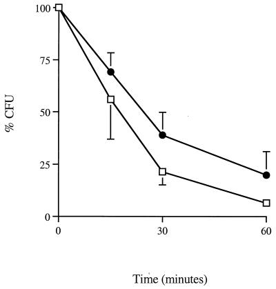
The killing of E. coli by eosinophils was measured by viable count technique (Fig. 1). The killing of the bacteria reached a maximum after 1 h of incubation. Addition of the eosinophil-activating agents PAF (1 μM) or IL-5 (1 nM) (4, 26) did not further increase the killing efficiency (data not shown). The vast majority of bacteria were killed and not sublethally injured (data not shown).
FIG. 1.
Killing of E. coli by eosinophils under aerobic conditions at ratios of 1:1 (□) and 5:1 (●) as measured by viable counts. At the indicated time points, aliquots of the bacterial cell suspension were taken and assayed for viable bacteria. Bacterial viability is expressed as percent survival in the presence of eosinophils, where 100% represents the number of colonies formed at the indicated time points in the absence of eosinophils. Results are means ± SEMs of four independent experiments.
Aerobic versus anaerobic conditions.
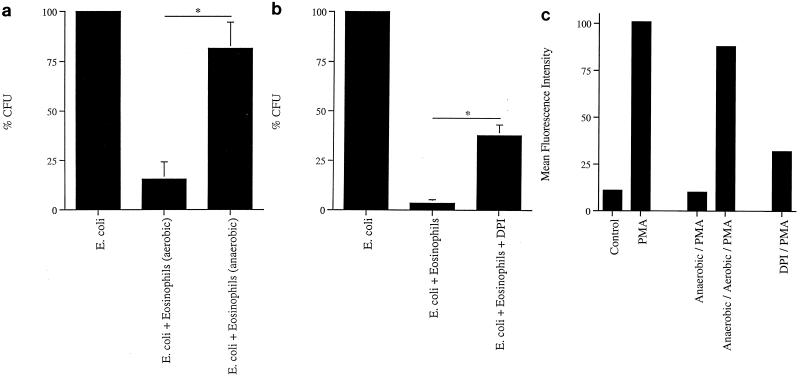
Anaerobic conditions and pharmacological uncoupling of the NADPH oxidase under aerobic conditions reduced eosinophil killing of E. coli significantly compared with incubation of eosinophils and bacteria under aerobic conditions (Fig. 2a and b). These findings suggest that superoxide formed by NADPH oxidase is important for bacterial killing in this system.
FIG. 2.
Killing of E. coli by eosinophils is O2 dependent. (a) Eosinophils were preincubated under aerobic and anaerobic conditions for 30 min before the addition of E. coli. After 60 min of incubation, viable bacteria were counted. Results are means ± SEMs of four independent experiments. ∗, P < 0.05 (Student's t test for paired observations). (b) Pharmacological uncoupling of NADPH oxidase by DPI. Eosinophils were preincubated at 37°C for 10 min in the absence or presence of DPI (10 μM) before incubation with E. coli in the continued absence or presence of DPI for 60 min. ∗, P < 0.05 (Student's t test for paired observations). Results are means plus SEMs of five independent experiments. (c) EPO activity as determined by DCF. Eosinophils were loaded with DCF and incubated under aerobic or anaerobic conditions for 30 min, under anaerobic followed by aerobic conditions for 30 min each, or in the presence of DPI (10 μM) under aerobic conditions for 30 min; cells were subsequently stimulated with PMA (100 ng/ml) for 15 min. The resulting mean fluorescence intensity was determined by flow cytometry and reflects the oxidase activity in the cells. Results of one representative independent experiment out of three are shown.
To determine the efficiency of anaerobic conditions and DPI in inhibiting the oxidase activity, cells were loaded with DCF, and the oxidase-dependent fluorescence was measured (13) (Fig. 2c). PMA was used to induce a respiratory burst in the cells. Anaerobic conditions and DPI both decreased the PMA-induced respiratory burst. When the cells were first incubated for 30 min under anaerobic conditions and then subjected to aerobic conditions for 30 min before stimulation with PMA, a respiratory burst almost of the order of that in stimulated cells held under aerobic conditions was seen. This shows that decreased killing during anaerobic conditions was not attributable to poor viability of the cells.
Inhibition of NO synthesis.
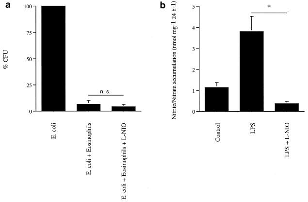
No significant reduction in eosinophil killing of E. coli was seen in the presence of the NO synthesis inhibitor l-NIO (Fig. 3a). This suggests that the generation of NO does not contribute to the killing of bacteria in this system. No NO production could be detected from resting or IL-5-stimulated eosinophils after 24 h of incubation, as determined by the Griess reaction (five separate experiments; data not shown). However, both constitutive and LPS-stimulated NO production from segments of rat aorta was inhibited by the same concentration of l-NIO (Fig. 3b).
FIG. 3.
Killing of E. coli by eosinophils during inhibited NO production. (a) Eosinophils were incubated with bacteria in the absence or presence of the NO synthase inhibitor l-NIO (100 μM). Means ± SEMs from three independent experiments are shown. n. s., not significant. (b) The efficiency of l-NIO (100 μM) in inhibiting NO synthase and thereby NO production was determined by measurement of the NO metabolites NO2− and NO3− by the Griess reaction. Segments of rat thoracic aorta were incubated in the absence or presence of LPS (100 ng/ml) and in the presence of LPS plus l-NIO (100 μM). Results are means plus SEMs from five independent experiments. ∗, P < 0.05 (Student's t test for paired observations).
Antibacterial activity of eosinophil protein extracts.
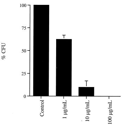
Protein extracts from purified eosinophils killed bacteria in a dose-dependent manner (Fig. 4). The 50% effective dose was approximately 3 μg/ml after 60 min of incubation with the bacteria.
FIG. 4.
Presence of bactericidal activity against E. coli in eosinophil crude protein extracts. Bacteria were incubated with extracts at the indicated concentrations for 60 min. Viable bacteria were assayed by viable counts.
Involvement of superoxide and eosinophil peroxidase (EPO) in bacterial killing.
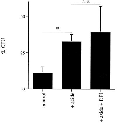
EPO was inhibited by the addition of azide (5 mM) (Fig. 5). Azide at this concentration did not kill bacteria itself, nor did it affect the viability of eosinophils as judged by trypan blue exclusion. The presence of azide significantly decreased the killing of E. coli, whereas the subsequent addition of DPI did not further decrease the killing.
FIG. 5.
Superoxide acts in conjunction with peroxide in eosinophil killing of E. coli. Eosinophils were incubated with E. coli in medium alone (control), after addition of azide (5 mM), and in the presence of the combination of azide (5 mM) and DPI (5 μM). ∗, P < 0.05; n. s., not significant (Student's t test for paired observations).
Phagocytosed versus bacteria bound to the cell surface.
To determine whether the bacteria were phagocytosed or bound to the surface of eosinophils, E. coli cells labeled with the fluorescent dye BODIPY were used. After 60 min of incubation under aerobic conditions, 11.6% ± 3.2% (mean ± standard error of the mean [SEM] for three separate experiments) of the bacteria were phagocytosed, as determined by quenching of extracellular BODIPY by trypan blue. There was no difference between the numbers of phagocytosed bacteria during aerobic and anaerobic incubation.
Treatment of eosinophils with cytochalasin B, an agent that disrupts the cytoskeleton and thereby inhibits phagocytosis, did not affect the bacterial killing efficiency of eosinophils (data not shown).
Taken together, these data suggest that the bacterial killing occurs mainly on the surface of the cells.
DISCUSSION
In the present study we show that eosinophils can kill E. coli and that the killing, to a large degree, is oxygen dependent under the conditions used. We also show that there is oxygen-independent bactericidal activity in eosinophil protein extracts. Furthermore, our data suggest that the bactericidal activity of eosinophils is dependent on the interaction between superoxide generated by the NADPH oxidase and the granule-bound peroxidase EPO.
Several studies have shown that eosinophils are less capable of killing bacteria than neutrophils (2, 10, 32). However, many of these were performed at a time when it was difficult to obtain cells of high purity. In addition, some data regarding the bactericidal capacities of eosinophils are conflicting. Some investigators have reported that eosinophils are as efficient phagocytes as are neutrophils (11), especially after stimulation by certain cytokines (3). More recently, it has been shown that they can, at least under certain circumstances, show bactericidal activities comparable to those of neutrophils (17, 38).
The microbicidal systems of granulocytes can be divided into oxygen-dependent and -independent systems (15, 24). The production of superoxide by the membrane-bound NADPH oxidase, of eosinophils is three to six times greater than that of neutrophils (12). The product of the oxidase system, superoxide, seems to have limited cytotoxicity, and the hydrogen peroxide that is formed by dismutation of superoxide is bactericidal only at high concentrations (19). Eosinophils are exceptionally rich in peroxidase, e.g., EPO, which is present in the matrix of eosinophil granules at 15 μg/106 eosinophils (9, 35). In systems using purified EPO, superoxide, and hydrogen peroxide, the generation of potent cytotoxic species such as the hydroxyl radical in eosinophils has been observed (31). When combined with H2O2 and halides (for example, chloride, iodide, or bromide), EPO catalyzes the generation of highly cytotoxic hypochalous acids (22). Recently, thiocyanate was suggested to be an important substrate in this reaction, generating hypothiocyanite and cyanate (1). Our findings obtained by using azide to block the heme group of EPO and DPI to block the NADPH oxidase system suggest that the oxidase system acting in conjunction with EPO is important for the bactericidal activity of eosinophils. This finding is in agreement with the finding that myeloperoxidase and NADPH oxidase act in conjunction in neutrophils (18), but it is in contrast to an earlier report showing that EPO did not contribute to the bactericidal activity of intact eosinophils (7).
The bactericidal activity of EPO is pH dependent (22). During incubation, the pH of the medium decreased, but there was no difference between the pHs of the media under aerobic and anaerobic conditions (data not shown), arguing against the possibility that such an effect contributed to the difference in bacterial killing.
It has been suggested that eosinophils can produce NO through a constitutive expression of inducible NO synthase (14). NO can react with superoxide and form peroxynitrate, which possesses potent cytotoxic properties against, for example, E. coli (6). Therefore, eosinophil NO production together with superoxide production by the oxidase system may present a powerful source of cytotoxicity (33). In our experiments, however, we were not able to detect NO production by the eosinophils, nor did we see any effects of the inducible NO synthase inhibitor l-NIO on bacterial killing by eosinophils. The reason for this could be the source of cells or the means of detection used. These methodological problems have been reported in experiments with neutrophils (37).
We found potent bactericidal activity in protein extracts from eosinophils. The oxygen-independent antibacterial effects of eosinophils may be attributable to several granule-bound proteins that have, in purified forms, been shown to possess bactericidal effects (25). The characteristic proteins making up the bulk content of eosinophil granules are the highly cationic proteins eosinophil cationic protein (ECP), EPO, eosinophil-derived neurotoxin (EDN), and major basic protein. ECP and eosinophil-derived neurotoxin possess RNase activity, but the bactericidal activity of at least ECP seems to be independent of its RNase activity (34). The effect of ECP on bacteria may instead come from its pore formation (40). Interestingly, the cytotoxicity of ECP can be potentiated by the presence of a functioning oxidase system (39), but the mechanism behind this has not been characterized.
Other granule proteins possessing antibacterial activity have been reported to be present in eosinophil granules. Among these are the 14-kDa secretory phospholipase A2 (5), the bactericidal/permeability-increasing protein (8), and the bacterial cell wall-degrading enzyme N-acetylmuramyl-l-alanine amidase (21). The contribution of each of these is difficult to determine, but together they could represent a powerful and, to a large degree, oxygen-independent bactericidal system. They may also be important for bactericidal activities after release to the extracellular environment.
Since IL-5 is important for eosinophil differentiation, increased susceptibility to infection in IL-5 knockout mice could be expected. Surprisingly, IL-5-deficient mice still produce basal levels of eosinophils that appear to be morphologically and functionally normal (30). In the case of host defense against parasitic infestation, the basal levels of eosinophils has been shown to compromise defense against several helminths. In addition, IL-5 deficient mice also appear to have functional deficiencies in B-1 B lymphocytes and in immunoglobulin A production (30). To our knowledge, the susceptibility to bacterial infection of IL-5 knockout mice has not been investigated. Furthermore, the regulation of the immune systems in mice is different at several levels from that in humans, and therefore firm conclusions with regard to the roles of eosinophils in mice and humans may be difficult to make.
Our study suggests that eosinophils can be efficient in host defense against invasion by gram-negative bacteria and that oxygen-dependent killing, i.e., superoxide acting in conjunction with EPO, may be the most important bactericidal effector function of these cells. This can be important at mucosal interfaces and in the mucosa of, for example, the large intestine, where the conditions are aerobic.
ACKNOWLEDGMENTS
This work was supported by grants from the Th C Bergh Foundation, the Magnus Bergvall Foundation, the Gorthon Foundation, the Greta & Johan Kock Foundations, the Malmö General Hospital Cancer Foundation, the Tore Nilsson Foundation, the Swedish Asthma and Allergy Association's Research Foundation, the Swedish Society of Medicine, the Zoéga Foundation, and the Alfred Österlund Foundation.
REFERENCES
- 1.Arlandson M, Decker T, Roongta V A, Bonilla L, Mayo K H, MacPherson J C, Hazen S L, Slungaard A. Eosinophil peroxidase oxidation of thiocyanate: characterization of major reaction products and a potential sulfhydryl-targeted cytotoxicity system. J Biol Chem. 2001;276:215–224. doi: 10.1074/jbc.M004881200. [DOI] [PubMed] [Google Scholar]
- 2.Baehner R L, Johnston R B J. Metabolic and bactericidal activities of human eosinophils. Br J Haematol. 1974;20:277–285. doi: 10.1111/j.1365-2141.1971.tb07038.x. [DOI] [PubMed] [Google Scholar]
- 3.Blom M, Tool A T, Kok P T, Koenderman L, Roos D, Verhoeven A J. Granulocyte-macrophage colony-stimulating factor, interleukin-3 (IL-3), and IL-5 greatly enhance the interaction of human eosinophils with opsonized particles by changing the affinity of complement receptor type 3. Blood. 1994;83:2978–2984. [PubMed] [Google Scholar]
- 4.Blom M, Tool A T, Roos D, Verhoeven A J. Priming of human eosinophils by platelet-activating factor enhances the number of cells able to bind and respond to opsonized particles. J Immunol. 1992;149:3672–3677. [PubMed] [Google Scholar]
- 5.Blom M, Tool A T, Wever P C, Wolbink G J, Brouwer M C, Calafat J, Egesten A, Knol E F, Hack C E, Roos D, Verhoeven A J. Human eosinophils express, relative to other circulating leukocytes, large amounts of secretory 14-kD phospholipase A2. Blood. 1998;91:3037–3043. [PubMed] [Google Scholar]
- 6.Brunelli L, Crow J P, Beckman J S. The comparative toxicity of nitric oxide and peroxynitrite to Escherichia coli. Arch Biochem Biophys. 1995;316:327–334. doi: 10.1006/abbi.1995.1044. [DOI] [PubMed] [Google Scholar]
- 7.Bujak J S, Root R K. The role of peroxidase in the bactericidal activity of human blood eosinophils. Blood. 1974;43:727–736. [PubMed] [Google Scholar]
- 8.Calafat J, Janssen H, Tool A, Dentener M A, Knol E F, Rosenberg H F, Egesten A. The bactericidal/permeability-increasing protein (BPI) is present in specific granules of human eosinophils. Blood. 1998;91:4770–4775. [PubMed] [Google Scholar]
- 9.Carlson M, Peterson C G, Venge P. Human eosinophil peroxidase: purification and characterization. J Immunol. 1985;134:1875–1879. [PubMed] [Google Scholar]
- 10.Cline M J. Microbicidal activity of human eosinophils. J Reticuloendothel Soc. 1972;12:332–339. [PubMed] [Google Scholar]
- 11.DeChatelet L R, Migler R A, Shirley P S, Muss H B, Szejda P, Bass D A. Comparison of intracellular bactericidal activities of human neutrophils and eosinophils. Blood. 1978;52:609–617. [PubMed] [Google Scholar]
- 12.DeChatelet L R, Shirley P S, McPhail L C, Huntley C C, Muss H B, Bass D A. Oxidative metabolism of the human eosinophil. Blood. 1977;50:525–535. [PubMed] [Google Scholar]
- 13.DeLeo F R, Allen L A, Apicella M, Nauseef W M. NADPH oxidase activation and assembly during phagocytosis. J Immunol. 1999;163:6732–6740. [PubMed] [Google Scholar]
- 14.Del Poso V, de Arruda-Chaves E, de Andrés B, Cárdaba B, López-Farré A, Gallardo S, Cortegano I, Vidarte L, Jurado A, Sastre J, Palomino P, Lahoz C. Eosinophils transcribe and translate messenger RNA for inducible nitric oxide synthase. J Immunol. 1997;158:859–864. [PubMed] [Google Scholar]
- 15.Elsbach P, Weiss J, Levy O. Oxygen-independent antimicrobial systems of phagocytes. In: Gallin J I, Snyderman R, editors. Inflammation: basic principles and clinical correlates. Baltimore, Md: Lippincott Williams & Wilkins Co.; 1999. pp. 801–817. [Google Scholar]
- 16.Erle D J, Briskin M J, Butcher E C, Garcia-Pardo A, Lazarovits A I, Tidswell M. Expression and function of the MAdCAM-1 receptor, integrin alpha 4 beta 7, on human leukocytes. J Immunol. 1994;153:517–528. [PubMed] [Google Scholar]
- 17.Fabian I, Kletter Y, Mor S, Geller-Bernstein C, Ben-Yaakov M, Volovitz B, Golde D W. Activation of human eosinophil and neutrophil functions by haematopoietic growth factors: comparisons of IL-1, IL-3, IL-5 and GM-CSF. Br J Haematol. 1992;80:137–143. doi: 10.1111/j.1365-2141.1992.tb08890.x. [DOI] [PubMed] [Google Scholar]
- 18.Hampton B M, Kettle A J, Winterbourn C C. Involvement of superoxide and myeloperoxidase in oxygen-dependent killing of Staphylococcus aureus by neutrophils. Infect Immun. 1996;64:3512–3517. doi: 10.1128/iai.64.9.3512-3517.1996. [DOI] [PMC free article] [PubMed] [Google Scholar]
- 19.Hampton B M, Kettle A J, Winterbourn C C. Inside the neutrophil phagosome: oxidants, myeloperoxidase, and bacterial killing. Blood. 1998;92:3007–3017. [PubMed] [Google Scholar]
- 20.Hansel T T, De Vries I J M, Iff T, Rihs S, Wandzilak M, Blaser K, Walker C. An improved immunomagnetic procedure for isolation of highly purified human blood eosinophils. J Immunol Methods. 1991;145:105–110. doi: 10.1016/0022-1759(91)90315-7. [DOI] [PubMed] [Google Scholar]
- 21.Hoijer A M, Melief M J, Calafat J, Roos D R, van den Beemd W, van Dongen J J, Hazenberg M P. Expression and intracellular localization of the human N-acetylmuramyl-l-alanine amidase, a bacterial cell wall-degrading enzyme. Blood. 1997;90:1246–1254. [PubMed] [Google Scholar]
- 22.Jong E C, Henderson W R, Klebanoff S J. Bactericidal activity of eosinophil peroxidase. J Immunol. 1980;124:1378–1382. [PubMed] [Google Scholar]
- 23.Kato M, Kephart G M, Talley N J, Wagner J M, Sarr M G, Bonno M, McGovern T W, Gleich G J. Eosinophil infiltration and degranulation in normal human tissue. Anat Rec. 1998;252:418–425. doi: 10.1002/(SICI)1097-0185(199811)252:3<418::AID-AR10>3.0.CO;2-1. [DOI] [PubMed] [Google Scholar]
- 24.Klebanoff S J. Oxygen metabolites from phagocytes. In: Gallin J I, Snyderman R, editors. Inflammation: basic principles and clinical correlates. Baltimore, Md: Lippincott Williams & Wilkins Co.; 1999. pp. 721–768. [Google Scholar]
- 25.Lehrer R I, Szklarek D, Barton A, Ganz T, Hammann K J, Gleich G J. Antibacterial properties of major basic protein and eosinophil cationic protein. J Immunol. 1989;142:4428–4434. [PubMed] [Google Scholar]
- 26.Lopez A F, Sanderson C J, Gamble J R, Campbell H D, Young I G, Vadas M A. Recombinant human interleukin 5 is a selective activator of human eosinophil function. J Exp Med. 1988;167:219–224. doi: 10.1084/jem.167.1.219. [DOI] [PMC free article] [PubMed] [Google Scholar]
- 27.Makiyama K, Kanzaki S, Yamasaki K, Zee-Iriarte W, Tsuji Y. Activation of eosinophils in the pathophysiology of ulcerative colitis. J Gastroenterol. 1995;30:64–69. [PubMed] [Google Scholar]
- 28.Mandic-Mulec I, Weiss J, Zychlinsky A. Shigella flexneri is trapped in polymorphonuclear leukocyte vacuoles and efficiently killed. Infect Immun. 1997;65:110–115. doi: 10.1128/iai.65.1.110-115.1997. [DOI] [PMC free article] [PubMed] [Google Scholar]
- 29.Mannion B A, Weiss J, Elsbach P. Separation of sublethal and lethal effects of polymorphonuclear leukocytes on Escherichia coli. J Clin Investig. 1990;86:631–641. doi: 10.1172/JCI114755. [DOI] [PMC free article] [PubMed] [Google Scholar]
- 30.Matthaei K I, Foster P, Young I G. The role of interleukin-5 (IL-5) in vivo: studies with IL-5 deficient mice. Mem Inst Oswaldo Cruz. 1997;92:63–68. doi: 10.1590/s0074-02761997000800010. [DOI] [PubMed] [Google Scholar]
- 31.McCormick M L, Roeder T L, Railsback M A, Britigan B E. Eosinophil peroxidase-dependent hydroxyl radical generation by human eosinophils. J Biol Chem. 1994;269:27914–27919. [PubMed] [Google Scholar]
- 32.Mickenberg I D, Root R K, Wolff S M. Bactericidal and metabolic properties of human eosinophils. Blood. 1972;39:67–80. [PubMed] [Google Scholar]
- 33.Oliviera S H, Fonseca S G, Romao P R, Figueiredo F, Ferreira S H, Cunha F Q. Microbicidal activity of eosinophils is associated with activation of the arginine-NO pathway. Parasite Immunol. 1998;20:405–412. doi: 10.1046/j.1365-3024.1998.00159.x. [DOI] [PubMed] [Google Scholar]
- 34.Rosenberg H F. Recombinant human eosinophil cationic protein. Ribonuclease activity is not essential for cytotoxicity. J Biol Chem. 1995;270:7876–7881. doi: 10.1074/jbc.270.14.7876. [DOI] [PubMed] [Google Scholar]
- 35.Rosenberg H F. The eosinophil. In: Gallin J I, Snyderman R, editors. Inflammation: basic principles and clinical correlates. Baltimore, Md: Lippincott Williams & Wilkins Co.; 1999. pp. 61–77. [Google Scholar]
- 36.Rosenkranz A R, Schmaldienst S, Stuhlmeier K M, Chen W, Knapp W G, Zlabinger J. A microplate assay for the detection of oxidative products using 2′,7′-dichlorofluorescin-diacetate. J Immunol Methods. 1992;156:39–45. doi: 10.1016/0022-1759(92)90008-h. [DOI] [PubMed] [Google Scholar]
- 37.Wheeler A M, Smith S D, Garcia-Cardena G, Nathan C F, Weiss R M, Sessa W C. Bacterial infection induces nitric oxide synthase in human neutrophils. J Clin Investig. 1997;99:110–116. doi: 10.1172/JCI119121. [DOI] [PMC free article] [PubMed] [Google Scholar]
- 38.Yazdanbakhsh M, Eckmann C M, Bot A A, Roos D. Bactericidal action of eosinophils from normal human blood. Infect Immun. 1986;53:192–198. doi: 10.1128/iai.53.1.192-198.1986. [DOI] [PMC free article] [PubMed] [Google Scholar]
- 39.Yazdanbakhsh M, Tai P-C, Spry C J F, Gleich G J, Roos D. Synergism between eosinophil cationic protein and oxygen metabolites in killing of schistosomula of Schistosoma mansoni. J Immunol. 1987;138:3443–3447. [PubMed] [Google Scholar]
- 40.Young J D, Peterson C G, Venge P, Cohn Z A. Mechanism of membrane damage mediated by human eosinophil cationic protein. Nature. 1986;321:613–616. doi: 10.1038/321613a0. [DOI] [PubMed] [Google Scholar]


